Next Issue
Volume 7, February
Previous Issue
Volume 6, October


Prosthesis, Volume 6, Issue 6 (December 2024) – 24 articles

Cover Story (view full-size image): 
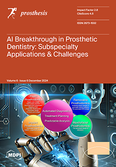
Prosthodontics focuses on diagnosing, rehabilitating, and maintaining oral function for patients with missing teeth and maxillofacial tissues. This area of dentistry includes four branches, fixed, removable, implant, and maxillofacial prosthodontics, each using evolving technologies for precision. Digital tools such as CAD, CAM, intraoral scanners, and 3D printing have revolutionized prosthesis design. AI, powered by machine learning, enhances diagnostics, treatment planning, and workflow. It aids in crown design, denture fabrication, implant placement, and maxillofacial prostheses. Challenges to AI adoption include data quality, transparency, integration, ethics, and financial costs. However, AI promises personalized prosthetics and improved patient care, requiring continuous professional development and global collaboration to advance. View this paper
  • Issues are regarded as officially published after their release is announced to the table of contents alert mailing list.
  • You may sign up for e-mail alerts to receive table of contents of newly released issues.
  • PDF is the official format for papers published in both, html and pdf forms. To view the papers in pdf format, click on the "PDF Full-text" link, and use the free Adobe Reader to open them.
Order results
Result details
Section
Select all
Export citation of selected articles as:
16 pages, 2606 KiB  
Article
Effectiveness of a New Microprocessor-Controlled Knee–Ankle–Foot System for Transfemoral Amputees: A Randomized Controlled Trial
by Christelle Requena, Joseph Bascou, Isabelle Loiret, Xavier Bonnet, Marie Thomas-Pohl, Clément Duraffourg, Laurine Calistri and Hélène Pillet
Prosthesis 2024, 6(6), 1591-1606; https://doi.org/10.3390/prosthesis6060115 - 18 Dec 2024
Cited by 1 | Viewed by 1587
Abstract
Background: Advances in prosthetic technology, especially microprocessor-controlled knees (MPKs), have helped enhance gait symmetry and reduce fall risks for individuals who have undergone transfemoral amputation. However, challenges remain in walking in constrained situations due to the limitations of passive prosthetic feet, lacking ankle [...] Read more.
Background: Advances in prosthetic technology, especially microprocessor-controlled knees (MPKs), have helped enhance gait symmetry and reduce fall risks for individuals who have undergone transfemoral amputation. However, challenges remain in walking in constrained situations due to the limitations of passive prosthetic feet, lacking ankle mobility. This study investigates the benefits of SYNSYS®, a new microprocessor-controlled knee–ankle–foot system (MPKA_NEW), designed to synergize knee and ankle movements. Methods: A randomized crossover trial was conducted on 12 male participants who had undergone transfemoral amputation who tested both the MPKA_NEW and their usual MPK prosthesis. Biomechanical parameters were evaluated using quantitative gait analysis in various walking conditions. Participants also completed self-reported questionnaires on their quality of life, locomotor abilities, and prosthesis satisfaction. Results: The MPKA_NEW showed a significant reduction in the risk of slipping and tripping compared to standard MPK prostheses, as evidenced by increased flat-foot time and minimum toe clearance during gait analysis. The MPKA_NEW also improved physical component scores in quality-of-life assessments (Short-Form 36 General Health Questionnaire), suggesting enhanced stability and reduced cognitive load during walking. Conclusions: The MPKA_NEW offers significant improvements in gait safety and quality of life for people who have undergone TFA, particularly in challenging conditions. Further studies are needed to assess the long-term benefits and adaptability across diverse amputee populations. Full article
Show Figures

Figure 1

5 pages, 332 KiB  
Brief Report
An Economic Analysis of Metal-on-Metal Versus Ceramic-on-Ceramic Hip Resurfacing: Costs and Clinical Implications
by Georgios Orfanos, Thomas W. Wainwright and Robert G. Middleton
Prosthesis 2024, 6(6), 1586-1590; https://doi.org/10.3390/prosthesis6060114 - 18 Dec 2024
Viewed by 1174
Abstract
Background: Ceramic may be an alternative bearing surface that could be used to deal with the negative sequalae associated with the use of metal-on-metal (MoM) hip resurfacing implants, keeping the benefits of hip resurfacing without the risks. The aim of this study is [...] Read more.
Background: Ceramic may be an alternative bearing surface that could be used to deal with the negative sequalae associated with the use of metal-on-metal (MoM) hip resurfacing implants, keeping the benefits of hip resurfacing without the risks. The aim of this study is to evaluate the additional financial and logistical implications that MoM hip resurfacing has, in comparison to its ceramic-on-ceramic (CoC) counterpart. Methods: Two different follow up protocols were analysed, according to current practice guidance, on how to follow up with hip resurfacing procedures. Reference costs were identified using the latest national cost collection data and data from the local biochemistry department. The results incorporated the number of hip resurfacings performed nationally. Results: The non-MoM protocol yielded a per operation lifetime follow up cost of £802.50, whereas the MoM protocol yielded a cost of £2132.83 at 25 years. Nationally, according to the 2023 data, this amounted to £561,750 versus £1,492,981 per year, respectively. MoM hip resurfacing is 2.6 times more expensive to be followed up with when compared to the standard protocol. Conclusion: The cost of the follow up of MoM bearings far exceeds that of non-MoM bearings. If CoC hip resurfacing is as clinically effective as MoM hip resurfacing, then the economics favour CoC bearing for resurfacing. Full article
(This article belongs to the Special Issue State of Art in Hip, Knee and Shoulder Replacement (Volume 2))
Show Figures

Figure 1

11 pages, 2016 KiB  
Article
An In Vitro Study on the Accuracy of a Splint for the Transfer of Dental Implants
by Rommy Chalacan Galindo, Angel Tul Tipantuña, Maria Flores Araque, Lupe Poussin and Byron Velasquez Ron
Prosthesis 2024, 6(6), 1575-1585; https://doi.org/10.3390/prosthesis6060113 - 17 Dec 2024
Cited by 1 | Viewed by 1196
Abstract
The purpose of this study is to measure the positional accuracy of transfers during the splinting printing process using four biomaterials (two acrylic resins and two bis acrylic resins). Materials and Methods: A master model was constructed for the acrylic simulation of edentulous [...] Read more.
The purpose of this study is to measure the positional accuracy of transfers during the splinting printing process using four biomaterials (two acrylic resins and two bis acrylic resins). Materials and Methods: A master model was constructed for the acrylic simulation of edentulous mandibles with four multiunit analogs (Bioinnovation, Sao Paulo, Brazil) positioned between the lateral incisor and premolar areas. Eighty samples (n = 80) were created in total. Four different dental materials, Duralay (Reliance, Chicago, IL, USA); Pattern Resin LS (GC, Tokyo, Japan) with a ratio of one part liquid and two parts powder; Structur (Vocco, Colonia, Germany); and Protemp (3 M ESPE, Neus, Germany), were used as splints with five samples each. Measurements were obtained for distances between points A, B, C, and D in sequential order through an INSIZE digital calibrator (Inzise, Taiwan, Seng, China). Results: The results were as follows: Distance A: better performance was observed for Pattern Resin (mean = 38.59 mm) and Duralay (mean = 38.58 mm) compared to the base (mean = 38.59 mm); distance B: Pattern Resin (mean = 19.08 mm) and Duralay (mean = 19.07 mm) were compared to the base (mean = 19.08 mm); distance C: no significant differences in relation to the base (prom = 18.85 mm) were shown for the three materials of Pattern Resin (prom = 18.86 mm), Structur (prom = 18.85 mm), and Duralay (mean = 18.85 mm); and distance D: Pattern Resin (mean = 20.46 mm) and Duralay (mean = 20.46 mm) provided lower performance compared to the base (mean = 20.47 mm), ANOVA (Prob = 0.333 > 0.05). Conclusions: Pattern resins and Duralay acrylic resins exhibit better dimensional stability when used as splints for the transfer of multiple dental implants. Full article
(This article belongs to the Collection Oral Implantology: Current Aspects and Future Perspectives)
Show Figures

Figure 1

14 pages, 4474 KiB  
Article
Enhancing Tensile Bond Strength of Glass Fiber Posts Using Chitosan as a Coupling Agent: A Novel Approach for Improved Dental Restorations
by Noha Taymour, Mohammed Hashim Albin Hejji, Mohammed Faihan Alotaibi, Rakan Abdullah Alzahrani, Ali Mohammed Almarzooq, Ashwin C. Shetty and Shimaa Rifaat
Prosthesis 2024, 6(6), 1561-1574; https://doi.org/10.3390/prosthesis6060112 - 16 Dec 2024
Cited by 2 | Viewed by 1111
Abstract
Objectives: This study was designed to assess the effectiveness of chitosan as a coupling agent for improving the tensile bond strength of fiber posts. Methods: A total of 91 single-rooted mandibular teeth were root canal-filled. Post spaces were created and categorized into seven [...] Read more.
Objectives: This study was designed to assess the effectiveness of chitosan as a coupling agent for improving the tensile bond strength of fiber posts. Methods: A total of 91 single-rooted mandibular teeth were root canal-filled. Post spaces were created and categorized into seven groups: Group A (Control), Group B (Silane), Group C (Chitosan), Group D (37% Phosphoric acid + Silane), Group E (37% Phosphoric acid + Chitosan), Group F (10% Hydrogen Peroxide + Silane), and Group G (10% Hydrogen Peroxide + Chitosan). Posts were cemented and tensile bond strength was measured, while the morphological structure of the fiber posts was analyzed using Scanning Electron Microscopy. One-way (ANOVA) and Tukey’s multiple comparison tests were performed at a level of significance of 5%. The percentages of fracture patterns among the groups were compared. Results: 10% Hydrogen peroxide + Chitosan exhibited the significantly highest tensile bond strength (p < 0.001). Adhesive failures were more frequent in Groups A, B, C, and D, whereas cohesive failures within the resin cement were predominant in Groups E, F, and G. Conclusions: The protocol of using 10% hydrogen peroxide followed by a chitosan coupling agent significantly improved tensile bond strengths for glass fiber posts, which highlights the potential of using chitosan as a natural biopolymer and an alternative to synthetic coupling agents to develop more effective bonding strategies for dental restorations. Full article
(This article belongs to the Special Issue Advancements in Adhesion Techniques and Materials in Prosthodontics)
Show Figures

Figure 1

8 pages, 2107 KiB  
Technical Note
Modular Stems in Revision Hip Arthroplasty: A Three-Step Technique
by Francesco Pirato, Jacopo Vittori, Federico Dettoni, Matteo Bruzzone, Roberto Rossi and Umberto Cottino
Prosthesis 2024, 6(6), 1553-1560; https://doi.org/10.3390/prosthesis6060111 - 16 Dec 2024
Viewed by 906
Abstract
Background: Complications such as periprosthetic fractures necessitate challenging revision surgeries. In particular, femoral stem revisions can be complicated by poor bone quality, making primary stability and leg length restoration difficult to achieve. Modular fluted tapered stems (MFTSs) have emerged as a viable option [...] Read more.
Background: Complications such as periprosthetic fractures necessitate challenging revision surgeries. In particular, femoral stem revisions can be complicated by poor bone quality, making primary stability and leg length restoration difficult to achieve. Modular fluted tapered stems (MFTSs) have emerged as a viable option for these complex cases. This study aims to describe a reproducible three-step technique for femoral stem revision using MFTSs. The technique focuses on (1) obtaining distal primary stability, (2) restoring leg length, and (3) ensuring overall implant stability. Materials and methods: We conducted a retrospective analysis of ten patients who underwent revision THA using this three-step technique, with a minimum follow-up of 12 months. The mean patient age was 70.7 years, and the average follow-up was 24.2 months. Limits were the small sample size, the lack of clinical outcomes and the short-term follow-up. Results: There was no subsidence, a mean leg length discrepancy of 4 mm (p: 0.604), and no dislocations. However, heterotopic ossifications (HOs) were observed in 25% of patients, although no trochanteric migrations occurred. One patient experienced an intraoperative femoral fracture, which was successfully treated. Conclusions: This three-step approach can break down the revision procedure, making it more accessible to surgeons. The findings suggest that this technique is effective in achieving reliable outcomes in femoral stem revisions, potentially improving the standard of care for patients requiring complex THA revisions. Full article
Show Figures

Figure 1

24 pages, 4713 KiB  
Review
Infective Endocarditis After Transcatheter Aortic Valve Replacement: A Narrative Review
by Francesco Nappi
Prosthesis 2024, 6(6), 1529-1552; https://doi.org/10.3390/prosthesis6060110 (registering DOI) - 12 Dec 2024
Viewed by 1295
Abstract
Prosthetic valve endocarditis (PVE) has undergone significant changes over the past five decades and is currently affecting an aging population, with an increasing prevalence in patients with transcatheter valve implants. The introduction of transcatheter aortic valve replacement (TAVR) represents a significant advance in [...] Read more.
Prosthetic valve endocarditis (PVE) has undergone significant changes over the past five decades and is currently affecting an aging population, with an increasing prevalence in patients with transcatheter valve implants. The introduction of transcatheter aortic valve replacement (TAVR) represents a significant advance in the field of interventional cardiology and cardiac surgery. The incidence of IE after TAVR has remained stable, with rates similar to those reported after surgical aortic valve replacement. This is despite significant refinements in the TAVR procedure, with less invasive handling and its extension to younger and healthier patients. TAVR should be considered as a potential treatment option for patients with PVE, despite some differences. In terms of evolutionary advances, there have been notable and significant developments in the fields of microbiology and imaging diagnostics. The 2023 Duke-International Society for Cardiovascular Infectious Diseases diagnostic criteria for infective endocarditis now incorporate significant advances in molecular biology and fluorine-18 fluorodeoxyglucose positron emission tomography/computed tomography. This has led to a significant enhancement in diagnostic sensitivity for PVE while maintaining the same level of specificity in validation studies. PVE is a deadly disease. A multidisciplinary endocarditis treatment team in a cardiac center is essential to improve outcomes. The availability of novel surgical options allows clinicians to offer an increasing number of patients the opportunity to avoid surgical intervention. Some patients will complete antimicrobial treatment at home. Those with prosthetic valves are eligible for antibiotic prophylaxis before dental procedures. Post-TAVR infective endocarditis (IE) is a subcategory of prosthetic valve endocarditis. This condition presents a particularly complex scenario, characterized by a distinctive clinical and microbiological profile, a high prevalence of IE-related complications, an ambiguous role of cardiac surgery, and a poor prognosis for the majority of patients with TAVR IE. The number of TAVR procedures is set to skyrocket in the coming years, which will undoubtedly lead to a significant rise in the number of people at risk of this life-threatening complication. This review will provide an overview of this rare complication in light of the advent of IE following TAVR. It is crucial to gain a comprehensive understanding of the disease and its associated complications to enhance clinical outcomes. Full article
Show Figures

Figure 1

19 pages, 14533 KiB  
Article
Evaluation of the Integration of Topological Optimisation in the Process Chain for Manufacturing Customised Orthopaedic Devices via Additive Manufacturing
by Francesca Sala, Gianluca Danilo D’Urso and Claudio Giardini
Prosthesis 2024, 6(6), 1510-1528; https://doi.org/10.3390/prosthesis6060109 - 11 Dec 2024
Viewed by 1256
Abstract
Purpose: The effectiveness of the customised solutions compared to the conventional ones and the emergence of advanced production technologies, such as Additive Manufacturing (AM) techniques, strengthened the trend towards an enhanced individualization of the clinical treatments. In the present research, the value of [...] Read more.
Purpose: The effectiveness of the customised solutions compared to the conventional ones and the emergence of advanced production technologies, such as Additive Manufacturing (AM) techniques, strengthened the trend towards an enhanced individualization of the clinical treatments. In the present research, the value of topological optimisation (TO) in the manufacturing process of tailor-made orthopaedic appliance (upper-limb orthosis) was analysed. Methodology: From the morphology of a patient’s arm, orthotic models were developed. Nonparametric optimization (Simulia Tosca) was performed, based on the Finite Element Analysis (FEA) program (Abaqus), and contributed to the development of TO orthotic models with diverse levels of volume reduction fraction. The modelling and manufacturing framework for customising orthotic solutions was evaluated with a discussion on the feasibility of lightweight and high-performance products, encompassing production time and cost. Pilot products were produced with a Material Extrusion (MEX) printer. Findings: TO proved to be a practical and valuable approach for the advanced customisation of orthopaedic devices, offering lightweight solutions able to withstand stresses also during patient rehabilitation and remission. From the rapid prototyping perspective, specific strategies must be adopted to prevent the escalation of production costs and time. Originality: The research delves into the overall benefit of implementing an advanced modelling technique within the context of manufacturing highly customised orthoses, analysing how TO activity impacts the rapid prototyping process. Beyond product evaluation, the analysis explores broader implications, including the assessment of feasibility and the development of strategies for integrating the approach into clinical workflows and hospital settings. Full article
Show Figures

Figure 1

19 pages, 6647 KiB  
Article
The Design and Application of an Advanced System for the Diagnosis and Treatment of Flatfoot Based on Infrared Thermography and a Smart-Memory-Alloy-Reinforced Insole
by Ali F. Abdulkareem, Auns Q. Al-Neami, Tariq J. Mohammed and Hayder R. Al-Omairi
Prosthesis 2024, 6(6), 1491-1509; https://doi.org/10.3390/prosthesis6060108 - 9 Dec 2024
Cited by 1 | Viewed by 1351
Abstract
Background: Flatfoot deformity is a common condition in children and teenagers that may increase the risk of knee, hip, and back pain. Most of the insoles suggested to treat flatfoot symptoms are not designed to adapt to foot temperature during walking, and they [...] Read more.
Background: Flatfoot deformity is a common condition in children and teenagers that may increase the risk of knee, hip, and back pain. Most of the insoles suggested to treat flatfoot symptoms are not designed to adapt to foot temperature during walking, and they are either too soft to provide support or hard enough to be uncomfortable. Purpose: This study aims to develop an advanced solution to diagnose and treat flexible flatfoot (FFT) using infrared thermography measurements and a hybrid insole reinforced by nitinol (NiTiCu) smart-memory-alloy wires (SMAWs), this super-elastic alloy can return back to its pre-deformed shape when heated, which helps to reduce the local high-temperature points caused by the uneven pressure of FFT. This approach achieves a more uniform thermal distribution across the foot, which makes the hybrid insole more comfortable. Methods: The study involved 16 subjects, divided into two groups of eight flat-footed and eight normal. The procedure includes two parts, namely, designing a prototype insole with SMAW properties based on thermography measurement by using SolidWorks, and evaluating this design using Ansys. Second, a hybrid insole reinforced with SMAWs is customized for flatfoot subjects. The thermography measurement differences between the medial and lateral sides of the metatarsophalangeal line are compared for the normal and flatfoot groups before and after wearing the suggested design. Results: The results show that our approach safely diagnosed FFT and significantly improved the thermal distribution in FFT subjects by more than 80% after wearing the suggested design. A paired t-test reported significant (p-value > 0.001) thermal decreases in the high-temperature points after using the SMAW insole, which was closely approximated to the normal subjects. Conclusions: the SMAW-reinforced insole is comfortable and suitable for treating FFT deformity, and infrared thermography is an effective tool to evaluate FFT deformity. Full article
(This article belongs to the Special Issue Recent Advances in Foot Prosthesis and Orthosis)
Show Figures

Figure 1

12 pages, 9673 KiB  
Technical Note
Impression Reference Technique for the Open Flap Digital Workflow in the Immediate Loading Rehabilitation of the Upper and Lower Jaws
by Gerardo Pellegrino, Gabriele Anselmo, Carlo Barausse, Stefano Ratti, Cristiana Breccia, Edoardo Mancuso, Amerigo Giudice, Pietro Di Bene and Pietro Felice
Prosthesis 2024, 6(6), 1479-1490; https://doi.org/10.3390/prosthesis6060107 - 3 Dec 2024
Viewed by 1001
Abstract
Immediate loading implant surgery has emerged as a significant advancement in the rehabilitation of edentulous patients. This approach requires meticulous planning and precise execution to ensure successful outcomes. Transferring established intermaxillary and occlusal relationships to the definitive prostheses can be challenging. However, with [...] Read more.
Immediate loading implant surgery has emerged as a significant advancement in the rehabilitation of edentulous patients. This approach requires meticulous planning and precise execution to ensure successful outcomes. Transferring established intermaxillary and occlusal relationships to the definitive prostheses can be challenging. However, with a digital approach, this procedure can be standardized in cases of fully guided surgery with a flapless technique or by using disposable guides. Conversely, when extensive open flap implant surgery is required, such as in the treatment of severely atrophic patients (e.g., with zygomatic implants or simultaneous bone augmentation), the digital workflow can be demanding. The impression reference technique was proposed to enhance the digital workflow for immediate loading in zygomatic implant rehabilitation. This study aims to describe the impression reference technique, applied to both upper and lower jaws for immediate loading open flap rehabilitations, using standard implants. Full article
(This article belongs to the Collection Oral Implantology: Current Aspects and Future Perspectives)
Show Figures

Figure 1

20 pages, 11970 KiB  
Article
AI-Enhanced Analysis to Investigate the Feasibility of EMG Signals for Prosthetic Hand Force Control Incorporating Anthropometric Measures
by Deepak Chandra Joshi, Pankaj Kumar, Rakesh Chandra Joshi and Santanu Mitra
Prosthesis 2024, 6(6), 1459-1478; https://doi.org/10.3390/prosthesis6060106 - 2 Dec 2024
Cited by 2 | Viewed by 2512
Abstract
Background/Objectives: The potential application of electromyography (EMG) as a method for precise force control in prosthetic devices is investigated, expanding on its traditional use in gesture detection. Variability in EMG signals among individuals is influenced by physiological factors such as muscle mass, body [...] Read more.
Background/Objectives: The potential application of electromyography (EMG) as a method for precise force control in prosthetic devices is investigated, expanding on its traditional use in gesture detection. Variability in EMG signals among individuals is influenced by physiological factors such as muscle mass, body fat percentage, and subcutaneous fat, as well as demographic variables like age, gender, height, and weight. This study aims to evaluate how these factors impact EMG signal quality and force output. Methods: EMG data was normalized using the maximum voluntary contraction (MVC) method, recorded at 100%, 50%, and 25% of MVC with simultaneous grip force measurement. Physiological parameters, including fat percentage, subcutaneous fat, and muscle mass, were analyzed. An extreme gradient boosting algorithm was applied to model the relationship between EMG amplitude and grip force. Results: The findings demonstrated significant linear correlations, with r2 coefficients reaching up to 0.93 and 0.83 in most cases. Muscle mass and fat levels were identified as key determinants of EMG variability, with significance coefficients ranging from 0.36592 to 0.0856 for muscle mass and 0.281918 to 0.06001 for fat levels. Conclusions: These results underscore the potential of EMG to enhance force control in prosthetic limbs, particularly in tasks such as grasping, holding, and releasing objects. Incorporating body composition factors into EMG-based prediction algorithms offers a refined approach to improving the precision and functionality of prosthetic control systems for complex motor tasks. Full article
Show Figures

Figure 1

11 pages, 3467 KiB  
Article
Adaptation of 3D-Printed and Milled Titanium Custom Post and Core
by Abdulaziz A. Alzaid, Sarah Bukhari, Mathew T. Kattadiyil, Hatem Alqarni, Abdulaziz A. AlHelal, Khalid K. Alanazi, Montry S. Suprono, Rami Jekki and Erik F. Sahl
Prosthesis 2024, 6(6), 1448-1458; https://doi.org/10.3390/prosthesis6060105 - 2 Dec 2024
Viewed by 1055
Abstract
Background/Objectives: The purpose of this in vitro study was to evaluate and compare the internal adaptation and cement film thickness of cast-gold custom post and core (CPC), three-dimensionally (3D)-printed titanium (Ti) CPC, and milled Ti CPC. Methods: Forty-eight 3D printed resin models, simulating [...] Read more.
Background/Objectives: The purpose of this in vitro study was to evaluate and compare the internal adaptation and cement film thickness of cast-gold custom post and core (CPC), three-dimensionally (3D)-printed titanium (Ti) CPC, and milled Ti CPC. Methods: Forty-eight 3D printed resin models, simulating a tooth prepared to receive a CPC, were fabricated. Models were randomly assigned to one of three groups (n = 16 per group): (A) cast-gold CPC (control group), (B) 3D-printed Ti CPC, and (C) milled Ti CPC. Following the manufacturing of CPCs, each CPC was cemented using dual-cure polymerizing resin cement. Then, each model/post-and-core assembly was sectioned at the coronal, middle, and apical thirds of the post at a specific point. Each section was photographed using a microscope in a standardized setting (25×). The pixel count for cement surface area was calculated for each image using Adobe Photoshop software. Descriptive statistics of the mean and standard deviation of the cement film thickness around posts were calculated. Kruskal–Wallis and Dwass–Steel–Critchlow–Fligner tests were used for statistical analysis, with a significance level of α = 0.05. Results: Pairwise comparisons in the coronal section revealed a statistically significant difference (p < 0.05) between groups A and B and groups B and C. In the middle section, there was a statistically significant difference (p < 0.05) between groups A and B only. In the apical section, there was a statistically significant difference (p < 0.05) between all groups. Conclusions: Within the limitation of the present study, neither 3D printed nor milled Ti CPC could achieve comparable cement film thickness to cast-gold CPC in all three sections. Cast-gold CPC cement film thickness was found to be more reduced and consistent, thus having superior internal adaptation to 3D-printed and milled Ti CPCs. Full article
Show Figures

Figure 1

11 pages, 514 KiB  
Article
Micro-CT Structure Analysis on Dental Implants: Preliminary In Vitro Trial
by Fulvia Galletti, Tommaso D’Angelo, Luca Fiorillo, Paola Lo Giudice, Natasha Irrera, Giuseppina Rizzo and Gabriele Cervino
Prosthesis 2024, 6(6), 1437-1447; https://doi.org/10.3390/prosthesis6060104 - 29 Nov 2024
Viewed by 1197
Abstract
Introduction: This preliminary in vitro study aims to evaluate the application of micro-CT in analyzing the microstructural coupling between dental implant fixtures and prosthetic abutments, with an emphasis on understanding the effectiveness and limitations of this technique in dental implantology. Materials and Methods: [...] Read more.
Introduction: This preliminary in vitro study aims to evaluate the application of micro-CT in analyzing the microstructural coupling between dental implant fixtures and prosthetic abutments, with an emphasis on understanding the effectiveness and limitations of this technique in dental implantology. Materials and Methods: A search of PubMed, MEDLINE, and the Cochrane Library up to May 2024 identified eight relevant studies that examined different facets of dental implantology, such as osseointegration, implant stability, and the comparative accuracy of micro-CT versus other imaging techniques. A comparative micro-CT radiographic analysis was performed on five different implant fixtures with respective prosthetic and healing abutments, by using SkyScan1174 micro-CT. Results: The reviewed studies demonstrated that micro-CT is reliable for assessing bone quality, implant stability, and the microstructural integrity of dental implants. Micro-computed tomography (micro-CT) studies reveal bone–implant contact (BIC) ratios of 40–80%, bone volume per total volume (BV/TV) values of 20–60%, and detect microgaps as small as 0.3 µm, highlighting its high-resolution capability (5–10 µm) for detailed implant analysis. The comparative analysis of the implant fixtures analyzed the implant–abutment connection, highlighting the relevance of implant design for ensuring stability. Conclusions: Micro-CT analysis has proven to be a valuable tool for evaluating the intricate microstructural properties of dental implants, offering insights into implant stability, bone quality, and osseointegration. The literature reviewed highlights consistent findings that underscore micro-CT’s accuracy and reliability in capturing high-resolution data, suggesting its potential as a standard imaging modality in implant research and clinical assessment. Full article
(This article belongs to the Special Issue Innovative Prosthetic Devices Applied to the Human Body)
Show Figures

Figure 1

17 pages, 6882 KiB  
Article
The Impact of Incorporating Grapefruit Seed Skin Particles into 3D-Printed Acrylic Resin on Mechanical Properties
by Mira Mohideen Sulaiman, Abdalbseet Ahmad Fatalla and Julfikar Haider
Prosthesis 2024, 6(6), 1420-1436; https://doi.org/10.3390/prosthesis6060103 - 29 Nov 2024
Cited by 1 | Viewed by 1173
Abstract
Background: Grapefruit seed skin particles (GSSPs) have antifungal properties due to the presence of flavonoids. Therefore, it has the potential to display antifungal characteristics when added to acrylic resin, but it could affect the mechanical properties of the resin. This study investigated the [...] Read more.
Background: Grapefruit seed skin particles (GSSPs) have antifungal properties due to the presence of flavonoids. Therefore, it has the potential to display antifungal characteristics when added to acrylic resin, but it could affect the mechanical properties of the resin. This study investigated the effects of adding GSSPs on the mechanical characteristics of 3D-printed denture base resins. Purpose: The aim of the present study was to determine the effects of the addition of GSSPs to 3D-printed acrylic at different concentrations on the degree of conversion (DC), surface hardness, flexural strength, and tensile strength. Methods: In this study, 90 samples were printed with acrylic resin via a Digital Light Processing (DLP) printer. Thirty square samples were used for the surface hardness test. Thirty rectangular samples were used for the flexural strength test, and thirty dumbbell-shaped samples were used for the tensile strength test. These materials were prepared by adding different concentrations of GSSPs (0.0 wt.%, 5.0 wt.%, and 7.0 wt.%), which were determined by a pilot study to be the most effective in 3D denture base resins. The Durometer Shore Hardness Scale (DSHS) was used to measure the surface hardness, and a universal testing machine was employed to gauge the flexural strength and tensile strength. Field emission scanning electron microscopy (FE-SEM) was employed for particle size analysis and fracture behavior determination. Results: Compared with those of the control group, the degree of conversion (DC), surface hardness, flexural strength, and tensile strength of the treated groups significantly improved after the addition of 5.0 wt.% and 7.0 wt.% GSSPs. The FE‒SEM images revealed a decrease in porosity as the concentration of GSSPs increased with a brittle fracture behavior. Conclusions: The addition of GSSPs to 3D-printed acrylic is recommended because of their significant positive impacts on the mechanical properties of 3D-printed denture base resin. Full article
Show Figures

Figure 1

10 pages, 902 KiB  
Systematic Review
Microbial Adhesion to Poly Methyl Methacrylate (PMMA) Denture Base Resins Containing Zinc Oxide (ZnO) Nanostructures: A Systematic Review of In Vitro Studies
by Nawal M. Majrashi, Mohammed S. Al Qattan, Noor S. AlMubarak, Kawther Zahar Alzahir and Mohammed M. Gad
Prosthesis 2024, 6(6), 1410-1419; https://doi.org/10.3390/prosthesis6060102 - 27 Nov 2024
Viewed by 1311
Abstract
Background: Denture stomatitis is an inflammatory condition involving swelling and redness of the oral mucosa beneath a denture. Among various available treatments, zinc oxide nanoparticles (ZnONPs) and nano-wire nanostructures have been suggested as potential future therapies. However, there is a lack of information [...] Read more.
Background: Denture stomatitis is an inflammatory condition involving swelling and redness of the oral mucosa beneath a denture. Among various available treatments, zinc oxide nanoparticles (ZnONPs) and nano-wire nanostructures have been suggested as potential future therapies. However, there is a lack of information in the literature about the effectiveness of ZnONPs regarding microbial adhesion to different denture base resins. Here, we review studies on the effect of ZnONP use on microbial adhesion to denture base resins to answer the following study question: “Does incorporating ZnONPs into denture base resins reduce microbial adhesion?” Methods: Following the Preferred Reporting Items for Systematic Reviews and Meta-Analyses (PRISMA) guidelines, an electronic and manual search ranging from Jan 2000 to May 2024 was performed using PubMed, Web of Science, and Scopus databases to answer the study question. All full-length English-language articles investigating the effects of ZnO nanostructures on Candida albicans adhesion to polymethyl methacrylate (PMMA) denture base resins were included. The extracted data were tabulated for qualitative and quantitative analysis of the included studies. Results: Of the 479 studies reviewed, 7 studies successfully met the eligibility criteria. All included studies utilized PMMA as the denture base material with different polymerization methods. C. albicans was the most extensively studied microbial species, with various count methods used. Six studies concluded a statistically significant impact of ZnONPs on decreasing C. albicans adhesion to the denture base. However, one study reported the opposite. Conclusions: Incorporating ZnONPs into PMMA denture base resin has a positive impact on reducing C. albicans adherence and could be recommended for denture stomatitis treatment. However, further studies are needed to cover the notable gap in data regarding the safety and effectiveness of ZnO nanostructures. Full article
Show Figures

Figure 1

10 pages, 1330 KiB  
Article
Experience Using a New High-Density Polyethylene-Based Porous Orbital Implant: Explorative 1-Year Performance and Safety Results
by Antonio Manuel Garrido-Hermosilla, Irene Martínez-Alberquilla, María Concepción Díaz-Ruiz, Raquel Monge-Carmona, Mariola Méndez-Muros, Víctor Sánchez-Margalet, Estanislao Gutiérrez-Sánchez, María Isabel Relimpio-López and Enrique Rodríguez-de-la-Rúa-Franch
Prosthesis 2024, 6(6), 1400-1409; https://doi.org/10.3390/prosthesis6060101 - 27 Nov 2024
Cited by 1 | Viewed by 1070
Abstract
OBJECTIVES: To describe our experience with a new high-density porous polyethylene orbital implant post-enucleation and evisceration and comment on their performance and safety. METHODS: Patients with an indication of enucleation or evisceration with orbital implantation were included in a prospective study. [...] Read more.
OBJECTIVES: To describe our experience with a new high-density porous polyethylene orbital implant post-enucleation and evisceration and comment on their performance and safety. METHODS: Patients with an indication of enucleation or evisceration with orbital implantation were included in a prospective study. All patients were implanted with OCULFIT orbital implants (AJL Ophthalmic S.A.) and followed up over 12 months. Anatomical and functional parameters, motility, and aesthetic appearance were evaluated. Patient satisfaction on a scale of 0 (very bad) to 5 (excellent), complications and success rates were reported. RESULTS: Overall, 16 enucleated and 17 eviscerated eyes were analyzed. Orbital implant motility was good for 93.8% and 100% of enucleated and eviscerated patients, respectively. No shortened fornixes were found after external prosthesis placement, and palpebral fissure, orbital volume, and lower eyelid laxity were symmetric with the contralateral eye for the vast majority. The aesthetic appearance was good for 87.5% and 100% of enucleated and eviscerated patients at the last visit with no differences between groups. Excellent satisfaction was reported for 100% and 94.1% of enucleated and eviscerated patients. Anatomical and functional success rates were 78.8% and 81.8%, respectively. No exposure, infection or complications related to surgery were reported during the follow-up. CONCLUSIONS: After 1 year of follow-up, OCULFIT orbital implants provide excellent patient satisfaction and performance in terms of motility and symmetry with the contralateral eye after external prosthesis placement with no complications related to the surgical procedure. No differences between enucleated and eviscerated eyes were found, and aesthetic results were remarkable for both groups. Conclusions should be interpreted with caution due to the small sample size. Full article
(This article belongs to the Special Issue Innovative Prosthetic Devices Applied to the Human Body)
Show Figures

Figure 1

17 pages, 268 KiB  
Article
Impact of Shade Selection on Prosthodontic Outcomes: A Comparative Analysis of Dental Students’ and Practitioners’ Awareness
by Mohammed M. Gad, Sujood S. Al Shehab, Zainab H. Al Dawood, Rabab H. Al Zaher, Soban Q. Khan, Noha Taymour and Amr A. Mahrous
Prosthesis 2024, 6(6), 1383-1399; https://doi.org/10.3390/prosthesis6060100 - 25 Nov 2024
Viewed by 1307
Abstract
Background: Shade selection is a critical procedure of any esthetic prosthodontic treatment process. Despite its importance, there is a concern regarding the awareness and knowledge of shade selection among dental students and practitioners. Purpose: To evaluate the knowledge of dental students and practitioners [...] Read more.
Background: Shade selection is a critical procedure of any esthetic prosthodontic treatment process. Despite its importance, there is a concern regarding the awareness and knowledge of shade selection among dental students and practitioners. Purpose: To evaluate the knowledge of dental students and practitioners regarding shade selection principles and investigate the differences in aesthetic prioritization between fixed and removable prostheses. Additionally, to investigate the emphasis placed on aesthetics in removable prosthetic cases compared to fixed prostheses, and to examine how this emphasis differs between dental students and experienced practitioners. Methods: A cross-sectional questionnaire-based study was conducted involving students and dental practitioners. The questionnaire consisted of multiple-choice questions designed to evaluate participants’ knowledge of shade selection principles and methods used in contemporary dental clinical practice and the perceived impact on treatment outcomes in Saudi Arabia. This study involved 554 participants, divided into three groups: 117 who treated fixed cases, 51 who treated removable cases, and 398 who treated both fixed and removable cases. The collected data was tabulated for data analysis using the Mann–Whitney U test and the Kruskal–Wallis test to generate the significance per category. Results: The study revealed no significant differences in knowledge and practice scores across the demographic factors for participants who exclusively treated either fixed or removable cases, suggesting a uniformity in expertise and application. However, among participants who managed both fixed and removable cases, significant variations emerged based on gender, age, and educational or professional level. Notably, a statistically significant disparity (p < 0.05) was observed in the comprehension of digital shade-matching technology, with practicing dentists exhibiting a higher level of awareness compared to dental students. Conclusions: The study concluded that age, gender, and education level significantly impact the knowledge and practice of shade selection. The findings highlighted a concerning gap in the awareness and knowledge regarding shade selection by dental students compared to practitioners, which emphasizes the need for enhanced educational training programs focusing on shade selection techniques and their implications for prosthodontic outcomes. Full article
(This article belongs to the Section Prosthodontics)
11 pages, 4525 KiB  
Article
Influence of Simulated Skin Color on the Accuracy of Face Scans
by Ido Brintouch, Aisha Ali, Georgios E. Romanos and Rafael A. Delgado-Ruiz
Prosthesis 2024, 6(6), 1372-1382; https://doi.org/10.3390/prosthesis6060099 - 20 Nov 2024
Viewed by 1225
Abstract
Aims: this study aims to investigate the impact of simulated skin color and the use of fiducial markers on the accuracy of 3D facial scans, comparing two types of structured light scanners under constant ambient lighting conditions. Materials and Methods: Three mannequins with [...] Read more.
Aims: this study aims to investigate the impact of simulated skin color and the use of fiducial markers on the accuracy of 3D facial scans, comparing two types of structured light scanners under constant ambient lighting conditions. Materials and Methods: Three mannequins with different skin colors—black, white, and pink—were scanned using two light based hand-held scanners (infrared light and blue-light). Each mannequin was scanned with and without fiducial markers placed on defined anatomical landmarks. A total of one hundred thirty-two scans were performed and converted into standard tessellation language (STL) files. STL files from each scanner were compared to their respective control scans using point cloud comparison software. Accuracy was evaluated based on root mean square (RMS) values. Descriptive statistics summarized the data, and a t-test was performed to assess differences in RMS values between scans with and without fiducial markers for each scanner type. Results: The infrared light scanner showed the highest accuracy for the white mannequin, as evidenced by lower RMS values compared to the pink and black mannequins. Adding fiducial markers significantly enhanced scan accuracy for the pink and black mannequins. The blue-light scanner achieved accuracy for the white and pink mannequins comparable to that of the infrared scanner. However, it was unable to scan the black mannequin, even with the use of markers. Conclusions: Within the limitations of this study, simulated skin color significantly affects the accuracy of facial 3D scans. Scans of lighter (white) tones demonstrate higher accuracy compared to darker tones. Fiducial markers enhance the accuracy for an infrared scanner; however, a blue-light scanner is unable to capture dark simulated skin, even with the addition of fiducial markers. Full article
Show Figures

Figure 1

13 pages, 4316 KiB  
Article
Comparison of Full-Arch Intraoral Scans Immediately After Implant Insertion Versus Healed Tissue: A Multicentric Clinical Study
by Francesco Bagnasco, Maria Menini, Paolo Pesce, Armando Crupi, Umberto Gibello, Francesca Delucchi, Massimo Carossa and Francesco Pera
Prosthesis 2024, 6(6), 1359-1371; https://doi.org/10.3390/prosthesis6060098 - 20 Nov 2024
Cited by 4 | Viewed by 1624
Abstract
Objectives: The purpose of this clinical study is to compare implant full-arch intraoral scans taken immediately after implant placement with those obtained after tissue healing in patients rehabilitated with implant-supported fixed prostheses. Methods: Between September 2023 and March 2024, a total [...] Read more.
Objectives: The purpose of this clinical study is to compare implant full-arch intraoral scans taken immediately after implant placement with those obtained after tissue healing in patients rehabilitated with implant-supported fixed prostheses. Methods: Between September 2023 and March 2024, a total of 19 patients with compromised residual dentition (6 women; 13 men) were rehabilitated using 4-to-6 immediately loaded post-extraction implants. These implants supported fixed full-arch screw-retained prostheses either in the lower jaw (9 patients) or upper jaw (10 patients). Intraoral scans were taken immediately after implant placement (termed “immediate scan”). After a healing period of four months, the provisional prosthesis was removed, and a second intraoral scan was performed using the same scan bodies and scan pattern as the initial scan (termed “delayed scan”). The two scans were overlaid, and the discrepancies between them were measured. Results: The average discrepancy between the immediate and delayed scans was 0.1905 mm. Our statistical analysis revealed larger discrepancies for implants placed in the posterior areas, with the implant in site 1.6 (Implant 1) showing a discrepancy of 0.2326 mm, and the implant in site 2.6 (Implant 4) showing a discrepancy of 0.2124 mm (p = 0.05). No statistically significant difference was observed when comparing patients treated in the upper and lower jaws. Conclusions: Within the limitations of the study and based on this result, clinicians should be aware that an immediate post-surgical intraoral digital scan for implant-supported full-arch rehabilitations may result in a higher risk of imprecision. Furthermore, according to the results of the study, the accuracy of the digital impression on implant full-arch rehabilitations seems to be influenced by the clinician’s skills. Further studies with larger sample sizes are required to confirm our results. Full article
(This article belongs to the Collection Oral Implantology: Current Aspects and Future Perspectives)
Show Figures

Figure 1

19 pages, 2250 KiB  
Article
Mechanical Behavior of Oil-Saturated Silicone Membranes for Adipose Tissue Synthesis in Clinical and Theatrical Prosthesis
by Richard Arm, Arash Shahidi, Andreea Pislaru, Kalana Marasinghe, Richard Bibb and Theodore Hughes-Riley
Prosthesis 2024, 6(6), 1340-1358; https://doi.org/10.3390/prosthesis6060097 - 19 Nov 2024
Cited by 1 | Viewed by 1400
Abstract
Emulating very soft tissues with synthetic materials is important for clinical prosthetists who want to improve compliance in maxillofacial and breast prosthesis. It is equally important for theatrical prosthetists wanting to model bariatric conditions and soft organs for surgical or palpation training. Polydimethylsiloxane [...] Read more.
Emulating very soft tissues with synthetic materials is important for clinical prosthetists who want to improve compliance in maxillofacial and breast prosthesis. It is equally important for theatrical prosthetists wanting to model bariatric conditions and soft organs for surgical or palpation training. Polydimethylsiloxane (PDMS) gels, which are often used in medical model construction, are stiff and highly elastic compared to the friable soft tissues found in the body. Silicone oil is known to soften PDMS gels, but it is not known precisely how oil dispersal affects these gels and what proportion of oil is needed to simulate very soft tissue membranes like adipose tissue. In this work, internationally agreed test standards were used to mechanically characterize a range of PDMS gel membranes saturated with different amounts of silicone oil to determine whether materials with behavioral similarities to adipose tissue could be created. Mechanical properties like hardness, elasticity, strength, viscoelastic behavior and cure-time are presented in this study, which are all key factors required by the creators of such membranes. Results were compared to identical tests on porcine fat and data in the literature for porcine and human fat. The data revealed a strong correlation between increases in oil content and decreases in membrane hardness, strength and elastic modulus. It was also found that increases in oil content caused proportional increases in cure time, while membranes with equal amounts of oil and gel were best at mimicking characteristics of human and porcine fat, like hardness and elasticity. Full article
(This article belongs to the Special Issue Innovative Prosthetic Devices Applied to the Human Body)
Show Figures

Figure 1

11 pages, 574 KiB  
Article
Trends in Hip Arthroplasty Cementation: Insights from an Italian Registry of 142,113 Patients
by Loris Perticarini, Luca Andriollo, Roberta Righini, Rudy Sangaletti and Francesco Benazzo
Prosthesis 2024, 6(6), 1329-1339; https://doi.org/10.3390/prosthesis6060096 - 5 Nov 2024
Cited by 3 | Viewed by 1235
Abstract
Introduction: The use of cementation in total hip arthroplasty (THA) has evolved over the years, with a notable shift toward cementless and hybrid techniques. The aim of this study is to evaluate data from an Italian registry on the type of fixation used [...] Read more.
Introduction: The use of cementation in total hip arthroplasty (THA) has evolved over the years, with a notable shift toward cementless and hybrid techniques. The aim of this study is to evaluate data from an Italian registry on the type of fixation used in THA implants and their corresponding survival rates in a large patient cohort with long-term follow-up, and to compare these data with the most recent literature. Methods: From January 2000 to December 2020, a total of 137,256 primary THA procedures were performed, of which 10.4% were fixed with stem cementation. There were 4857 revision implants, of which 11.4% were implanted with stem cementation. Results: The trend showing a significant shift toward cementless fixation methods is particularly pronounced in younger patients, with cementless implants comprising 99.5% of procedures in patients under 40 years of age. As patient age increases, the use of cemented or hybrid fixation methods becomes more common. This trend aligns with existing literature, in which cementation is often preferred for elderly patients due to concerns about bone quality and the potential for better immediate fixation provided by cemented stems. At 10 years, the implant that showed the highest survival rate was the fully cemented implant, with a rate of 94.8%. This study also reports the causes of failure based on the different fixation techniques. Conclusions: The development of standardized guidelines that consider patient-specific factors such as age, bone quality, and comorbidities in the choice of fixation type could help reduce variability in practice and improve overall outcomes in hip arthroplasty. Full article
(This article belongs to the Special Issue State of Art in Hip, Knee and Shoulder Replacement (Volume 2))
Show Figures

Figure 1

4 pages, 184 KiB  
Editorial
Digital Technologies, Materials and Telemedicine in Dentistry
by Giuseppe Minervini
Prosthesis 2024, 6(6), 1325-1328; https://doi.org/10.3390/prosthesis6060095 - 4 Nov 2024
Viewed by 1785
Abstract
As we delve into this Special Issue on prosthesis, it is evident that digital dentistry continues to revolutionize the field, offering improved precision, efficiency, and patient satisfaction [...] Full article
(This article belongs to the Special Issue Digital Technologies, Materials and Telemedicine in Dentistry)
15 pages, 2460 KiB  
Article
A Customized Distribution of the Coefficient of Friction of the Porous Coating in the Short Femoral Stem Reduces Stress Shielding
by Konstantina Solou, Anna Vasiliki Solou, Irini Tatani, John Lakoumentas, Konstantinos Tserpes and Panagiotis Megas
Prosthesis 2024, 6(6), 1310-1324; https://doi.org/10.3390/prosthesis6060094 - 31 Oct 2024
Viewed by 1122
Abstract
Stress shielding and aseptic loosening have been identified as adverse effects of short-stem total hip arthroplasty resulting in hardware failure. However, there is a gap in research regarding the impact of stress shielding in customized porous coatings. The purpose of this study was [...] Read more.
Stress shielding and aseptic loosening have been identified as adverse effects of short-stem total hip arthroplasty resulting in hardware failure. However, there is a gap in research regarding the impact of stress shielding in customized porous coatings. The purpose of this study was to optimize the distribution of the coefficients of friction in the porous coating of a metaphyseal femoral stem to minimize stress shielding. Static structural analysis of an implanted short, tapered-wedge stem with a titanium porous coating was performed with the use of Analysis System Mechanical Software under axial loading. To limit computational time, we randomly sampled only 500 of the possible combinations of coefficients of friction. Results indicate that the coefficient of friction in the distal lateral porous coating significantly affected the mid-distal medial femoral surface and lateral femoral surface. The resultant increased proximal strains resulted from an increased coefficient of friction in lateral porous coating and a reduction in the coefficient of friction in medial mid-distal coating. These findings suggest that a customized porous coating distribution may produce strain patterns that are biomechanically closer to intact bone, thereby reducing stress shielding in short femoral stems. Full article
(This article belongs to the Special Issue State of Art in Hip, Knee and Shoulder Replacement (Volume 2))
Show Figures

Figure 1

10 pages, 14793 KiB  
Case Report
Prosthetically Driven Approach to Post-Ablative Maxilla Rehabilitation with an Implant-Supported Overdenture: A Case Report
by Tine Malgaj, Andrej Kansky, Alenka Ludvig Ribič, Tom Kobe, Blaž Berce and Peter Jevnikar
Prosthesis 2024, 6(6), 1300-1309; https://doi.org/10.3390/prosthesis6060093 - 31 Oct 2024
Cited by 1 | Viewed by 1631
Abstract
This clinical report details the prosthetic rehabilitation of a patient following maxillary ablation to remove a peripheral giant cell granuloma. After a hemimaxillectomy, the defect was reconstructed with a fasciocutaneous radial free flap. Implant placement was digitally planned using the dual scanning technique, [...] Read more.
This clinical report details the prosthetic rehabilitation of a patient following maxillary ablation to remove a peripheral giant cell granuloma. After a hemimaxillectomy, the defect was reconstructed with a fasciocutaneous radial free flap. Implant placement was digitally planned using the dual scanning technique, and implants were inserted with the aid of an acrylic surgical guide. After implant osseointegration, a removable implant-supported overdenture with an electroplated secondary structure retained on a titanium bar was fabricated. At the 3-year follow-up, peri-implant tissues remained healthy, showing only mild gingival hyperplasia around the bar, while no prosthesis retention loss or significant technical complications were observed. In conclusion, the reconstruction of the moderate maxillary defect using a soft-tissue flap provided satisfactory functional and esthetic outcomes, significantly enhancing patient satisfaction. However, the limited bone availability necessitated precise implant planning to ensure adequate biomechanical support for the overdenture. Full article
(This article belongs to the Special Issue Prosthetic Rehabilitation in Oral Cancer Patients)
Show Figures

Figure 1

19 pages, 1306 KiB  
Review
AI in Prosthodontics: A Narrative Review Bridging Established Knowledge and Innovation Gaps Across Regions and Emerging Frontiers
by Laura Iosif, Ana Maria Cristina Țâncu, Oana Elena Amza, Georgiana Florentina Gheorghe, Bogdan Dimitriu and Marina Imre
Prosthesis 2024, 6(6), 1281-1299; https://doi.org/10.3390/prosthesis6060092 - 28 Oct 2024
Cited by 3 | Viewed by 7185
Abstract
As the discipline of prosthodontics evolves, it encounters a dynamic landscape characterized by innovation and improvement. This comprehensive analysis underscores future developments and transformative solutions across its various subspecialties: fixed, removable, implant, and maxillofacial prosthodontics. The narrative review examines the latest advancements in [...] Read more.
As the discipline of prosthodontics evolves, it encounters a dynamic landscape characterized by innovation and improvement. This comprehensive analysis underscores future developments and transformative solutions across its various subspecialties: fixed, removable, implant, and maxillofacial prosthodontics. The narrative review examines the latest advancements in prosthetic technology, focusing on several critical areas. The integration of artificial intelligence and machine learning into prosthetic design and fitting processes is revolutionizing the field, serving as a common thread that links these innovative technologies across all subspecialties. This includes advancements in automated diagnostics, predictive analysis, and treatment planning. Furthermore, the review offers a forward-looking perspective on how these innovations are influencing each prosthetic dentistry domain, patient outcomes, and current clinical practices. By thoroughly analyzing contemporary research and emerging technologies, the study illustrates how these advancements represent a growing focal point of interest in developing countries, such as Romania, with the potential to redefine the trajectory of prosthetic rehabilitation and enhance patient care not only within this country but also beyond. Full article
Show Figures

Figure 1

Previous Issue
Next Issue
Back to TopTop