Article

Micro-CT Structure Analysis on Dental Implants: Preliminary In Vitro Trial

1
Department of Biomedical and Dental Sciences and Morphofunctional Imaging, University of Messina, Via Consolare Valeria, 1, 98125 Messina, Italy
2
Multidisciplinary Department of Medical-Surgical and Odontostomatological Specialties, University of Campania “Luigi Vanvitelli”, 80121 Naples, Italy
3
Department of Dental Cell Research, Dr. D.Y. Patil Dental College and Hospital, Dr. D.Y. Patil Vidyapeeth, Pimpri, Pune 411018, India
4
Department of Clinical and Experimental Medicine, University of Messina, Via Consolare Valeria, 1, 98125 Messina, Italy
5
Department of Medical and Surgical Science, Section of Dentistry, University of Catania, 95123 Catania, Italy
*
Authors to whom correspondence should be addressed.
Prosthesis 2024, 6(6), 1437-1447; https://doi.org/10.3390/prosthesis6060104
Submission received: 14 October 2024 / Revised: 25 November 2024 / Accepted: 27 November 2024 / Published: 29 November 2024
(This article belongs to the Special Issue Innovative Prosthetic Devices Applied to the Human Body)

Abstract

:
Introduction: This preliminary in vitro study aims to evaluate the application of micro-CT in analyzing the microstructural coupling between dental implant fixtures and prosthetic abutments, with an emphasis on understanding the effectiveness and limitations of this technique in dental implantology. Materials and Methods: A search of PubMed, MEDLINE, and the Cochrane Library up to May 2024 identified eight relevant studies that examined different facets of dental implantology, such as osseointegration, implant stability, and the comparative accuracy of micro-CT versus other imaging techniques. A comparative micro-CT radiographic analysis was performed on five different implant fixtures with respective prosthetic and healing abutments, by using SkyScan1174 micro-CT. Results: The reviewed studies demonstrated that micro-CT is reliable for assessing bone quality, implant stability, and the microstructural integrity of dental implants. Micro-computed tomography (micro-CT) studies reveal bone–implant contact (BIC) ratios of 40–80%, bone volume per total volume (BV/TV) values of 20–60%, and detect microgaps as small as 0.3 µm, highlighting its high-resolution capability (5–10 µm) for detailed implant analysis. The comparative analysis of the implant fixtures analyzed the implant–abutment connection, highlighting the relevance of implant design for ensuring stability. Conclusions: Micro-CT analysis has proven to be a valuable tool for evaluating the intricate microstructural properties of dental implants, offering insights into implant stability, bone quality, and osseointegration. The literature reviewed highlights consistent findings that underscore micro-CT’s accuracy and reliability in capturing high-resolution data, suggesting its potential as a standard imaging modality in implant research and clinical assessment.

1. Introduction

X-Ray microcomputed tomography (micro-CT) is an advanced and powerful imaging technique that enables the quantitative visualization, inspection, and investigation of the internal structure of different materials, including rocks, in three dimensions [1]. Micro-CT allows researchers to obtain comprehensive and fundamental quantitative information in 3D directly, thus including data on the distribution, size, shape, and orientation of pores and fractures, minerals, grains, and inclusions within the material. Such detailed insights are crucial for understanding the complex internal structures and properties of the materials under investigation, which were missed by traditional 2D techniques [2].
In particular, micro-CT may provide advanced tissue engineering and regeneration information in the context of bioengineering. For instance, a precise evaluation of scaffolds used for tissue growth might be provided following this kind of analysis: the knowledge of scaffolds porosity, pore size distribution, and interconnectivity may improve cell growth and flow [3,4,5,6]. Furthermore, the micro-CT approach can monitor tissue growth over time, thus providing insights about new tissue integration through scaffolds, tissue density, and volume measures. An additional significant micro-CT application in the context of bioengineering is based on the characterization of biomaterials: the detailed assessment of materials used in implants and prosthetics, with the identification of internal structures and possible defects that could compromise their performance, may facilitate use in clinical practice by avoiding failures. Moreover, micro-CT is crucial for studying the degradation patterns of biodegradable materials, ensuring that their breakdown in the body occurs predictably and safely. In vascular imaging, micro-CT is used to observe complex microvascular networks within tissues. This capability is essential for understanding how engineered tissues will be vascularized for survival and functionality.
Moreover, micro-CT facilitates the study of the mechanical properties and structural integrity of blood vessels, aiding in the design of vascular grafts and stents. In medicine, micro-CT is also extensively employed in orthopedics to analyze bone structure and density. This application provides detailed images of bone microarchitecture, critical for diagnosing bone diseases, assessing fracture risk, and evaluating osteoporosis therapies’ effectiveness. Micro-CT can visualize the intricate internal structure of bones, including the trabecular and cortical components, offering insights that are impossible with conventional imaging techniques [7]. In cardiovascular research, micro-CT provides a detailed examination of heart structures and vascular systems and could also be used to detect congenital heart defects and assess the efficacy of surgical interventions [8].
Additionally, micro-CT can visualize and quantify atherosclerotic plaques within arteries, aiding in studying cardiovascular diseases and developing new treatments. Micro-CT also plays a vital role in oncology, providing detailed images of tumors and surrounding tissues to evaluate tumor growth, monitor the response to treatments, and study the distribution of drug delivery systems within tumors. This information is crucial for developing effective cancer therapies and improving patient outcomes [9].
In dentistry, micro-CT is used to examine the detailed structure of teeth and surrounding tissues, including the analysis of enamel, dentin, and pulp chambers. It can accurately identify caries, cracks, and other dental diseases with high precision. This detailed imaging capability is invaluable for planning and evaluating dental treatments, such as implants and root canal therapies. The ability to visualize these structures in three dimensions and non-destructively makes micro-CT an invaluable tool for diagnosing dental conditions and planning treatments. For instance, it can help assess the extent of tooth decay, understand root canal morphology, and evaluate teeth’ structural integrity before performing restorative procedures [10].
This preliminary study from this in vitro study aims to verify the uses of the micro-CT method in dentistry by reviewing the literature and presenting further results that could be obtained from the microstructural analysis of dental implants by evaluating the coupling between implant fixture and abutment prosthetics.

2. Materials and Methods

2.1. Study Approach and Criteria

This study applied micro-CT imaging as the primary method to assess specific characteristics of dental implants related to osseointegration, stability, and bone quality. Studies included for background analysis were selected based on their use of micro-CT and their provision of quantitative data on these implant parameters. Only peer-reviewed journal articles were considered.

2.2. Sources and Search Parameters

Relevant articles were identified through targeted searches in primary scientific databases, including PubMed, MEDLINE, and the Cochrane Library, up to May 2024. Additional sources comprised gray literature and reference lists from selected studies.

2.3. Data Extraction and Analysis

Critical data from relevant studies were extracted, focusing on imaging techniques, measured implant parameters (e.g., bone volume fraction, stability), and principal findings. This analysis served to support the contextual background of the present study. Data discrepancies were resolved through consensus among the authors.

2.4. Micro-CT Imaging in the Present Study
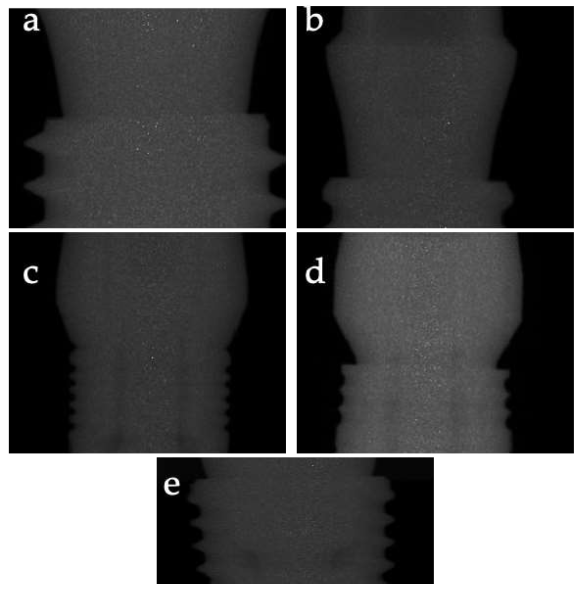
The present investigation involved a comparative micro-CT analysis of five different dental implant types to evaluate the coupling between implant fixtures and prosthetic abutments under various mechanical conditions. The implants underwent non-destructive double scans to limit image noise and artifacts, enabling an in-depth structural and interfacial integrity evaluation.

2.4.1. Sample

Different fixtures with coupling with prosthetic abutments and healing abutments were evaluated. In particular, the sample comprises five different implants (1 sample for each type, after a comparative visive analysis with an optic microscope from 2 samples for each type) with respective prosthetic and healing abutments. Since the investigation is not destructive, a double scan was carried out on each sample to limit image problems (noise disturbance or micro-movements). All dental implants showed internal connections.
The dental implants were the following:
  • OsstemImplant (Seoul, Republic of Korea); TSIII, Regular connection;
  • Megagen Implant Co., Ltd. (Seoul, Republic of Korea); AnyOne, Regular connection;
  • Schütz Dental GmbH (Rosbach, Germany); Diagram, Regular connection;
  • FDS76 (Reggio Calabria, Italy); K2 Regular connection;
  • FDS76 (Reggio Calabria, Italy); V4 Regular connection.

2.4.2. Micro-CT

The dental implants were evaluated using µCT (SkyScan1174; Bruker; Kontich, Belgium). They were scanned parallel to the sagittal and coronal planes.
The scanning parameters were set as follows: camera pixel size (15.45 µm), tube voltage (50 kV), tube current (800 µA), image pixel size (11.1 µm), exposure (500 ms), rotation step (0.8°), 180° tomographic rotation with random movement, and filter (Al 1 mm).
Image reconstruction was performed by using NRecon software version 2.0 (Bruker; Kontich, Belgium), through Hamming-filtered back projection, with the following parameters: smoothing (4); smoothing kernel (2—Gaussian); ring artifact correction (4); beam hardening correction (41%); grey thresholds (82–255).
µCT images were resliced parallel to the axial plane of the dental implant, and the microarchitecture analysis was performed on consecutive slices or with multiplanar image reconstructions.

3. Results

3.1. Study Selection

The search identified 256 records. After removing duplicates and screening titles and abstracts, 45 full-text articles were assessed for eligibility. Eight studies met the inclusion criteria and were included in the review. The flow diagram summarizes the selection process (Table 1).

3.2. Study Characteristics

The included studies varied in their objectives, methods, and outcomes. Yu et al. [11] investigated the effect of fluoride exposure on osseointegration in rabbits. Min et al. [12] analyzed metal artifacts in Cone Beam Computed Tomography (CBCT) images using micro-CT as a reference. González-García et al. [13] assessed the reliability of CBCT for determining bone density at implant sites. Neldam et al. [14] used synchrotron micro-CT to evaluate osseointegration. Kapishnikov et al. [15] examined the microgap at the implant-sleeve connection under mechanical loading. Hsu et al. [16] studied the effects of bone stiffness and bone–implant contact (BIC) on implant stability. Parsa et al. [17] evaluated bone quality by using multiple CT modalities. Bissinger et al. [18] compared 3D micro-CT and 2D histomorphometry for assessing osseointegration.

3.3. Risk of Bias in Studies

Most studies were assessed for low risk of bias. The primary sources of bias included a lack of blinding and small sample sizes. Table 2 details the risk of bias assessments.

3.4. Results of Individual Studies

  • Yu et al. [11] found that fluoride exposure significantly reduced bone mineral density (BMD) and BIC ratios in rabbits;
  • Min et al. [12] reported that metal artifacts in CBCT images could be effectively quantified using micro-CT as a reference;
  • González-García et al. [13] demonstrated a strong correlation between radiographic bone density from CBCT and bone volumetric fraction from micro-CT;
  • Neldam et al. [14] showed that synchrotron micro-CT provides high-resolution images that accurately depict bone microarchitecture;
  • Kapishnikov et al. [15] observed that static compressive loads significantly increased the microgap size at the implant–sleeve connection;
  • Hsu et al. [16] found a positive correlation between bone stiffness, BIC, and initial implant stability;
  • Parsa et al. [17] reported strong correlations between bone volume fraction and bone density;
  • Bissinger et al. [18] found strong correlations between 3D micro-CT and 2D histomorphometry in evaluating osseointegration (Table 3).

3.5. Results of Syntheses

Due to heterogeneity, a meta-analysis was not feasible for all outcomes. A narrative synthesis indicated that micro-CT is reliable for assessing bone quality and implant stability. Variations in study design and conditions (e.g., fluoride exposure and mechanical loading) affected the outcomes, highlighting the importance of standardized protocols in future research.
Yu et al. [11]: In rabbit models, the study used micro-CT to evaluate the bone mineral density (BMD) and bone–implant contact (BIC) ratios. The abstract did not detail specific settings such as voxel size, scanning parameters, or reconstruction algorithms. However, micro-CT images were used to illustrate the differences in BMD between the fluoride exposure group and the control group over time. Min et al. [12]: This study exploited micro-CT to measure six trabecular microstructural parameters: trabecular thickness (TbTh), trabecular spacing (TbSp), bone volume per total volume (BV/TV), bone surface per total volume (BS/TV), connectivity density (CD), and fractal dimension (FD). Micro-CT measurements were used as the gold standard for comparing CBCT images. The specific micro-CT settings, such as resolution or scan times, were not provided in the abstract. González-García et al. [13]: micro-CT was used to assess the bone volumetric fraction (BV/TV) and other osseous micro-structural variables of bone biopsies collected from implant sites in the maxillary bones. The micro-CT settings included the parameters necessary to evaluate trabecular thickness (Tb.Th), trabecular number (Tb.N), and trabecular separation (Tb.Sp). The abstract did not provide specific details on voxel size or scanning parameters.
Neldam et al. [14]: This study employed high-resolution synchrotron radiation micro-CT (SRmicro-CT), considered the gold standard for evaluating bone microarchitecture. The settings included a voxel size of 5 μm, which provided high-resolution images with excellent contrast and signal-to-noise ratio. The study assessed the peri-implant bone volume fraction at various distances from the implant surface in a goat mandible model. Kapishnikov et al. [15]: Micro-CT was used to determine the microgap at the implant–sleeve connection of a novel two-piece dental implant under different mechanical conditions. The abstract did not detail the specific settings, such as voxel size or scanning parameters. The study measured the mean gap differences under cyclic and static compressive loads. Hsu et al. [16]: This study utilized micro-CT to calculate the three-dimensional bone-to-implant contact ratio (BIC%) and assess the dental implant’s initial stability. The settings included the necessary parameters to evaluate the elastic modulus and trabecular structure of artificial sawbone models. The abstract did not provide specific details on resolution or other scanning settings. Parsa A et al. [17]: Micro-CT was used to evaluate the bone volume fraction (BV/TV) and trabecular bone density in human mandibular cadavers. The specific settings included the use of the SkyScan 1173 micro-CT system.
Image analysis was performed using software tools like Amira, 3Diagnosis, Geomagic, and CTAn. The abstract did not provide specific details on voxel size or scan parameters. Bissinger et al. [18]: The study compared 3D micro-CT with 2D histomorphometry to evaluate dental implant osseointegration in the maxilla of minipigs. The micro-CT settings included a voxel size of 10 μm. Measurements of the bone implant contact (BIC), inner ring, and outer ring distances were used to correlate with histomorphometry values. The settings allowed for high-resolution imaging to accurately depict the bone microarchitecture around the implants. Yu et al. [11] reported that micro-computed tomography (micro-CT) measurements reveal a range of bone–implant contact (BIC) ratios from 40% to 80%, with higher values indicating enhanced osseointegration, particularly under optimized implant surface treatments and controlled loading conditions.
Similarly, Min et al. [12] demonstrated that bone volume per total volume (BV/TV), a critical indicator of bone density surrounding the implant, can vary significantly between 20% and 60%, depending on bone quality and regional bone density. Kapishnikov et al. [15] further highlighted micro-CT’s precision in detecting structural variations, showing that it can measure microgaps at the implant–abutment interface with an accuracy down to 0.3 ± 0.15 µm under compressive loads, thereby emphasizing differences linked to implant design and mechanical stability. Micro-CT’s high resolution, typically ranging between 5 and 10 µm in voxel size, allows it to capture these fine structural details with exceptional clarity and accuracy. This precision surpasses conventional imaging techniques, as demonstrated in comparative studies, where micro-CT provided a 10- to 20-fold improvement in detail. These findings underscore micro-CT’s potential as an advanced and reliable imaging tool for detailed evaluation of osseointegration, implant stability, and microstructural integrity in dental implantology.

3.6. Certainty of Evidence

The overall certainty of the evidence was moderate to high, with consistent findings across studies supporting the use of micro-CT in dental implant research. Further research with larger sample sizes and standardized methodologies is needed to confirm these findings.

3.7. Additional Results

A preliminary qualitative analysis of the micro-CT images was conducted, focusing on evaluating dental implant connections. The analysis revealed no significant differences or issues in the coupling between the implant fixture and the prosthetic abutment across the samples examined. The micro-CT images showed a consistent and secure interface between the implant components, with no apparent microgaps, deformations, or mechanical failures (Figure 1). These findings suggest that the studied implant designs are structurally sound and capable of providing stable connections, which is essential for the long-term success of dental implants.

4. Discussion

Micro-computed tomography has emerged as a pivotal tool for evaluating and analyzing dental implants due to its non-destructive nature and ability to produce high-resolution three-dimensional images of complex internal structures. In dental implantology, micro-CT allows for the detailed assessment of the bone–implant interface, osseointegration, and the mechanical integrity of implant connections [19]. This imaging technique provides invaluable insights into dental implants’ structural adaptations and potential failures, which are critical for optimizing implant design and improving patient outcomes. The importance of such studies lies in their ability to inform clinicians and researchers about the microstructural interactions between the implant and surrounding bone, which directly impact the longevity and success of dental implants.
Micro-CT can evaluate the bone quality and density in potential implant sites, which is crucial for determining site suitability for implant placement and planning the appropriate size and type of implant [20]. By providing detailed images of the bone structure, micro-CT helps clinicians assess whether sufficient bone volume and density might support an implant, thereby reducing the risk of implant failure. Moreover, micro-CT is used to analyze implant materials’ microstructure and interactions with biological tissues in developing new dental implants. Researchers need to examine the surface topography of implants with different surface treatments and coatings affecting implant integration in the surrounding bone to optimize implant design for better osseointegration [10].
In this context, micro-CT is a nondestructive, valuable tool for investigating the underlying causes of failure. By examining the implant and surrounding bone in detail, clinicians can identify factors such as poor osseointegration, peri-implantitis (inflammation of the tissue around the implant), or mechanical failures. This information is invaluable for improving clinical practice and implant design to prevent future failures.
Micro-CT is also used to evaluate bone regeneration in grafted areas before implant placement. The detailed images of the grafted site allow for the assessment of new bone formation and integration with the native bone. This is critical for ensuring that the grafted site is suitable for supporting a dental implant [21].
For research purposes, micro-CT may provide important information regarding the properties of new biomaterials that could be used in implants and bone grafts. By analyzing the internal structure and porosity, researchers can optimize their properties to enhance their performance in clinical applications. Yu et al. [11] conducted a study to investigate the influence of fluoride exposure on the osseointegration of titanium dental implants in rabbits. The study revealed that fluoride exposure significantly reduced bone mineral density (BMD) and bone–implant contact (BIC) ratios, particularly in the later stages of osseointegration. These data suggest that high fluoride levels negatively affect the quality of bone surrounding implants and hinder successful bone integration. Min et al. [12] quantitatively analyzed metal artifacts in dental implant CBCT images using correlation with micro-CT. The study assessed various microstructural parameters and found that metal artifacts could be effectively quantified. The subject’s angular position influenced the correlation of these parameters, thus highlighting the importance of imaging angle in improving CBCT image quality. González-García et al. [13] evaluated the reliability of cone-beam computed tomography (CBCT) for assessing bone density at dental implant sites. The study found a strong positive correlation between radiographic bone density (RBD) from CBCT and bone volumetric fraction (BV/TV) from micro-CT. This evidence indicates that CBCT may be considered a reliable pre-operative tool for determining bone density and planning dental implants. Neldam et al. [14] described a refined method using high-resolution synchrotron radiation micro-CT (SRmicro-CT) to evaluate osseointegration and peri-implant bone volume fraction after titanium dental implant insertion. The study demonstrated that SRmicro-CT provides high-resolution images that accurately depict bone microarchitecture and bone-to-implant contact, offering more precise measurements than traditional methods. Kapishnikov et al. [15] investigated the microgap at the implant–sleeve connection of a novel two-piece dental implant through micro-CT use. The study assessed the microgap under various mechanical conditions, and an increase in the microgap size, particularly in the direction of force application, was observed following static compressive loads, thus highlighting the importance of considering mechanical behavior in implant design to ensure stability.
Hsu et al. [16] examined the effects of bone stiffness and three-dimensional bone-to-implant contact (BIC) on the primary stability of dental implants. The results obtained following micro-CT and resonance frequency analysis demonstrated that primary implant stability is positively correlated with the elasticity of cancellous bone and the 3D BIC ratio and suggest that bone stiffness and BIC are critical factors in achieving initial implant stability. Parsa et al. [17] analyzed the correlation between bone volume fraction (BV/TV) and radiographic bone density (Hounsfield units) by using multislice CT (MSCT), micro-CT, and cone-beam CT (CBCT). The study found strong correlations between CBCT and MSCT density and between CBCT and micro-CT BV/TV measurements. It supported the hypothesis that CBCT is reliable for assessing bone quality at implant sites. Bissinger et al. [18] compared 3D micro-CT and 2D histomorphometry to analyze dental implant osseointegration in the maxilla of minipigs. The study found strong correlations between micro-CT and histomorphometry in evaluating bone–implant contact (BIC) and peri-implant bone volume. Micro-CT’s non-destructive nature makes this technique advantageous for assessing 3D BIC, providing a comprehensive understanding of implant integration [22,23].
The results from this preliminary analysis underscore the importance of using advanced imaging techniques like micro-CT in dental research. As shown in Figure 1 (detail of the abutment implant connection), the connection appears intact and coincident. The internal connection screw seems to be ideally housed in all examples; the coils of the latter create retention on the internal connection of the implant without creating interference or damage to the latter. It is necessary to report that the connection screw was tightened with the torque parameters required by the company, and this occurred without friction or problems. In analyzing all the micro-TC cuts of which this image is only a significant extract, in all the examples shown, no solution of continuity was seen between the “intra-connection” space and the external space. This should testify to the total absence of a possibility of communication and passage of liquids or microorganisms between the oral environment and the inside of the fixture [24]. Different studies have shown how the implant connection, mainly the flat-to-flat one, can create deformations under load, which lead to the opening (creation of space) of the connection itself [25,26]. From this point of view, it would be fascinating to continue investigating. By providing detailed visualizations of the internal structures of dental implants, micro-CT enables a more comprehensive understanding of implant behavior in vivo [27]. This kind of study is crucial for validating the mechanical stability of current implant designs and guiding the development of next-generation implants that can improve performance and longevity. The ability to detect and analyze subtle structural variations before they manifest as clinical failures represents a significant advantage, thus allowing for early interventions and adjustments in clinical practice. Ultimately, these studies may improve patient care by ensuring dental implants are safe and effective over the long term.

Limitations

The limitations of this study and the use of micro-computed tomography (micro-CT) in dental research include several noteworthy aspects. First, the relatively small sample size restricts the generalizability of the findings, as a broader range of implant types and patient conditions would be needed to confirm the applicability of these results across various clinical settings. Furthermore, the high cost of micro-CT technology limits its availability to specialized research centers, as the equipment and technical expertise required make it financially and logistically challenging for widespread clinical use. This restriction also impacts routine application, as micro-CT’s accessibility remains limited compared to other imaging modalities in dental clinics. Additionally, while micro-CT provides non-destructive imaging, the higher levels of radiation involved, especially for the high-resolution scans necessary for detailed bone and implant interface analysis, present challenges in in vivo applications where cumulative radiation exposure could become a concern. Another significant drawback is the susceptibility to metal artifacts, as metallic components in dental implants can distort micro-CT images, particularly at the critical bone–implant interface. This leads to potential inaccuracies in assessing bone volume and bone–implant contact. This issue of artifacts can affect data reliability and interpretation accuracy, particularly when precise measurements are essential. Micro-CT’s high-resolution capabilities, while beneficial for in vitro or ex vivo studies, are less practical in routine clinical applications, where alternatives like cone-beam computed tomography (CBCT) are often preferred due to their lower cost, reduced radiation exposure, and more streamlined integration into clinical workflows despite their comparatively lower resolution. Micro-CT imaging and subsequent data reconstruction are highly time-consuming, requiring advanced processing software and technical expertise. This further complicates its use in routine diagnostics where rapid imaging results are often essential. These limitations indicate that, although micro-CT offers unparalleled imaging resolution for detailed dental research, significant barriers related to cost, practicality, and clinical utility must be addressed before it can achieve broader acceptance and application in research and clinical practice.

5. Conclusions

Micro-CT is a powerful tool in dentistry. It offers detailed, high-resolution, three-dimensional images essential for diagnosing, planning treatment, and evaluating dental implants. This study reinforces the utility of micro-computed tomography (micro-CT) as a precise imaging tool for assessing dental implants’ microstructural properties, including bone–implant contact, implant stability, and interface quality. Future studies should focus on expanding sample sizes and establishing standardized imaging protocols to enhance data reliability. Additionally, technological advancements that address metal artifact reduction and radiation dose optimization could make micro-CT more practical and broaden its application in clinical settings.

Author Contributions

Conceptualization, L.F. and T.D.; methodology, N.I., T.D. and L.F.; software, N.I., T.D. and L.F.; validation, G.R. and G.C.; formal analysis, N.I., T.D. and L.F.; investigation, N.I., T.D. and L.F.; resources, G.R. and N.I.; data curation, T.D.; writing—original draft preparation, L.F.; writing—review and editing, T.D., F.G. and N.I.; supervision, G.C.; visualization, P.L.G.; project administration, G.R. All authors have read and agreed to the published version of the manuscript.

Funding

This research received no external funding.

Institutional Review Board Statement

Not applicable.

Informed Consent Statement

Not applicable.

Data Availability Statement

Data are available on request from the corresponding author.

Conflicts of Interest

The authors declare no conflicts of interest.

References

  1. Hipsley, C.A.; Aguilar, R.; Black, J.R.; Hocknull, S.A. High-throughput microCT scanning of small specimens: Preparation, packing, parameters and post-processing. Sci. Rep. 2020, 10, 13863. [Google Scholar] [CrossRef]
  2. Batista, R.; Moreira, A.; Oliveira, S.J.; Mesquita, P.; Sampaio-Fernandes, J.; Figueiral, M.H. Deformation of implant retaining screws-Study with stereoscopic microscopy and microCT. J. Esthet. Restor. Dent. 2022, 34, 1147–1155. [Google Scholar] [CrossRef] [PubMed]
  3. Roatesi, I.; Roatesi, S. Modeling of Dental Implant Osseointegration Progress by Three-Dimensional Finite Element Method. Appl. Sci. 2020, 10, 5561. [Google Scholar] [CrossRef]
  4. Brånemark, P.I. Osseointegration and its experimental background. J. Prosthet. Dent. 1983, 50, 399–410. [Google Scholar] [CrossRef]
  5. Romanos, G.E.; Schesni, A.; Nentwig, G.-H.; Winter, A.; Sader, R.; Brandt, S. Impact of Implant Diameter on Success and Survival of Dental Implants: An Observational Cohort Study. Prosthesis 2023, 5, 888–897. [Google Scholar] [CrossRef]
  6. Shopova, D.; Mihaylova, A.; Yaneva, A.; Bakova, D.; Dimova-Gabrovska, M. Biofabrication Approaches for Peri-Implantitis Tissue Regeneration: A Focus on Bioprinting Methods. Prosthesis 2024, 6, 372–392. [Google Scholar] [CrossRef]
  7. Mannino, F.; D’Angelo, T.; Pallio, G.; Ieni, A.; Pirrotta, I.; Giorgi, D.A.; Scarfone, A.; Mazziotti, S.; Booz, C.; Bitto, A.; et al. The Nutraceutical Genistein-Lycopene Combination Improves Bone Damage Induced by Glucocorticoids by Stimulating the Osteoblast Formation Process. Nutrients 2022, 14, 4296. [Google Scholar] [CrossRef]
  8. Matos, J.D.M.d.; Queiroz, D.A.; Nakano, L.J.N.; Andrade, V.C.; Ribeiro, N.d.C.R.; Borges, A.L.S.; Bottino, M.A.; Lopes, G.d.R.S. Bioengineering Tools Applied to Dentistry: Validation Methods for In Vitro and In Silico Analysis. Dent. J. 2022, 10, 145. [Google Scholar] [CrossRef]
  9. Zangooei, M.H.; Margolis, R.; Hoyt, K. Multiscale computational modeling of cancer growth using features derived from microCT images. Sci. Rep. 2021, 11, 18524. [Google Scholar] [CrossRef]
  10. Ghavami-Lahiji, M.; Davalloo, R.T.; Tajziehchi, G.; Shams, P. Micro-computed tomography in preventive and restorative dental research: A review. Imaging Sci. Dent. 2021, 51, 341–350. [Google Scholar] [CrossRef]
  11. Yu, Y.J.; Zhu, W.Q.; Xu, L.N.; Ming, P.P.; Shao, S.Y.; Qiu, J. Osseointegration of titanium dental implant under fluoride exposure in rabbits: Micro-CT and histomorphometry study. Clin. Oral Implant. Res. 2019, 30, 1038–1048. [Google Scholar] [CrossRef] [PubMed]
  12. Min, C.K.; Kim, K.A. Quantitative analysis of metal artefacts of dental implant in CBCT image by correlation analysis to micro-CT: A microstructural study. Dentomaxillofacial Radiol. 2021, 50, 20200365. [Google Scholar] [CrossRef] [PubMed]
  13. González-García, R.; Monje, F. The reliability of cone-beam computed tomography to assess bone density at dental implant recipient sites: A histomorphometric analysis by micro-CT. Clin. Oral Implant. Res. 2012, 24, 871–879. [Google Scholar] [CrossRef] [PubMed]
  14. Neldam, C.A.; Lauridsen, T.; Rack, A.; Lefolii, T.T.; Jørgensen, N.R.; Feidenhans’l, R.; Pinholt, E.M. Application of high resolution synchrotron micro-CT radiation in dental implant osseointegration. J. Cranio-Maxillofac. Surg. 2015, 43, 682–687. [Google Scholar] [CrossRef] [PubMed]
  15. Kapishnikov, S.; Gadyukov, A.; Chaushu, G.; Chaushu, L. Micro-CT Analysis of Microgap at a Novel Two-Piece Dental Implant Comprising a Replaceable Sleeve In Vitro. Int. J. Oral Maxillofac. Implant. 2021, 36, 451–459. [Google Scholar] [CrossRef]
  16. Hsu, J.T.; Huang, H.L.; Tsai, M.T.; Wu, A.Y.; Tu, M.G.; Fuh, L.J. Effects of the 3D bone-to-implant contact and bone stiffness on the initial stability of a dental implant: Micro-CT and resonance frequency analyses. Int. J. Oral Maxillofac. Surg. 2013, 42, 276–280. [Google Scholar] [CrossRef]
  17. Parsa, A.; Ibrahim, N.; Hassan, B.; van der Stelt, P.; Wismeijer, D. Bone quality evaluation at dental implant site using multislice CT, micro-CT, and cone beam CT. Clin. Oral Implant. Res. 2015, 26, e1–e7. [Google Scholar] [CrossRef]
  18. Bissinger, O.; Probst, F.A.; Wolff, K.D.; Jeschke, A.; Weitz, J.; Deppe, H.; Kolk, A. Comparative 3D micro-CT and 2D histomorphometry analysis of dental implant osseointegration in the maxilla of minipigs. J. Clin. Periodontol. 2017, 44, 418–427. [Google Scholar] [CrossRef]
  19. Vinhas, A.S.; Salazar, F.; Mendes, J.M.; Silva, A.S.; Ríos-Carrasco, B.; Ríos-Santos, J.V.; Gil, J.; Herrero-Climent, M.; Aroso, C. SEM Analysis and Micro-CT Evaluation of Four Dental Implants after Three Different Mechanical Requests—In Vitro Study. Materials 2024, 17, 434. [Google Scholar] [CrossRef]
  20. Kowalski, J.; Puszkarz, A.K.; Radwanski, M.; Sokolowski, J.; Cichomski, M.; Bourgi, R.; Hardan, L.; Sauro, S.; Lukomska-Szymanska, M. Micro-CT Evaluation of Microgaps at Implant-Abutment Connection. Materials 2023, 16, 4491. [Google Scholar] [CrossRef]
  21. Takechi, M.; Ishioka, Y.; Ninomiya, Y.; Ono, S.; Tada, M.; Nakagawa, T.; Sasaki, K.; Murodumi, H.; Shigeishi, H.; Ohta, K. Morphological Evaluation of Bone by CT to Determine Primary Stability—Clinical Study. Materials 2020, 13, 2605. [Google Scholar] [CrossRef] [PubMed]
  22. Fiorillo, L.; D’Amico, C.; Ronsivalle, V.; Cicciù, M.; Cervino, G. Single Dental Implant Restoration: Cemented or Screw-Retained? A Systematic Review of Multi-Factor Randomized Clinical Trials. Prosthesis 2024, 6, 871–886. [Google Scholar] [CrossRef]
  23. Capparé, P.; Vinci, R.; Di Stefano, D.A.; Traini, T.; Pantaleo, G.; Gherlone, E.F.; Gastaldi, G. Correlation between Initial BIC and the Insertion Torque/Depth Integral Recorded with an Instantaneous Torque-Measuring Implant Motor: An in vivo Study. Clin. Implant. Dent. Relat. Res. 2015, 17, e613–e620. [Google Scholar] [CrossRef] [PubMed]
  24. Berberi, A.; Dagher, M.; Kanj, W.; Amine, E.; Aramouni, P. Micromovement Evaluation of Original and Compatible Abutments at the Implant–abutment Interface. J. Contemp. Dent. Pract. 2016, 17, 907–913. [Google Scholar] [CrossRef] [PubMed]
  25. Camps-Font, O.; Rubianes-Porta, L.; Valmaseda-Castellón, E.; Jung, R.E.; Gay-Escoda, C.; Figueiredo, R. Comparison of external, internal flat-to-flat, and conical implant abutment connections for implant-supported prostheses: A systematic review and network meta-analysis of randomized clinical trials. J. Prosthet. Dent. 2023, 130, 327–340. [Google Scholar] [CrossRef]
  26. Cicciù, M.; Bramanti, E.; Cecchetti, F.; Scappaticci, L.; Guglielmino, E.; Risitano, G. FEM and Von Mises analyses of different dental implant shapes for masticatory loading distribution. Oral Implantol. 2014, 7, 1–10. [Google Scholar]
  27. Lo Russo, L.; Guida, L.; Mariani, P.; Ronsivalle, V.; Gallo, C.; Cicciù, M.; Laino, L. Effect of Fabrication Technology on the Accuracy of Surgical Guides for Dental-Implant Surgery. Bioengineering 2023, 10, 875. [Google Scholar] [CrossRef]
Figure 1. Dental implant connection details. (a) type 1, (b) type 2, (c) type 3, (d) type 4, (e) type 5.
Prosthesis 06 00104 g001
Table 1. Manuscript selection flow chart.
StageDescription
IdentificationRecords identified through database searching (n = 256)
ScreeningRecords after duplicates removed (n = 200)
EligibilityFull-text articles assessed for eligibility (n = 45)
EligibilityFull-text articles excluded (n = 37)
IncludedStudies included in qualitative synthesis (n = 8)
Table 2. Risk of bias table.
StudyRandom Sequence GenerationAllocation ConcealmentBlinding of Participants and PersonnelBlinding of Outcome AssessmentIncomplete Outcome DataSelective ReportingOther Bias
Yu YJ et al. (2019) [11]LowLowUnclearUnclearLowLowLow
Min CK et al. (2021) [12]UnclearUnclearUnclearLowLowLowLow
González-García R et al. (2013) [13]LowLowUnclearUnclearLowLowLow
Neldam CA et al. (2015) [14]LowLowLowLowLowLowLow
Kapishnikov S et al. (2021) [15]LowLowLowLowLowLowLow
Hsu JT et al. (2013) [16]UnclearUnclearUnclearUnclearLowLowLow
Parsa et al. (2013) [17]LowLowLowLowLowLowLow
Bissinger O et al. (2017) [18]LowLowLowLowLowLowLow
Table 3. Main results data.
Author and YearType of StudySample Size and TypeMain ResultsStatistical Results
Yu YJ et al. (2019) [11]Animal Study24 rabbitsFluoride exposure significantly reduced BMD and BIC ratios in rabbits.Bone volume around the implants increased in a time-dependent manner in both groups.
Min CK et al. (2021) [12]Experimental StudyPolyurethane synthetic bone blocksMetal artifacts in CBCT images quantified effectively using micro-CT.Spearman correlation coefficients for microstructural parameters varied with alpha angle changes.
González-García R et al. (2013) [13]Observational Study39 bone biopsies from 31 patientsStrong correlation between RBD from CBCT and Bone Volume/Total Volume (BV/TV) from micro-CT.Pearson’s correlation coefficient (r = 0.858, p < 0.001) between RBD and BV/TV.
Neldam CA et al. (2015) [14]Experimental StudyGoat mandible modelHigh-resolution synchrotron micro-CT accurately depicts bone microarchitecture.Peri-implant bone volume fraction increased to 50% and leveled out at 80% at 400 μm distance.
Kapishnikov S et al. (2021) [15]Experimental StudyIn vitroStatic compressive loads significantly increased the microgap size at the implant–sleeve connection.Mean gap difference after cyclic compressive load was 0.3 ± 0.15 μm.
Hsu JT et al. (2013) [16]Experimental StudyArtificial sawbone modelsPositive correlation between bone stiffness, BIC, and initial implant stability.Regression correlation coefficient was 0.96 for correlations of ISQ with elasticity of cancellous bone and 3D BIC%.
Parsa A et al. (2013) [17]Observational Study20 human mandibular cadaversStrong correlations between CBCT and Multislice Computed Tomography (MSCT) density, and CBCT and micro-CT BV/TV measurements.Excellent correlation observed between MSCT Hounsifield Unit (HU) and micro-CT BV/TV (r = 0.91).
Bissinger O et al. (2017) [18]Animal Study54 implants in 14 minipigsStrong correlations between 3D micro-CT and 2D histomorphometry in evaluating osseointegration.Strong correlations (p < 0.0001) for BIC, inner ring, and outer ring between micro-CT and histomorphometry.
Disclaimer/Publisher’s Note: The statements, opinions and data contained in all publications are solely those of the individual author(s) and contributor(s) and not of MDPI and/or the editor(s). MDPI and/or the editor(s) disclaim responsibility for any injury to people or property resulting from any ideas, methods, instructions or products referred to in the content.

