Review

Infective Endocarditis After Transcatheter Aortic Valve Replacement: A Narrative Review

Department of Cardiac Surgery, Centre Cardiologique du Nord, 93200 Saint-Denis, France
Prosthesis 2024, 6(6), 1529-1552; https://doi.org/10.3390/prosthesis6060110 (registering DOI)
Submission received: 24 September 2024 / Revised: 28 November 2024 / Accepted: 4 December 2024 / Published: 12 December 2024

Abstract

:
Prosthetic valve endocarditis (PVE) has undergone significant changes over the past five decades and is currently affecting an aging population, with an increasing prevalence in patients with transcatheter valve implants. The introduction of transcatheter aortic valve replacement (TAVR) represents a significant advance in the field of interventional cardiology and cardiac surgery. The incidence of IE after TAVR has remained stable, with rates similar to those reported after surgical aortic valve replacement. This is despite significant refinements in the TAVR procedure, with less invasive handling and its extension to younger and healthier patients. TAVR should be considered as a potential treatment option for patients with PVE, despite some differences. In terms of evolutionary advances, there have been notable and significant developments in the fields of microbiology and imaging diagnostics. The 2023 Duke-International Society for Cardiovascular Infectious Diseases diagnostic criteria for infective endocarditis now incorporate significant advances in molecular biology and fluorine-18 fluorodeoxyglucose positron emission tomography/computed tomography. This has led to a significant enhancement in diagnostic sensitivity for PVE while maintaining the same level of specificity in validation studies. PVE is a deadly disease. A multidisciplinary endocarditis treatment team in a cardiac center is essential to improve outcomes. The availability of novel surgical options allows clinicians to offer an increasing number of patients the opportunity to avoid surgical intervention. Some patients will complete antimicrobial treatment at home. Those with prosthetic valves are eligible for antibiotic prophylaxis before dental procedures. Post-TAVR infective endocarditis (IE) is a subcategory of prosthetic valve endocarditis. This condition presents a particularly complex scenario, characterized by a distinctive clinical and microbiological profile, a high prevalence of IE-related complications, an ambiguous role of cardiac surgery, and a poor prognosis for the majority of patients with TAVR IE. The number of TAVR procedures is set to skyrocket in the coming years, which will undoubtedly lead to a significant rise in the number of people at risk of this life-threatening complication. This review will provide an overview of this rare complication in light of the advent of IE following TAVR. It is crucial to gain a comprehensive understanding of the disease and its associated complications to enhance clinical outcomes.

1. Introduction

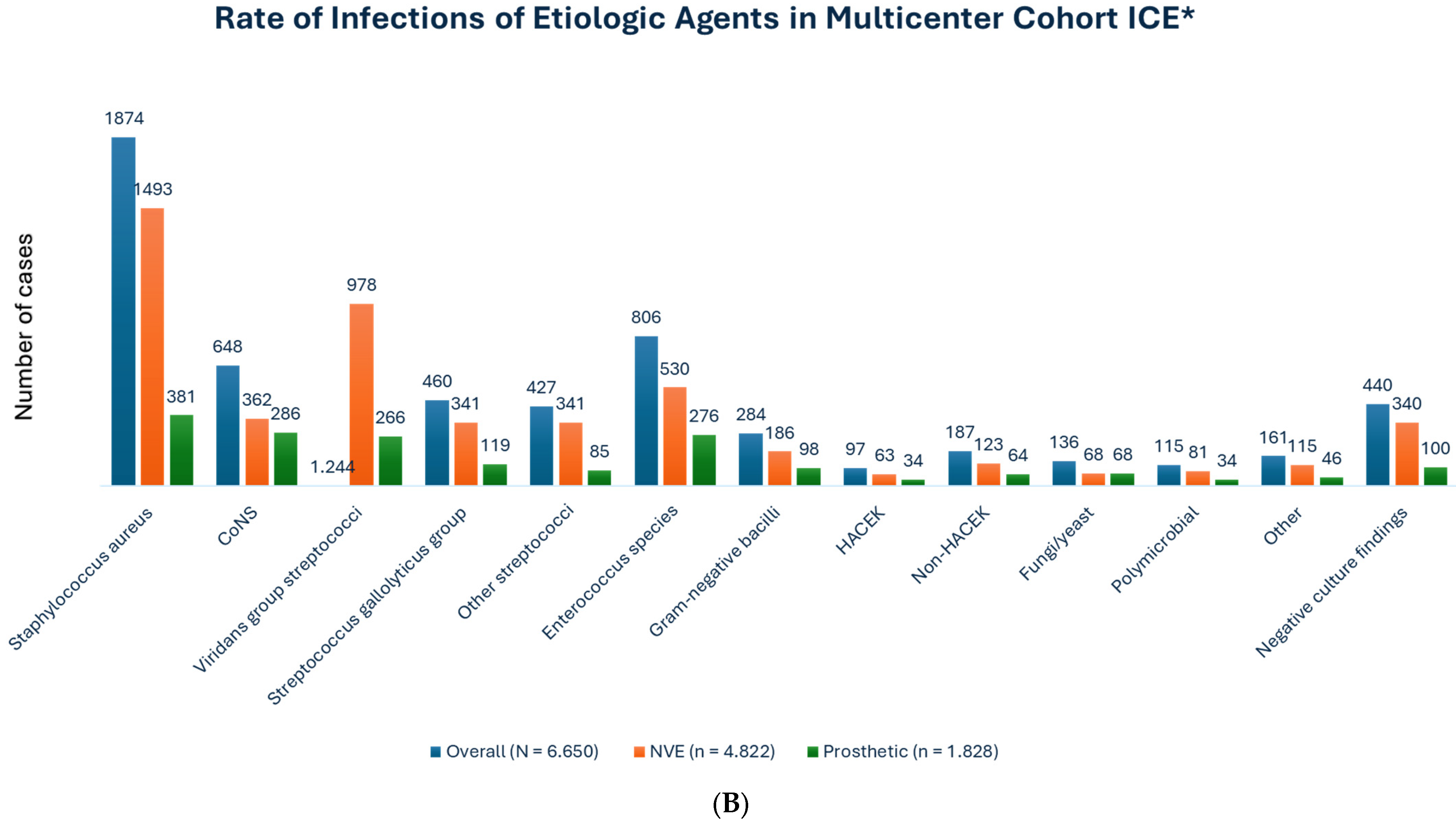
Infective endocarditis (IE) in the postoperative period, occurring as a complication of valve replacement surgery, has an incidence rate ranging from 1 to 6 percent. This complication is significantly associated with elevated mortality [1,2,3,4,5]. Transcatheter aortic valve replacement (TAVR) represents a promising therapeutic avenue for patients with severe aortic valve stenosis (AVS) deemed to be at low, intermediate, and elevated or prohibitive surgical risk, for whom the standard surgical approach is contraindicated according to the latest international guidelines [6,7,8,9,10,11,12,13]. The documented incidence of IE within the year following TAVR ranges from 0.5% to 3.1%. A reported incidence of 1.5% has been documented. There is a higher prevalence of Enterococcus faecalis-localized pathogens, which often necessitates explantation of the infected device and appropriate treatment (Figure 1) [12,14]. This is comparable to the rates of infective endocarditis observed after standard surgical valve replacement [15,16]. It is possible that increased familiarity with surgical techniques, greater age, and the presence of foreign materials (e.g., stent struts) within the composition of transcatheter valve prostheses may have collectively elevated the risk of infectious endocarditis in patients subjected to this treatment. The existing data on infective endocarditis subsequent to TAVR are limited to case reports and relatively small series with restricted follow-up [12,14,17,18,19], which demonstrate in-hospital complication and mortality rates as high as 87% and 47%, respectively. Valve explant rates in these patients remain relatively low, at less than 10%. It is consequently pertinent to undertake a further examination of the factors associated with infective endocarditis subsequent to TAVR, along with an investigation into the clinical characteristics and outcomes of such instances. In view of the emergence of IE subsequent to TAVR in this context, the present study aims to present a comprehensive description of this rare complication. It is therefore crucial to gain a detailed understanding of the illness and its associated complications in order to improve the quality of treatment and increase the likelihood of positive clinical outcomes.
In recent years, there has been a notable shift in the clinical profile of patients undergoing TAVR. Initially, these procedures were performed on patients with prohibitive or high surgical risk. However, there has been a significant transition towards less complex and younger patients with lower surgical risk. In a similar manner, TAVR has gradually transitioned towards less invasive and minimalist procedures, which have the potential to reduce the risk of bacteremia during the early post-procedural period and subsequently the risk of IE.
As a consequence of this sustained advancement, the principal attributes of patients who develop IE subsequent to TAVR are also undergoing modification. The objective of this review is to present the most recent evidence-based insights into the epidemiology, clinical features, diagnosis, management, outcomes, and prevention of this entity, encompassing the latest research and future projections on this subject area that is undergoing rapid evolution.

2. IE in the 21st Century: An Ongoing Evolutionary Challenge

The 21st century has seen IE evolve further still. It is now acquired in over 25% of cases [20,21]. At the same time, advances in cardiology have led to significant changes in the patient profile and the symptoms exhibited by those affected. The advent of cardiac implantable electronic devices (CIEDs) has coincided with a notable increase in the incidence of IE associated with complex devices [22]. Similarly, TAVR has emerged as a transformative approach to the management of valvular heart disease, with higher rates of IE compared to surgically implanted prosthetic valves [14,23,24]. The key challenges posed by contemporary IE in developed countries were identified, as well as the underlying factors preventing the full impact of diagnostic and treatment advances on the disease. The latest data definitively show the impact of revised antibiotic prophylaxis guidelines [25,26,27,28] and the current status of molecular and imaging diagnostic strategies [3,29,30,31,32,33,34,35,36,37].
Given the lack of significant advancement in the prognostic outlook for IE, there is a clear need to focus on enhancing service provision and surgical outcomes. Furthermore, data on IE in three patient populations are of critical interest for investigation, in order to reflect the ongoing evolution of the disease. These patient cohorts represent some of the most challenging cases, including those with TAVR endocarditis [12,14,17,18,19,23,24], those with stroke [3,38,39,40,41,42,43], and those with CIED infection [44,45,46,47,48]. Further examination is required to gain a deeper understanding of the interconnectivity between these three discrete populations.

The Clinical Problem: Insight into Epidemiology, Pathophysiology, Clinical Future, and Microbiological Characteristics

A bacterial infection of a TAVR device is a distinct possibility. This process is facilitated by the material that makes up the device, which causes platelet aggregation. The material is present in the stent, prosthetic leaflets, and other parts of the device. Fibrin adheres to the device, forming a microthrombotic lesion called a bacterial vegetation. If the host response is inadequate, pathogens will proliferate in situ, promoting additional platelet and fibrin deposition to form the infective vegetation that is the defining feature of IE. This structured vegetation provides a defensive microenvironment that is inaccessible to the action of neutrophils and host defense molecules [1,5,21,49]. Vegetation is contaminated with pathogens at extremely high densities (i.e., 109 to 1010 colony-forming units [CFUs] per gram of vegetation), which unequivocally supports high-grade bacteremia and continued growth of the vegetation, which breaks down and easily fragments into the bloodstream. The four mechanisms responsible for most of the clinical features of IE and its complications are maintained by three factors: high bacterial density, growing vegetation, and the friability and fragmentation of the growing vegetation. Pathophysiology is characterized by valvular destruction, paravalvular extension of infection, and heart failure. Once more, microvascular and large vessel embolization, as well as the metastatic infection of target organs with involvement of the brain, kidneys, spleen, and lungs, result in a serious clinical picture. There are two main types of immunologic disorder: hypocomplementemic glomerulonephritis and false-positive serologic results for rheumatoid factor, antineutrophil antibodies, and syphilis [1,5,21,49].
It is important to be aware that noncardiac risk factors such as poor dental health, diabetes, hemodialysis, chronic liver disease, neoplastic disease, compromised immunity, and indwelling intravascular devices may be present in patients receiving TAVR. Fever and a murmur are the two definitive signs of infective endocarditis, present in approximately 90% and 75% of patients, respectively [1,5,21,49,50,51]. PVE can be acute and progress rapidly to congestive heart failure, pulmonary embolism, stroke, severe sepsis, or septic shock. It can also be subacute, presenting with non-specific symptoms including low-grade fever, shortness of breath, back pain, malaise, chills, sweating, arthralgia, and decreased loss over the course of weeks or months [21,49,50,51]. It is crucial to note that TAVR-infected patients may experience microembolic or immunologic phenomena. It is observed that 5–10% of patients present with the following: splinter hemorrhages, conjunctival hemorrhages, Osler nodes presenting as distal vasculitis lesions of the fingers and toes, Janeway lesions with vasculitis alterations of the palms and soles, and Roth spots with hemorrhagic retinal involvement [1,5,21,49,50,51].
The etiologies of PVE are shown in Figure 2 [21,52,53,54].
There is a significant difference between the microbiological profiles of early PVE (within 12 months post-cardiac surgery) and late PVE (after 12 months). In the case of early infection, the primary source of pathogens is always those acquired in a healthcare setting. This includes nosocomial infections, which typically manifest within the first two months following surgery. These pathogens include Staphylococcus epidermidis, which is often methicillin-resistant, as well as Staphylococcus aureus and, less frequently, Enterococcus species, Gram-negative rods (Enterobacter, Serratia, and Pseudomonas species), and Candida species [50,55]. Late periprosthetic valve endocarditis, which occurs more than twelve months after implantation, is a community-acquired complication. The distribution of microorganisms is identical to that observed in native valve infections. The transition between the initial, nosocomial phase and the subsequent, late community phase will almost certainly occur within the first year following valve implantation, as evidenced by the observations of several investigators [50,56]. It is important to discuss the phenomenon of both nosocomial and intraoperative acquired PVE, which manifests clinically later in the course of infection due to Cutibacterium acnes, a low-virulent anaerobic bacterium that causes inducible lens infections [50,57].
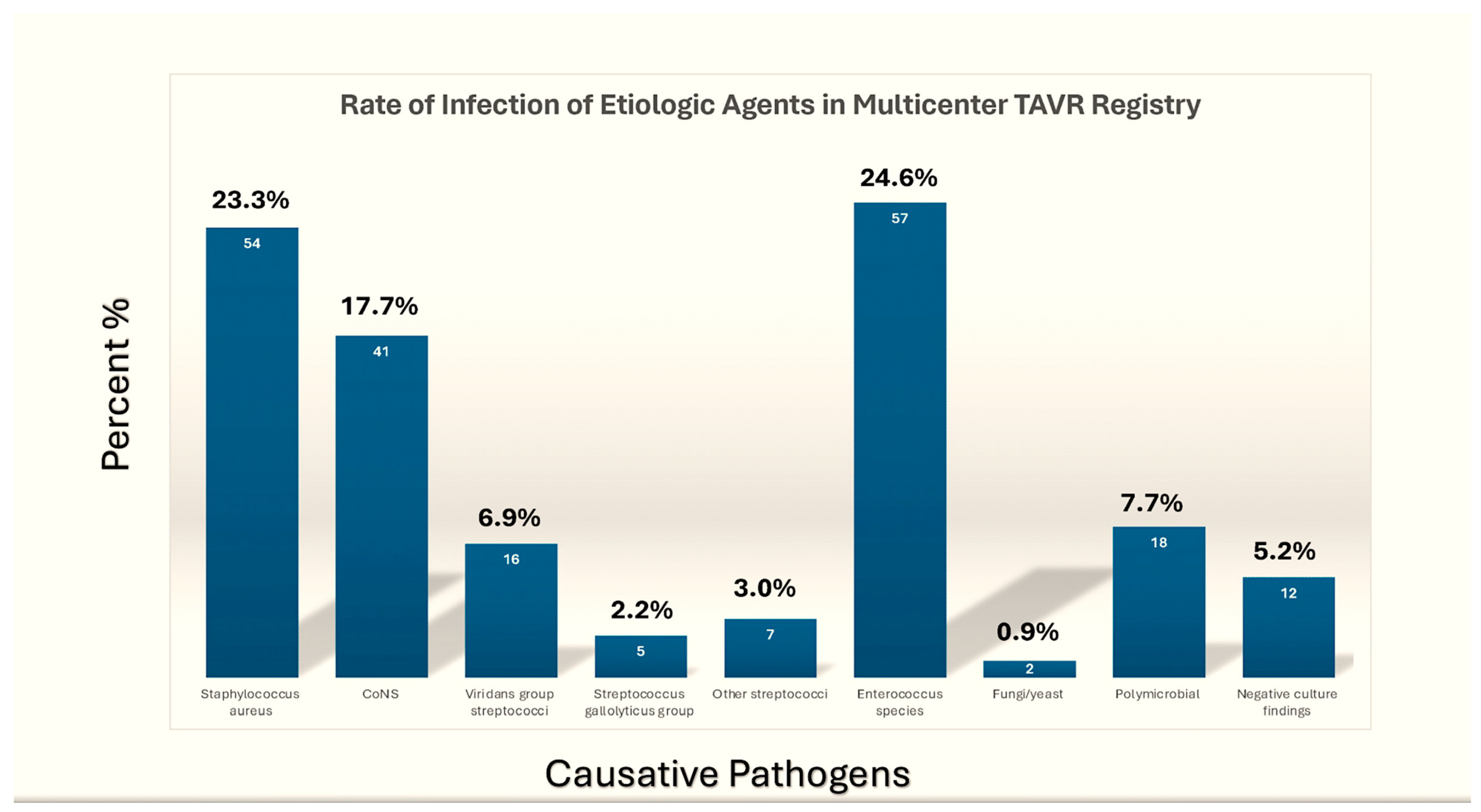
It is well established that TAVR procedures are associated with a high prevalence of healthcare-associated infections. These are predominantly caused by staphylococcal and, in particular, enterococcal pathogens, which have been reported to account for up to 30% of the causative agents in some series [50,54,58,59] (Figure 3).
The high incidence of early enterococcal infection following TAVR procedures is undoubtedly due to the frequent utilization of a femoral approach during these procedures [50,60]. It is clear that the causative microorganism was Staphylococcus aureus in 23.8 percent to 24.6 percent of cases [53,54], while the main pathogen was Enterococcus faecalis in 25 percent of cases [53].
Despite their prevalence as a cause of PVE in TAVR recipients, with a rate of 17.7% [50,53,59], coagulase-negative staphylococci are relatively uncommon in native valve infective endocarditis. However, this is not the case for S. lugdunensis, which is clinically similar to S. aureus [21,49,50,59]. It is noteworthy that in patients undergoing TAVR, HACEK species (Haemophilus species, Aggregatibacter [formerly Actinobacillus] species, Cardiobacterium species, Eikenella corrodens, and Kingella species), fungal, polymicrobial, and, on rare occasions, aerobic Gram-negative bacilli have been isolated in less than 2%. Culture negatives were identified in up to 5.2% of cases [49,50,51,52,53,54,55,56,57,58,59].
There are clear differences. The incidence of healthcare-associated IE was significantly higher in the S. aureus IE population than in the non-S. aureus IE population (53.2% vs. 40.7%). There is a significant difference in the prevalence of IE episodes involving a TAVR prosthesis between the non-S. aureus IE cohort and the S. aureus IE cohort (63.4% vs. 51.1%). Furthermore, S. aureus IE patients are more likely to develop implantable cardiac device infections, other than TAVR prostheses (9.2% vs. 2.6%) [50,54,59].
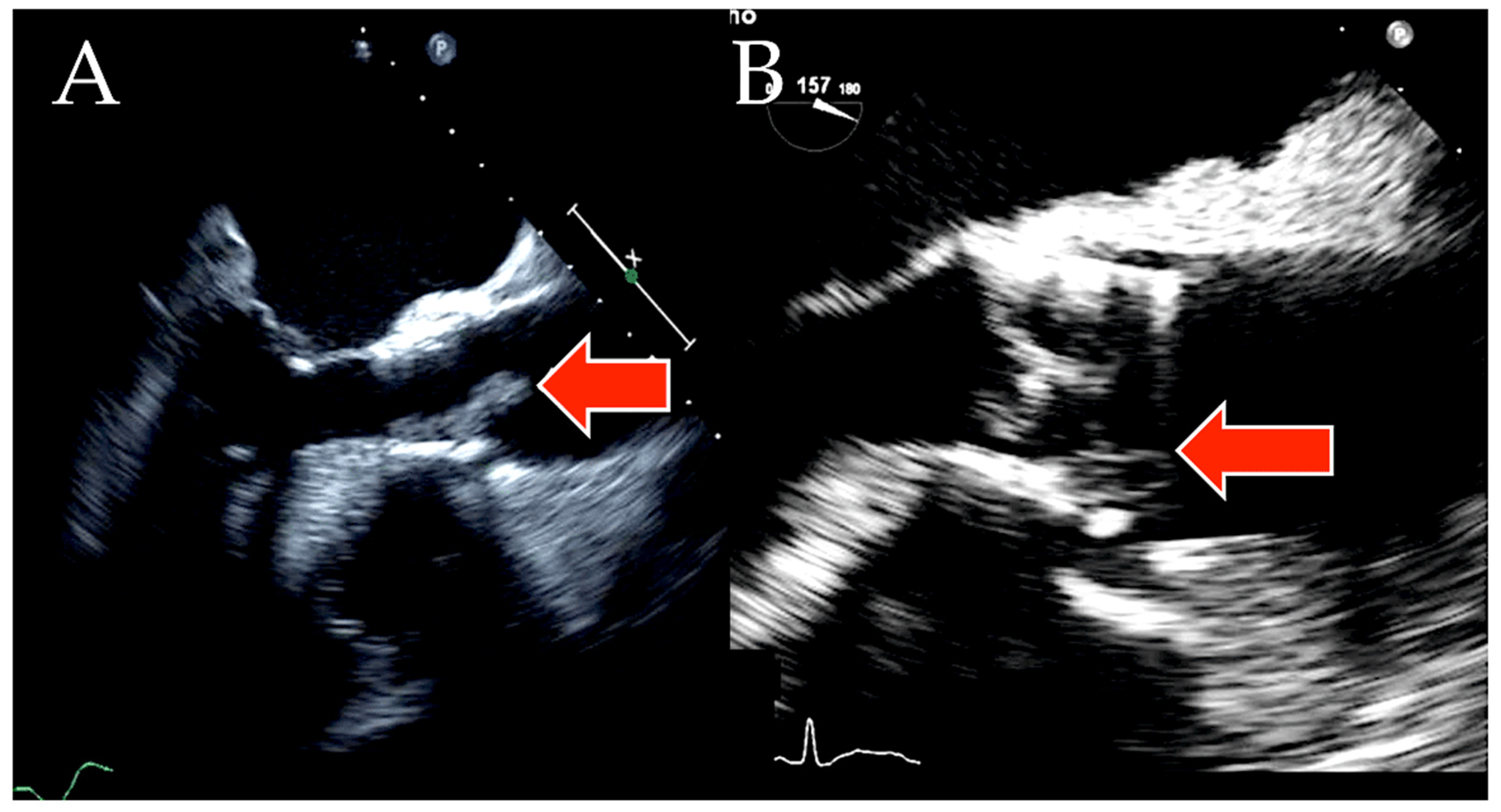
Echocardiography definitively showed the presence of vegetations in a significant proportion of patients. Vegetation is found on either the stent or the leaflets of the device (Figure 4).
Reguiero et al. definitively reported a prevalence of 67.6% of vegetation, with a mean (SD) size of 10.7 mm (7.5 mm). The authors confirmed that vegetations are anchored to the stent frame in 18.2% of patients and to the leaflets of the transcatheter valve in 47.9% of patients. It has been established that there is a significantly greater prevalence of vegetation in the stent of self-expandable devices in comparison to the stent balloon-expandable devices. The occurrence of vegetation was 26.2% in patients receiving self-expandable valves and 10.6% in those receiving balloon-expandable valves. The proportions are reversed when comparing balloon versus self-expandable devices. There is a significant difference in the incidence of vegetation at the valve leaflet level between patients who received a balloon-expandable valve (58.8%) and those who received a self-expandable valve (36.2%) [53].
Reguiero et al. [53] also found that concomitant mitral valve involvement was recorded in 20.0%, tricuspid in 4.4%, and pacing devices in 6.0%. Infection spreading from the TAVR site to other valves is not uncommon. Severe mitral regurgitation, new moderate to severe aortic regurgitation, and periannular complications have been observed. These include the presence of abscesses, fistulas, and pseudoaneurysms [14,23,50,53,54,59].
Particular attention is devoted to Enterococcus faecalis infective endocarditis given the high frequency with which this pathogen colonizes TAVR devices. This enterococcal pathogen accounts for approximately 5–8% of hospital-acquired bacteremia and 5–20% of all infective endocarditis cases and is the most clinically important of the Enterococcus species. Prosthetic valve infections caused by E. faecalis lead to endocarditis, accounting for up to 30% of TAVR recipients. Without effective antibiotic treatment, the infection can cause damage to the valves and result in mortality [1,5,59,60,61,62,63]. The emergence of resistance to multiple antibiotics has underscored the importance of gaining novel insights into enterococcal endocarditis, a condition with significant clinical implications. In stark contrast to group A streptococci or Staphylococcus aureus, which are notably virulent pathogens that depend on the secretion of a multitude of hemolysins and toxins to successfully counteract innate immune responses [64,65], E. faecalis relies on a relatively limited number of virulence determinants for its pathogenicity [66].
The initial stage of E. faecalis infection is thought to be primarily initiated by the attachment and colonization of host tissue surfaces, as evidenced by emerging research [66,67]. This hypothesis is corroborated by the detection of primarily Gram-positive pathogens, which points to an important function played by proteins in the adhesive matrix molecules (MSCRAMM) family, which could be potential targets for the development of new and effective immunotherapies [68]. Sillanpää and colleagues have identified seventeen proteins from the E. faecalis V583 genome that exhibit cell-wall-anchoring motifs and MSCRAMM-like structural features [68,69,70,71].

3. New Challenges in the Field of IE: The Management of IE Following TAVR

3.1. Causative Pathogens in the TAVR-IE Trials

TAVR has transformed the outlook for patients with aortic stenosis who were previously considered inoperable or at high risk for surgery. However, today’s TAVR patients are often frail, undergoing multiple medical interventions, and may be at high risk for bacteremia and IE. Nevertheless, the technology is expected to spread to intermediate and low-risk populations over time. The TAVR endocarditis population presents a significant challenge for cardiologists and surgeons in managing contemporary IE. The question is clear: what is the best strategy to manage PVE in patients who are elderly and at high risk for surgery but who are expected to do poorly with medical management? We must consider the impact of surgery on this population.
A small number of cases of TAVR endocarditis were reported in the landmark Placement of Aortic Transcatheter Valve (PARTNER) trials [8,9]. Real-world observational data cohorts are now beginning to illuminate the occurrence and outcomes of TAVR endocarditis. A single-center study from Germany reported 55 cases of TAVR endocarditis, representing a cumulative incidence of 3.02% (1.82% per patient/year). Of these cases, 42% (23 of 55) were identified as healthcare-acquired [23]. The results of the multivariate analysis clearly indicate that chronic hemodialysis and peripheral arterial disease are significant risk factors for the subsequent occurrence of TAVR endocarditis. Chronic hemodialysis exhibits an HR of 8.37 and a 95% CI of 2.54 to 27.63, while peripheral arterial disease displays an HR of 3.77 and a 95% CI of 1.88 to 7.58. Staphylococcus aureus was the cause of 38% of cases, enterococci of 31%, CoNS of 9%, and Streptococci of 9.1%. In seven cases, an infectious process affecting the prosthetic valve was superimposed on a primary valve infection [23].
A further report outlined the cases of 53 patients diagnosed with TAVR endocarditis in a multicenter U.S. registry [14]. This represented a cumulative incidence of 0.67% at a mean follow-up period of 1.1 years. In the initial post-procedure period spanning the first year, a prevalence of 0.5% for TAVR endocarditis was observed, with a median time to occurrence of 6 months. A majority of patients (70% or greater) presented with a fever, while 77% exhibited a discernible vegetation on echocardiographic imaging. An antecedent procedure was found to be the likely cause of bacteremia in around 50% of patients, with antibiotic prophylaxis being used in 59% of cases. The most prevalent causative agents of infection were identified as CoNS (25%), Staphylococcus aureus (21%), and Enterococcus (21%). The self-expanding CoreValve system (Medtronic, Minneapolis, MN, USA) was identified as an independent risk factor for IE (hazard ratio [HR]: 3.1; 95% CI: 1.37 to 7.14). This finding will be validated in other series [14].
Raguiero and colleagues analyzed the data from the Infective Endocarditis after TAVR International Registry, which included reports from 47 centers across the globe, encompassing a total of 250 cases. The overall prevalence was 1.1% per person/year, with a median time of presentation of 5.3 months following the procedure. In a multivariate analysis, the following predictive factors were identified: younger age (hazard ratio, HR: 0.97 per year; 95% confidence interval, CI: 0.94 to 0.99), male sex (HR: 1.69; 95% CI: 1.13 to 2.52), diabetes mellitus (HR: 1.52; 95% CI: 1.02 to 2.29), and moderate-to-severe aortic regurgitation (HR: 2.05; 95% CI: 1.28 to 3.28) [53]. The primary infectious agents were identified as enterococci (24.6%) and S. aureus (23.3%). We observed an in-hospital mortality rate of 36%, with a two-year mortality rate of 67%. Further patient- and device-related factors that contribute to an increased risk of endocarditis will undoubtedly be identified. This will also facilitate a greater understanding of the nature of endocarditis. The apparently high incidence is undoubtedly due to an elevated risk in the initial postoperative months. To enable a comparison with the outcomes of surgical valve replacement, a longer follow-up period is essential [53].
In a recent study, del Val and colleagues analyzed data from the IE after TAVR International Registry. Their study cohort comprised patients with confirmed IE following TAVR from 59 centers in 11 countries. Patients were classified into two groups based on microbiological etiology: non-S. aureus infective endocarditis (non-S. aureus IE) and S. aureus infective endocarditis (S. aureus IE) [54]. S. aureus IE was identified in 141 patients out of 573 (24.6%). In the majority of cases (115/141, 81.6%), the infecting strain was methicillin-sensitive S. aureus. The use of self-expanding valves was more prevalent than that of balloon-expandable valves in patients presenting with an early diagnosis of S. aureus IE. The presence of major bleeding and sepsis complicating TAVR, as well as the emergence of neurologic symptoms or systemic embolism at the time of admission, and the presence of IE with cardiac device involvement (excluding the TAVR prosthesis) were identified as risk factors for S. aureus IE (p < 0.05 for all). In the cohort of patients with IE subsequent to TAVR, the probability of S. aureus IE rose from 19% in the absence of those risk factors to 84.6% when three risk factors were identified [54].
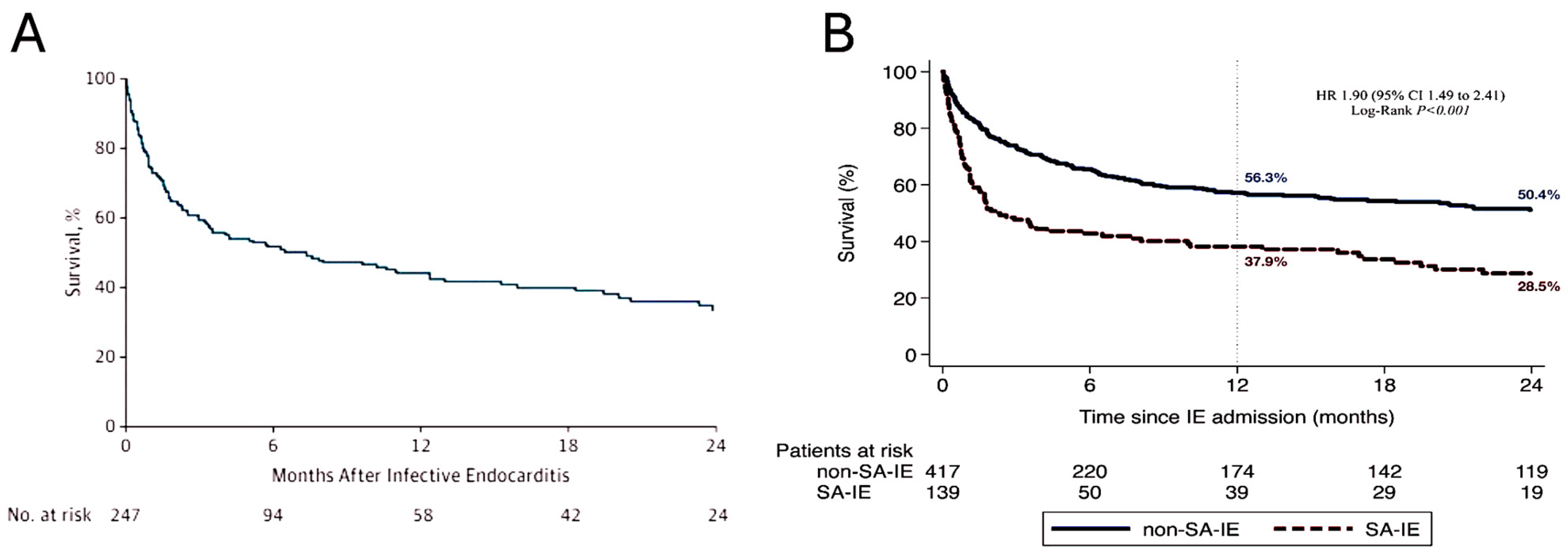
Patients with S. aureus IE had significantly higher in-hospital (47.8% vs. 26.9%; p < 0.001) and 2-year (71.5% vs. 49.6%; p < 0.001) mortality rates compared to those with non-S. aureus IE. Surgical intervention during the initial episode of S. aureus IE reduces mortality at the follow-up stage compared to medical therapy alone. The adjusted hazard ratio was 0.46 (95% CI 0.22–0.96, p = 0.038), demonstrating a statistically significant reduction in mortality [54] (Table 1).
The optimal treatment of TAVR endocarditis presents significant challenges, which must be overcome [50,59]. It is yet to be proven whether transcatheter techniques can manage this condition without the removal of the infected implant. Prior to undergoing TAVR, these patients were often deemed to pose a high or extremely high risk for surgical intervention. A review of the studies conducted to date revealed that approximately 20 percent of patients underwent either open-heart surgery or a transcatheter valve-in-valve procedure. In stark contrast, outcomes with antibiotic therapy alone are markedly unfavorable, with observed in-hospital and one-year mortality rates spanning a wide range from 47% to 64% and 66% to 75%, respectively. These data show that we must develop better prophylactic strategies with respect to valve design and the eradication of bacteremia [50,59].

3.2. Medical Management

It can reasonably be concluded that, with the exception of fungal PVE, no other cause of PVE should be regarded as sufficient justification for a decision to proceed with a mandatory surgical approach. Recent years have yielded the most significant research. This research employs methodologically sound designs with appropriate adjustments for selection and immortalization biases and has reached a clear consensus: surgical indications should be made on an individualized basis. This conclusion has been reached based on observations indicating that a subset of patients will experience favorable clinical outcomes with exclusive medical treatment. Surgical intervention is more beneficial for patients who are more likely to require surgery. Therefore, any patient who does not have a clear indication for surgical intervention beyond the etiological agent should be initially designated for exclusive medical management.
The recommendations for antimicrobial therapy in the early critical phase of PVE (first two weeks of treatment) remain unchanged [2,43]. However, the 2023 ESC IE guidelines introduce a significant new amendment to clinical practice. Non-complicated PVE patients starting intravenous antibiotic therapy at the hospital can complete their treatment at home. This can either be intravenously as outpatient parenteral antibiotic therapy (OPAT) or orally. These recommendations are based on two key sources. The first is the new OPAT-GAMES criteria, which provide a framework for outpatient parenteral antibiotic therapy (OPAT) management [72]. Second is the POET trial, which has shown the potential of partial oral treatment of endocarditis [73].
The study by Pericas et al. [72] provides a comprehensive analysis of data from the Spanish GAMES registry (GAMES Registry of Acute Medications for the Treatment of Endocarditis). The study included 429 patients with IE who completed antibiotic therapy under an OPAT regimen. Patients with a history of injectable substance use were not included in the study. Contrary to what is set out in both American and European clinical guidelines, a total of 117 study participants exhibited the presence of this condition despite the presence of PVE being an exclusion criterion for outpatient management. The study demonstrated that there was no elevated mortality rate or increased incidence of hospital readmission. The research team confidently presented a novel proposal for the identification of patients with PVE who would benefit from OPAT treatment. This new approach, called OPAT-GAMES, builds on the existing criteria for patient selection. In an evaluation study recently conducted by Pericas and colleagues [72], the efficacy of the aforementioned criteria was substantiated using the GAMES registry. This was proven by the fact that patients undergoing outpatient parenteral antimicrobial therapy for infective endocarditis had a comparable incidence of complications and hospital readmissions to that observed in the general population. Furthermore, dalbavancin, a long-acting glycopeptide, is an optimal choice for OPAT consolidation treatment [56] (Table 2).
Table 2 presents data from Pericà et al. [72]. The term “highly difficult-to-treat microorganisms” is employed to describe those requiring intravenous antibiotic combinations that cannot be administered via OPAT or that necessitate rigorous monitoring of drug levels in either the blood or other bodily fluids, due to their potential toxicity or narrow therapeutic index (e.g., methicillin-resistant Staphylococcus aureus). Furthermore, there are strains of Staphylococcus aureus that are resistant to both penicillin and vancomycin, as well as enterococci that are resistant to vancomycin and other drugs, including daptomycin and linezolid. Furthermore, there are multidrug-resistant and extensively drug-resistant Gram-negative rods, as well as highly penicillin-resistant viridans group streptococci. It should be noted that there are fungal strains that are resistant to penicillin, with the exception of those belonging to the Candida species. Abbreviation: OPAT: outpatient parenteral antibiotic treatment. Please note that the following abbreviations are used throughout this document.

3.3. Surgery on Bioprosthetic Valves for PVE in Patients with TAVR

In cases where prosthetic valve dysfunction is the cause of heart failure and an elevated risk of embolization, the best course of action is surgical intervention. Surgical management remains unchanged. In select cases, other surgical options are available, including the use of a homograft and heart transplant [74]. Nevertheless, the high prevalence of comorbidities and the inherent surgical risks mean there is a subset of patients, particularly those with transcatheter valve implants, who will decline surgical intervention, even when it is indicated [53,75]. It can be assumed that the extremely low number of valve surgeries is a result of the high surgical risk profile observed in these patients. In addition, there are technical challenges involved in removing a fixed stent frame from the aorta.
Transcatheter interventions are an effective alternative to surgery for severe residual valvular dysfunction in high-risk patients. These include paravalvular leak closure, mitral edge-to-edge repair, and aortic and mitral valve-in-valve replacement. It is important to note that the majority of these interventional techniques have been successfully employed in patients without active infection [76,77]. If it is medically feasible, treatment should be delayed until there are no remaining signs of active infection and the antibiotic course has been completed. The evidence base for treatment during the active phase of endocarditis is largely anecdotal, with only a limited number of cases reported [78]. The decision to prescribe long-term oral suppressive antimicrobial therapy must be made on a case-by-case basis, with consideration given to the pathogen and input from an infectious disease specialist.

4. Discussion

Prosthetic valve endocarditis has been identified as the most serious form of infective endocarditis, with a prevalence ranging from 1% to 6% in patients with prosthetic valves. This condition accounts for 10% to 30% of all cases of infective endocarditis [1,2,3,4,5]. The incidence of infective endocarditis following TAVR, as observed by Reguiro et al. [53], was comparable to that reported for infective endocarditis associated with surgical prosthetic valve implants. Consequently, these findings lend support to the hypothesis that the reduction in prosthetic valve infective endocarditis following TAVR, despite TAVR’s comparatively less invasive nature in comparison to surgical valve replacement, has not led to a commensurate reduction in infective endocarditis [53]. Another crucial finding of the aforementioned study was to substantiate the elevated incidence of morbidity and mortality associated with infective endocarditis following TAVR. Additionally, the study yielded novel insights pertaining to the timing, causative organisms, and predictive factors of IE within this specific population. This knowledge may assist clinicians in identifying patients at heightened risk and facilitate the implementation of suitable preventive measures [53]. Although the median age of patients in the population was 80 years in the early randomized controlled trials [6,7,8,12], patients who were younger in subsequent RCTs were at an elevated risk of infective endocarditis following TAVR [10,12,13]. The underlying cause of this phenomenon remains elusive. It is notable that younger patients who are deemed to be at a high or prohibitive risk for surgical intervention may present with a greater comorbidity profile than their older counterparts. This may be correlated with an elevated likelihood of developing IE. Similarly, sex-based discrepancies in the prevalence of comorbid conditions and outcomes among patients who undergo TAVR procedures may contribute to the observed higher risk of infective endocarditis among male patients [79]. A growing body of evidence from previous research indicates a link between diabetes and an enhanced likelihood of developing IE [80].

4.1. Risk Factors

A number of risk factors have been identified, which can be classified into the following categories: procedural and patient-related. Those pertaining to the procedure include moderate and severe AR, increased residual peak gradient, the valve-in-valve procedure, and TAVR complications such as bleeding. Similarly, both self and expanded devices have been found to be equally risky. It is clear that the most significant patient-related risk factors are attributable to younger age, sex (where males are more susceptible), comorbidities, prior IE, and BMI.
There is a definitive association between the presence of residual moderate or severe aortic regurgitation and a reduction in survival rates following transcatheter aortic valve replacement [81]. Abnormalities of blood flow, turbulence, and shear stress are the defining features of aortic regurgitation. This will inevitably lead to an increased propensity for platelets and fibrin to deposit within the affected tissue. This results in a condition called non-bacterial thrombotic endocarditis, which can cause the formation of vegetations [82].
These detrimental effects undoubtedly play an instrumental role in the occurrence and maintenance of device infection [83]. Furthermore, there is a clear link between a higher degree of valve calcification and the development of aortic regurgitation following TAVR. This could potentially contribute to an elevated risk of infective endocarditis [84]. The results clearly show that a more assertive strategy, including antibiotic prophylaxis (in line with current guidelines), is the best way to reduce the risk of bacteremia. This involves eliminating unnecessary procedures and reinforcing preventive measures for patients with significant aortic regurgitation following TAVR.
Prior studies have identified certain periprocedural TAVR features, including transcatheter valve type and the use of orotracheal intubation, as potential risk factors for infective endocarditis [14]. Nevertheless, these associations have not been consistently confirmed by larger studies, as the current investigation definitively demonstrates. It included a significantly larger cohort of patients. The investigation conducted by Reguiero et al. [53] revealed a significant discrepancy in infective endocarditis occurrence rates within the 60-day postoperative period following TAVR. This study observed a significantly higher rate of 29%, in stark contrast to the 14% reported by the International Collaboration on Endocarditis (ICE) Prospective Cohort Study [85]. It is reasonable to conclude that the aforementioned discrepancies are a direct result of the heightened risk profile and the considerable burden of instrumentation present in the TAVR population.
A more judicious approach is imperative. This entails avoiding superfluous invasive examinations and reinforcing the significance of maintaining aseptic conditions throughout any invasive procedure. The objective is to minimize the likelihood of exposure to sources of bacteremia. Previous research on prosthetic valve infective endocarditis has been contradicted by multiple studies which have indicated that enterococci are the most common causative microorganisms [53,60,61]. These studies confirm the comparable prevalence of S. aureus and coagulase-negative staphylococci infections, as previously reported [85]. In contrast, another study by de Val and colleagues found that around 25% of infective endocarditis cases following TAVR were caused by Staphylococcus aureus. Furthermore, this strain of bacteria was unequivocally linked with markedly elevated rates of in-hospital mortality and mortality occurring after a delayed period. Specific characteristics were identified as a significant predictor of S. aureus IE, offering valuable insights into the formulation of prompt antibiotic regimens [54].
It is well established that enterococcal infective endocarditis is associated with advanced age, chronic disease, and aortic valve calcification. This undoubtedly explains the greater rates of enterococcal infections in patients with TAVR compared to their surgically implanted prosthetic valve counterparts with IE. Pre-procedure antibiotic prophylaxis and careful infection control during and after the procedure are also essential, given the use of trans-femoral access for TAVR (enterococci are a common groin pathogen) and the high rate of enterococcal infective endocarditis within 60 days of the procedure. Given the increased risk of enterococcal endocarditis, antibiotics must be chosen in patients suspected of having endocarditis while awaiting blood culture results.
These findings highlight the need for a reassessment of the use of antibiotics in these patients. Beta-lactam antibiotics are commonly administered as antibiotic prophylaxis for TAVR in most patients, but they are ineffective at protecting against enterococcal infections. Glycopeptides and aminoglycosides are the preferable alternative in such patients [1,5,50,53,54,55,59,60,61,62,63].
The occurrence of vegetation is consistent with a previous surgical PVE series [85]. It is notable that the rate of vegetation on the stent frame was almost three times higher in patients who received a self-expanding valve than in those who received a balloon-expandable valve [53,62] (Figure 1 and Figure 5).
The larger stent frame of self-expanding devices is the most likely explanation for these differences. Furthermore, it is important to highlight the role of pacemaker devices in some cases. This is particularly the case given the high rate of pacemaker implantation following TAVR. When evaluating a patient with clinical suspicion of infective endocarditis after TAVR, it is essential to consider these transcatheter valve-specific features [1,86,87].

4.2. Outcomes

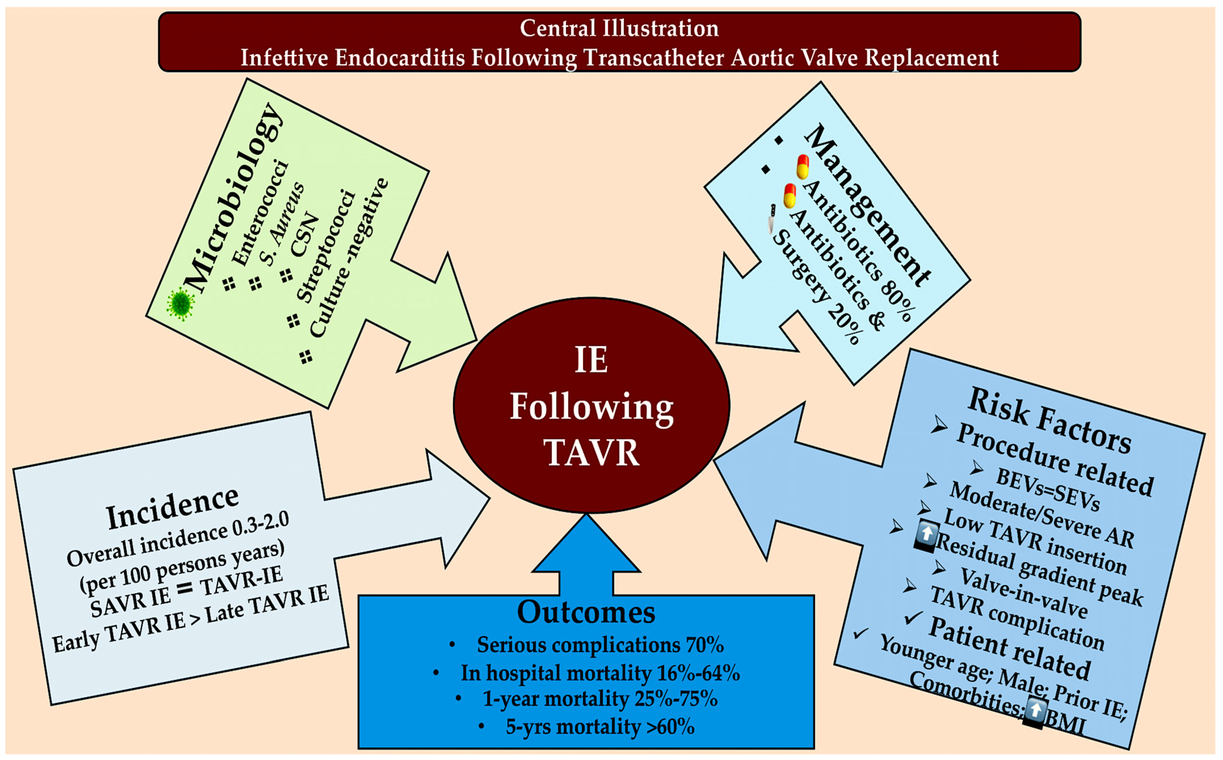
It is clear that there are serious complications in almost 70% of patients who have developed IE after TAVR. The in-hospital mortality rate ranges from 16% to 64%, while the one-year mortality rate is between 27% and 75%. The five-year mortality rate is well over 60% [55] (Figure 6).
The study results definitively show that heart failure at the time of admission and acute kidney injury during infective endocarditis are associated with an elevated risk of in-hospital mortality when combined with an elevated surgical pre-procedural risk, irrespective of surgical therapy administration. Heart failure is the strongest predictor of in-hospital mortality. Its significant impact on patients with infective endocarditis who have undergone TAVR is confirmed.
Furthermore, elevated logistic EuroSCORE was linked with an elevated risk of in-hospital mortality, emphasizing the influence of comorbidities on outcomes following infective endocarditis in this high-risk population. A study of a large cohort of TAVR recipients revealed a significantly elevated in-hospital mortality rate, reaching 36%. The microorganism responsible for IE also exerts an influence on the in-hospital mortality rate. It has been observed that the in-hospital mortality rate in patients with S. aureus IE is higher than that which occurred with non-S. aureus IE (47.8% vs. 26.9%) [50,53,54,59,88].
Raguiero and colleagues [53] definitively established a mortality rate of 67% at the two-year follow-up. This rate is clearly higher than the observed incidence of mortality associated with prosthetic surgical valve endocarditis, which ranged from 27% to 61% in previous studies [89,90]. This is undoubtedly due to the advanced age and elevated comorbidity burden of the TAVR population. However, the observed mortality rate remains higher than that documented two years following TAVR in patients at high surgical risk. In fact, it is 22% and 34% in the US pivotal trial for the self-expanding valve and the Placement of Aortic Transcatheter Valves (PARTNER) trial, respectively [91,92]. This clearly demonstrates that patients with infective endocarditis following TAVR have an extremely poor prognosis.
The study conducted by del Val and colleagues definitively showed that patients with IE caused by S. aureus had a significantly elevated mortality rate (71.5%) in comparison to those with other causative agents (49.6%) at two-year follow-up (p < 0.001 by log-rank test). These findings can be attributed to the following determinants: the elevated risk profile indicated by the logistic EuroSCORE, the lack of surgical intervention during the initial infective endocarditis hospitalization, and IE-related complications, including septic shock and persistent bacteremia, collectively elevate the mortality risk for patients infected with S. aureus [54].
In patients with IE and severe valve dysfunction or large vegetations, early surgical intervention is the most effective way to reduce the risk of in-hospital death and embolic events [93]. This is in line with existing guidelines, which recommend surgical intervention with debridement and valve replacement for patients with severe valve dysfunction, heart failure, cardiac abscesses, highly resistant organisms, or persistent bacteremia, particularly when the latter two are coupled with the former four.
Lalani and colleagues [71] found that approximately 50% of patients in a contemporary cohort of patients with PVE underwent valve surgery. The 10.8% rate of surgical treatment involving valve explantation is clearly distinct from that observed in a large cohort of patients who received TAVR. Despite the exceedingly high prevalence of patients with at least one indication for surgical intervention (>80%, in line with current guidelines), the observed surgical rate is significantly below the figure of 50% reported by Lalani et al. [71]. A further large cohort study found no differences between groups in surgical management rates according to S. aureus IE vs. non-S. aureus IE (21.0% vs. 19.5%) [29], thereby corroborating the data of previous investigations [53].
The extremely low incidence of valve surgery is undoubtedly due to the high or prohibitive surgical risk profile observed in these patients, coupled with the technical challenges associated with removing a stent frame that has become firmly attached to the aorta. However, contrary to previous studies, there was no statistically significant association found between valve surgery and reduced mortality [50,53,54].
For instance, Reguiero et al. discovered that patients who underwent surgery during their hospitalization for infective endocarditis did not exhibit a reduced risk of in-hospital mortality compared to those who did not require surgery (29.7%; 95% CI, 15.0% to 44.4% for surgery vs. 37.1%; 95% CI, 30.6% to 43.6% for no surgery; difference, −7.4%; 95% CI, −21.3% to 0.8%; OR, 0.72; 95% CI, 0.33 to 1.53; p = 0.39) [53]. It is probable that the observed outcome was influenced by a number of factors. Firstly, it is evident that there is a clear selection bias, with TAVR patients having a higher surgical risk than the general population. Secondly, the limited number of patients who underwent valve explantation represents a significant limiting factor [50,53,59]. Further studies should investigate the potential benefits of surgical therapy in this challenging group of patients. Please refer to the central illustration.

4.3. Infective Endocarditis Caused by Staphylococcus Aureus After Transcatheter Aortic Valve Replacement: A Nightmare That Will Not End

A recently published study revealed a striking finding: approximately 25% of cases of IE after TAVR were caused by Staphylococcus aureus. Patients with S. aureus-associated IE had an elevated risk for TAVR-related complications such as acute kidney injury, stroke, major bleeding, and sepsis. Furthermore, these patients were more likely to require a prolonged intensive care unit (ICU) stay following TAVR [94,95]. In addition, severe periprocedural TAVR bleeding or sepsis, neurologic symptoms or systemic emboli upon admission, and IE involving cardiac devices other than the TAVR prosthesis were identified as factors associated with S. aureus IE. The presence of two or more of these factors at the index hospitalization demonstrated a high probability of S. aureus IE, reaching over 80% in patients with three or more factors. A number of studies have demonstrated that, in comparison to infections caused by non-S. aureus pathogens, IE caused by S. aureus is associated with a higher incidence of associated complications [94,95,96]. Del Val and colleagues observed that these patients had a very high in-hospital mortality rate of up to 50% and a follow-up mortality rate of >70% at 2 years. In their report, the authors demonstrated that the absence of surgical intervention during the initial hospitalization of patients with S. aureus IE was associated with an elevated risk of mortality [54].
S. aureus PVE represents the most severe subtype of IE, accounting for 10% to 30% of all IE cases [97]. The incidence of post-TAVR IE has been reported to range from 0.6% to 3.4% annually [53,98,99]. Despite advances in TAVR procedures and devices, the overall incidence of IE has remained relatively constant [100]. There is currently a lack of data available that directly compares the incidence of IE following SAVR and TAVR.
A recent study analyzing a pooled cohort of three randomized clinical trials showed a lower incidence of IE after TAVR compared with SAVR [96], although most studies reported similar incidence rates [95,96]. It is noteworthy that the microbiological profile is distinct between SAVR-IE and TAVR-IE. There is a discrepancy between studies regarding the causative pathogen responsible for IE. While enterococci account for only w10% of SAVR IE [54], previous analyses using the same cohort of patients identified this microorganism as the leading cause of TAVR IE [53,100]. Rates of IE due to Staphylococcus aureus remain higher in patients undergoing TAVR, reaching one in four patients [54]. These results are consistent with those reported in SAVR IE registries. However, the proportion of patients with late S. Aureus IE after TAVR was slightly higher than those with late S. aureus IE following the open surgery [85]. This result may be linked to the unique profile of the TAVR patient population, which leads to a higher frequency of invasive diagnostic and therapeutic procedures during the follow-up period [54].
The diagnosis of PVE is a challenging process. Therefore, an early diagnosis is of the utmost importance, as delayed treatment is associated with significantly worse clinical outcomes. This is particularly relevant in patients with suspected IE after a TAVR procedure. Initially, TAVR recipients represent a unique population with a high prevalence of comorbidities. Consequently, patients commonly present with atypical symptoms. Prior research indicates that the modified Duke criteria demonstrate reduced diagnostic efficacy for IE following TAVR in comparison to native valve endocarditis, predominantly due to an elevated prevalence of negative blood cultures and inconclusive echocardiographic results [50,53,59,98]. Additionally, IE in patients who have undergone TAVR has been linked to a considerable prevalence of severe IE-related complications and unfavorable outcomes. At the one-year follow-up, the mortality rate approaches 50%. To date, no factors have been identified that are consistently correlated with S. aureus as the etiological agent of post-TAVR infective endocarditis. It is of particular significance that del Val and colleagues [54,59] observed an association between TAVR complications and the presence of major bleeding or sepsis, the emergence of neurologic symptoms or systemic embolism at the time of IE index admission, and the manifestation of infection involving implantable cardiac devices other than the TAVR prosthesis. These factors were identified as being independently associated with IE sustained by S. aureus in recipients of a TAVR. It can be reasonably inferred that these conclusions may have significant clinical repercussions, particularly in light of the fact that the presence of these factors, particularly when they occur in conjunction with one another, is associated with an exceedingly high probability of S. aureus IE, exceeding 80% in patients who present with three risk factors [54,59,100] (Figure 6B).
Therefore, in patients exhibiting two or more risk factors for S. aureus IE following TAVR and suspected IE, prompt treatment is recommended, with an initial focus on appropriate antibiotic regimens to cover S. aureus while awaiting blood culture results. Given the inconsistencies in the data pertaining to the primary microorganisms responsible for IE, with notable discrepancies between enterococci [53,59,60,61] and staphylococci [54,59,100], further investigation is necessary to ascertain whether this approach can effectively reduce the incidence of IE-related complications and enhance clinical outcomes. It is noteworthy that no correlation was identified between prosthesis type and the overall risk of IE. However, findings from multiple studies indicate that patients with early onset IE caused by S. aureus were more likely to have a self-expanding prosthesis in comparison to those who received a balloon-expandable prosthesis [14,53,54,94]. The higher proportion of patients receiving a permanent pacemaker after the TAVR procedure in the self-expanding population (25.4%) compared to the balloon expanding population (10.4%) may contribute to an elevated risk of bacteremia and subsequent risk of IE due to S. aureus [48,101,102,103].
There is a clear link between S. aureus IE following TAVR and unfavorable outcomes, including a high mortality rate. This conclusion is supported by the findings of multiple studies that have definitively shown the pathogenic potential of S. aureus IE. Patients with IE caused by S. aureus have a worse outlook and are more likely to die during their hospital stay or later on than those with IE caused by other microorganisms. The in-hospital mortality rates for S. aureus and non-S. aureus IE were 47.8% and 26.9%, respectively. The late mortality rates were 71.5% and 49.6% for S. aureus and non-S. aureus IE, respectively. The mortality rate observed in the present study was significantly higher than that documented in patients with surgical prosthetic-valve S. aureus IE, with an in-hospital mortality rate of approximately 35% [104,105]. This discrepancy is undoubtedly due to the elevated baseline risk of TAVR patients, which is attributable to advanced age and comorbidities. Therefore, any complication will place them at a significantly heightened risk of mortality relative to their surgical counterparts. The incidence of acute renal failure, acute heart failure, and septic shock during the index hospitalization in patients with S. aureus IE was approximately twice that observed in the non-S. aureus-IE population.
PVE management entails the implementation of intricate antibiotic regimens, in conjunction with surgical procedures such as valve explantation, in a select cohort of patients. Previous studies have indicated that approximately half of the patients with native or prosthetic valves who develop IE secondary to S. aureus undergo surgical intervention during their initial hospitalization for IE [21,106,107]. The extant guidelines counsel the utilization of combined and prolonged antibiotic regimens [43,100]. The findings revealed considerable heterogeneity in the antibiotic regimens prescribed to patients with S. aureus IE following TAVR [54]. One potential explanation for this finding is the significant prevalence of antimicrobial resistance, renal/hepatic toxicity, and drug interactions observed in the TAVR population. These factors may limit the applicability of these recommendations in real-world settings. Furthermore, the rate of surgical intervention in patients with S. aureus IE was notably low. While surgical treatment of PVE caused by S. aureus should be considered (if there is a low likelihood of control with the use of antimicrobial therapy), the rate of surgery was comparable to that of patients with non-S. aureus IE [54,59,100]. Prior research has been unable to substantiate the assertion that patients with TAVR-IE who undergo surgical intervention exhibit superior outcomes when compared to those who receive antibiotic treatment alone [108]. It is crucial to acknowledge that in patients with S. aureus IE following TAVR, surgical intervention may confer a protective effect when accounting for perioperative mortality risk (logistic EuroSCORE) and severe IE-related complications (acute renal failure, septic shock, persistent bacteremia) [54]. Nevertheless, the role of surgical treatment in TAVR patients remains a topic of ongoing debate and discussion, and these results should therefore be interpreted with the utmost scrutiny and caution. Historically, a considerable percentage of TAVR recipients exhibited absolute contraindications that made surgical treatment unadvisable. It can therefore be concluded that the general recommendation for surgical intervention in native or prosthetic valve IE cannot be extended to patients undergoing TAVR. It should be noted that the demographic profile of TAVR recipients is changing rapidly, with an increasing proportion of patients deemed low-risk surgical candidates, which could lead to a greater number of patients with TAVR-IE being referred for surgery in the near future. It is therefore essential that dedicated studies be conducted in order to further validate our findings and establish surgical indications in TAVR-IE patients.
The number of individuals at risk for IE following TAVR is expected to increase exponentially due to the broader application of this treatment to younger patients with greater life expectancies. Consequently, strategies aimed at limiting the occurrence of IE and improving clinical outcomes for this population are of heightened significance. Prevention should be the cornerstone of addressing this potentially lethal disease. Firstly, TAVR should continue to evolve towards more streamlined (and less invasive) procedures, with the aim of facilitating earlier patient ambulation and reducing hospital stays. Secondly, it is of paramount importance to establish evidence-based recommendations regarding the most suitable antimicrobial prophylaxis, in addition to aseptic techniques, for use prior to certain invasive procedures. Moreover, it is imperative to emphasize the significance of minimizing healthcare-associated processes that could potentially give rise to a bloodstream infection among this demographic. Thirdly, the development of novel strategies for the prevention of Staphylococcus aureus bacteremia and prosthetic infection would address a significant unmet medical need. The development of prosthetic devices incorporating innovative antibacterial biomaterials that prevent bacterial adhesion to prosthetic surfaces may prove to be an important strategy for reducing infective endocarditis in cases of bloodstream infection.

4.4. Impact of Imaging Techniques

TEE should be performed in the majority of cases, even in instances where TTE has provided sufficient indications for a definitive and conclusive diagnosis. In cases of suspected endocarditis in patients with prosthetic valves, TTE is simply not sensitive enough to be relied upon. In fact, the minimum detection rate is only 36–69%. In such circumstances, the most appropriate course of action is to proceed with a TEE [2,109,110,111,112]. In cases where the heart device is infected, TEE must be conducted. Furthermore, in cases where complications are suspected, a repetition of the TTE scan is essential. A baseline TTE scan must be conducted after the completion of the therapeutic regimen for monitoring and follow-up purposes [4,109,110,111,112].
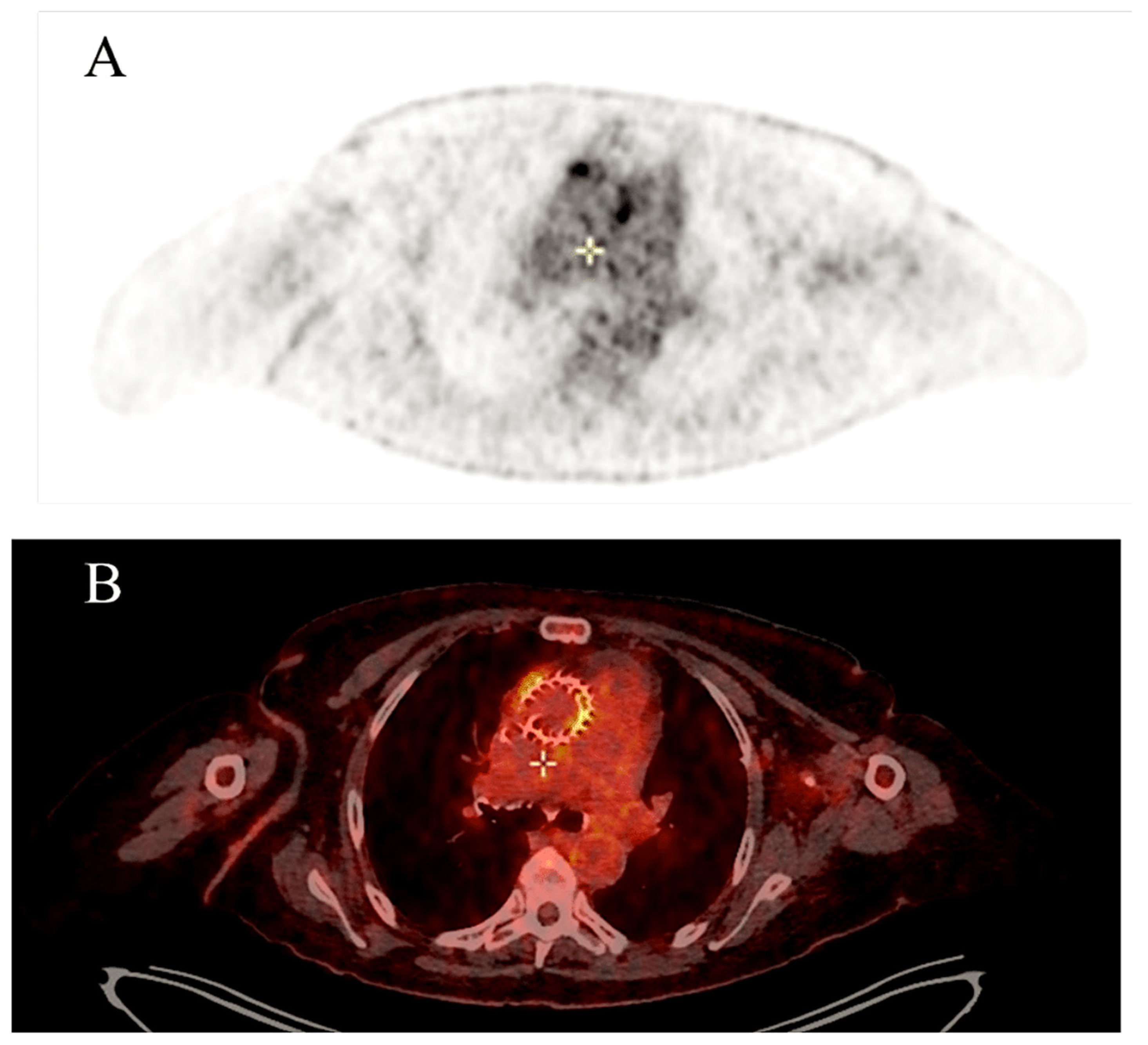
The 2023 edition of the Duke-International Society for Cardiovascular Infectious Diseases (ISCVIID) diagnostic criteria incorporates fluorine-18 fluorodeoxyglucose (18F-FDG) positron emission tomography (PET)/computed tomography (CT) as both a major and minor diagnostic tool. This is due to the efficacy of this imaging technique in reclassifying cases from a “possible” infective endocarditis (IE) diagnosis to an “established” IE diagnosis [4].
While distinguishing between postoperative inflammatory uptake patterns and infection can be challenging, advances in digital positron emission tomography (PET) in conjunction with electrocardiogram-gated computed tomography angiography (CTA) are effectively addressing the shortcomings associated with early infective endocarditis within the initial three months of valve implantation. This is because postoperative inflammatory uptake patterns often manifest as diffuse and homogeneous processes, which are indistinguishable from infection. Infection, on the other hand, tends to manifest as intense focal or multifocal processes with heterogeneous patterns [113]. 18F-FDG-PET/CT is a valuable tool for identifying extracardiac phenomena associated with septic embolism, in addition to its utility in cardiac imaging. Furthermore, it can be used to provide an alternative diagnosis for patients who have been classified as having “rejected” infective endocarditis [114]. Although radiolabeled leukocyte single-photon emission CT (SPECT)/CT has high specificity, it is no longer the preferred imaging modality. This is due to its relatively higher technical complexity, lower sensitivity, and resolution. The preferred modality is now 18F-FDG-PET/CT [115,116]. The central illustration is shown as Figure 7 below.

5. Conclusions

The number of patients undergoing bioprosthetic TAVR has grown. This increase has made it clear that the field of PVE must evolve further, particularly in the areas of preventative measures, early detection techniques, and effective treatment strategies. These advancements are of paramount importance, given the current mortality rate of 40% to 50% within the first year. As with IE in native valves, comprehensive clinical trials are essential to gaining further insight into this life-threatening infection. Based on the current assessment of ongoing and planned investigative efforts, it is clear that future modifications to PVE-TAVR prevention, diagnosis, and management strategies will emerge, particularly in response to enterococcal and staphylococcal infection concerns.

Funding

This research received no external funding.

Institutional Review Board Statement

Not applicable.

Informed Consent Statement

Not applicable.

Data Availability Statement

Data are contained within the article.

Acknowledgments

I would like to express my gratitude to Marianna Di Palma (IBM Group France) for her invaluable assistance in creating the charts.

Conflicts of Interest

The author declares no conflicts of interest.

References

  1. Nappi, F.; Spadaccio, C.; Mihos, C. Infective endocarditis in the 21st century. Ann. Transl. Med. 2020, 8, 1620. [Google Scholar] [CrossRef] [PubMed]
  2. Delgado, V.; Ajmone Marsan, N.; de Waha, S.; Bonaros, N.; Brida, M.; Burri, H.; Caselli, S.; Doenst, T.; Ederhy, S.; Erba, P.A.; et al. 2023 ESC Guidelines for the management of endocarditis. Eur. Heart J. 2023, 44, 3948–4042. [Google Scholar] [PubMed]
  3. Habib, G.; Lancellotti, P.; Antunes, M.J.; Bongiorni, M.G.; Casalta, J.P.; Del Zotti, F.; Dulgheru, R.; El Khoury, G.; Erba, P.A.; Iung, B.; et al. 2015 ESC Guidelines for the management of infective endocarditis: The Task Force for the Management of Infective Endocarditis of the European Society of Cardiology (ESC)—Endorsed by: European Association for Cardio-Thoracic Surgery (EACTS), the European Association of Nuclear Medicine (EANM). Eur. Heart J. 2015, 36, 3075–3128. [Google Scholar] [PubMed]
  4. Fowler, V.G.; Durack, D.T.; Selton-Suty, C.; Athan, E.; Bayer, A.S.; Chamis, A.L.; Dahl, A.; Di Bernardo, L.; Durante-Mangoni, E.; Duval, X.; et al. The 2023 Duke-International Society for Cardiovascular Infectious Diseases Criteria for Infective Endocarditis: Updating the Modified Duke Criteria. Clin. Infect. Dis. 2023, 77, 518–526. [Google Scholar] [CrossRef] [PubMed]
  5. Nappi, F.; Martuscelli, G.; Bellomo, F.; Avtaar Singh, S.S.; Moon, M.R. Infective Endocarditis in High-Income Countries. Metabolites 2022, 12, 682. [Google Scholar] [CrossRef]
  6. Otto, C.M.; Nishimura, R.A.; Bonow, R.O.; Carabello, B.A.; Erwin, J.P., III; Gentile, F.; Jneid, H.; Krieger, E.V.; Mack, M.; McLeod, C.; et al. 2020 ACC/AHA guideline for the management of patients with valvular heart disease: A report of the American College of Cardiology/American Heart Association joint committee on clinical practice guidelines. J. Am. Coll. Cardiol. 2021, 77, e25–e197. [Google Scholar] [CrossRef]
  7. Vahanian, A.; Beyersdorf, F.; Praz, F.; Milojevic, M.; Baldus, S.; Bauersachs, J.; Capodanno, D.; Conradi, L.; De Bonis, M.; De Paulis, R.; et al. 2021 ESC/EACTS Guidelines for the management of valvular heart disease. Eur. Heart J. 2022, 43, 561–632. [Google Scholar] [CrossRef]
  8. Leon, M.B.; Smith, C.R.; Mack, M.; Miller, D.C.; Moses, J.W.; Svensson, L.G.; Tuzcu, E.M.; Webb, J.G.; Fontana, G.P.; Makkar, R.R.; et al. Transcatheter aortic-valve implantation foraortic stenosis in patients who cannot undergo surgery. N. Engl. J. Med. 2010, 363, 1597–1607. [Google Scholar] [CrossRef]
  9. Smith, C.R.; Leon, M.B.; Mack, M.J.; Miller, D.C.; Moses, J.W.; Svensson, L.G.; Tuzcu, E.M.; Webb, J.G.; Fontana, G.P.; Makkar, R.R.; et al. Transcatheter versus surgical aortic-valve replacement in high-risk patients. N. Engl. J. Med. 2011, 364, 2187–2198. [Google Scholar] [CrossRef]
  10. Aung, T.; Poon, K.; Horvath, R.; Coulter, C.; Walters, D.L. A case series of medically managed infective endocarditis after transcatheter aortic valve replacement. Scand. J. Infect. Dis. 2013, 45, 489–493. [Google Scholar] [CrossRef]
  11. Latib, A.; Naim, C.; De Bonis, M.; Sinning, J.M.; Maisano, F.; Barbanti, M.; Parolari, A.; Lorusso, R.; Testa, L.; Actis Dato, G.M.; et al. TAVR-associated prosthetic valve infective endocarditis: Results of a large, multicenter registry. J. Am. Coll. Cardiol. 2014, 64, 2176–2178. [Google Scholar] [CrossRef] [PubMed]
  12. Olsen, N.T.; De Backer, O.; Thyregod, H.G.; Vejlstrup, N.; Bundgaard, H.; Søndergaard, L.; Ihlemann, N. Prosthetic valve endocarditis after transcatheter aortic valve implantation. Circ. Cardiovasc. Interv. 2015, 8, e001939. [Google Scholar] [CrossRef] [PubMed]
  13. Puls, M.; Eiffert, H.; Hünlich, M.; Schöndube, F.; Hasenfuß, G.; Seipelt, R.; Schillinger, W. Prosthetic valve endocarditis after transcatheter aortic valve implantation: The incidence in a single-centre cohort and reflections on clinical, echocardiographic and prognostic features. EuroIntervention 2013, 8, 1407–1418. [Google Scholar] [CrossRef] [PubMed]
  14. Amat-Santos, I.J.; Messika-Zeitoun, D.; Eltchaninoff, H.; Kapadia, S.; Lerakis, S.; Cheema, A.N.; Gutiérrez-Ibanes, E.; Munoz-Garcia, A.J.; Pan, M.; Webb, J.G.; et al. Infective endocarditis after transcatheter aortic valve implantation: Results from a large multicenter registry. Circulation 2015, 131, 1566–1574. [Google Scholar] [CrossRef] [PubMed]
  15. Calderwood, S.B.; Swinski, L.A.; Waternaux, C.M.; Karchmer, A.W.; Buckley, M.J. Risk factors for the development of prosthetic valve endocarditis. Circulation 1985, 72, 31–37. [Google Scholar] [CrossRef]
  16. Agnihotri, A.K.; McGiffin, D.C.; Galbraith, A.J.; O’Brien, M.F. The prevalence of infective endocarditis after aortic valve replacement. J. Thorac. Cardiovasc. Surg. 1995, 110, 1708–1720. [Google Scholar] [CrossRef]
  17. Amat-Santos, I.J.; Ribeiro, H.B.; Urena, M.; Allende, R.; Houde, C.; Bédard, E.; Perron, J.; DeLarochellière, R.; Paradis, J.M.; Dumont, E.; et al. Prosthetic valve endocarditis after transcatheter valve replacement: A systematic review. JACC Cardiovasc. Interv. 2015, 8, 334–346. [Google Scholar] [CrossRef]
  18. Braghieri, L.; Kaur, S.; Black, C.K.; Cremer, P.C.; Unai, S.; Kapadia, S.R.; Mentias, A. Endocarditis after Transcatheter Aortic Valve Replacement. J. Clin. Med. 2023, 12, 7042. [Google Scholar] [CrossRef]
  19. Martínez-Sellés, M.; Bouza, E.; Díez-Villanueva, P.; Valerio, M.; Fariñas, M.C.; Muñoz-García, A.J.; Ruiz-Morales, J.; Gálvez-Acebal, J.; Antorrena, I.; de la Hera Galarza, J.M.; et al. Incidence and clinical impact of infective endocarditis after transcatheter aortic valve implantation. EuroIntervention 2016, 11, 1180–1187. [Google Scholar] [CrossRef]
  20. Selton-Suty, C.; Célard, M.; Le Moing, V.; Doco-Lecompte, T.; Chirouze, C.; Iung, B.; Strady, C.; Revest, M.; Vandenesch, F.; Bouvet, A.; et al. Preeminence of Staphylococcus aureus in infective endocarditis: A 1-year population-based survey. Clin. Infect. Dis. 2012, 54, 1230–1239. [Google Scholar] [CrossRef]
  21. Murdoch, D.R.; Corey, G.R.; Hoen, B.; Miró, J.M.; Fowler, V.G., Jr.; Bayer, A.S.; Karchmer, A.W.; Olaison, L.; Pappas, P.A.; Moreillon, P.; et al. Clinical presentation, etiology and outcome of infective endocarditis in the 21st century: The International Collaboration on Endocarditis Prospective Cohort Study. Arch. Intern. Med. 2009, 169, 463–473. [Google Scholar] [CrossRef] [PubMed]
  22. Voigt, A.; Shalaby, A.; Saba, S. Rising rates of cardiac rhythm management device infections in the United States: 1996 through 2003. J. Am. Coll. Cardiol. 2006, 48, 590–591. [Google Scholar] [CrossRef] [PubMed]
  23. Mangner, N.; Woitek, F.; Haussig, S.; Schlotter, F.; Stachel, G.; Höllriegel, R.; Wilde, J.; Lindner, A.; Holzhey, D.; Leontyev, S.; et al. Incidence, predictors, and outcome of patients developing infective endocarditis following transfemoral transcatheter aortic valve replacement. J. Am. Coll. Cardiol. 2016, 67, 2907–2908. [Google Scholar] [CrossRef] [PubMed]
  24. Van Dijck, I.; Budts, W.; Cools, B.; Eyskens, B.; Boshoff, D.E.; Heying, R.; Frerich, S.; Vanagt, W.Y.; Troost, E.; Gewillig, M. Infective endocarditis of a transcatheter pulmonary valve in comparison with surgical implants. Heart 2015, 101, 788–793. [Google Scholar] [CrossRef] [PubMed]
  25. Van der Meer, J.T.; Van Wijk, W.; Thompson, J.; Vandenbroucke, J.P.; Valkenburg, H.A.; Michel, M.F. Efficacy of antibiotic prophylaxis for pre- vention of native-valve endocarditis. Lancet 1992, 339, 135–139. [Google Scholar] [CrossRef]
  26. van der Meer, J.M.; Thompson, J.; Valkenburg, H.A.; Michel, M.F. Epidemiology of bacterial endocarditis in the Netherlands. II. antecedent procedures and use of prophylaxis. Arch. Intern. Med. 1992, 152, 1869–1873. [Google Scholar] [CrossRef]
  27. Strom, B.L.; Abrutyn, E.; Berlin, J.A.; Kinman, J.L.; Feldman, R.S.; Stolley, P.D.; Levison, M.E.; Korzeniowski, O.M.; Kaye, D. Dental and cardiac risk factors for infective endocarditis: A population-based, case-control study. Ann. Intern. Med. 1998, 129, 761–769. [Google Scholar] [CrossRef]
  28. Lockhart, P.B.; Brennan, M.T.; Sasser, H.C.; Fox, P.C.; Paster, B.J.; Bahrani-Mougeot, F.K. Bacteremia associated with toothbrushing and dental extraction. Circulation 2008, 117, 3118–3125. [Google Scholar] [CrossRef]
  29. Habib, G.; Badano, L.; Tribouilloy, C.; Prendergast, B.; Vilacosta, I.; Moreillon, P.; de Jesus Antunes, M.; Thilen, U.; Lekakis, J.; Lengyel, M.; et al. Guidelines on the prevention, diagnosis, and treatment of infective endocarditis (new version 2009). Endorsed by the European Society of Clinical Microbiology and Infectious Diseases (ESCMID) and by the International Society of Chemotherapy (ISC) for Infection and Cancer. Eur. Heart J. 2009, 30, 2369–2413. [Google Scholar]
  30. Fagman, E.; Perrotta, S.; Bech-Hanssen, O.; Flinck, A.; Lamm, C.; Olaison, L.; Svensson, G. ECG-gated computed tomography: A new role for patients with suspected aortic prosthetic valve endocarditis. Eur. Radiol. 2012, 22, 2407–2414. [Google Scholar] [CrossRef]
  31. Saby, L.; Laas, O.; Habib, G.; Cammilleri, S.; Mancini, J.; Tessonnier, L.; Casalta, J.P.; Gouriet, F.; Riberi, A.; Avierinos, J.F.; et al. Positron emission tomography/computed tomography for diagnosis of prosthetic valve endocarditis: Increased valvular 18F-fluorodeoxyglucose uptake as a novel major criterion. J. Am. Coll. Cardiol. 2013, 61, 2374–2382. [Google Scholar] [CrossRef] [PubMed]
  32. Pizzi, M.N.; Roque, A.; Fernández-Hidalgo, N.; Cuéllar-Calabria, H.; Ferreira-González, I.; Gonzàlez-Alujas, M.T.; Oristrell, G.; Gracia-Sánchez, L.; González, J.J.; Rodríguez-Palomares, J.; et al. Improving the diagnosis of infective endocarditis in prosthetic valves and intracardiac devices with 18F-fluorodeoxyglucose positron emission tomography/computed tomography: Initial results at an infective endocarditis referral center. Circulation 2015, 132, 1113–1126. [Google Scholar] [CrossRef] [PubMed]
  33. Erba, P.A.; Conti, U.; Lazzeri, E.; Doria, R.; De Tommasi, S.M.; Bandera, F.; Tascini, C.; Menichetti, F.; Dierckx, R.A.; Signore, A.; et al. Added value of 99mTc-HMPAO-labeled leukocyte SPECT/CT in the characterization and management of patients with infectious endocarditis. J. Nucl. Med. 2012, 53, 1235–1243. [Google Scholar] [CrossRef] [PubMed]
  34. Rouzet, F.; Chequer, R.; Benali, K.; Lepage, L.; Ghodbane, W.; Duval, X.; Iung, B.; Vahanian, A.; Le Guludec, D.; Hyafil, F. Respective performance of 18F-FDG PET and radiolabeled leukocyte scintigraphy for the diagnosis of prosthetic valve endocarditis. J. Nucl. Med. 2014, 55, 1980–1985. [Google Scholar] [CrossRef]
  35. Duval, X.; Iung, B.; Klein, I.; Brochet, E.; Thabut, G.; Arnoult, F.; Lepage, L.; Laissy, J.P.; Wolff, M.; Leport, C.; et al. Effect of early cerebral magnetic resonance imaging on clinical decisions in infective endocarditis: A prospective study. Ann. Intern. Med. 2010, 152, 497–504. [Google Scholar] [CrossRef]
  36. Meshaal, M.S.; Kassem, H.H.; Samir, A.; Zakaria, A.; Baghdady, Y.; Rizk, H.H. Impact of routine cerebral CT angiography on treatment decisions in infective endocarditis. PLoS ONE 2015, 10, e0118616. [Google Scholar] [CrossRef]
  37. Iung, B.; Klein, I.; Mourvillier, B.; Olivot, J.M.; Détaint, D.; Longuet, P.; Ruimy, R.; Fourchy, D.; Laurichesse, J.J.; Laissy, J.P.; et al. Respective effects of early cerebral and abdominal magnetic resonance imaging on clinical decisions in infective endocarditis. Eur. Heart J. Cardiovasc. Imaging 2012, 13, 703–710. [Google Scholar] [CrossRef]
  38. Barsic, B.; Dickerman, S.; Krajinovic, V.; Pappas, P.; Altclas, J.; Carosi, G.; Casabé, J.H.; Chu, V.H.; Delahaye, F.; Edathodu, J.; et al. Influence of the timing of cardiac surgery on the outcome of patients with infective endocarditis and stroke. Clin. Infect. Dis. 2013, 56, 209–217. [Google Scholar] [CrossRef]
  39. Hasbun, R.; Vikram, H.R.; Barakat, L.A.; Buenconsejo, J.; Quagliarello, V.J. Complicated left-sided native valve endocarditis in adults: Risk classification for mortality. JAMA 2003, 289, 1933–1940. [Google Scholar] [CrossRef]
  40. Vilacosta, I.; Graupner, C.; San Román, J.A.; Sarriá, C.; Ronderos, R.; Fernández, C.; Mancini, L.; Sanz, O.; Sanmartín, J.V.; Stoermann, W. Risk of embolization after institution of antibiotic therapy for infective endocarditis. J. Am. Coll. Cardiol. 2002, 39, 1489–1495. [Google Scholar] [CrossRef]
  41. Thuny, F.; Di Salvo, G.; Belliard, O.; Avierinos, J.F.; Pergola, V.; Rosenberg, V.; Casalta, J.P.; Gouvernet, J.; Derumeaux, G.; Iarussi, D.; et al. Risk of embolism and death in infective endocarditis: Prognostic value of echocardiography: A prospective multicenter study. Circulation 2005, 112, 69–75. [Google Scholar] [CrossRef] [PubMed]
  42. García-Cabrera, E.; Fernández-Hidalgo, N.; Almirante, B.; Ivanova-Georgieva, R.; Noureddine, M.; Plata, A.; Lomas, J.M.; Gálvez-Acebal, J.; Hidalgo-Tenorio, C.; Ruíz-Morales, J.; et al. Neurological complications of infective endocarditis: Risk factors, outcome, and impact of cardiac surgery: A multicenter observational study. Circulation 2013, 127, 2272–2284. [Google Scholar] [CrossRef] [PubMed]
  43. Baddour, L.M.; Wilson, W.R.; Bayer, A.S.; Fowler, V.G., Jr.; Tleyjeh, I.M.; Rybak, M.J.; Barsic, B.; Lockhart, P.B.; Gewitz, M.H.; Levison, M.E.; et al. Infective endocarditis in adults: Diagnosis, antimicrobial therapy, and management of complications. Circulation 2015, 132, 1435–1486. [Google Scholar] [CrossRef] [PubMed]
  44. Voigt, A.; Shalaby, A.; Saba, S. Continued rise in rates of cardiovascular implantable electronic de- vice infections in the United States: Temporal trends and causative insights. Pacing Clin. Electrophysiol. 2010, 33, 414–419. [Google Scholar] [CrossRef]
  45. Johansen, J.B.; Jørgensen, O.D.; Møller, M.; Arnsbo, P.; Mortensen, P.T.; Nielsen, J.C. Infection after pacemaker implantation: Infection rates and risk factors associated with infection in a population-based cohort study of 46299 consecutive patients. Eur. Heart J. 2011, 32, 991–998. [Google Scholar] [CrossRef]
  46. Landolina, M.; Gasparini, M.; Lunati, M.; Iacopino, S.; Boriani, G.; Bonanno, C.; Vado, A.; Proclemer, A.; Capucci, A.; Zucchiatti, C.; et al. Long-term complications related to biventricular defibrillator implantation: Rate of surgical revisions and impact on survival: Insights from the Italian Clinical Service Database. Circulation 2011, 123, 2526–2535. [Google Scholar] [CrossRef]
  47. Sohail, M.R.; Henrikson, C.A.; Braid-Forbes, M.J.; Forbes, K.F.; Lerner, D.J. Mortality and cost associated with cardio- vascular implantable electronic device infections. Arch. Intern. Med. 2011, 171, 1821–1828. [Google Scholar] [CrossRef]
  48. Polyzos, K.A.; Konstantelias, A.A.; Falagas, M.E. Risk factors for cardiac implantable electronic device infection: A systematic review and meta-analysis. Europace 2015, 17, 767–777. [Google Scholar] [CrossRef]
  49. Cahill, T.J.; Prendergast, B.D. Infective endocarditis. Lancet 2016, 387, 882–893. [Google Scholar] [CrossRef]
  50. Cuervo, G.; Quintana, E.; Regueiro, A.; Perissinotti, A.; Vidal, B.; Miro, J.M.; Baddour, L.M. The Clinical Challenge of Prosthetic Valve Endocarditis: JACC Focus Seminar 3/4. J. Am. Coll. Cardiol. 2024, 83, 1418–1430. [Google Scholar] [CrossRef]
  51. Ambrosioni, J.; Hernandez-Meneses, M.; Téllez, A.; Pericàs, J.; Falces, C.; Tolosana, J.M.; Vidal, B.; Almela, M.; Quintana, E.; Llopis, J.; et al. The changing epidemiology of infective endocarditis in the twenty-first century. Curr. Infect. Dis. Rep. 2017, 19, 21. [Google Scholar] [CrossRef] [PubMed]
  52. Ramos-Martínez, A.; Domínguez, F.; Muñoz, P.; Marín, M.; Pedraz, Á.; Fariñas, M.C.; Tascón, V.; de Alarcón, A.; Rodríguez-García, R.; Miró, J.M.; et al. Clinical presentation, microbiology, and prognostic factors of prosthetic valve endocarditis. Lessons learned from a large prospective registry. PLoS ONE 2023, 18, e0290998. [Google Scholar] [CrossRef] [PubMed]
  53. Regueiro, A.; Linke, A.; Latib, A.; Ihlemann, N.; Urena, M.; Walther, T.; Husser, O.; Herrmann, H.C.; Nombela-Franco, L.; Cheema, A.N.; et al. Association between transcatheter aortic valve replacement and subsequent infective endocarditis and in-hospital death. JAMA 2016, 316, 1083–1092. [Google Scholar] [CrossRef] [PubMed]
  54. Del Val, D.; Abdel-Wahab, M.; Mangner, N.; Durand, E.; Ihlemann, N.; Urena, M.; Pellegrini, C.; Giannini, F.; Gasior, T.; Wojakowski, W.; et al. Infective Endocarditis Caused by Staphylococcus aureus After Transcatheter Aortic Valve Replacement. Can. J. Cardiol. 2022, 38, 102–112. [Google Scholar] [CrossRef]
  55. Del Val, D.; Panagides, V.; Mestres, C.A.; Miró, J.M.; Rodés-Cabau, J. Infective endocarditis after transcatheter aortic valve replacement: JACC state-of-the-art review. J. Am. Coll. Cardiol. 2023, 81, 394–412. [Google Scholar] [CrossRef]
  56. Hidalgo-Tenorio, C.; Sadyrbaeva-Dolgova, S.; Enríquez-Gómez, A.; Muñoz, P.; Plata-Ciezar, A.; Miró, J.M.; Alarcón, A.; Martínez-Marcos, F.J.; Loeches, B.; Escrihuela-Vidal, F.; et al. EN-DALBACEN 2.0 Cohort: Real-life study of dalbavancin as sequential/consolidation therapy in patients with infective endocarditis due to Gram-positive cocci. Int. J. Antimicrob. Agents 2023, 62, 106918. [Google Scholar] [CrossRef]
  57. Siciliano, R.F.; Randi, B.A.; Gualandro, D.M.; Sampaio, R.O.; Bittencourt, M.S.; da Silva Pelaes, C.E.; Mansur, A.J.; Pomerantzeff, P.M.A.; Tarasoutchi, F.; Strabelli, T.M.V. Early-onset prosthetic valve endocarditis definition revisited: Prospective study and literature review. Int. J. Infect. Dis. 2018, 67, 3–6. [Google Scholar] [CrossRef]
  58. Van Valen, R.; De Lind Van Wijngaarden, R.A.F.; Verkaik, N.J.; Mokhles, M.M.; Bogers, A.J.J.C. Prosthetic valve endocarditis due to Propionibacterium acnes. Interact. Cardiovasc. Thorac. Surg. 2016, 23, 150–155. [Google Scholar] [CrossRef]
  59. Panagides, V.; Abdel-Wahab, M.; Mangner, N.; Durand, E.; Ihlemann, N.; Urena, M.; Pellegrini, C.; Giannini, F.; Scislo, P.; Huczek, Z.; et al. Very early infective endocarditis after transcatheter aortic valve replacement. Clin. Res. Cardiol. 2022, 111, 1087–1097. [Google Scholar] [CrossRef]
  60. Widmer, D.; Widmer, A.F.; Jeger, R.; Dangel, M.; Stortecky, S.; Frei, R.; Conen, A. Prevalence of enterococcal groin colonization in patients undergoing cardiac interventions: Challenging antimicrobial prophylaxis with cephalosporins in patients undergoing transcatheter aortic valve replacement. J. Hosp. Infect. 2022, 129, 198–202. [Google Scholar] [CrossRef]
  61. Nappi, F. Current Knowledge of Enterococcal Endocarditis: A Disease Lurking in Plain Sight of Health Providers. Pathogens 2024, 13, 235. [Google Scholar] [CrossRef] [PubMed]
  62. Nappi, F.; Avtaar Singh, S.S.; Jitendra, V.; Fiore, A. Bridging Molecular and Clinical Sciences to Achieve the Best Treatment of Enterococcus faecalis Endocarditis. Microorganisms 2023, 11, 2604. [Google Scholar] [CrossRef] [PubMed]
  63. Nappi, F. Native Infective Endocarditis: A State-of-the-Art-Review. Microorganisms 2024, 12, 1481. [Google Scholar] [CrossRef] [PubMed]
  64. Novick, R.P. Autoinduction and signal transduction in the regulation of staphylococcal virulence. Mol. Microbiol. 2003, 48, 1429–1449. [Google Scholar] [CrossRef] [PubMed]
  65. Cunningham, M.W. Pathogenesis of group A streptococcal infections. Clin. Microbiol. Rev. 2000, 13, 470–511. [Google Scholar] [CrossRef]
  66. Pillar, C.M.; Gilmore, M.S. Enterococcal virulence—Pathogenicity island of E. faecalis. Front. Biosci. 2004, 9, 2335–2346. [Google Scholar] [CrossRef]
  67. Gilmore, M.S. The Enterococci: Pathogenesis, Molecular Biology, and Antibiotic Resistance; ASM Press: Washington, DC, USA, 2020; 439p. [Google Scholar]
  68. Sillanpaa, J.; Xu, Y.; Nallapareddy, S.R.; Murray, B.E.; Hook, M. A family of putative MSCRAMMs from Enterococcus faecalis. Microbiology 2004, 150, 2069–2078. [Google Scholar] [CrossRef]
  69. Paulsen, I.T.; Banerjei, L.; Myers, G.S.; Nelson, K.E.; Seshadri, R.; Read, T.D.; Fouts, D.E.; Eisen, J.A.; Gill, S.R.; Heidelberg, J.F.; et al. Role of mobile DNA in the evolution of vancomycin-resistant Enterococcus faecalis. Science 2003, 299, 2071–2074. [Google Scholar] [CrossRef]
  70. Chirouze, C.; Alla, F.; Fowler, V.G.; Sexton, D.J.; Corey, G.R.; Chu, V.H.; Wang, A.; Erpelding, M.L.; Durante-Mangoni, E.; Fernández-Hidalgo, N.; et al. Impact of early valve surgery on outcome of Staphylococcus aureus prosthetic valve infective endocarditis: Analysis in the international collaboration of endocarditis-prospective cohort study. Clin. Infect. Dis. 2015, 60, 741–749. [Google Scholar] [CrossRef]
  71. Lalani, T.; Chu, V.H.; Park, L.P.; Cecchi, E.; Corey, G.R.; Durante-Mangoni, E.; Fowler, V.G., Jr.; Gordon, D.; Grossi, P.; Hannan, M.; et al. In-hospital and 1-year mortality in patients undergoing early surgery for prosthetic valve endocarditis. JAMA Intern. Med. 2013, 173, 1495–1504. [Google Scholar] [CrossRef]
  72. Pericà, S.J.M.; Llopis, J.; González-Ramallo, V.; Goenaga, M.Á.; Muñoz, P.; García-Leoni, M.E.; Fariñas, M.C.; Pajarón, M.; Ambrosioni, J.; Luque, R.; et al. Outpatient parenteral antibiotic treatment for infective endocarditis: A prospective cohort study from the GAMES cohort. Clin. Infect. Dis. 2019, 69, 1690–1700. [Google Scholar] [CrossRef] [PubMed]
  73. Iversen, K.; Ihlemann, N.; Gill, S.U.; Madsen, T.; Elming, H.; Jensen, K.T.; Bruun, N.E.; Høfsten, D.E.; Fursted, K.; Christensen, J.J.; et al. Partial oral versus intravenous antibiotic treatment of endocarditis. N. Engl. J. Med. 2019, 380, 415–424. [Google Scholar] [CrossRef] [PubMed]
  74. Pettersson, G.B.; Hussain, S.T. Current AATS guidelines on surgical treatment of infective endocarditis. Ann. Cardiothorac. Surg. 2019, 8, 630–644. [Google Scholar] [CrossRef] [PubMed]
  75. Mangner, N.; del Val, D.; Abdel-Wahab, M.; Crusius, L.; Durand, E.; Ihlemann, N.; Urena, M.; Pellegrini, C.; Giannini, F.; Gasior, T.; et al. Surgical treatment of patients with infective endocarditis after transcatheter aortic valve implantation. J. Am. Coll. Cardiol. 2022, 79, 772–785. [Google Scholar] [CrossRef]
  76. Santos-Martínez, S.; Alkhodair, A.; Nombela-Franco, L.; Saia, F.; Muñoz-García, A.J.; Gutiérrez, E.; Regueiro, A.; Jimenez-Diaz, V.A.; Rivero, F.; Romaguera, R.; et al. Transcatheter aortic valve replacement for residual lesion of the aortic valve following “healed” infective endocarditis. J. Am. Coll. Cardiol. Interv. 2020, 13, 1983–1996. [Google Scholar] [CrossRef]
  77. Khan, M.H.; Biondi, N.L.; Zulfiqar, S.; Arif, I.; Das, M.; Budhiraja, M.; Aronow, W.S.; Sukhija, R.; Vimal, M. Challenging case of transcatheter mitral valve-in-valve-in-valve replacement. Am. J. Case Rep. 2023, 24, e938415. [Google Scholar] [CrossRef]
  78. Brankovic, M.; Hashemi, A.; Ansari, J.; Sharma, A. Transcatheter aortic valve replacement for aortic valve infective endocarditis: A systematic review and call for action. Cardiol. Ther. 2023, 12, 297–306. [Google Scholar] [CrossRef]
  79. O’Connor, S.A.; Morice, M.C.; Gilard, M.; Leon, M.B.; Webb, J.G.; Dvir, D.; Rodés-Cabau, J.; Tamburino, C.; Capodanno, D.; D’Ascenzo, F.; et al. Revisiting sex equality with transcatheter aortic valve replacement outcomes: A collaborative, patient-level meta-analysis of 11 310 patients. J. Am. Coll. Cardiol. 2015, 66, 221–228. [Google Scholar] [CrossRef]
  80. Movahed, M.R.; Hashemzadeh, M.; Jamal, M.M. Increased prevalence of infectious endocarditis in patients with type II diabetes mellitus. J. Diabetes Complicat. 2007, 21, 403–406. [Google Scholar] [CrossRef]
  81. Jerez-Valero, M.; Urena, M.; Webb, J.G.; Tamburino, C.; Munoz-Garcia, A.J.; Cheema, A.; Dager, A.E.; Serra, V.; Amat-Santos, I.J.; Barbanti, M.; et al. Clinical impact of aortic regurgitation after transcatheter aortic valve replacement: Insights into the degree and acuteness of presentation. JACC Cardiovasc. Interv. 2014, 7, 1022–1032. [Google Scholar] [CrossRef]
  82. Rodbard, S. Blood velocity and endocarditis. Circulation 1963, 27, 18–28. [Google Scholar] [CrossRef] [PubMed]
  83. Baddour, L.M.; Bettmann, M.A.; Bolger, A.F.; Epstein, A.E.; Ferrieri, P.; Gerber, M.A.; Gewitz, M.H.; Jacobs, A.K.; Levison, M.E.; Newburger, J.W.; et al. Nonvalvular cardiovascular device-related infections. Circulation 2003, 108, 2015–2031. [Google Scholar] [CrossRef] [PubMed]
  84. Athappan, G.; Patvardhan, E.; Tuzcu, E.M.; Svensson, L.G.; Lemos, P.A.; Fraccaro, C.; Tarantini, G.; Sinning, J.M.; Nickenig, G.; Capodanno, D.; et al. Incidence, predictors, and outcomes of aortic regurgitation after transcatheter aortic valve replacement: Meta-analysis and systematic review of literature. J. Am. Coll. Cardiol. 2013, 61, 1585–1595. [Google Scholar] [CrossRef] [PubMed]
  85. Wang, A.; Athan, E.; Pappas, P.A.; Fowler, V.G., Jr.; Olaison, L.; Paré, C.; Almirante, B.; Muñoz, P.; Rizzi, M.; Naber, C.; et al. Contemporary clinical profile and outcome of prosthetic valve endocarditis. JAMA 2007, 297, 1354–1361. [Google Scholar] [CrossRef]
  86. Scialla, S.; Martuscelli, G.; Nappi, F.; Singh, S.S.A.; Iervolino, A.; Larobina, D.; Ambrosio, L.; Raucci, M.G. Trends in Managing Cardiac and Orthopaedic Device-Associated Infections by Using Therapeutic Biomaterials. Polymers 2021, 13, 1556. [Google Scholar] [CrossRef]
  87. Nappi, F.; Iervolino, A.; Singh, S.S.A. The New Challenge for Heart Endocarditis: From Conventional Prosthesis to New Devices and Platforms for the Treatment of Structural Heart Disease. BioMed Res. Int. 2021, 2021, 7302165. [Google Scholar] [CrossRef]
  88. Leone, S.; Noviello, S.; Esposito, S. Combination antibiotic therapy for the treatment of infective endocarditis due to enterococci. Infection 2016, 44, 273–281. [Google Scholar] [CrossRef]
  89. Rieg, S.; von Cube, M.; Kaasch, A.J.; Bonaventura, B.; Bothe, W.; Wolkewitz, M.; Peyerl-Hoffmann, G.; Deppe, A.C.; Wahlers, T.; Beyersdorf, F.; et al. Investigating the Impact of Early Valve Surgery on Survival in Staphylococcus aureus Infective Endocarditis Using a Marginal Structural Model Approach: Results of a Large, Prospectively Evaluated Cohort. Clin. Infect. Dis. 2019, 69, 487–494. [Google Scholar] [CrossRef]
  90. Akowuah, E.F.; Davies, W.; Oliver, S.; Stephens, J.; Riaz, I.; Zadik, P.; Cooper, G. Prosthetic valve endocarditis: Early and late outcome following medical or surgical treatment. Heart 2003, 89, 269–272. [Google Scholar] [CrossRef]
  91. Reardon, M.J.; Adams, D.H.; Kleiman, N.S.; Yakubov, S.J.; Coselli, J.S.; Deeb, G.M.; Gleason, T.G.; Lee, J.S.; Hermiller, J.B., Jr.; Chetcuti, S.; et al. 2-Year outcomes in patients undergoing surgical or self-expanding transcatheter aortic valve replacement. J. Am. Coll. Cardiol. 2015, 66, 113–121. [Google Scholar] [CrossRef]
  92. Kodali, S.K.; Williams, M.R.; Smith, C.R.; Svensson, L.G.; Webb, J.G.; Makkar, R.R.; Fontana, G.P.; Dewey, T.M.; Thourani, V.H.; Pichard, A.D.; et al. Two-year outcomes after transcatheter or surgical aortic-valve replacement. N. Engl. J. Med. 2012, 366, 1686–1695. [Google Scholar] [CrossRef] [PubMed]
  93. Kang, D.-H.; Kim, Y.-J.; Kim, S.-H.; Sun, B.J.; Kim, D.H.; Yun, S.C.; Song, J.M.; Choo, S.J.; Chung, C.H.; Song, J.K.; et al. Early surgery versus conventional treatment for infective endocarditis. N. Engl. J. Med. 2012, 366, 2466–2473. [Google Scholar] [CrossRef] [PubMed]
  94. Butt, J.H.; Ihlemann, N.; de Backer, O.; Søndergaard, L.; Havers-Borgersen, E.; Gislason, G.H.; Torp-Pedersen, C.; Køber, L.; Fosbøl, E.L. Long-term risk of infective endocarditis after transcatheter aortic valve replacement. J. Am. Coll. Cardiol. 2019, 73, 1646–1655. [Google Scholar] [CrossRef]
  95. Alexis, S.L.; Malik, A.H.; George, I.; Hahn, R.T.; Khalique, O.K.; Seetharam, K.; Bhatt, D.L.; Tang, G.H.L. Infective endocarditis after surgical and transcatheter aortic valve replacement: A state of the art review. J. Am. Heart Assoc. 2020, 9, e017347. [Google Scholar] [CrossRef]
  96. Lanz, J.; Reardon, M.J.; Pilgrim, T.; Stortecky, S.; Deeb, G.M.; Chetcuti, S.; Yakubov, S.J.; Gleason, T.G.; Huang, J.; Windecker, S. Incidence and outcomes of infective endocarditis after transcatheter or surgical aortic valve replacement. J. Am. Heart Assoc. 2021, 10, e020368. [Google Scholar] [CrossRef]
  97. Habib, G.; Thuny, F.; Avierinos, J.-F. Prosthetic valve endocarditis: Current approach and therapeutic options. Prog. Cardiovasc. Dis. 2008, 50, 274–281. [Google Scholar] [CrossRef]
  98. Harding, D.; Cahill, T.J.; Redwood, S.R.; Prendergast, B.D. Infective endocarditis complicating transcatheter aortic valve implantation. Heart 2020, 106, 493–498. [Google Scholar] [CrossRef]
  99. Mentias, A.; Girotra, S.; Desai, M.Y.; Horwitz, P.A.; Rossen, J.D.; Saad, M.; Panaich, S.; Kapadia, S.; Sarrazin, M.V. Incidence, predictors, and outcomes of endocarditis after transcatheter aortic valve replacement in the United States. JACC Cardiovasc. Interv. 2020, 13, 1973–1982. [Google Scholar] [CrossRef]
  100. del Val, D.; Abdel-Wahab, M.; Linke, A.; Durand, E.; Ihlemann, N.; Urena, M.; Pellegrini, C.; Giannini, F.; Landt, M.; Auffret, V.; et al. Temporal trends, characteristics, and outcomes of infective endocarditis after transcatheter aortic valve replacement. Clin. Infect. Dis. 2021, 73, e3750–e3758. [Google Scholar] [CrossRef]
  101. Mousa Basha, M.; Al-Kassou, B.; Gestrich, C.; Weber, M.; Beiert, T.; Bakhtiary, F.; Nickenig, G.; Zimmer, S.; Shamekhi, J. Microbial growth on temporary pacemaker leads post-TAVR: Pathogen spectrum and clinical implications. Clin. Res. Cardiol. 2024. ahead-of-print. [Google Scholar] [CrossRef]
  102. Henrikson, C.A.; Sohail, M.R.; Acosta, H.; Johnson, E.E.; Rosenthal, L.; Pachulski, R.; Dan, D.; Paladino, W.; Khairallah, F.S.; Gleed, K.; et al. Antibacterial Envelope Is Associated with Low Infection Rates After Implantable Cardioverter-Defibrillator and Cardiac Resynchronization Therapy Device Replacement: Results of the Citadel and Centurion Studies. JACC Clin. Electrophysiol. 2017, 3, 1158–1167. [Google Scholar] [CrossRef] [PubMed]
  103. Korantzopoulos, P.; Sideris, S.; Dilaveris, P.; Gatzoulis, K.; Goudevenos, J.A. Infection control in implantation of cardiac implantable electronic devices: Current evidence, controversial points, and unresolved issues. Europace 2016, 18, 473–478. [Google Scholar] [CrossRef] [PubMed]
  104. Fernández Guerrero, M.L.; González López, J.J.; Goyenechea, A.; Fraile, J.; de Górgolas, M. Endocarditis caused by Staphylococcus aureus: A reappraisal of the epidemiologic, clinical, and pathologic manifestations with analysis of factors determining outcome. Medicine 2009, 88, 1–22. [Google Scholar] [CrossRef] [PubMed]
  105. Nadji, G.; Remadi, J.P.; Coviaux, F.; Mirode, A.A.; Brahim, A.; Enriquez-Sarano, M.; Tribouilloy, C. Comparison of clinical and morphological characteristics of Staphylococcus aureus endocarditis with endocarditis caused by other pathogens. Heart 2005, 91, 932–937. [Google Scholar] [CrossRef] [PubMed]
  106. Tornos, P.; Iung, B.; Permanyer-Miralda, G.; Baron, G.; Delahaye, F.; Gohlke-Bärwolf, C.; Butchart, E.G.; Ravaud, P.; Vahanian, A. Infective endocarditis in Europe: Lessons from the Euro Heart Survey. Heart 2005, 91, 571–575. [Google Scholar] [CrossRef] [PubMed]
  107. Kiefer, T.; Park, L.; Tribouilloy, C.; Cortes, C.; Casillo, R.; Chu, V.; Delahaye, F.; Durante-Mangoni, E.; Edathodu, J.; Falces, C.; et al. Association between valvular surgery and mortality among patients with infective endocarditis complicated by heart failure. JAMA 2011, 306, 2239–2247. [Google Scholar] [CrossRef]
  108. Mangner, N.; Leontyev, S.; Woitek, F.J.; Kiefer, P.; Haussig, S.; Binner, C.; Mende, M.; Schlotter, F.; Stachel, G.; Höllriegel, R.; et al. Cardiac surgery compared with antibiotics only in patients developing infective endocarditis after transcatheter aortic valve replacement. J. Am. Heart Assoc. 2018, 7, e010027. [Google Scholar] [CrossRef]
  109. Horgan, S.J.; Mediratta, A.; Gillam, L.D. Cardiovascular imaging in infective endocarditis. Circ. Cardiovasc. Imaging 2020, 13, 8956. [Google Scholar] [CrossRef]
  110. Dilsizian, V.; Budde, R.P.J.; Chen, W.; Mankad, S.V.; Lindner, J.R.; Nieman, K. Best practices for imaging cardiac device–related infections and endocarditis: A JACC: Cardiovascular Imaging expert panel statement. J. Am. Coll. Cardiol. Imaging 2022, 15, 891–911. [Google Scholar] [CrossRef]
  111. Lo Presti, S.; Elajami, T.K.; Zmaili, M.; Reyaldeen, R.; Xu, B. Multimodality imaging in the diagnosis and management of prosthetic valve endocarditis: A contemporary narrative review. World J. Cardiol. 2021, 13, 254–270. [Google Scholar] [CrossRef]
  112. Montané, B.; Chahine, J.; Fiore, A.; Alzubi, J.; Alnajjar, H.; Mutti, J.; Grimm, R.A.; Griffin, B.P.; Xu, B. Diagnostic performance of contemporary transesophageal echocardiography with modern imaging for infective endocarditis. Cardiovasc. Diagn. Ther. 2023, 13, 25–37. [Google Scholar] [CrossRef] [PubMed]
  113. Roque, A.; Pizzi, M.N.; Fernández-Hidalgo, N.; Permanyer, E.; Cuellar-Calabria, H.; Romero-Farina, G.; Ríos, R.; Almirante, B.; Castell-Conesa, J.; Escobar, M.; et al. Morpho-metabolic post-surgical patterns of non-infected prosthetic heart valves by [18vF] FDG PET/CTA: “normality” is a possible diagnosis. Eur. Heart J. Cardiovasc. Imaging 2020, 21, 24–33. [Google Scholar] [CrossRef] [PubMed]
  114. Granados, U.; Fuster, D.; Pericas, J.M.; Llopis, J.L.; Ninot, S.; Quintana, E.; Almela, M.; Paré, C.; Tolosana, J.M.; Falces, C.; et al. Diagnostic accuracy of 18F-FDG PET/CT in infective endocarditis and implantable cardiac electronic device infection: A cross-sectional study. J. Nucl. Med. 2016, 57, 1726–1732. [Google Scholar] [CrossRef] [PubMed]
  115. Wahadat, A.R.; Tanis, W.; Swart, L.E.; Scholtens, A.; Krestin, G.P.; van Mieghem, N.M.; Schurink, C.A.; van der Spoel, T.I.; van den Brink, F.S.; Vossenberg, T.; et al. Added value of 18F-FDG-PET/CT and cardiac CTA in suspected transcatheter aortic valve endocarditis. J. Nucl. Cardiol. 2021, 28, 2072–2082. [Google Scholar] [CrossRef]
  116. Régis, C.; Thy, M.; Mahida, B.; Deconinck, L.; Tubiana, S.; Iung, B.; Duval, X.; Rouzet, F. Absence of infective endocarditis relapse when end-oftreatment fluorodeoxyglucose positron emission tomography/computed tomography is negative. Eur. Heart J. Cardiovasc. Imaging 2023, 24, 1480–1488. [Google Scholar] [CrossRef]
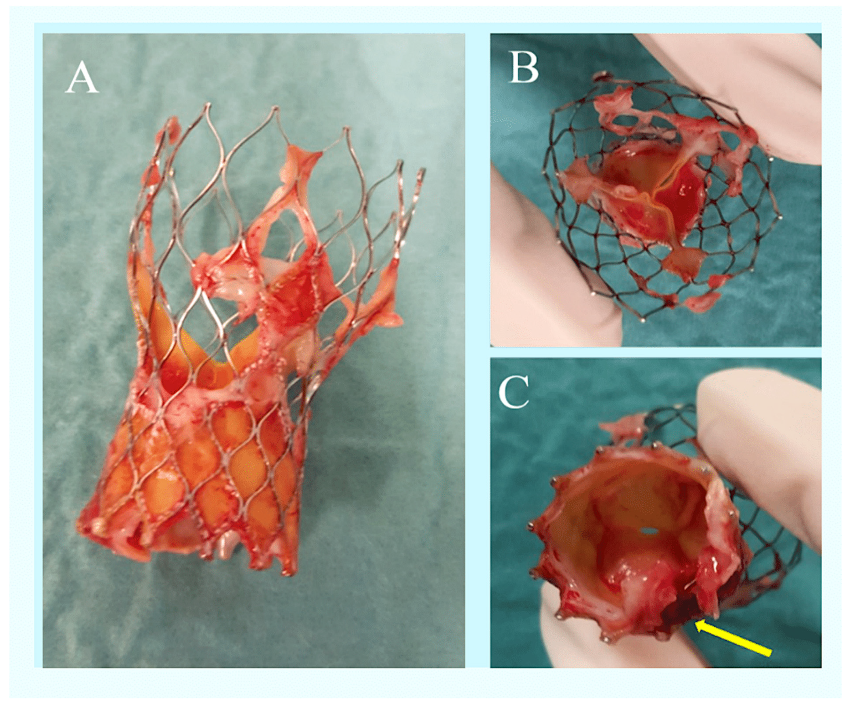
Figure 1. A TAVR was removed due to an Enterococcus faecalis infection. The images depict a case of infective endocarditis resulting from an Enterococcus faecalis infection in a patient who underwent TAVR. Panel (A) illustrates the TAVR prosthesis following its removal, demonstrating the presence of vegetations attached to the frame. Panel (B) depicts the aorta, while panel (C) presents a ventricular view, showcasing evidence of valvular destruction (yellow arrow) in correspondence with the abscess within the intervalvular fibrosa. Abbreviation: TAVR, transcatheter aortic valve replacement [1,5].
Prosthesis 06 00110 g001
Figure 2. Pathogens causing TAVR-IE. This illustration demonstrates the prevalence of etiological causative microorganisms reported in a multicenter cohort of the ICE, as indicated by the percentage of cases (A) and the number of cases (B). The different columns represent the overall population (blue), NVE group (orange), and the PVE group (green). Abbreviations: CoNS, coagulase-negative staphylococci; HACEK, Haemophilus species, Aggregatibacter actino mycetemcomitans, Aggregatibacter aphrophilus (formerly Haemophilus aphrophilus and Haemophilus paraphrophilus), Cardiobacterium hominis, Eikenella corrodens, and Kingella species; ICE*, International Collaboration on Endocarditis; NVE, native valve endocarditis; PVE, prosthetic valve endocarditis [2,4,5,21,52]. x, causative pathogens; y, percentage and number of cases.
Prosthesis 06 00110 g002aProsthesis 06 00110 g002b
Figure 3. Percentage of causative pathogens in TAVR-IE. The illustration presents the prevalence of causative microorganisms identified in a multicenter cohort study of the TAVR. Abbreviations: CoNS, coagulase-negative staphylococci; HACEK, Haemophilus species, Aggregatibacter actino mycetemcomitans, Aggregatibacter aphrophilus (formerly Haemophilus aphrophilus and Haemophilus paraphrophilus), Cardiobacterium hominis, Eikenella corrodens, and Kingella species; ICE, International Collaboration on Endocarditis; NVE, native valve endocarditis; PVE, prosthetic valve endocarditis; values are n (patients) [8,9,10,11,12,13,50,51,52,53,59,60]. x, causative pathogens; y, percent of cases.
Prosthesis 06 00110 g003
Figure 4. Transesophageal echocardiography images of TAVR-IE. The images display two distinct cases (LVOT view) exhibiting a substantial vegetation (red arrow) situated at the level of the valve leaflet (A) or stent frame (B) of a balloon-expandable transcatheter valve. Abbreviation: LVOT, left-ventricle outflow tract.
Prosthesis 06 00110 g004
Figure 5. Images depict patients with TAVR IE caused by Enterococcus faecalis. The preoperative FDG-PET/CT imaging assessment revealed multifocal uptake of the prosthetic transcatheter valve. The axial slices from the attenuation-corrected FDG-PET (A) and fused attenuation-corrected FDG-PET/CT (B) are presented for reference. Abbreviations: CT, computed tomography; FDG, fluorodeoxyglucose; PET, positron emission tomography; TAVR, transcatheter aortic valve replacement [62].
Prosthesis 06 00110 g005
Figure 6. Survival curve for patients with IE after transcatheter aortic valve replacement. (A) depicts the Kaplan–Meier survival curve over the 24-month follow-up period following infective endocarditis following transcatheter aortic valve replacement. The median follow-up period was 10.5 months (interquartile range, 3–21 months). (B) presents the Kaplan–Meier estimated survival curve at the two-year follow-up, comparing patients with and without Staphylococcus aureus IE. Abbreviations: CI, confidence interval; HR, hazard ratio.
Prosthesis 06 00110 g006
Figure 7. Central illustration: a summary of the incidence, risk factors, microbiological profile, management, and outcomes of infective endocarditis (IE) after transcatheter aortic valve replacement (TAVR) is presented. Abbreviations: AR, aortic regurgitation; BEV, balloon-expandable valve; BMI, body mass index; CNS, coagulase-negative staphylococci; S. aureus, Staphylococcus aureus; SAVR, surgical aortic valve replacement; SEV, self-expanding valve.
Prosthesis 06 00110 g007
Table 1. Infective endocarditis in TAVR studies.
First Author,
Year (Ref. ϕ)
No. of TAVR-IE PatientsMicrobiology1-Yr Incidence of TAVR-IE In-Hospital Mortality1-Yr Mortality
PARTNER B, 2010
Leon et al.
NEJM [8]
2 (cohort of 179)Not indicated1.12%γNot indicated100%
PARTNER A, 2011
Smith et al.
NEJM [9]
3 (cohort of 344)Not indicated0.87%γNot indicated33%
Aung et al., 2013
SJID [10]
4 (cohort of 132)Enterococci (75%),
oral streptococci (25%)
3.0%0%0%
Latib et al., 2014
JACC [11]
29 (cohort of 2572)Enterococci (21%),
CoNS (17%),
S. aureus (14%),
oral streptococci (3.4%)
0.89%γ45%Not indicated
Olsen et al., 2015
CCI 2015 [12]
18 (cohort of 509)Enterococci (33%),
S. aureus (17%),
oral streptococci (17%), CoNS (11%)
3.1%11%Not indicated
Puls et al., 2013
EuroIntervention [13]
5 (cohort of 180)Enterococcus (40%),
oral streptococci (20%)
S. aureus (20%)
E. coli (20%)
2.78%40%40%
Mangner et al., 2016
JACC [23]
55 (cohort of 1820)S. aureus (38%),
enterococci (31%),
CoNS (9.1%),
oral streptococci (3.6%)
2.25%γ64%75%
Amat-Santos et al.,
2015
Circulation [14]
53 (cohort of 7944)CoNS (24%),
Staphylococcus aureus (21%),
enterococci (21%),
oral streptococci (5.7%)
0.5%47%66%
Raguiero et al., 2016
JAMA [53]
250 (cohort of 20,006)Enterococcus (25%),
S. aureus (24%),
CoNS (17%)
1.1% per person/year36%66.7% (2-yr mortality)
del Val et al., 2022
CJC [54]
604 (cohort of 40,345)Non-S. aureus (432)
S. aureus (141)
Non-S. aureus 6.3 months vs. S. aureus 4.7 monthsS. aureus group (47.8% vs. 26.9%)S. aureus group (71.5% vs. 49.6%)
Abbreviations: CoNS, coagulase-negative staphylococci; IE, infective endocarditis; PARTNER, Placement of Aortic Transcatheter Valve; TAVR, transcatheter aortic valve replacement. γ Calculated/estimated. ϕ; Reference.
Table 2. GAMES investigators’ criteria for using OPAT in prosthetic valve endocarditis patients.
RecommendationIndicationsApplications
A rapid transfer to OPAT is to be initiated at the 10-day mark following admission.This indication covers all cases caused by viridans or bovis (gallolyticus) group streptococci or Enterococcus faecalis, provided that the patient is not undergoing cardiac surgery.The blood cultures taken at 72 h yielded negative results. There were no severe clinical complications, no anticoagulation issues, and a TEE ruled out severe aortic regurgitation and prosthetic dysfunction.
The transfer to OPAT is postponed for a minimum of three weeks after admission/surgery.This indication applies to all cardiac surgery cases that do not fall into any of the following two categories:
  • Those caused by microorganisms that are highly difficult to treat.
  • Those that present with severe complications.
  • The blood cultures taken at 72 h yielded negative results. There were no severe clinical complications, no anticoagulation issues, and a TEE ruled out severe aortic regurgitation and prosthetic dysfunction.
  • There are no severe sequelae or clinical complications.
  • There is a need for frequent and/or complex treatments.
Disclaimer/Publisher’s Note: The statements, opinions and data contained in all publications are solely those of the individual author(s) and contributor(s) and not of MDPI and/or the editor(s). MDPI and/or the editor(s) disclaim responsibility for any injury to people or property resulting from any ideas, methods, instructions or products referred to in the content.