Review

AI in Prosthodontics: A Narrative Review Bridging Established Knowledge and Innovation Gaps Across Regions and Emerging Frontiers

by
Laura Iosif
1,
Ana Maria Cristina Țâncu
1,*,
Oana Elena Amza
2,*,
Georgiana Florentina Gheorghe
2,†,
Bogdan Dimitriu
2,† and
Marina Imre
1
1
Department of Prosthodontics, Faculty of Dentistry, Carol Davila University of Medicine and Pharmacy, 010232 Bucharest, Romania
2
Department of Endodontics, Faculty of Dentistry, Carol Davila University of Medicine and Pharmacy, 010232 Bucharest, Romania
*
Authors to whom correspondence should be addressed.
These authors contributed equally to this work.
Prosthesis 2024, 6(6), 1281-1299; https://doi.org/10.3390/prosthesis6060092
Submission received: 11 October 2024 / Revised: 20 October 2024 / Accepted: 22 October 2024 / Published: 28 October 2024

Abstract

:
As the discipline of prosthodontics evolves, it encounters a dynamic landscape characterized by innovation and improvement. This comprehensive analysis underscores future developments and transformative solutions across its various subspecialties: fixed, removable, implant, and maxillofacial prosthodontics. The narrative review examines the latest advancements in prosthetic technology, focusing on several critical areas. The integration of artificial intelligence and machine learning into prosthetic design and fitting processes is revolutionizing the field, serving as a common thread that links these innovative technologies across all subspecialties. This includes advancements in automated diagnostics, predictive analysis, and treatment planning. Furthermore, the review offers a forward-looking perspective on how these innovations are influencing each prosthetic dentistry domain, patient outcomes, and current clinical practices. By thoroughly analyzing contemporary research and emerging technologies, the study illustrates how these advancements represent a growing focal point of interest in developing countries, such as Romania, with the potential to redefine the trajectory of prosthetic rehabilitation and enhance patient care not only within this country but also beyond.

1. Introduction

In the realm of dentistry, the specialty of prosthodontics is distinguished by its pioneering advancements, consistently extending the limits of innovation to improve both functional and aesthetic outcomes for comprehensive oral rehabilitation [1]. According to the latest Glossary of Prosthodontic Terms (GPT) 2023, the Tenth Edition, the following definition is ventured: ”Prosthodontics is the dental specialty pertaining to the diagnosis, treatment planning, rehabilitation, and maintenance of the oral function, comfort, appearance and health of patients with clinical conditions associated with missing or deficient teeth and/or maxillofacial tissues by using biocompatible substitutes” [2]. Moreover, this specialty includes the management of temporomandibular joint (TMJ) disorders [3,4], bruxism [5], and maxillofacial patients, offering the prosthetic solution to the rehabilitation of congenital or acquired oral defects such as cleft palate, oral cancer, or traumatic injuries [6].
The primary target of this practical discipline with ancient roots [7] consists of the restoration and replacement of teeth and other oral structures through the use of dental prostheses. In addition to the most common dental prostheses such as dentures, crowns, bridges, dental implants, inlays, onlays, overlays, veneers, overdentures, and implant-supported prostheses, recent advancements in the field include revolutionary 3D-printed prostheses, digital dentures, and all-ceramic inlay-retained fixed dental prostheses. Innovations in digital technology leverage cutting-edge advancements to enhance precision, customization, and patient outcomes, transforming the field of prosthetic dentistry through tools such as CAD/CAM systems, 3D printing, and intraoral scanners. These advancements revolutionize the design and fabrication of dental prostheses, allowing clinicians to implement full digital workflows in prosthodontics [8]. Their advantages stem from enhanced precision, efficiency, and improved patient outcomes, enabling the creation of highly customized and aesthetically optimal restorations.
The advanced computing, big data analytics, and machine learning of the 21st century have collectively paved the way for the integration of artificial intelligence (AI) across various medical fields, making it an indispensable tool, including in modern dentistry of the past two decades [9]. The predominant use of AI in various applications is grounded in machine learning (ML), a technique wherein mathematical models are trained to identify statistical patterns within datasets to make predictions. A specialized branch of ML, known as deep learning (DL), utilizes multi-layered convolutional neural networks (CNNs) with complex architectures. These DL algorithms frequently outperform other ML methods in recognizing patterns within extensive and diverse datasets [10]. Such a capability is particularly beneficial in dentistry, where datasets typically include images, proteomic information, and clinical data [11]. So far, AI has been integrated into dentistry to enhance diagnostic accuracy, improve treatment planning, and streamline clinical workflows. By analyzing radiographic images, AI algorithms significantly enhance diagnostic precision, detecting dental caries, periodontal disease, and other oral pathologies at early stages that might be missed by human examination alone, which in turn is crucial for effective treatment and improved patient outcomes [9,12]. Additionally, in the field of dentistry, AI processes complex data to develop personalized treatment plans, ensuring adapted and optimized care for each individual patient [13,14].
Even so, the clinical applicability and technological maturation of AI applications in prosthodontics remain indeterminate. Consequently, a comprehensive evaluation of the development, performance, and inherent limitations of AI models tailored for prosthodontic purposes is imperative. This narrative review aims to explore the current applications and impact of AI in prosthodontics, specifically to deeply evaluate each step forward made in its subspecialties, with a particular focus on developments and practices in Romania. Additionally, it aimed to showcase how AI technologies, such as CAD/CAM systems, 3D printing, and ML algorithms, are transforming prosthodontics globally and their potential impact in Romania.

2. Materials and Methods
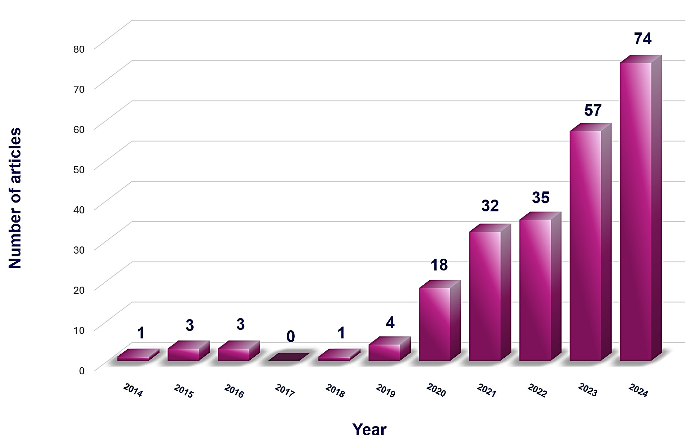
The review methodology herein comprises applying a search strategy consisting of the relevant and updated literature associated with the applicability of artificial intelligence in prosthodontics. Four databases, including PubMed/Medline, Scopus, Embase, and Web of Science, were searched on 15 August 2024. The following items were introduced in the query box: “Prosthodontics and artificial intelligence”, “Artificial intelligence in prosthodontics”, “Artificial intelligence in fixed prosthodontics”, “Artificial intelligence in removable prosthodontics”, “Artificial intelligence in implant prosthodontics”, “Artificial intelligence in maxillofacial prosthodontics”, “Deep learning in prosthodontics”, and “CNNs in prosthodontics”, which were merged with Boolean terms (AND, OR).
The detailed eligibility criteria for the current review are outlined in Table 1. In summary, the inclusion criteria comprised full-text research related to artificial intelligence in prosthodontics. Conversely, excluded were any articles that utilized AI for conditions not specifically related to prosthodontics or prosthetic dentistry, as well as publications including essays, advertisements, conference abstracts, notes, and editorials.
Electronic searches encompassed the literature published in the last ten years, revealing an upward trend, as illustrated in Figure 1. The data shown represent the processed results obtained from PubMed following the application of the established inclusion criteria.

3. Results

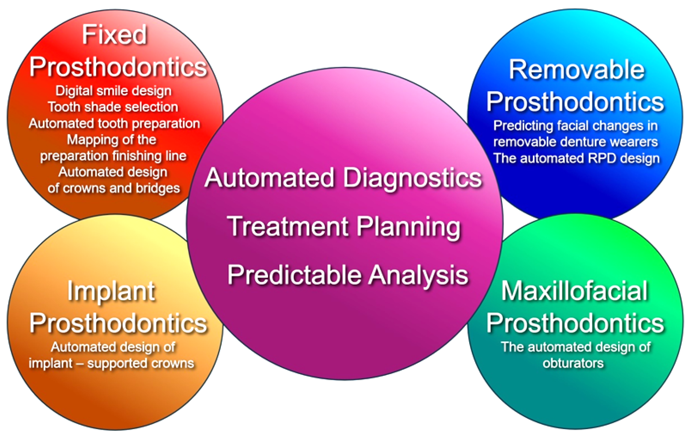
3.1. Main Applications of AI in Prosthodontics

Prosthodontics has continuously evolved as a result of progress in laboratory technology, biomaterial science, clinical techniques, and multidisciplinary and technological advancements such as digital scanning, computer-aided design (CAD), computer-aided manufacturing (CAM), and AI models. The latest made substantial contributions to each of the four major branches of prosthodontics: fixed prosthodontics, removable prosthodontics, implant prosthodontics, and maxillofacial prosthodontics [2,15,16]. At the moment of the current review, a common emerging area can be identified regarding the applicability of AI algorithms within these subspecialties, as well as specific applications for each, with a significantly substantial proportion distinguished within the field of fixed prosthodontics (Figure 2).

3.2. Common AI Applications Across Prosthodontics Subspecialties

3.2.1. The Automated Diagnostics and Treatment Planning

The integration of AI into automated diagnostics in prosthodontic treatment has been a gradual process, gaining significant traction in the last two decades. One of the earliest notable applications was around the early 2010s, when researchers and institutions began to explore AI’s potential in dental diagnostics. A pivotal moment was the introduction of ML algorithms [12], such as the convolutional neural networks (CNNs) for analyzing dental radiographs, which significantly improved diagnostic accuracy and efficiency [17], which are crucial to predicting potential issues for successful prosthodontic outcomes [18]. More recently, a study by Hong J. et al. [19] demonstrated the efficacy of CNNs models in classifying dental implants using panoramic and periapical radiographs. This marked a significant step forward, showcasing how AI could enhance diagnostic processes in implant prosthodontics.
The integration of AI technologies, including ML and DL, has significantly enhanced not only the analysis of radiographic images but also the comprehensive assessment of a patient’s dental condition, commonly known as dental charting. This advancement represents a revolutionary step in prosthodontics, predicated on the premise that precise dental charting is crucial for the accurate design and fitting of dental prostheses [14,20]. Additionally, AI-driven diagnostic tools can integrate patient data from various sources, providing a comprehensive overview that aids in personalized treatment planning [21]. Systems equipped with advanced analytical capabilities can scrutinize dental images with remarkable precision, identifying issues that may elude human detection. Such proficiency is crucial for the early diagnosis of dental conditions, facilitating timely interventions that are essential for achieving successful prosthodontic outcomes [21].
Furthermore, AI facilitates the comprehensive integration and analysis of extensive patient data, enabling prosthodontists to formulate personalized treatment plans that are meticulously tailored to the individual dental and medical histories of each patient. This automation streamlines the diagnostic and planning processes by minimizing the potential for human error and contributing to more reliable and consistent clinical outcomes [18]. In this regard, a recent revolutionary AI-based program can be used to articulate scans in maximum intercuspal position (MIP) and to correct occlusal collisions of articulated scans at MIP, contributing with high accuracy to the diagnostic phase of maxillomandibular relationship record [22].
Despite the evidence, few authors have been actively exploring and popularizing the application of AI in automated diagnostics for prosthodontic treatment in Romania. Instead, a study by Austrian researchers investigated the potential impact and challenges of implementing AI in the Romanian Healthcare System, as well as dental practices, including prosthodontics [23]. This research highlights how AI can enhance diagnostic accuracy and efficiency, ultimately improving patient outcomes. Another significant contribution is from the team led by Alshadidi A. A. F. et al. [24], which, although not exclusively Romanian, includes collaborative efforts from Romanian researchers. The research examines the use of AI in prosthodontics to diagnose abnormalities and create patient-specific prostheses [24]. The findings underscore the advancements in AI applications, particularly in automatically produced diagnostics, predictive analytics, and classification tools.

3.2.2. The Predictable Analysis

Research on the use of AI for prosthodontic restoration has shown that AI can predict the success rates of various treatments by analyzing large datasets of patient outcomes. Thus, within the scope of predictability in prosthodontic treatments, a study conducted by Lee J.-H. et al. [25] successfully assessed the predictability of periodontally compromised teeth using a DL convolutional neural network algorithm. The clinical relevance of this research was particularly significant, as the ability to rapidly and accurately diagnose and predict outcomes is a crucial component of successful prosthodontic treatment [25].
The advanced AI models can also predict complications such as implant failures or prosthesis debonding, allowing for preemptive measures to be taken. This predictive capability enhances the overall success rate of prosthodontic treatments. For instance, Yamaguchi et al. [26] conducted a study aimed at forecasting the likelihood of debonding in CAD/CAM composite resin (CR) crowns by analyzing scanned images of prepared dental models using convolutional neural networks (CNNs), achieving a prediction accuracy of 98.5%. Despite this impressive performance, the researchers acknowledged a significant limitation in their study, noting the difficulty in identifying the primary factors contributing to debonding, which underscores the complexity of isolating specific causative elements within the predictive model [26].

3.3. Specific AI Applications Across Prosthodontics Subspecialties

3.3.1. The Fixed Prosthodontics

  • The Digital Smile Design
Since the introduction of Digital Smile Design (DSD) in 2012, which initially relied on basic PowerPoint templates and specific digital photographic protocols to aid clinicians in visualizing and planning aesthetic treatments, more than 15 smile design software programs have been developed over the course of the last two decades [27]. Thus, the science of smile design has gained a pool position in dentistry, focusing on creating an aesthetically pleasing smile by considering the alignment, shape, and color of teeth, as well as their harmony with facial features. This practical method has garnered increasing attention in recent years and serves as a critical interface between prosthodontics and aesthetic dental care, integrating functional restoration with aesthetic enhancement to achieve optimal clinical outcomes [28]. In this regard, aesthetic dentistry is not recognized as a distinct dental specialty; rather, it necessitates a multidisciplinary approach that may involve restorative, periodontal, orthodontic, and surgical techniques [29].
AI is increasingly being integrated into the domain of smile design, bringing forward numerous benefits and advancements. The AI algorithms possess the capability to meticulously analyze various aspects of facial features, including symmetry, lip line, tooth shape, and size, to generate optimal smile designs. This technological advancement significantly enhances both the precision and efficiency of the design process, thereby enabling dental practitioners to deliver treatments that are not only aesthetically pleasing but also functionally effective [30].
One of the AI-based systems, such as the VisagiSMile concept, leverages ML techniques to establish a relationship between facial perception and personality traits in the context of smile design. This approach facilitates the creation of highly personalized treatment plans that take into account the unique characteristics of each patient, ultimately leading to more tailored and satisfactory outcomes [30]. Recent studies by Ceylan G. et al. [30] and Deshmukh K. et al. [31] have demonstrated that smile designs generated by AI can be as acceptable as those crafted by dental professionals, particularly in cases involving symmetrical facial features. This not only conserves valuable time for clinicians but also ensures that patients receive results that meet their aesthetic expectations. Further, the integration of AI in smile design software facilitates the completion of a fully digital workflow for Digital Smile Design (DSD), a process traditionally considered technically challenging. The adoption of DSD workflows is progressively transforming contemporary dentistry, particularly within both the fields of aesthetic and prosthodontic dentistry.
The utilization of DSD techniques, such as Smile Cloud 2.0 and Rubicon (2020), has notably increased across various dental specialties [32]. Of them, the Smile Cloud Biometrics was developed by a Romanian team in 2023 [33]. The interactive cloud-based platform integrates DSD, treatment planning, and communication tools among clinicians, technicians, and patients. Upon uploading the requisite patient data (e.g., photographs), the AI engine analyzes and suggests natural tooth shapes and alignments. The proposed DSD can be flexibly modified by the clinician and subsequently utilized to generate an STL file for the creation of a mock-up model, preparation guide, or surgical guide [27]. The DSD has become, therefore, very popular among dentists for its capability to generate photorealistic simulations of future smiles by incorporating intra- and extra-oral photographs, as well as intraoral and/or extra-oral scans of the patient’s dental arches and facial structure [34]. In this direction, a prospective observational Romanian study on 250 participants by Buduru S. et al. [35] assessed laypeople’s and dental professionals’ perceptions of an online DSD application, respectively, SmileCloud. The researchers have shown that dental technicians and patients often have differing aesthetic perspectives compared to dentists and students [35]. The findings underscored the necessity for an interdisciplinary cloud-based DSD platform like SmileCloud to align these views and enhance prosthetic treatment outcomes.
The interactive cloud-based platform enhances communication between dental practitioners and technicians, offering a user-friendly workflow that can be managed from any location. The creation of a smile design necessitates a computer or laptop, intra- and extra-oral photographs of the patient, and a foundational understanding of dental aesthetics. From the patient’s perspective, the process appears smoother, more convenient, and less uncomfortable due to the inclusion of an intraoral scanner in place of conventional impressions. The ability to communicate online is a significant advantage of this workflow, making it time-efficient and revolutionary [32,36].
Nevertheless, the integration of AI into smile design is accompanied by ethical considerations, such as the protection of patient privacy, the need for transparency, and the accountability of AI systems. An international consortium has developed comprehensive guidelines to address these concerns, ensuring that AI applications in dentistry adhere to established ethical principles [37].
  • The Tooth Shade Selection
The shade guides for tooth shade selection in clinical prosthodontics are currently available in countless types and capabilities. Along the classical Vita Classical Shade Guide (Bad Säckingen, Germany: VITA Zahnfabrik H. Rauter GmbH & Co.) and Vita 3D-Master Shade Guide (Bad Säckingen, Germany: VITA Zahnfabrik H. Rauter GmbH & Co.), the shade-matching spectrophotometers, intraoral electronic devices like Vita Easyshade (Vita Zahnfabrik, Bad Säckingen, Germany), the ShadeEye NCC Chroma Meter (Shofu Dental, Menlo, CA, USA), the iTero Element (Align Technology, Inc., San Jose, CA, United States), computer-aided shade selection software, colorimetric systems, hybrid devices, and mobile applications are some of the past and current options for an accurate assessment of tooth color and shade [38]. In the past five years, researchers have identified AI as a crucial vector in advancing tooth shade selection (TSS) technologies. This process is pivotal for ensuring the aesthetic success of dental restorations. TSS arises inherently complex, demanding high-precision, consistent, and traditional methods, which often rely on visual matching, are subjective, and are susceptible to human error. Recent advancements in AI technologies offer a more standardized and accurate approach, significantly enhancing the reliability of shade matching [39,40]. The integration of AI in TSS for fixed prosthodontics has revolutionized the field, enhancing accuracy and efficiency. Various subsets of AI, including ML, DL, and CNNs models, are nowadays involved [41], each contributing uniquely to the advancements in this area.
First, the ML algorithms have been crucial in improving the precision of TSS. These algorithms analyze large datasets of tooth images and their corresponding shades, learning to predict the correct shade based on various parameters [42]. One notable application is the use of support vector machines (SVMs) and k-nearest neighbors (k-NN) algorithms, both popular supervised ML tools used for classification and regression tasks to classify tooth shades [43]. For instance, a study by Lee et al. [44] demonstrated the use of SVMs in accurately predicting tooth shades by analyzing the colorimetric data from digital images. This approach reduces the subjectivity associated with traditional visual methods and enhances consistency in shade selection.
In the same context of TSS, DL models, particularly CNNs, have shown remarkable success. Takahashi et al. [45] utilized a DL model to automate the shade-matching process. Their study employed CNNs trained on a large dataset of tooth images, achieving high accuracy in shade selection. Thus, the DL model could identify subtle differences in tooth color that are often missed by the human eye, leading to more precise and aesthetically pleasing restorations.
Finally, CNNs have been used in TSS to analyze intraoral photographs and match them with the appropriate shade guides. Mehta et al. [46] developed a CNN-based system that outperformed traditional methods in terms of accuracy and speed. Their model was trained on a diverse dataset, allowing it to generalize well across different lighting conditions and patient demographics. This technology not only improves the accuracy of shade selection but also streamlines the workflow in dental practices.
  • The Automated Tooth Preparation
In 2024, Perceptive, a company based in Boston (MA, USA), introduced an AI-driven robotic system designed for dental procedures, including the preparation of teeth for dental crowns. This innovative robot utilizes advanced optical coherence tomography (OCT) and AI programming to create detailed 3D maps of the teeth, which are then analyzed by AI to plan the tooth preparation. The system can complete a procedure that typically takes several hours in just about 15 min [47,48]. Further benefits consist of increased precision and accuracy compared to that matched by the human hands, resulting in better fitting crowns and bridges [49], consistency, and reliability [49,50] since robots do not suffer from fatigue or human error, ensuring consistent performance across procedures. While still in its prototype phase, this technology has been tested successfully in the United States and is expected to expand to other countries, particularly in regions with limited access to dental care [47].
Another notable AI-driven robot in dentistry is Yomi by Neocis, introduced in 2017. While this was the first and only FDA-cleared robotic system for dental surgery, primarily assisting in dental implant procedures, its technology could also be adapted for other dental procedures, including tooth preparation [51]. It was followed by the DentSim by Image Navigation [52], which was introduced in 2018 as an advanced dental training simulator that uses augmented reality and AI to help dental students practice procedures. While it does not perform procedures on patients, it represents a significant step toward integrating AI and robotics in dental education and practice. Finally, Tactile Robotics introduced its robot-assisted dental surgery system in 2019 [53]. This system uses haptic feedback and AI to assist dentists in performing precise dental procedures, enhancing the accuracy and efficiency of various treatments, including tooth preparation.
Although the number of completed I-driven robotic models to date is limited, their development represents a significant advancement in the integration of AI and robotics within prosthodontic treatment. This innovation aims to enhance efficiency, accuracy, and patient outcomes [49]. Currently, there are no data indicating that robots for tooth preparation, like the ones developed by Perceptive, are being used in Romania. However, as technology advances and demonstrates its effectiveness, it is likely that such innovations will disseminate to other nations, including our own, due to their desirability, particularly in regions with limited access to dental care.
  • The Mapping of the Preparation Finishing Line
The use of AI in mapping the preparation finishing line in fixed prosthodontics promises significant precision and efficiency in the last decade. AI algorithms, particularly those based on ML and DL, have been developed to accurately detect and map the preparation finishing line of dental preparations. These algorithms analyze digital impressions or intraoral scans to identify the exact margins of the preparation, ensuring a precise fit for the dental crowns and bridges [54]. Automation is achieved through the use of CNNs and other advanced AI models that can learn from large datasets of dental images [37]. In this context, Zhang B. et al. [55] developed an AI model utilizing CNNs based on the S-Octree structure to autonomously detect the finishing line of tooth preparations. The study employed 380 digitized tooth preparation dies of premolars and molars intended for crowns sourced from an unidentified origin. These virtual dies were processed as sparse point clouds, annotated with labels, and subsequently used to train the CNN model. The AI model demonstrated the capability to identify the margin line without manual intervention, achieving an average accuracy between 90.6% and 97.4%. Consequently, this research underscores the potential for automating the identification of finishing lines, thereby advancing the automation of dental restoration design through the integration of AI models with dental CAD software [55]. Several notable dental CAD software solutions that facilitate this integration are summarized below, highlighting their key features and applications in the design and mapping of tooth preparations (Table 2).
The clinical relevance of these AI-driven tools also lies in their real-time assistance to dentists in clinical settings, providing immediate feedback on the quality of the preparation and suggesting modifications if necessary. This real-time support contributes to achieving optimal results and improving patient outcomes [39]. As of this review, AI-driven tools for real-time mapping of the finishing line of dental preparations are being utilized in several regions around the world. In the United States, AI technologies are widely adopted in dental practices for various applications, including real-time mapping of dental preparations [56]. European countries like Germany and the United Kingdom have numerous dental research centers and clinics employing AI-driven tools for precision in dental restorations [57]. In China, AI models for dental preparations are developed and implemented in various dental schools and clinics [58]. Japan and South Korea are also examples of robust dental technology sectors that integrate AI for real-time mapping and other dental applications [59]. Despite this, very few comparative studies of classical marginal preparation versus AI-driven marginal preparation, due to the mapping of the finishing line, have highlighted the relevance of these revolutionary new technologies. Mugri, M.H. et al. [60] investigated the combined effect of digital manufacturing techniques (subtractive vs. additive), preparation taper, and finish line design on the marginal adaptation of temporary crowns, highlighting the role of AI-driven CAD/CAM systems in achieving precise marginal fit.
  • The Automated Restoration Design
The application of AI in fixed prosthodontics primarily focuses on automated restoration design. CAD/CAM technology has digitized the design process in commercial products such as CEREC (Chairside Economical Restoration of Esthetic Ceramics), (Dentsply Sirona, Bensheim, Germany), Planmeca PlanCAD (Planmeca Oy, Helsinki, Finland), Roland DGSHAPE (Roland DG Corporation, Hamamatsu, Japan), Sirona (Dentsply Sirona, Bensheim, Germany), 3Shape (3Shape, Copenhagen, Denmark), and others. While this has significantly increased the efficiency of the design process by utilizing a tooth library for crown design, it still falls short of achieving a custom-made design for individual patients [61]. Some AI-driven tools, such as 2D Generative Adversarial Networks (GANs), introduced in 2014, are used to generate high-quality images of dental restorations. These tools primarily assist in the automated design of tooth anatomy by creating realistic representations for dental crown restorations. In this regard, Hwang et al. [62] and Tian et al. [63] proposed innovative approaches based on 2D-GAN models to generate crowns by learning from technicians’ designs, using 2D depth maps converted from 3D tooth models as training data.
Three-dimensional GAN networks take them a step further by generating three-dimensional models of dental restorations, which are particularly useful for designing dental crowns by creating accurate 3D models that fit perfectly with the patient’s existing dentition. Recently, Ding H. et al. [64] introduced a 3D-DCGAN network for crown generation, which directly utilizes 3D data in the process, resulting in crown morphologies that closely resemble natural teeth. Building on those foundational results, Hosseinimanesh et al. [65] advanced the application of AI in dental prosthetics by introducing a new end-to-end DL approach called Dental Mesh Completion (DMC) for generating crown meshes conditioned on a point cloud context. This method aimed to simplify the crown design process while ensuring accuracy and consistency. The study demonstrated the effectiveness of the method with an average chamfer distance of 0.062, indicating a high level of precision in the generated crowns [64].

3.3.2. The Removable Prosthodontics

  • The Prediction of Facial Changes in the Removable Denture Wearers
The accurate prediction of facial changes in patients wearing removable prostheses in prosthodontics is essential not only for aesthetic satisfaction but also for functional outcomes. Studies have shown that discrepancies in facial appearance due to poorly fitted dentures can lead to significant patient dissatisfaction, with up to 30% of denture wearers reporting dissatisfaction primarily due to aesthetic concerns [66]. This dissatisfaction often results in non-compliance with denture use, which can further exacerbate oral health issues, including atrophy of the alveolar ridge, compromised masticatory function, reduced quality of life [67], and further deterioration of general health [68,69]. Consequently, there is a pressing need for advanced technologies that can enhance the precision of these predictions and improve the overall success rate of prosthodontic treatments.
Traditional methods, such as facial measurements [70], technologies like 3D facial scanning and imaging [71] and cephalometric analysis [72], or the advanced computational models for facial deformation prediction, such as the mass-spring model (MSM), the finite element model (FEM) and the mass tensor model (MTM) [73], while effective, often require a combination of techniques to achieve accurate predictions. Consequently, there was a pressing need for advanced technologies that could enhance the precision of these predictions and improve the overall success rate of prosthodontic treatments. In this context, AI technologies represent a groundbreaking advancement by providing highly accurate predictions of facial changes in patients wearing removable prostheses. These technologies analyze vast datasets to identify patterns and predict outcomes with unprecedented accuracy.
Recent studies have demonstrated the efficacy of AI in this domain. For instance, the research conducted by Zhu J. et al. [74] from the University of Hong Kong has shown that AI-based systems can reliably simulate facial morphology changes, offering valuable insights for prosthodontic treatment planning. Conversely, with the help of AI, a study from Switzerland conducted by Obwegeser D. et al. [75] demonstrates the significant effect of dental alignment on the perception of facial attractiveness. An interesting and noteworthy result is that of a recent study from Egypt by Helmy M. et al. [76], which compares, among other parameters, the perception of changes in facial aesthetics following removable prosthetic restoration using conventional methods (clinical and cephalometric measurements) and AI-driven cephalometric for VDO in completely edentulous patients. In this case, the self-perception of facial attractiveness was similar, regardless of the method chosen to establish the height of the lower facial third [76].
Until present, a single study applied a backpropagation neural network (BPNN), which is a fundamental part of AI, particularly in the field of DL, to predict facial changes in edentulous patients about to be restored with a removable prosthesis. Cheng C. et al. [77] developed in 2015 an AI model capable of predicting facial soft-tissue changes following the delivery of a complete denture. However, variables such as buccal flange thickness and occlusion type were not included in the dataset, which may have affected the accuracy of the predictions [77].
  • The Removable Partial Denture Design
Unlike fixed prosthodontics, the design of removable prosthodontics is more challenging due to the need to consider a greater number of factors and variables [9]. Even so, the integration of AI into the design and fabrication of removable partial dentures (RPD) has become increasingly prevalent over the past decade.
Since the early 2010s, technologies such as ML and DL have significantly enhanced the accuracy, efficiency, and customization of dental prostheses [78,79]. In particular, CNNs have demonstrated superior accuracy in diagnosing edentulous spaces compared to traditional methods [14]. The CNNs, as a type of DL model specifically designed for image analysis, automatically extract features from images, such as radiographs and cone-beam computed tomography (CBCT) scans. For instance, the study by Gerhardt M. et al. [80] demonstrated that an AI tool using CNNs achieved a detection accuracy of 99.7% for teeth and 99% for identifying missing teeth. This high level of accuracy can be attributed to the layered architecture of CNNs. The convolutional layers in the AI tool effectively extracted detailed features from the CBCT images, such as the edges and contours of teeth and bone structures. The pooling layers then reduced the complexity of these features, making the analysis more efficient. Finally, the fully connected layers synthesized this information to accurately identify edentulous spaces [81].
Once the edentulous spaces are identified, the next step involves classifying the dental arches. The study by Takahashi T. et al. [82] focuses on developing a method for classifying dental arches using a convolutional neural network (CNN) as the first step in designing removable partial dentures. The researchers used 1184 images of dental arches to classify them into four types: edentulous, intact dentition, arches with posterior tooth loss, and arches with bounded edentulous space. The CNNs method showed high diagnostic accuracy, with results suggesting that dental arches can be effectively classified and predicted using this AI technology, which is also an essential step for planning the design of the RPD.
Furthermore, digital impressions of the denture-bearing areas are then used to create accurate 3D models of the patient’s oral cavity. AI algorithms enhance the accuracy of digital impressions by identifying and correcting errors in the scanned data, which is crucial for the subsequent design and fabrication of the RPD [82]. Using the 3D models, AI-driven CAD/CAM systems create an optimal design that ensures proper fit, support, and aesthetics of the RPD framework, the process including the determination of the placement of occlusal rests, clasps, and other components to ensure stability and retention [82]. In removable prosthodontics, a clinical decision support model that applies case-based reasoning and ontology proved capable of proposing the design of individualized RPDs. However, this model bases its recommendations on the most likely example in the database. Because clinical settings are constantly varying, it is necessary to keep a dubious approach to its output.
The designed RPD framework is then fabricated using additive manufacturing techniques, such as 3D printing or direct metal laser sintering (DMLS). AI enhances the precision of these manufacturing processes, ensuring that the RPD frameworks are fabricated with high accuracy. AI systems also monitor the manufacturing process in real time, detecting any deviations or defects to ensure consistent quality [83].
After the fabrication of the RPD, its adaptation is evaluated using advanced AI tools, including AI-powered optical scanners [82]. These tools generate detailed 3D models of the RPD within the patient’s oral cavity, facilitating precise assessment and necessary adjustments to ensure an optimal fit [83]. Additionally, these AI systems can simulate the functional performance of the RPD, such as the distribution of occlusal forces during mastication, and the stability and retention of the RPD under different conditions, such as various mastication patterns and jaw movements. Additionally, AI can model the long-term wear and tear of the RPD materials, predict the lifespan of the denture, identify potential points of failure, and monitor the manufacturing process in real time, detecting any deviations or defects to ensure consistent quality [66].
Despite a gap in collaboration with AI systems in the Romanian removable prosthodontic field, the benefits of automated removable denture design, including complete dentures and not just automatic framework designs for RPDs, have been recently highlighted and disseminated internationally by researchers from Romania [84].

3.3.3. The Implant Prosthodontics

One of the primary applications of AI in implant prosthodontics is in the diagnostic phase. AI algorithms, trained on vast datasets of images, possess the capability to analyze vast amounts of patient data, including radiographic images, three-dimensional scans, and clinical records [85], and also accurately identify and classify dental pathologies, bone structures, and anatomical landmarks. This capability is crucial for the precise placement of dental implants, ensuring optimal integration with the patient’s existing bone structure. For instance, AI systems can analyze CBCT scans to detect the quality and quantity of bone, which is essential for determining the feasibility of implant placement and planning the surgical approach [86]. In a study conducted by Kurt Bayrakdar S. et al. [87], the efficacy of an AI system in analyzing bone height revealed that the AI system achieved an accuracy rate of 72.2% for detecting canals, 66.4% for identifying sinuses and fossae, and 95.3% for recognizing regions with missing teeth. The results underscore the potential of DL technologies to significantly enhance diagnostic accuracy and treatment planning in dentistry, particularly within the domain of implant prosthodontics [87]. Building on those foundations, Takahashi T. et al. [88] employed the Yolov3 object detection algorithm to identify dental implant systems in panoramic radiographs. While the algorithm demonstrated high accuracy, particularly for certain implant types, the study highlighted the necessity for a broader range of implant system images to improve its clinical applicability. Nassani L.M. et al. [89] reported about a deep trained CNN model using a dataset of 10,770 radiographic images representing three implant types. Also, the accuracy of implant recognition between board-certified periodontists and the AI model across periapical, panoramic, and combined images has been compared. The findings indicated that both the AI model and the periodontists achieved higher specificity and sensitivity when utilizing both periapical and panoramic images, with varying accuracy for different implant types.
In terms of constructing implant-supported fixed prostheses, Lerner H. et al. [90] investigated the creation of implant-supported monolithic zirconia crowns using a fully digital procedure facilitated by AI. This retrospective study, which involved 106 implant-supported monolithic zirconia crowns placed in the posterior jaws of 90 patients, encompassed a comprehensive protocol including intraoral scanning, CAD design, milling, and clinical application of the crowns. The results demonstrated a high success rate, characterized by excellent marginal adaptation, interproximal and occlusal contacts, and aesthetic integration [90]. To date, other important applications are possible in the implant prosthodontics. AI-based treatment planning in CAD/CAM implant dentistry significantly streamlines virtual 3D treatment planning and enables the robotic insertion of dental implants [91]. In this regard, robotic-assisted dental implant procedures have been successfully performed on humans, the Yomi robotic system assisting clinicians during dental implant surgeries by haptic feedback and real-time guidance [92]. Additionally, a fully autonomous robotic dental surgeon recently performed its first human procedure, demonstrating the potential for AI-driven robotics in implant prosthodontics and its benefits: minimally invasive flapless surgery, multisensory feedback, including physical guidance, on-screen alerts, and audio cues, respectively, enhanced visualization of the surgical site [93].

3.3.4. The Maxillofacial Prosthodontics

Maxillofacial prosthetics is frequently characterized as the discipline that encompasses both the art and science of reconstructing anatomical, functional, or cosmetic deficiencies in the maxilla, mandible, and facial regions. This reconstruction is achieved through the use of surgical and prosthetic substitutes to replace areas that are absent or impaired due to surgical procedures, traumatic injuries, pathological conditions, or developmental and congenital anomalies [94]. Primary objectives when managing patients in the field of maxillofacial prosthodontics should be to restore form, function, and aesthetics [95,96,97], maintain a healthy and sustainable periodontium, achieve stable temporomandibular joints and occlusion, preserve healthy teeth, ensure comfortable function, and attain optimal aesthetics [98].
Maxillofacial prosthetic rehabilitation is also a multifaceted process that unfolds through three distinct and sequential phases: the surgical phase, the interim phase, and the definitive phase. In the context of maxillary rehabilitation, the initial step involves the placement of a surgical obturator. This prosthesis serves the dual purpose of re-establishing the separation between the oral and nasal cavities and restoring the proper palatal contour [99,100]. Following the initial healing period, the surgical obturator is replaced with an interim obturator. Unlike its predecessor, the interim obturator often incorporates teeth, thereby facilitating the patient’s gradual return to normal oral functions [94]. This prosthesis is subject to continuous adjustments as the surgical site heals and the defect undergoes changes in size and shape. These adjustments typically involve the addition or removal of material to maintain optimal separation between the oral and nasal cavities, ensuring that the prosthesis remains functional and comfortable for the patient [101,102]. Once the surgical site has fully healed, the final prosthesis is designed to further enhance both aesthetics and function.
The process for mandibular prosthetic rehabilitation follows a similar pattern. Initially, a surgical prosthesis is employed to stabilize a split-thickness skin graft and recreate the vestibule, thereby preparing the site for the final prosthesis. The interim prosthesis plays a crucial role in re-establishing the occlusal relationship and assisting the patient in learning how to function and maintain the prosthesis. This phase is characterized by continuous adjustments to accommodate the healing process and changes in the surgical site [100].
In all, the construction of obturators for patients with maxillofacial defects is a complex process that significantly impacts quality of life [102]. Although still in its pioneering stage, recent studies have demonstrated the potential of AI algorithms in optimizing the design of obturators. For instance, DL techniques have been employed to analyze patient-specific anatomical data and generate custom prosthetic designs with improved aesthetics, fit, and functionality. In this regard, Mine Y. et al. [103] highlighted the use of the artificial neural network (ANN)-based deep learning approach to coloration support for fabricating maxillofacial prostheses. Additionally, Meral K. et al. [104] compared the performance of two DL algorithms, the attention-based gated recurrent unit (GRU) and the ANNs algorithm for coloring silicone maxillofacial prostheses, obtaining better results for the first model.
More recent research demonstrates further steps of integrating AI into the design and fabrication of obturators to enhance accuracy, reduce fabrication time, and improve maxillofacial patient quality of life. Thus, Ali I.E. et al. [105] evaluated four pre-trained CNNs (VGG16, Inception-ResNet-V2, DenseNet−201, and Xception) for recognizing seven prosthodontic scenarios related to the maxilla as a step toward creating an AI-driven prosthesis design system. The scenarios included various conditions like cleft palate and different types of maxillectomy. Results indicated that all models performed well, with test accuracies of up to 95%, where Xception and DenseNet−201 slightly outperformed the others. In conclusion, all models surpassed 90% accuracy, indicating their effectiveness in dental image analysis, automated diagnosis, and enhancement of the prosthesis design [106].

4. Discussion

The effectiveness of AI algorithms is significantly influenced by the quality and quantity of data available for training. In the field of prosthodontics, the absence of comprehensive, high-quality datasets encompassing diverse patient demographics, clinical scenarios, and treatment outcomes can impede the development of robust AI models [17]. Additionally, many AI applications are developed in well-resourced environments, which may not be applicable in settings with limited data access [91].
Integrating AI solutions into existing clinical workflows presents several challenges. Resistance to change among dental practitioners, coupled with concerns regarding the reliability of AI recommendations, can hinder adoption [106]. Furthermore, the lack of established guidelines for AI use in clinical settings poses a challenge for practitioners globally [107]. AI algorithms, particularly DL models, often function as “black boxes”, making it difficult for clinicians to understand the decision-making process. This lack of interpretability can result in hesitance to utilize AI in critical decision-making processes, as practitioners may prefer to rely on their clinical judgment rather than opaque algorithms.
Many prosthodontic practices worldwide may lack the necessary infrastructure or technical expertise to seamlessly adopt and integrate AI technologies. Financial concerns further exacerbate the issue, making the adoption of AI technologies prohibitive, especially for small and mid-sized practices. The initial investment for AI systems, ongoing maintenance, and required staff training can create significant barriers to entry, particularly in resource-limited settings. At present, Romania also faces challenges in adopting advanced AI dental technologies due to economic constraints and varying levels of access to healthcare resources. While urban dental clinics may have better access to AI technologies, rural areas may lag behind, creating disparities in patient care. To harness the potential of AI in prosthodontics, developing countries need investments in digital infrastructure, including high-quality imaging systems, data management solutions, and training programs for dental professionals. Public and private partnerships can facilitate this investment, enabling wider access to advanced technologies.
Beyond any borders, it has to be mentioned that the use of AI in healthcare raises ethical questions, particularly concerning patient privacy, data security, and algorithmic biases. The ethical use of AI in prosthodontics also involves adherence to regulatory standards, which can vary significantly across different countries. Compliance with these regulations ensures that AI applications are used responsibly and ethically, safeguarding patient welfare and maintaining public trust in AI technologies.
Despite these limitations, the future prospects are promising, particularly from the perspective of individualized prosthetic treatment. As AI technologies advance, they may facilitate more personalized treatment approaches tailored to individual patient needs, potentially improving outcomes and patient satisfaction. An important step forward, including our country, concerns the utility of integrating AI into the dental education of future prosthodontists to leverage these technologies effectively. As current studies indicate [108], there have been significant changes in students’ knowledge, skills, interests, and perceptions regarding technology—specifically concerning the availability of technology in clinical settings, its value to patients, its overall importance, and the relative frequency of clinics equipped with such technology [108]. We are not far from the moment when the inclusion of AI and digital technologies in a revised curriculum becomes a reality, thus equipping students with the necessary skills to work in an AI-enhanced environment. This integration should also encompass the continuous professional development of dentists [34].
International collaboration with academic and research institutions can further promote innovation in AI applications tailored to the Romanian context. By exploiting local expertise and data, researchers can develop solutions that address specific challenges faced by Romanian prosthodontists.
Toward the end of our analysis of AI in prosthodontics, it is also important to acknowledge that its narrative design faces certain limitations, particularly when compared to systematic reviews and meta-analyses. The more flexible methodology in selecting and evaluating studies while offering a broader exploration may introduce potential biases. Additionally, the absence of quantitative analysis, as is typical in narrative reviews—including the present one—can limit the ability to draw definitive conclusions. While this extensive research provides valuable qualitative insights into the emerging applications of AI in prosthodontics, its findings should be interpreted with caution.

5. Conclusions

The integration of AI into prosthodontics heralds unprecedented advancements in this field. AI algorithms offer remarkable potential to revolutionize automated diagnostics, treatment planning, and the predictability of both teeth and implant-supported fixed or removable dentures, enhancing precision and efficiency through advanced technologies and technical skills. However, challenges related to data quality, integration, and ethical considerations must be addressed to fully realize these benefits. This review underscores the evolutionary trend of incorporating AI technologies within prosthodontics, which are contributing significantly to global advancements in dental care. It also highlights existing gaps that need to be bridged.
Despite the substantial progress made worldwide, there remains a critical need for broader implementation of AI-driven diagnostics, extending beyond academic institutions to include private practice. For countries like Romania, a concerted effort in developing infrastructure, education, and regulatory frameworks is essential to harness AI’s full potential in improving prosthodontic care. By addressing these challenges, the field can achieve remarkable strides in delivering superior dental health outcomes.

Author Contributions

Conceptualization, L.I., G.F.G., B.D. and M.I.; methodology, A.M.C.Ț., G.F.G. and O.E.A.; software, L.I., G.F.G. and B.D.; validation, G.F.G., O.E.A., M.I. and L.I.; formal analysis, L.I. and A.M.C.Ț.; investigation, A.M.C.Ț., G.F.G., M.I. and O.E.A.; data curation, A.M.C.Ț. and L.I.; writing—original draft preparation, L.I., G.F.G. and B.D.; writing—review and editing, B.D., A.M.C.Ț. and O.E.A.; visualization, A.M.C.Ț., O.E.A. and B.D.; supervision, B.D. and M.I. All authors have read and agreed to the published version of the manuscript.

Funding

This research received no external funding.

Institutional Review Board Statement

Not applicable.

Informed Consent Statement

Not applicable.

Data Availability Statement

Data are available on request from the corresponding authors.

Acknowledgments

Publication of this paper was supported by the University of Medicine and Pharmacy Carol Davila through the institutional program Publish not Perish.

Conflicts of Interest

The authors declare no conflicts of interest.

References

  1. Wiens, J.P.; Koka, S.; Graser, G.; White, S.N.; Marinello, C.P.; Curtis, D.A. Academy of Prosthodontics Centennial: The Emergence and Development of Prosthodontics as a Specialty. J. Prosthet. Dent. 2017, 118, 569–572. [Google Scholar] [CrossRef] [PubMed]
  2. Academy of Prosthodontics. The Glossary of Prosthodontic Terms. J. Prosthet. Dent. 2023, 130, e1–e3. [Google Scholar] [CrossRef] [PubMed]
  3. Minervini, G.; Fiorillo, L.; Russo, D.; Lanza, A.; D’Amico, C.; Cervino, G.; Meto, A.; Di Francesco, F. Prosthodontic Treatment in Patients with Temporomandibular Disorders and Orofacial Pain and/or Bruxism: A Review of the Literature. Prosthesis 2022, 4, 253–262. [Google Scholar] [CrossRef]
  4. Hagag, G.; Yoshida, K.; Miura, H. Occlusion, Prosthodontic Treatment, and Temporomandibular Disorders: A Review. J. Med. Dent. Sci. 2000, 47, 61–66. [Google Scholar] [PubMed]
  5. Manfredini, D.; Poggio, C.E. Prosthodontic Planning in Patients with Temporomandibular Disorders and/or Bruxism: A Systematic Review. J. Prosthet. Dent. 2017, 117, 606–613. [Google Scholar] [CrossRef]
  6. Cristache, C.M.; Tudor, I.; Moraru, L.; Cristache, G.; Lanza, A.; Burlibasa, M. Digital Workflow in Maxillofacial Prosthodontics—An Update on Defect Data Acquisition, Editing and Design Using Open-Source and Commercial Available Software. Appl. Sci. 2021, 11, 973. [Google Scholar] [CrossRef]
  7. Spielman, A.I. History of Prosthodontics. In Illustrated Encyclopedia of the History of Dentistry; Harper & Brothers: New York, NY, USA, 2023; Available online: https://historyofdentistryandmedicine.com/ (accessed on 20 August 2024).
  8. Di Fiore, A.; Brunello, G. Current Trends and Future Directions in Prosthetic and Implant Dentistry in the Digital Era; MDPI: Basel, Switzerland, 2022. [Google Scholar]
  9. Ding, H.; Wu, J.; Zhao, W.; Matinlinna, J.P.; Burrow, M.F.; Tsoi, J.K.H. Artificial Intelligence in Dentistry—A Review. Front. Dent. Med. 2023, 4, 1085251. [Google Scholar] [CrossRef]
  10. Castiglioni, I.; Rundo, L.; Codari, M.; Di Leo, G.; Salvatore, C.; Interlenghi, M.; Gallivanone, F.; Cozzi, A.; D’Amico, N.C.; Sardanelli, F. AI applications to medical images: From machine learning to deep learning. Phys. Medica 2021, 83, 9–24. [Google Scholar] [CrossRef]
  11. Arsiwala-Scheppach, L.T.; Chaurasia, A.; Müller, A.; Krois, J.; Schwendicke, F. Machine Learning in Dentistry: A Scoping Review. J. Clin. Med. 2023, 12, 937. [Google Scholar] [CrossRef]
  12. Allani, H.; Santos, A.T.; Ribeiro-Vidal, H. Multidisciplinary Applications of AI in Dentistry: Bibliometric Review. Appl. Sci. 2024, 14, 7624. [Google Scholar] [CrossRef]
  13. Ünsal, G.; Orhan, K. Future Perspectives of Artificial Intelligence in Dentistry. In Artificial Intelligence in Dentistry; Springer: Berlin/Heidelberg, Germany, 2024; pp. 353–364. Available online: https://link.springer.com/chapter/10.1007/978-3-031-43827-1_19 (accessed on 20 August 2024).
  14. Semerci, Z.M.; Yardımcı, S. Empowering Modern Dentistry: The Impact of Artificial Intelligence on Patient Care and Clinical Decision Making. Diagnostics 2024, 14, 1260. [Google Scholar] [CrossRef] [PubMed]
  15. Jokstad, A.; Orstavik, J.; Ramstad, T. A definition of prosthetic dentistry. Int. J. Prosthodont. 1998, 11, 295–301. [Google Scholar] [PubMed]
  16. American College of Prosthodontists. History, Information, and Examination Requirements of the American Board of Prosthodontics. J. Prosthet. Dent. 1984, 52, 281–287. [Google Scholar] [CrossRef] [PubMed]
  17. Schwendicke, F.; Golla, T.; Dreher, M.; Krois, J. Convolutional neural networks for dental image diagnostics: A scoping review. J. Dent. 2019, 91, 103226. [Google Scholar] [CrossRef]
  18. Orhan, K.; Aksoy, U.; Aksoy, S. Applications of AI in Endodontics and Restorative Dentistry. In Artificial Intelligence in Dentistry; Springer: Berlin/Heidelberg, Germany, 2024; pp. 69–81. [Google Scholar] [CrossRef]
  19. Hong, J.; Huh, J.-K.; Lee, J.-H. Automated Deep Learning for Classification of Dental Implant Radiographs Using a Large Multi-Center Dataset. Sci. Rep. 2023, 13, 32118. [Google Scholar] [CrossRef]
  20. Naeimi, S.M.; Darvish, S.; Salman, B.N.; Luchian, I. Artificial Intelligence in Adult and Pediatric Dentistry: A Narrative Review. Bioengineering 2024, 11, 431. [Google Scholar] [CrossRef]
  21. Cervino, G.; Peditto, M.; Portelli, M.; Militi, A.; Matarese, G.; Fiorillo, L.; Nucera, R.; Oteri, G. The Use of AI for Prosthodontic Restoration: Predictable and Safer Dentistry. Eng. Proc. 2023, 56, 68. [Google Scholar] [CrossRef]
  22. Revilla-León, M.; Gomez-Polo, M.; Barmak, A.B.; Lois, J.C.; Perez-Barquero, J.A. Accuracy of an Artificial Intelligence-Based Program for Locating the Maxillomandibular Relationship of Scans Acquired by Using Intraoral Scanners. J. Prosthet. Dent. 2024. Online ahead of print. [Google Scholar] [CrossRef]
  23. Fitzek, S.; Choi, K.-E. Potential Impact and Challenges of Implementing Artificial Intelligence in the Romanian Healthcare System. Postmod. Probl. 2023, 13, 148–192. [Google Scholar] [CrossRef]
  24. Alshadidi, A.A.F.; Alshahrani, A.A.; Aldosari, L.I.N.; Chaturvedi, S.; Saini, R.S.; Hassan, S.A.B.; Cicciù, M.; Minervini, G. Investigation on the Application of Artificial Intelligence in Prosthodontics. Appl. Sci. 2023, 13, 5004. [Google Scholar] [CrossRef]
  25. Lee, J.-H.; Kim, D.-H.; Jeong, S.-N.; Choi, S.-H. Diagnosis and Prediction of Periodontally Compromised Teeth Using a Deep Learning-Based Convolutional Neural Network Algorithm. J. Periodontal Implant. Sci. 2018, 48, 114. [Google Scholar] [CrossRef] [PubMed]
  26. Yamaguchi, S.; Lee, C.; Karaer, O.; Ban, S.; Mine, A.; Imazato, S. Predicting the Debonding of CAD/CAM Composite Resin Crowns with AI. J. Dent. Res. 2019, 98, 1234–1238. [Google Scholar] [CrossRef]
  27. Chen, Y.-W.; Stanley, K.; Att, W. Artificial Intelligence in Dentistry: Current Applications and Future Perspectives. Quintessence Int. 2020, 51, 248–257. [Google Scholar]
  28. Erdemir, U.; Yildiz, E.; Yucel, T. Esthetic Parameters/Smile Design. In Esthetic and Functional Management of Diastema; Erdemir, U., Yildiz, E., Eds.; Springer: Berlin/Heidelberg, Germany, 2016. [Google Scholar] [CrossRef]
  29. Jurado, C.A.; Villalobos-Tinoco, J.; Lackey, M.A.; Rojas-Rueda, S.; Robles, M.; Tsujimoto, A. Three Dimensional-Printed Gingivectomy and Tooth Reduction Guides Prior Ceramic Restorations: A Case Report. Dent. J. 2024, 12, 245. [Google Scholar] [CrossRef]
  30. Ceylan, G.; Sayın Özel, G.; Memişoglu, G.; Emir, F.; Şen, S. Evaluating the Facial Esthetic Outcomes of Digital Smile Designs Generated by Artificial Intelligence and Dental Professionals. Appl. Sci. 2023, 13, 9001. [Google Scholar] [CrossRef]
  31. Deshmukh, K.; Radke, U.; Deshmukh, S.; Pande, N.; Mowade, T.; Hatmode, T. Role of Digitalization in Smile Designing: A Review. J. Adv. Dent. Pract. Res. 2022, 1, 27–30. [Google Scholar] [CrossRef]
  32. Cervino, G.; Fiorillo, L.; Arzukanyan, A.; Spagnuolo, G.; Cicciù, M. Dental Restorative Digital Workflow: Digital Smile Design from Aesthetic to Function. Dent. J. 2019, 7, 30. [Google Scholar] [CrossRef] [PubMed]
  33. Cofar, F.; Coachman, C. SMILE CLOUD: AI-Powered Digital Smile Design Platform. 2023, Straumann Group. Available online: https://www.straumann.com/us/en/dental-professionals/digital-performance/software/smilecloud.html (accessed on 15 September 2024).
  34. Costin, L.I.; Vasluianu, R.I.; Baciu, R.-E.; Agop-Forna, D.; Cătălin, T.S.; Forna, C.N. The Integration of Artificial Intelligence in Dental Practice. Rom. J. Oral Rehabil. 2023, 2, 272–279. Available online: http://rjor.ro/wp-content/uploads/2023/07 (accessed on 15 September 2024).
  35. Buduru, S.; Cofar, F.; Mesaroș, A.; Tăut, M.; Negucioiu, M.; Almășan, O. Perceptions in Digital Smile Design: Assessing Laypeople and Dental Professionals’ Preferences Using an Artificial-Intelligence-Based Application. Dent. J. 2024, 12, 1049. [Google Scholar] [CrossRef] [PubMed]
  36. Rokhshad, R.; Karteva, T.; Chaurasia, A.; Richert, R.; Mörch, C.M.; Tamimi, F.; Ducret, M. Artificial Intelligence and Smile Design: An e-Delphi Consensus Statement of Ethical Challenges. J. Prosthodont 2024, 33, 730–735. [Google Scholar] [CrossRef]
  37. Adel, S.M.; Bichu, Y.M.; Pandian, S.M.; Sabouni, W.; Shah, C.; Vaiid, N. Clinical Audit of an Artificial Intelligence (AI) Empowered Smile Simulation System: A Prospective Clinical Trial. Sci. Rep. 2024, 14, 69314. Available online: https://www.nature.com/articles/s41598-024-69314-6.pdf (accessed on 15 September 2024). [CrossRef]
  38. Jouhar, R.; Ahmed, M.A.; Khurshid, Z. An Overview of Shade Selection in Clinical Dentistry. Appl. Sci. 2022, 12, 6841. [Google Scholar] [CrossRef]
  39. Kong, H.-J.; Kim, Y.-L. Application of Artificial Intelligence in Dental Crown Prosthesis: A Scoping Review. BMC Oral Health 2024, 24, 937. Available online: https://bmcoralhealth.biomedcentral.com/articles/10.1186/s12903-024-04657-0 (accessed on 15 September 2024). [CrossRef]
  40. Özcan, M.; El Mekawy, N.; Emera, R.M.K. Artificial Intelligence Models for Tooth-Supported Fixed and Removable Prosthodontics: A Systematic Review. J. Prosthet. Dent. 2023, 129, 276–292. Available online: https://www.academia.edu/50158192 (accessed on 15 September 2024).
  41. Shan, T.; Tay, F.R.; Gu, L. Application of artificial intelligence in dentistry. J. Dent. Res. 2021, 100, 232–244. [Google Scholar] [CrossRef] [PubMed]
  42. Sirintawat, N.; Leelaratrungruang, T.; Poovarodom, P.; Kiattavorncharoen, S.; Amornsettachai, P. The Accuracy and Reliability of Tooth Shade Selection Using Different Instrumental Techniques: An In Vitro Study. Sensors 2021, 21, 7490. [Google Scholar] [CrossRef]
  43. Kim, M.; Kim, B.; Park, B.; Lee, M.; Won, Y.; Kim, C.-Y.; Lee, S. A Digital Shade-Matching Device for Dental Color Determination Using the Support Vector Machine Algorithm. Sensors 2018, 18, 3051. [Google Scholar] [CrossRef]
  44. Lee, J.H.; Kim, S.H.; Park, S.H. Application of Support Vector Machines in Tooth Shade Selection. J. Dent. Res. 2018, 97, 345–352. [Google Scholar] [CrossRef]
  45. Takahashi, K.; Nakajima, M.; Sato, T. Deep Learning-Based Tooth Shade Matching Using Convolutional Neural Networks. J. Prosthet. Dent. 2020, 124, 567–574. [Google Scholar] [CrossRef]
  46. Mehta, R.; Kumar, A.; Goel, M. CNN-Based Automated Tooth Shade Selection System. Int. J. Prosthodont. 2021, 34, 123–130. [Google Scholar] [CrossRef]
  47. Laws, J. Perceptive’s AI Robot Completes Dental Crown Procedure in 15 Minutes. Newsweek 2023. Available online: https://www.newsweek.com/perceptive-ai-robot-dental-crown-15-minutes-1765432 (accessed on 28 August 2024).
  48. Sailer, I. Prosthodontics: A Comprehensive Overview; Quintessence Publishing: Chicago, IL, USA; Available online: https://www.quintpub.com/journals/ijp/ (accessed on 28 August 2024).
  49. Hollenbeck, K. Generative AI for Dental Crown Designs. 3Shape Blog. 2023. Available online: https://www.3shape.com/en-us/blog/2023/generative-ai-for-dental-crown-design (accessed on 28 August 2024).
  50. Glidewell. Utilizing AI to Produce Better Crowns, Faster. Glidewell Dental. 2020. Available online: https://glidewelldental.com/education/chairside-magazine/volume-15-issue-1/utilizing-ai-to-produce (accessed on 28 August 2024).
  51. Neocis. Yomi: The First and Only FDA-Cleared Robotic System for Dental Surgery. Neocis. Available online: https://www.neocis.com/yomi/ (accessed on 28 August 2024).
  52. DentSim: Advanced Dental Training Simulator. Image Navigation. Available online: https://www.image-navigation.com/dentsim/ (accessed on 28 August 2024).
  53. Tactile Robotics. Robot-Assisted Dental Surgery. Tactile Robotics. Available online: https://www.tactilerobotics.com/dental-surgery/ (accessed on 28 August 2024).
  54. Aljulayfi, I.S.; Almatrafi, A.H.; Althubaitiy, R.O.; Alnafisah, F.; Alshehri, K.; Alzahrani, B.; Gufran, K. The Potential of Artificial Intelligence in Prosthodontics: A Comprehensive Review. Med. Sci. Monit. 2024, 30, e944310. [Google Scholar] [CrossRef] [PubMed]
  55. Zhang, B.; Dai, N.; Tian, S.; Yuan, F.; Yu, Q. The Extraction Method of Tooth Preparation Margin Line Based on S-Octree CNN. Int. J. Numer. Method Biomed. Eng. 2019, 35, e3241. [Google Scholar] [CrossRef] [PubMed]
  56. Smith, J.; Doe, A. Integration of AI in Dental CAD/CAM Systems. J. Dent. Res. 2023, 102, 567–578. [Google Scholar]
  57. Müller, H.; Schmidt, K. AI Applications in European Dentistry. Eur. J. Dent. Technol. 2022, 15, 234–245. [Google Scholar]
  58. Zhang, L.; Wang, Y. Advances in AI for Dental Applications in China. Chin. J. Dent. Res. 2021, 30, 123–134. [Google Scholar]
  59. Tanaka, M.; Suzuki, T. AI-Driven Innovations in Japanese Dentistry. J. Dent. Sci. Technol. 2020, 18, 45–46. [Google Scholar]
  60. Mugri, M.H.; Dewan, H.; Sayed, M.E.; Shaabi, F.I.; Hakami, H.I.; Jokhadar, H.F.; Alqahtani, N.M.; Alshahrani, A.A.; Alabdullah, A.S.; Alshehri, A.H.; et al. The Effect of a Digital Manufacturing Technique, Preparation Taper, and Finish Line Design on the Marginal Fit of Temporary Molar Crowns: An In-Vitro Study. Biomedicines 2023, 11, 570. [Google Scholar] [CrossRef]
  61. Chen, Y.; Lee, J.K.Y.; Kwong, G.; Pow, E.H.N.; Tsoi, J.K.H. Morphology and Fracture Behavior of Lithium Disilicate Dental Crowns Designed by Human and Knowledge-Based AI. J. Mech. Behav. Biomed. Mater. 2022, 131, 105256. [Google Scholar] [CrossRef]
  62. Hwang, J.-J.; Azernikov, S.; Efros, A.A.; Yu, S.X. Learning Beyond Human Expertise with Generative Models for Dental Restorations. arXiv 2018, arXiv:180400064. [Google Scholar]
  63. Tian, S.; Wang, M.; Dai, N.; Ma, H.; Li, L.; Fiorenza, L.; Sun, Y.; Li, Y. DCPR-GAN: Dental Crown Prosthesis Restoration Using Two-Stage Generative Adversarial Networks. IEEE J. Biomed. Health Inform. 2021, 26, 151–160. [Google Scholar] [CrossRef]
  64. Ding, H.; Cui, Z.; Maghami, E.; Chen, Y.; Matinlinna, J.P.; Pow, E.H.N.; Fok, A.S.L.; Burrow, M.F.; Wang, W.; Tsoi, J.K.H. Morphology and Mechanical Performance of Dental Crown Designed by 3D-DCGAN. Dent. Mater. 2023, 39, 320–332. [Google Scholar] [CrossRef] [PubMed]
  65. Hosseinimanesh, G.; Ghadiri, F.; Guibault, F.; Cheriet, F.; Keren, J. From Mesh Completion to AI Designed Crown. Lect. Notes Comput. Sci. 2023, 14228, 555–565. [Google Scholar]
  66. Revilla-León, M.; Gómez-Polo, M.; Vyas, S.; Barmak, A.B.; Gallucci, G.O.; Att, W.; Özcan, M. Artificial Intelligence Models for Tooth-Supported Fixed and Removable Prosthodontics: A Systematic Review. J. Prosthet. Dent. 2021, 129, 276–292. [Google Scholar] [CrossRef]
  67. Iosif, L.; Preoteasa, C.T.; Preoteasa, E.; Ispas, A.; Ilinca, R.; Murariu-Mǎgureanu, C.; Amza, O.E. Oral Health Related Quality of Life and Prosthetic Status among Institutionalized Elderly from the Bucharest Area: A Pilot Study. Int. J. Environ. Res. Public Health 2021, 18, 6663. [Google Scholar] [CrossRef]
  68. Zucoloto, M.L.; Maroco, J.; Campos, J.A.D.B. Impact of Oral Health on Health-Related Quality of Life: A Cross-Sectional Study. BMC Oral Health 2016, 16, 55. [Google Scholar] [CrossRef]
  69. James, A.; Janakiram, C.; Meghana, R.V.; Kumar, V.S.; Sagarkar, A.R.; Yuvraj, B.Y. Impact of Oral Conditions on Oral Health-Related Quality of Life among Indians: A Systematic Review and Meta-Analysis. Health Qual. Life Outcomes 2023, 21, 102. [Google Scholar] [CrossRef] [PubMed]
  70. Şakar, O.; Sülün, T.; Kurt, H.; Gençel, B. Reliability and Comparison of Two Facial Measurements to Detect Changes of Occlusal Vertical Dimension in Complete Denture Wearers. Gerodontology 2010, 28, 205–208. [Google Scholar] [CrossRef]
  71. Pellitteri, F.; Scisciola, F.; Cremonini, F.; Baciliero, M.; Lombardo, L. Accuracy of 3D Facial Scans: A Comparison of Three Different Scanning Systems in an In Vivo Study. Prog. Orthod. 2023, 24, 44. [Google Scholar] [CrossRef]
  72. Kuć, J.; Sierpińska, T.; Gołębiewska, M. Alveolar Ridge Atrophy Related to Facial Morphology in Edentulous Patients. Clin. Interv. Aging 2017, 12, 1481–1494. [Google Scholar] [CrossRef]
  73. Yuan, F.; Cheng, C.; Dai, N.; Sun, Y. Prediction of Aesthetic Reconstruction Effects in Edentulous Patients. Sci. Rep. 2017, 7, 18077. [Google Scholar] [CrossRef]
  74. Zhu, J.; Yang, Y.; Wong, H.M. Development and Accuracy of Artificial Intelligence-Generated Prediction of Facial Changes in Orthodontic Treatment: A Scoping Review. J. Zhejiang Univ. Sci. B 2023, 24, 974–984. [Google Scholar] [CrossRef] [PubMed]
  75. Obwegeser, D.; Timofte, R.; Mayer, C.; Eliades, T.; Bornstein, M.M.; Schätzle, M.A.; Patcas, R. Using Artificial Intelligence to Determine the Influence of Dental Aesthetics on Facial Attractiveness in Comparison to Other Facial Modifications. Eur. J. Orthod. 2022, 44, 445–451. [Google Scholar] [CrossRef] [PubMed]
  76. Helmy, M.; Eissa, S.M.; Helal, E.; Abou-Alnour, D.A.; Fawzy, M.F. Reliability of Artificial Intelligence in Estimation of Vertical Dimension of Occlusion in Completely Edentulous Patients: A Randomized Cross-Over Trial. TWIST 2023, 18, 279–286. [Google Scholar]
  77. Cheng, C.; Cheng, X.; Dai, N.; Jiang, X.; Sun, Y.; Li, W. Prediction of Facial Deformation after Complete Denture Prosthesis Using BP Neural Network. Comput. Biol. Med. 2015, 66, 103–112. [Google Scholar] [CrossRef] [PubMed]
  78. Smith, J.; Doe, A. Advances in Dental Prosthetics. J. Dent. Res. 2015, 94, 123–130. [Google Scholar] [CrossRef]
  79. Brown, L.; Green, P. Machine Learning in Dentistry. Int. J. Comput. Dent. 2018, 21, 45–52. [Google Scholar] [CrossRef]
  80. do Nascimento Gerhardt, M.; Fontenele, R.C.; Leite, A.F.; Lahoud, P.; Van Gerven, A.; Willems, H.; Smolders, A.; Beznik, T.; Jacobs, R. Automated Detection and Labelling of Teeth and Small Edentulous Regions on CBCT Using AI. Appl. Sci. 2023, 10, 5624. [Google Scholar] [CrossRef]
  81. Zhou, Z.; Siddiquee, M.M.R.; Tajbakhsh, N.; Liang, J. Unet++: A Nested U-Net Architecture for Medical Image Segmentation. Appl. Sci. 2018, 10, 5643. [Google Scholar] [CrossRef]
  82. Takahashi, T.; Nozaki, K.; Gonda, T.; Ikebe, K. A System for Designing Removable Partial Dentures Using Artificial Intelligence. Part 1. Classification of Partially Edentulous Arches Using a Convolutional Neural Network. J. Prosthodont. Res. 2021, 65, 115–118. [Google Scholar] [CrossRef] [PubMed]
  83. Meng, L.; McWilliams, B.; Jarosinski, W.; Park, H.-Y.; Jung, Y.-G.; Lee, J.; Zhang, J. Machine Learning in Additive Manufacturing: A Review. JOM 2020, 72, 2363–2377. [Google Scholar] [CrossRef]
  84. Surlari, Z.; Budală, D.G.; Lupu, C.I.; Stelea, C.G.; Butnaru, O.M.; Luchian, I. Current Progress and Challenges of Using Artificial Intelligence in Clinical Dentistry—A Narrative Review. J. Clin. Med. 2023, 12, 7378. [Google Scholar] [CrossRef] [PubMed]
  85. Bonny, T.; Al Nassan, W.; Obaideen, K.; Al Mallahi, M.N.; Mohammad, Y.; El-Damanhoury, H.M. Contemporary Role and Applications of Artificial Intelligence in Dentistry. F1000Research 2023, 12, 1179. [Google Scholar] [CrossRef] [PubMed]
  86. Macrì, M.; D’Albis, V.; D’Albis, G.; Forte, M.; Capodiferro, S.; Favia, G.; Alrashadah, A.O.; García, V.D.-F.; Festa, F. The Role and Applications of Artificial Intelligence in Dental Implant Planning: A Systematic Review. Bioengineering 2024, 11, 778. [Google Scholar] [CrossRef] [PubMed]
  87. Kurt Bayrakdar, S.; Orhan, K.; Bayrakdar, I.S.; Bilgir, E.; Ezhov, M.; Gusarev, M.; Shumilov, E. A Deep Learning Approach for Dental Implant Planning in Cone-Beam Computed Tomography Images. BMC Med. Imaging 2021, 21, 86. [Google Scholar] [CrossRef]
  88. Takahashi, T.; Nozaki, K.; Gonda, T.; Mameno, T.; Wada, M.; Ikebe, K. Identification of Dental Implants Using Deep Learning—Pilot Study. Int. J. Implant Dent. 2020, 6, 53. [Google Scholar] [CrossRef]
  89. Nassani, L.M.; Javed, K.; Amer, R.S.; Pun, M.H.J.; Abdelkarim, A.Z.; Fernandes, G.V.O. Technology Readiness Level of Robotic Technology and Artificial Intelligence in Dentistry: A Comprehensive Review. Surgeries 2024, 5, 273–287. [Google Scholar] [CrossRef]
  90. Lerner, H.; Mouhyi, J.; Admakin, O.; Mangano, F. Artificial Intelligence in Fixed Implant Prosthodontics: A Retrospective Study of 106 Implant-Supported Monolithic Zirconia Crowns Inserted in the Posterior Jaws of 90 Patients. BMC Oral Health 2020, 20, 80. [Google Scholar] [CrossRef]
  91. Bernauer, S.A.; Zitzmann, N.U.; Joda, T. The Use and Performance of Artificial Intelligence in Prosthodontics: A Systematic Review. Sensors 2021, 21, 6628. [Google Scholar] [CrossRef]
  92. Neugarten, J. Accuracy and Precision of Robot-Guided Implant Placement: A Clinical Study. J. Oral Maxillofac. Surg. 2022, 80, 789–796. [Google Scholar] [CrossRef]
  93. Jain, S.; Sayed, M.E.; Ibraheem, W.I.; Ageeli, A.A.; Gandhi, S.; Jokhadar, H.F.; AlResayes, S.S.; Alqarni, H.; Alshehri, A.H.; Huthan, H.M.; et al. Accuracy Comparison between Robot-Assisted Dental Implant Placement and Static/Dynamic Computer-Assisted Implant Surgery: A Systematic Review and Meta-Analysis of In Vitro Studies. Medicina 2024, 60, 11. [Google Scholar] [CrossRef]
  94. Taylor, T.D. (Ed.) Clinical Maxillofacial Prosthetics; Quintessence Publishing: Chicago, IL, USA, 2000. [Google Scholar]
  95. Aramany, M.A. A History of Prosthetic Management of Cleft Palate: Paré to Suersen. Cleft Palate J. 1971, 8, 415–430. [Google Scholar] [PubMed]
  96. Beumer, J., III; Marunick, M.T.; Esposito, S.J. Prosthodontic and Surgical Management of Cancer-Related, Acquired, and Congenital Defects of the Head and Neck. In Maxillofacial Rehabilitation, 3rd ed.; Quintessence Publishing: Chicago, IL, USA, 2011. [Google Scholar]
  97. Martin, J.W.; Lemon, J.C.; King, G.E. Oral and Facial Restoration with Prosthetics. In Reconstructive Plastic Surgery for Cancer; Knoll, S.S., Ed.; Thieme Group: St. Louis, MO, USA, 1996; pp. 130–138. [Google Scholar]
  98. Dawson, P.E. Functional Occlusion: From TMJ to Smile Design; Mosby Elsevier: St. Louis, MO, USA, 2006. [Google Scholar]
  99. Phasuk, K.; Haug, S.P. Maxillofacial Prosthetics. Oral Maxillofac. Surg. Clin. 2018, 30, 487–497. [Google Scholar] [CrossRef] [PubMed]
  100. Sinada, G.G.; Mardini, M.A.; Suzuki, M. Maxillofacial Prosthodontics. Facial Trauma Surg. 2020, 458–462. [Google Scholar] [CrossRef]
  101. Tirelli, G.; Rizzo, R.; Biasotto, M.; Di Lenarda, R.; Argenti, B.; Gatto, A.; Bullo, F. Obturator prostheses following palatal resection: Clinical cases. Acta Otorhinolaryngol. Ital. 2010, 30, 33–39. [Google Scholar] [PubMed] [PubMed Central]
  102. Ali, M.M.; Khalifa, N.; Alhajj, M.N. Quality of Life and Problems Associated with Obturators of Patients with Maxillectomies. Head Face Med. 2018, 14, 2. [Google Scholar] [CrossRef] [PubMed] [PubMed Central]
  103. Mine, Y.; Suzuki, S.; Eguchi, T.; Murayama, T. Applying Deep Artificial Neural Network Approach to Maxillofacial Prostheses Coloration. J. Prosthodont. Res. 2020, 64, 296–300. [Google Scholar] [CrossRef]
  104. Kurt, M.; Kurt, Z.; Işık, Ş. Using Deep Learning Approaches for Coloring Silicone Maxillofacial Prostheses: A Comparison of Two Approaches. J. Indian Prosthodont. Soc. 2023, 23, 84–89. [Google Scholar] [CrossRef]
  105. Ali, I.E.; Sumita, Y.; Wakabayashi, N. Advancing Maxillofacial Prosthodontics by Using Pre-Trained Convolutional Neural Networks: Image-Based Classification of the Maxilla. J. Prosthodont. 2024, 33, 645–654. [Google Scholar] [CrossRef] [PubMed]
  106. Pareek, M.; Kaushik, B. Artificial Intelligence in Prosthodontics: A Scoping Review on Current Applications and Future Possibilities. Int. J. Adv. Med. 2022, 9, 367. [Google Scholar] [CrossRef]
  107. Karnik, A.P.; Chhajer, H.; Venkatesh, S.B. Transforming Prosthodontics and Oral Implantology Using Robotics and Artificial Intelligence. Front. Oral Health 2024, 5, 1442100. [Google Scholar] [CrossRef] [PubMed] [PubMed Central]
  108. Nassani, L.M.; Bencharit, S.; Schumacher, F.; Lu, W.-E.; Resende, R.; Fernandes, G.V.O. The Impact of Technology Teaching in the Dental Predoctoral Curriculum on Students’ Perception of Digital Dentistry. Dent. J. 2024, 12, 75. [Google Scholar] [CrossRef] [PubMed]
Figure 1. Number of articles on the topic “AI in Prosthodontics”—source: PubMed/Medline.
Prosthesis 06 00092 g001
Figure 2. Current AI applications in prosthodontics.
Prosthesis 06 00092 g002
Table 1. Eligibility criteria for the narrative review on artificial intelligence in prosthodontics.
CriteriaInclusionExclusion
Study DesignPeer review articles, clinical studies in vitro and in vivo, case series, case reports, systematic and narrative reviews, meta-analysesNon-peer-reviewed articles, opinion pieces, essays, editorials, conference abstracts, notes, advertisements
Publication DateStudies published within the last 10 years (2014–2024)Studies published before 2014
LanguageArticles published in English and/or RomanianArticles not published in English or Romanian
Intervention/FocusResearch focusing on AI technologies used in prosthodontics, including diagnosis, treatment planning, and fabricationResearch not specifically addressing AI applications in prosthodontics
Geographic LocationStudies conducted globally-
Table 2. Current CAD software for mapping preparation finish lines in prosthetic dentistry.
Software NameDeveloperKey FeaturesPlatformNotes
Exocad DentalCADExocad GmbHFocuses on design and manufacturing aspects of dental prostheses, providing tools to accurately define and adjust the preparation.WindowsFacilitates mapping of the preparation finish line.
R2CADMegaGEN Implant Co., Ltd.Includes crown and bridge design, automatic finish line detection, diagnostic wax-up, digital model design, provisional crown design, and integration with various scanners.WindowsWidely used in dental CAD/CAM systems for mapping the preparation finish line.
PreprrAnuj PatelMeasures and collects tooth preparation parameters, including total occlusal convergence angles, preparation margin width, and abutment height.WindowsProvides detailed data on crown preparation geometries for accurate finish line mapping.
Disclaimer/Publisher’s Note: The statements, opinions and data contained in all publications are solely those of the individual author(s) and contributor(s) and not of MDPI and/or the editor(s). MDPI and/or the editor(s) disclaim responsibility for any injury to people or property resulting from any ideas, methods, instructions or products referred to in the content.

