Article

Enhancing Tensile Bond Strength of Glass Fiber Posts Using Chitosan as a Coupling Agent: A Novel Approach for Improved Dental Restorations

by
Noha Taymour
1,
Mohammed Hashim Albin Hejji
2,
Mohammed Faihan Alotaibi
2,
Rakan Abdullah Alzahrani
2,
Ali Mohammed Almarzooq
2,
Ashwin C. Shetty
3 and
Shimaa Rifaat
4,*
1
Department of Substitutive Dental Sciences, College of Dentistry, Imam Abdulrahman Bin Faisal University, P.O. Box 1982, Dammam 31441, Saudi Arabia
2
College of Dentistry, Imam Abdulrahman Bin Faisal University, P.O. Box 1982, Dammam 31441, Saudi Arabia
3
Department of Dental Education, College of Dentistry, Imam Abdulrahman Bin Faisal University, P.O. Box 1982, Dammam 31441, Saudi Arabia
4
Department of Restorative Dental Sciences, College of Dentistry, Imam Abdulrahman Bin Faisal University, P.O. Box 1982, Dammam 31441, Saudi Arabia
*
Author to whom correspondence should be addressed.
Prosthesis 2024, 6(6), 1561-1574; https://doi.org/10.3390/prosthesis6060112
Submission received: 31 October 2024 / Revised: 20 November 2024 / Accepted: 6 December 2024 / Published: 16 December 2024
(This article belongs to the Special Issue Advancements in Adhesion Techniques and Materials in Prosthodontics)

Abstract

:
Objectives: This study was designed to assess the effectiveness of chitosan as a coupling agent for improving the tensile bond strength of fiber posts. Methods: A total of 91 single-rooted mandibular teeth were root canal-filled. Post spaces were created and categorized into seven groups: Group A (Control), Group B (Silane), Group C (Chitosan), Group D (37% Phosphoric acid + Silane), Group E (37% Phosphoric acid + Chitosan), Group F (10% Hydrogen Peroxide + Silane), and Group G (10% Hydrogen Peroxide + Chitosan). Posts were cemented and tensile bond strength was measured, while the morphological structure of the fiber posts was analyzed using Scanning Electron Microscopy. One-way (ANOVA) and Tukey’s multiple comparison tests were performed at a level of significance of 5%. The percentages of fracture patterns among the groups were compared. Results: 10% Hydrogen peroxide + Chitosan exhibited the significantly highest tensile bond strength (p < 0.001). Adhesive failures were more frequent in Groups A, B, C, and D, whereas cohesive failures within the resin cement were predominant in Groups E, F, and G. Conclusions: The protocol of using 10% hydrogen peroxide followed by a chitosan coupling agent significantly improved tensile bond strengths for glass fiber posts, which highlights the potential of using chitosan as a natural biopolymer and an alternative to synthetic coupling agents to develop more effective bonding strategies for dental restorations.

1. Introduction

Root canal-treated teeth often lack sufficient coronal structure to support an extra-coronal restoration due to the significant loss of tooth material during root canal therapy. In such scenarios, the use of posts becomes essential [1]. Traditionally, custom-cast posts and cores have been used; however, research has indicated that the disparity in elastic modulus between these metallic materials and the surrounding dentin can cause stress concentration in the cement layer, potentially leading to restoration failure [2]. Furthermore, root fractures are a frequent complication associated with these post systems [3]. To mitigate these issues, various prefabricated post systems have been developed and successfully implemented in clinical practice, offering benefits such as reduced chair time and lower costs for patients. Nonetheless, the need to continually assess different post systems remains critical for making informed, evidence-based clinical decisions [4].
Post retention is a key factor in ensuring the longevity of restorations applied to endodontically treated teeth. Retention values provide a straightforward and efficient means of evaluating the stability of posts [5]. Posts with higher retention are more capable of resisting dislodgement due to lateral occlusal forces, thereby enhancing the overall durability of the restoration [6]. The fiber posts’ retention within the root canal is contingent upon the bond strength among the various components of the composite ‘sandwich’ structure, which includes the post, cement, and dentin [7]. It is estimated that 60% of failures in fiber posts are adhesive failures at the GFP/cement interface. Therefore, establishing a strong bond between the glass fiber post and the resin cement is crucial. Numerous post surface treatment approaches have been documented to enhance the surface energy properties of fiber posts [8]. The bond strength at the GFP/cement interface can be enhanced through the chemical process of silanization of the post surface [9]. Despite the ongoing debate on this topic, some research indicates that enhancing the post surface through roughening techniques—such as micro-mechanical methods like sandblasting [10] or the application of chemical etchant—can enhance the GFP retention when used with resin cement [11]. However, the efficacy of the micro-mechanical methods remains inconclusive [12,13].
Silane is the most popular coupling agent used with glass fiber posts. It bonds chemically to the glass fiber posts through highly reactive functional groups [13]. Moreover, silane can hydrolyze quickly, generating many Silanol groups that can stay stable for several days [14]. Chemical etching of glass fiber posts with hydrogen peroxide or phosphoric acid produced surface irregularities by dissolving the epoxy resin and exposing fibers, which is the optimal surface for silanization [11,15]. Majeti et al. reported that the surface treatment of posts with 37% phosphoric acid for 15 s promoted greater bond strength and significantly improved the post’s ability to bond to the root dentin [16]. Furthermore, Mosharraf et al. showed that hydrogen peroxide with silane for 60 s generates higher bond strength, especially in the cervical region, with the lowest values in the untreated group and the apical region [17].
Chemical post surface conditioning alone may not be sufficient to establish a proper bond at the fiber post–resin cement interface. Several micro-mechanical treatment approaches have been developed to remove the surface layer of fiber posts, thereby exposing the glass fibers for effective silanization processes [13]. The combined approach, known as alternative etching methods, integrates both chemical and micro-mechanical techniques [18]. Some studies showed that common post surface treatment approaches carry the risk of damaging the glass fibers and compromising the structural integrity of the post [12,17,19].
Clinically applied chemical post surface treatments include coating the post with a silane preceded by acid etching of the post surface [20]. Silane application is believed to increase the GFP surface wettability, hence increasing the adhesion and chemically linking the methacrylate groups of the resin with the hydroxyl groups of the glass fibers [21]. While it is suggested that silane application may enhance the bonding ability of fiber posts, some studies have reported no significant effect [22,23].
Dental materials have undergone significant advancements over the years. The development of polymer-based materials has greatly expanded the scope of composite dentistry [24]. Modern composites are now designed to be biointeractive, addressing challenges such as secondary caries and mechanical failures. These innovations aim to enhance the durability and longevity of dental restorations, offering improved outcomes for patients [25].
Chitosan (1,4)-2-deoxy-b-D-glucan) is an alkaline derivative of chitin and is the second most abundant natural polymer after cellulose. It offers several advantages, including non-toxicity, excellent biocompatibility, biodegradability, and chemical stability. The chitosan molecule consists of dimer units connected by a β (1 → 4) glycosidic bond. The molecular structure of chitosan is distinguished by the repetition of these dimer units [26]. One of the notable advantages of chitosan is its insolubility in both water and diluted acids. Chitosan contains a high concentration of hydroxyl and amino groups, which endows it with significant chemical reactivity. Additionally, its molecular components can establish both intramolecular and intermolecular hydrogen bonds, contributing to its excellent film-forming capabilities [27]. Chitosan has been extensively applied in dentistry and biomaterials, demonstrating significant potential in tissue engineering, particularly in bone regeneration, guided tissue regeneration, and pulp regeneration [28,29]. Beyond its biological applications, its unique properties have been explored in dental restorations [30], where it has been used to modify glass ionomer cements [31] and dental adhesives, improving mechanical properties and introducing antibacterial effects [32]. Consequently, incorporating chitosan into the adhesive system not only facilitates effective reactions but also enhances the film-forming properties on the post surface, potentially leading to the development of adhesives with superior characteristics [33].
Despite extensive laboratory research on glass fiber post surface treatments, there is no agreement on the most effective one for achieving the best adhesive properties. There is a need for novel approaches to enhance the bond strength and reliability of GFP restorations. The current study was designed to assess and compare the tensile bond strength of GFPs treated with chitosan to other conventional surface treatments. The null hypothesis verified in this study was that there were no significant differences in bond strength between fiber posts and resin cement across the various surface treatment groups.

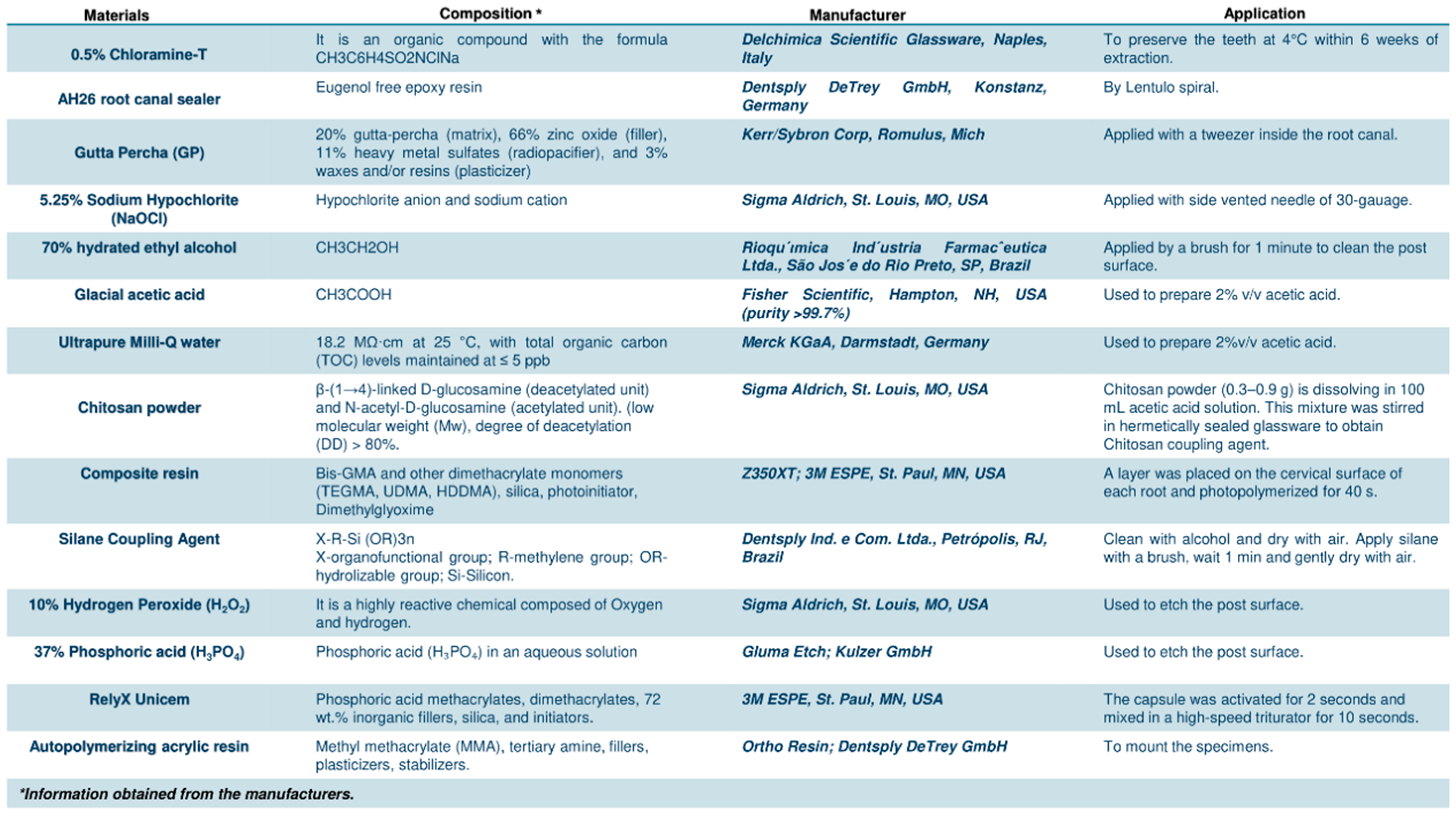
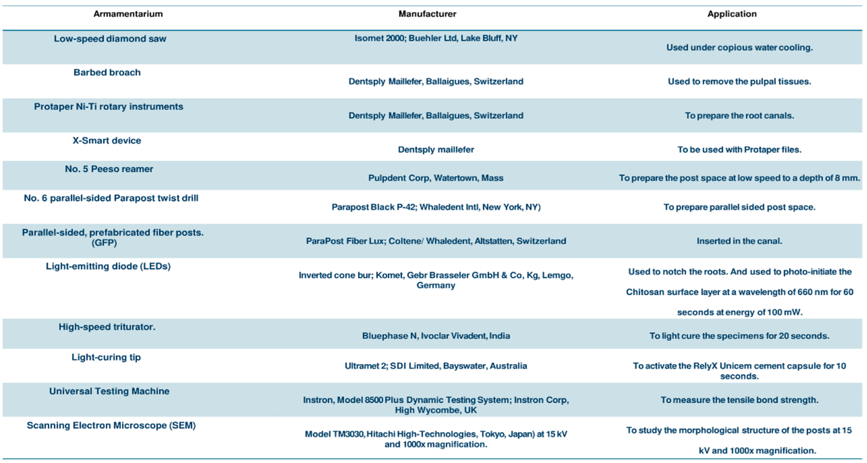
2. Materials and Methods

2.1. Ethical Approval

The current study was approved by the Institutional Review Board, College of Dentistry, Imam Abdulrahman Bin Faisal University, with the number (IRB-2024-02-177). During teeth collection, informed consent was secured from all participants.

2.2. Sample Size Estimation

G*Power software (version 3.1; University of Dusseldorf) was used to calculate the sample size. The effect size (d) was 0.5, α was 0.05, and 1-β (power) was 0.80. The suitable specimen size of 91 samples was calculated according to the tensile bond strength of the conventional GFPs from previous study [34].

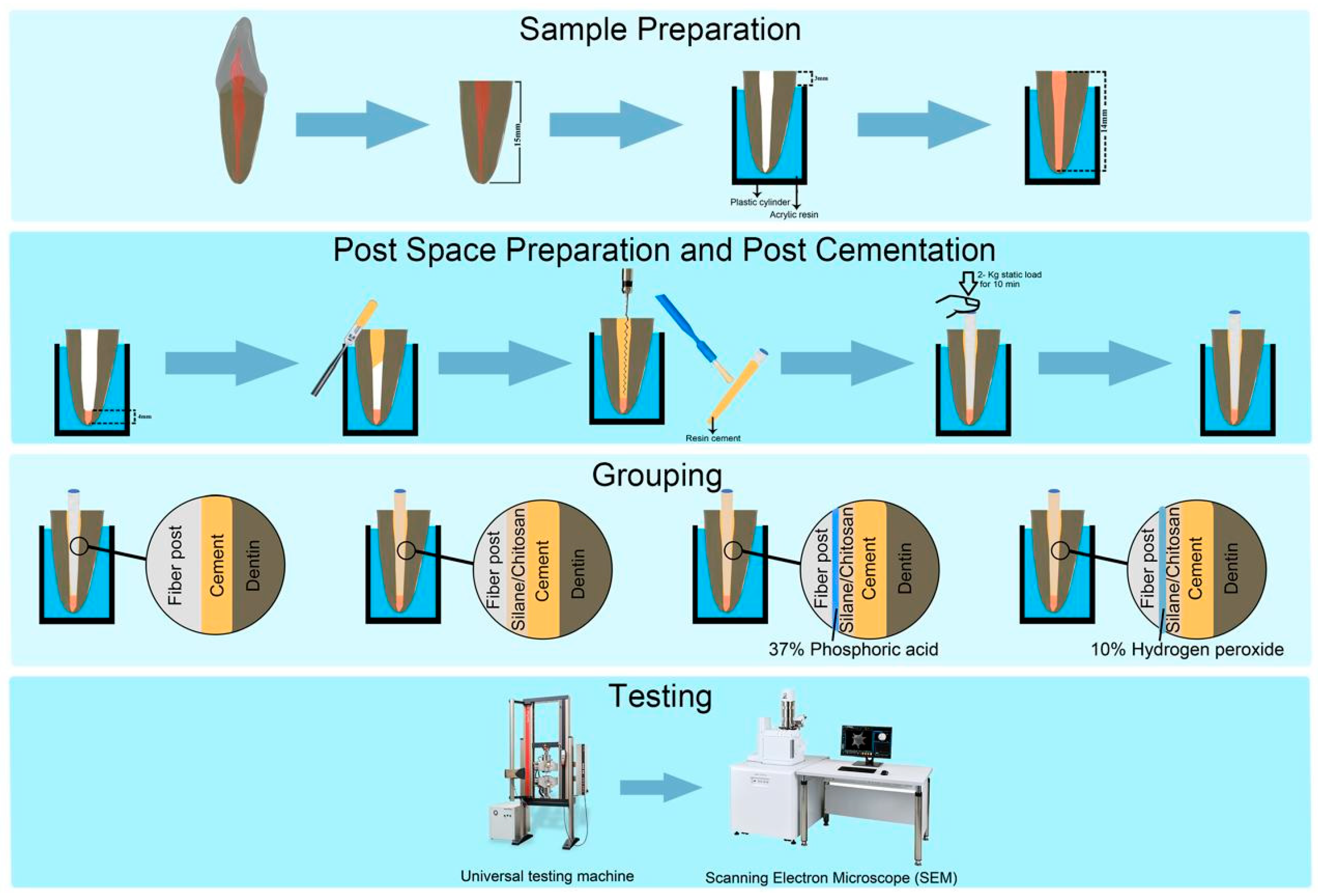
2.3. Teeth Selection and Post Space Preparation

For this study, 91 single-rooted, non-restored, and non-carious human mandibular premolar teeth of similar dimensions were selected from extracted specimens. Radiographic examination was conducted to verify that each tooth presented a straight single-rooted canal with a fully developed apex. Digital calipers were used to ensure that the root lengths were within a range of 14–16 mm and crown size of 10 mm mesiodistally and 10 mm buccolingually. No evidence of dental caries, cracks, or fractures was observed. Following extraction, teeth were preserved in a 0.5% Chloramine-T solution at 4 °C in a darkened environment. These specimens were used within 6–8 weeks of extraction [35]. One experienced operator performed the post space preparations to eliminate any bias. Prior to the study, the operator prepared 10 sample teeth not included in the study to ensure consistency. For the samples and the post space preparation, we adhered to the same protocol established in a previous study [36]. Teeth were cut 2 mm coronal to the CEJ, and the pulpal tissue was removed. Crown-down technique was used to prepare the canals with nickel–titanium rotary files. Root canals were cleaned with 5.25% NaOCl and obturated with lateral condensation technique using GP and AH26 root canal sealer. The obturated teeth roots were stored in 100% relative humidity for 7 days. Afterward, post spaces were prepared using reamers and drills to a depth of 8 mm and a diameter of 1.5 mm, followed by irrigation with NaOCl. Fiber posts were passively fitted into the post spaces, and moisture content was maintained throughout the procedure. The roots were notched with carbide burs, and the specimens were fixed in auto-polymerizing acrylic resin. Detailed information on the materials and equipment used in the current study is listed in Figure 1 and Figure 2.

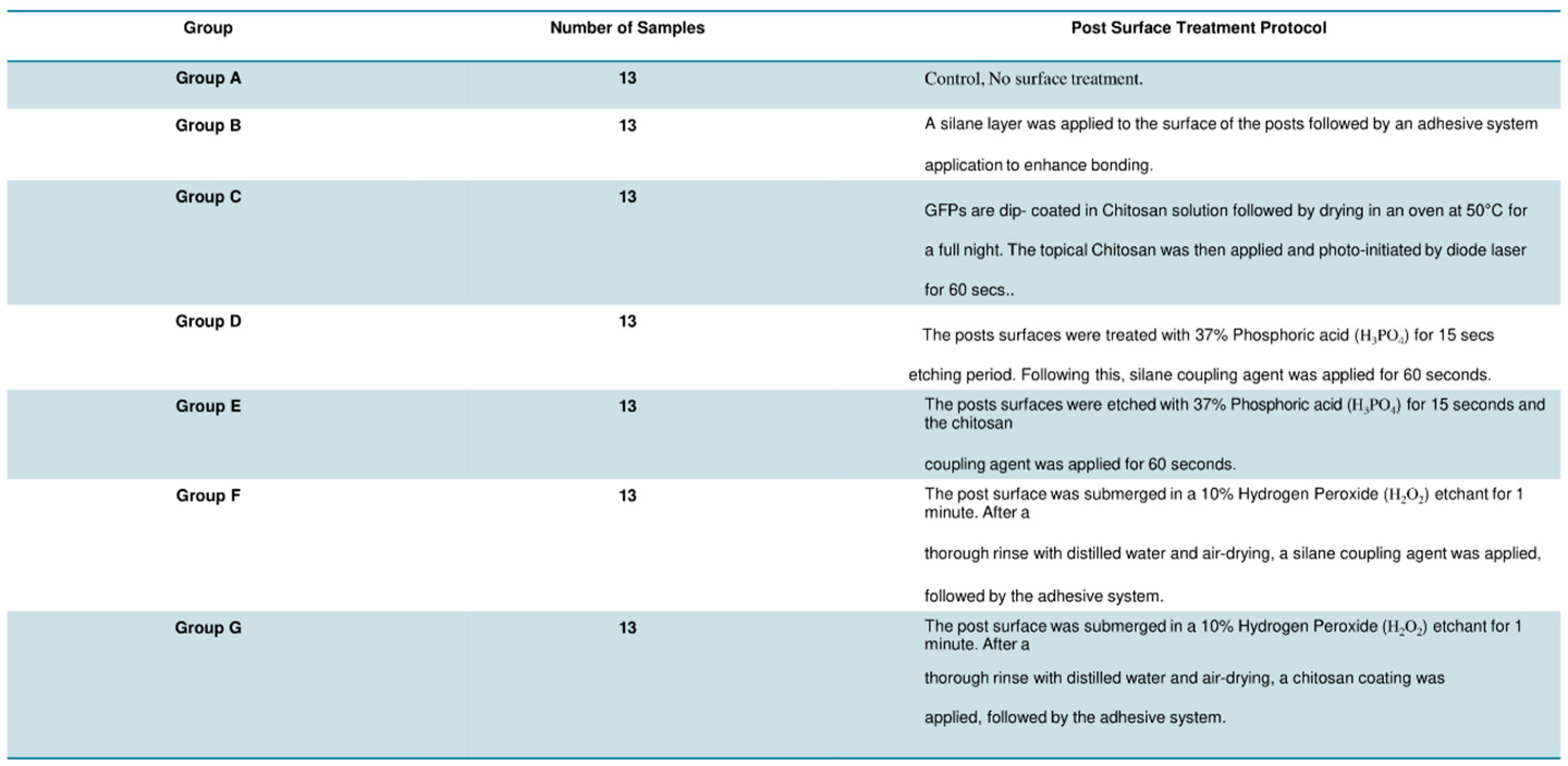
2.4. Sample Grouping and Post Surface Treatment

Prior to surface treatment, each post was tested within the root canals, cleaned with 70% ethyl alcohol, and subsequently air-dried. Samples were then categorized into seven equal groups (n = 13) according to the specific post surface treatment applied, as detailed in Figure 3.

2.5. Post Cementation

Prior to GFP cementation, post spaces were disinfected with 5.25% NaOCl, water-rinsed, and dried. The cementation process was performed using RelyX Unicem following the manufacturer’s instructions [34]. The cement was injected into the post space using a syringe elongation tip, beginning at the apical third and moving towards the cervical third until the cement overflowed, which helped to minimize bubble formation, which could affect adhesion. Resin cement was applied to the posts before they were inserted into the prepared root canal spaces with gentle finger pressure. The excess cement was carefully removed using an applicator tip to ensure a clean finish. After photopolymerization, a layer of composite resin was applied coronally to each root and light-cured for 40 s. The extended part of the post beyond the canal was left untrimmed. Afterward, roots were stored in distilled water at 37 °C for 24 h for tensile strength testing and failure mode analysis as shown in Figure 4.

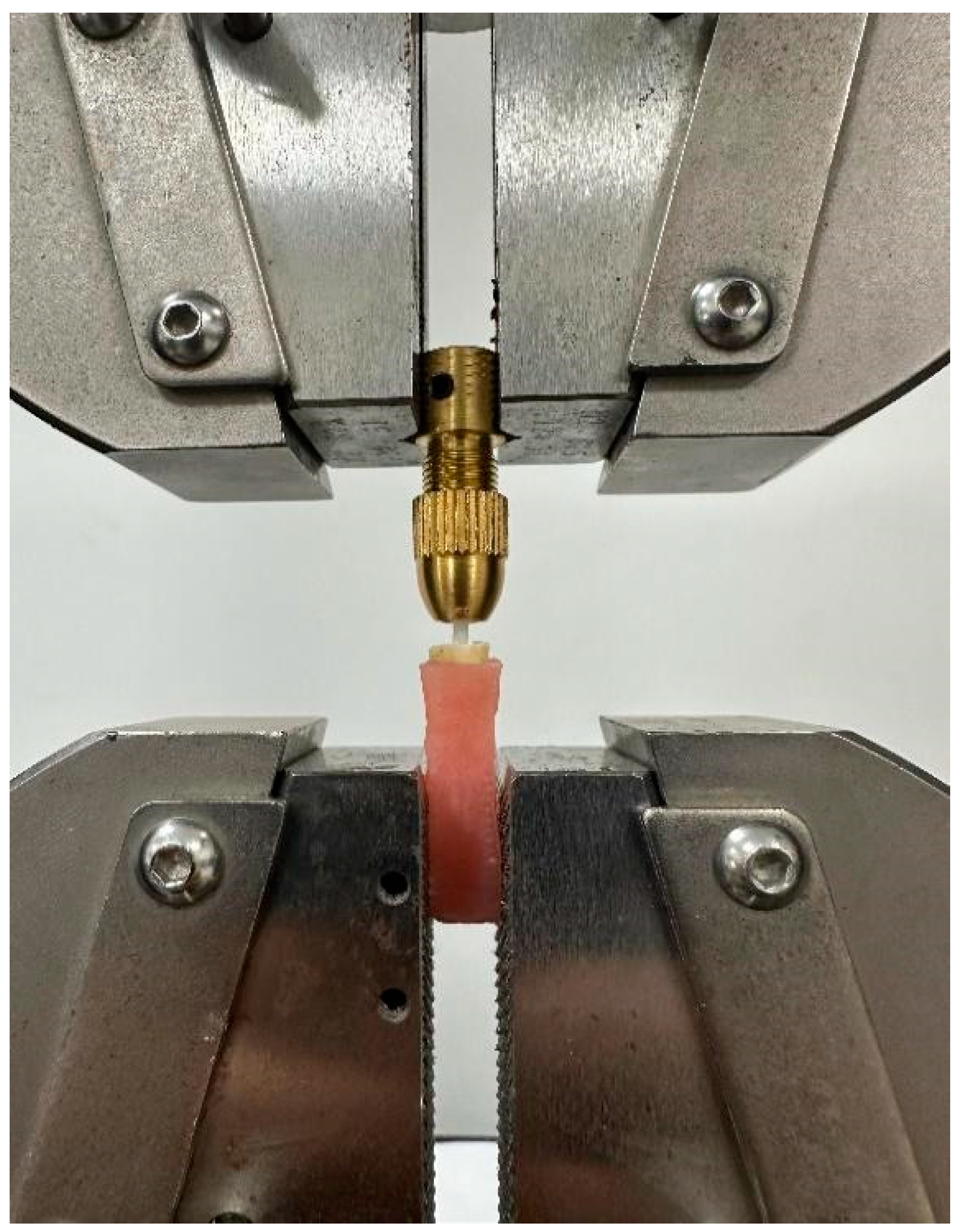
2.6. Tensile Bond Strength Testing

The tensile bond strength was assessed using a universal testing machine. A metallic pin was inserted through a hole located in the lower part of the acrylic resin cylinder to ensure proper alignment with the lower articulation of the universal testing machine. (Figure 5). The crosshead speed was applied to each sample via a hook that was connected to the upper part of the universal testing machine while the loop was applied at the coronal end of the post, and it was set at 0.5 mm/min. Testing continued until failure happened, defined as post separation from the root canal; the forces necessary to detach the posts were recorded. Samples where the posts separated from the acrylic core or broke within the root canal were excluded from the analysis.

2.7. Scanning Electron Microscopy and Failure Mode Analysis

The GFPs were sputter-coated with a thin layer of gold, mounted on stubs, and their surface morphological structure was captured using Scanning Electron Microscopy (SEM). The failure mode was determined as follows: adhesive failure, which indicates complete separation at the GFP/resin cement interface; cohesive failure, which refers to fractures occurring within the post or resin cement; and mixed failure, which involves characteristics of both types [37].
The laboratory testing was assessed by two evaluators who were directed to apply the same criteria for evaluating and analyzing the parameters of the current study. To verify the consistency and agreement between the two evaluators’ assessments, Cohen’s Kappa test was utilized. A Kappa value of 0.82 indicated a high degree of reliability between the evaluators [38].

2.8. Statistical Analysis

Statistical analysis of the resultant data was performed using a statistical software package (SPSS v16.0, SPSS Corp, Ill, Chicago, IL, USA). Shapiro–Wilk test showed normal distribution of the data (p > 0.05). One-way analysis of variance (ANOVA) was applied to compare the mean tensile strengths of various post surface treatments. Tukey’s multiple comparison tests were conducted to detect which post surface treatment group was significantly different. Cross-tabulation was used to compare the percentage of fracture patterns among the studied groups. All statistical results were set at a level of significance of 5%.

3. Results

In this study, one-way analysis of variance (ANOVA) showed a statistically significant difference in the bond strength among the tested groups (p < 0.001). The H2O2–Chitosan group (G) achieved the highest mean (SD) bond strength value (472.8 ± 34 N), significantly outperforming all other groups (p < 0.001). The least mean (SD) bond strength value was recorded for posts in Group A (Control) (Mean = 268 ± 70 N). The PA–Chitosan group (E) and the Chitosan group (C) also exhibited higher tensile bond strengths (330.6 ± 46 N and 300.3 ± 51 N, respectively) compared to the Control group (A) (268.0 ± 70 N) and the Silane group (B) (279.8 ± 49 N). Tukey’s post hoc test (multiple comparisons) showed statistically significant difference in the bond strength between Group A (Control) and the other test groups C (Chitosan), D (H3PO4–Silane), E (H3PO4–Chitosan), F (H2O2–Silane), and G (H2O2–Chitosan), where the p-values are <0.05; <0.05, <0.05, <0.05 and <0.001, respectively, as shown in Table 1.
Table 2 shows that adhesive failures were predominantly observed in the Control (76.9%), Silane (69.2%), and Chitosan-only groups (23.1%), indicating weaker bonding at the post–cement interface in these groups. Cohesive failures within the resin cement were more frequent in the PA + Chitosan (61.5%), H2O2 + Silane (53.8%), and H2O2 + Chitosan (76.9%) groups, correlating with their higher tensile bond strengths. Mixed failures were less common but occurred across groups, reflecting intermediate adhesion qualities. The data suggests that higher tensile bond strengths are associated with a shift from adhesive to cohesive failure modes, particularly in groups treated with chitosan and hydrogen peroxide. This indicates a stronger bond and improved integration at the interface.

SEM Analysis

The representative SEM images for different post surface treatment protocols after the debonding used in the study groups are shown in Figure 6.
The SEM images reveal a surface of intact post fibers with residual resin cement distributed across the post, indicating cohesive failure within the resin, as shown in Figure 6A–D. The roughened surface morphology, created by etching and enhanced by the Chitosan coupling agent, suggests a strong chemical bond and mechanical interlock, correlating with higher bond strength. The SEM images of the post surface of the groups showed a more continuous coat of resin cement (as shown in Figure 6F,G). The surfaces exhibited smoother morphologies with minimal residual resin, reflecting weaker mechanical interlocking and poorer chemical adhesion and resulting in lower bond strength performance (as shown in Figure 6A,B). A distinct crack shown in the post surface of Group F may suggest areas where the tensile force during debonding exceeded the cohesive strength of the resin cement, leading to cohesive failure within the resin layer.

4. Discussion

Fiberglass posts are the preferred choice for restoring teeth that have experienced significant coronal loss following root canal treatment, primarily because their modulus of elasticity closely resembles that of root dentin. This matching helps to minimize the external forces exerted on the dentinal walls, thereby lowering the risk of vertical root fractures [13,39]. Despite the advantages of fiberglass posts, previous research has revealed that the loss of adhesion at the cement post interface remains a concern that requires further investigation [40]. Physical methods like post design have been developed to enhance the post retention [41]. Given the interdependence of post retention on chemical factors and surface treatment, this study investigates the combined effects of coupling agent application and post surface conditioning [42]. The surface treatments selected for our study, particularly phosphoric acid and hydrogen peroxide as etchants and the silane coupling agent, were chosen due to their clinical relevance and known effectiveness in enhancing bond strength [13,15]. In the current study, the results indicated significant statistical differences in the tensile bond strength among the various experimental groups tested, which led to the rejection of this study’s null hypothesis.
Chitosan showed the best result when used as a coupling agent either solely or when preceded by an etchant for the GFP surface treatment (p < 0.05). Chitosan, acting as a bifunctional coupling agent, facilitates a chemical bond between the organic component of the resin cement and the inorganic component of the GFP, thereby enhancing the post’s wettability [43]. The reactive functional groups, such as amine (-NH2) and hydroxyl (-OH) groups, can form hydrogen bonds and covalent interactions with both the glass fiber post surface and resin cement components. Its amphiphilic nature allows chitosan to interact with both hydrophilic (glass fiber) and hydrophobic (resin) surfaces, thereby enhancing interfacial adhesion [44]. Additionally, chitosan exhibits a film-forming ability, creating a thin, uniform layer on the post surface that increases the surface area for bonding and improves stress distribution [45]. Chitosan’s hydroxyl groups can form hydrogen bonds with the silanol groups on the glass fiber surface, enhancing wettability and adhesion [46]. Furthermore, the amino groups of chitosan can react with functional groups in the resin cement (e.g., epoxy or carboxyl groups), resulting in strong chemical bonds [47]. These mechanisms collectively contribute to improved interfacial adhesion and enhanced tensile bond strength. Assis et al. [48] reported that the usage of chitosan can increase the mechanical properties by decreasing the formation of flaws and crack growth on the surface of resin cement. This was reported by Tan et al. [49] in 2018, who reported that when using chitosan as a coupling agent in phosphate glass fiber/polycaprolactone (PGF/PCL) composites, it increased the tensile strength up to 1.1–11.5%.
Furthermore, the chitosan coating can fill existing flaws and defects, thereby blunting the tips of any cracks present. Upon drying, the chitosan coating can induce compressive stresses on the fiber surface, which helps prevent the initiation and propagation of cracks. Additionally, previous research has suggested that chitosan can form entangled networks in acidic aqueous environments through self-crosslinking. This crosslinking process involves two structural units that may originate from the same chitosan polymer chain or from different chains, which leads to the formation of a cohesive network that enhances the overall integrity of the fiber surface [50]. This is evident in the SEM images (E, G), which demonstrate that the cohesive mode of failure is predominantly observed in chitosan-treated glass fiber posts.
Previous studies have indicated that single-surface treatment methods alone are insufficient for establishing a strong adhesive bond at the post–cement interface [51,52]. In the current study, 37% phosphoric acid and 10% hydrogen peroxide etchants were used to roughen the post surface enabling the coupling agent and primer to have more contact and interaction with the post fibers [15,53]. The results showed that glass fiber posts etched with 10% H2O2 showed higher tensile bond strength values (472.8 + 34 N) than those treated with 37% H3PO4 (330.6 + 46 N). This might be due to the dissolution of the resin matrix within the post through an oxidation mechanism, which creates surface roughness, exposes the fibers, and facilitates a strong chemical bond between the coupling agent and the post. This comes in accordance with Alshahrani et al. [10], who reported that the best bond strength was recorded when using an H2O2 etchant. However, the variability in the methodology employed, including differences in the type of post, manufacturer origin, tooth selection, technique, and filling material, may contribute to the observed differences in the results across studies [15]. All these findings can highlight the best tensile bond strength gained in this study when combining the H2O2 surface conditioning and chitosan coupling agent to reach the maximum tensile bond strength.
The findings of the current study demonstrated that the GFP etched with 10% H2O2 and coated with Chitosan predominantly exhibited cohesive failure within the resin cement, while the adhesive mode of failure at the post–cement interface was shown in the untreated fiber posts and the silane-coated fiber post. On the contrary, the mixed mode of failure was mostly demonstrated with the group of chitosan only, which highlighted the importance of using H2O2 etchant to enhance the micromechanical bonding in addition to the chemical bonding for the best tensile bonding results.
While the present study provides valuable results, it is challenging to directly correlate the in vitro experimental conditions with actual clinical situations. In the oral environment, teeth are subjected to a complex combination of forces, including compressive occlusal loads, and shear and bending stresses. This multifactorial loading scenario in the clinical setting may not be fully replicated by the experimental setup, which could limit the direct applicability of the in vitro findings. Another limitation is that the study exclusively used solid glass fiber posts. However, this study could serve as a foundation for advancing research on improved bonding protocols for various dental materials to enhance the longevity and reliability of dental restorations using different post designs. Additionally, it could explore different formulations of chitosan, including blends with other natural polymers or synthetic materials, to further optimize the bonding properties and mechanical strength of dental restorations.

5. Conclusions

Based on the findings of this study, the following conclusions were drawn:
  • The incorporation of chitosan as a coupling agent for the surface treatment of GFP demonstrates a significant enhancement in tensile bond strength when compared to traditional silane coupling agents.
  • The highest tensile bond strength was achieved when the posts underwent prior surface conditioning with a hydrogen peroxide etchant.
  • These findings suggest that utilizing chitosan in conjunction with appropriate surface conditioning can positively influence the longevity of glass fiber posts, reducing the likelihood of post failure.
  • This study highlights the potential of using chitosan as a natural biopolymer alternative to synthetic coupling agents to develop more effective bonding strategies for dental restorations.

6. Recommendations

It is recommended that dental practitioners consider incorporating chitosan-treated glass fiber posts into their restorative procedures, particularly in cases where maximum bond strength is crucial, such as in teeth with minimal remaining coronal structure or high occlusal loads. However, as with any new technique, we advise clinicians to stay informed about ongoing research and long-term clinical studies before fully adopting this approach.

Author Contributions

N.T.: Conceptualization, Formal analysis, Investigation, Methodology, Project administration, Supervision, Validation, Writing—original draft, review and editing. M.H.A.H.: Formal analysis, Investigation, Methodology, Writing—original draft. M.F.A.: Formal analysis, Investigation, Methodology, Writing—original draft. R.A.A.: Formal analysis, Investigation, Methodology, Writing—original draft. A.M.A.: Formal analysis, Investigation, Methodology, Writing—original draft. A.C.S.: Data curation, Software. S.R.: Investigation, Validation, Visualization, Writing—original draft, Writing—review and editing. All authors have read and agreed to the published version of the manuscript.

Funding

This research received no external funding.

Institutional Review Board Statement

This study was conducted in accordance with the Declaration of Helsinki and approved by the Institutional Review Board of College of Dentistry, Imam Abdulrahman Bin Faisal University (IRB-2024-02-177) (approval date: 25 February 2024).

Informed Consent Statement

Informed consent was obtained from all subjects involved in the study.

Data Availability Statement

Data related to this article can be found in the online version at https://10.6084/m9.figshare.27310095 (accessed on 26 October 2024).

Acknowledgments

The authors thank Omar Mukhtar, Department of Dental Education, College of Dentistry, Imam Abdulrahman Bin Faisal University for his valuable support in the current study. All authors gave their final approval and agreed to be accountable for all aspects of the work. We declare that the research is free of conflicts of interest.

Conflicts of Interest

The authors declare no conflicts of interest.

Correction Statement

This article has been republished with a minor correction to the Institutional Review Board Statement. This change does not affect the scientific content of the article.

References

  1. Tsintsadze, N.; Margvelashvili-Malament, M.; Natto, Z.S.; Ferrari, M. Comparing survival rates of endodontically treated teeth restored either with glass-fiber-reinforced or metal posts: A systematic review and meta-analyses. J. Prosthet. Dent. 2022, 131, 567–578. [Google Scholar] [CrossRef]
  2. Ben Suleiman, A.; Desai, S.; Tepperman, A.; Chvartszaid, D.; Malkhassian, G.; Habsha, E.; Barzilay, I.; Azarpazhooh, A. The Outcomes of Endodontically Treated Teeth Restored with Custom-Made Cast Post-and-Core Restorations: A Retrospective Cohort Study. J. Endod. 2024, 50, 316–328. [Google Scholar] [CrossRef]
  3. Oulghazi, I.; El Yamani, A.; Morchad, B. Factors influencing vertical radicular fractures in teeth supported by metallic dental core: A scoping review. Clin. Cosmet. Investig. Dent. 2024, 16, 101–114. [Google Scholar] [CrossRef]
  4. Sachi, S.; Varada, S.; Jitha, J.; Nair, S.V. Reattachment of Fractured Maxillary Central Incisor with Fibre Post. Kerala Dent. J. 2024, 47, 32–36. [Google Scholar] [CrossRef]
  5. de Matos, L.M.R.; Silva, M.L.; Cordeiro, T.O.; Cardoso, S.d.A.M.; Campos, D.E.S.; de Muniz, I.A.F.; Barros, S.A.d.L.; Seraidarian, P.I. Clinical and laboratorial performance of rehabilitation of endodontically treated teeth: A systematic review. J. Esthet. Restor. Dent. 2024, 36, 1281–1300. [Google Scholar] [CrossRef]
  6. Hawthan, M.; Larsson, C.; Chrcanovic, B.R. Survival of fixed prosthetic restorations on vital and nonvital teeth: A systematic review. J. Prosthodont. 2024, 33, 110–122. [Google Scholar] [CrossRef] [PubMed]
  7. Generali, L.; Veneri, F.; Forabosco, E.; Cavani, F.; Piergianni, V.; Sassatelli, P.; Checchi, V.; Pedullà, E. Push-out bond strength and SEM fractographical analysis of hollow fibre posts used with self-adhesive resin cement: A pilot study. Odontology 2024, 112, 158–168. [Google Scholar] [CrossRef]
  8. Cheniski, D.A.; Freire, A.; Camargo, E.S.; da Costa, R.G.; de Souza, E.M.; Rached, R.N. Bond strength of prefabricated and CAD-CAM milled glass fiber post-and-cores luted with conventional, universal, and self-adhesive composite resin cement. J. Prosthet. Dent. 2024, 131, 251.e1–251.e7. [Google Scholar] [CrossRef]
  9. Rajnics, Z.; Pammer, D.; Kőnig-Péter, A.; Turzó, K.; Marada, G.; Radnai, M. Push-Out Bond Strength of Glass Fiber Endodontic Posts with Different Diameters. Materials 2024, 17, 1492. [Google Scholar] [CrossRef] [PubMed]
  10. Alshahrani, A.; Albaqami, M.; Naji, Z.; Al-Khunein, Y.; Alsubaie, K.; Alqahtani, A.; Al-Thobity, A.M. Impact of different surface treatment methods on bond strength between fiber post and composite core material. Saudi Dent. J. 2021, 33, 334–341. [Google Scholar] [CrossRef]
  11. Souza, J.C.M.; Fernandes, V.; Correia, A.; Miller, P.; Carvalho, O.; Silva, F.; Özcan, M.; Henriques, B. Surface modification of glass fiber-reinforced composite posts to enhance their bond strength to resin-matrix cements: An integrative review. Clin. Oral Investig. 2022, 26, 95–107. [Google Scholar] [CrossRef] [PubMed]
  12. Mishra, L.; Khan, A.S.; Velo, M.M.d.A.C.; Panda, S.; Zavattini, A.; Rizzante, F.A.P.; Arbildo Vega, H.I.; Sauro, S.; Lukomska-Szymanska, M. Effects of Surface Treatments of Glass Fiber-Reinforced Post on Bond Strength to Root Dentine: A Systematic Review. Materials 2020, 13, 1967. [Google Scholar] [CrossRef] [PubMed]
  13. Simões, Í.I.N.; Moreira da Silva, E.; Penelas, A.G.; Poskus, L.T.; Botelho Dos Santos, G.; Guimarães, J.G.A. Evaluation of recementation protocols for dislodged glass fiber posts. Int. J. Prosthodont. 2023, 36, 650. [Google Scholar] [CrossRef] [PubMed]
  14. Machry, R.V.; Fontana, P.E.; Bohrer, T.C.; Valandro, L.F.; Kaizer, O.B. Effect of Different Surface Treatments of Resin Relined Fiber Posts Cemented With Self-adhesive Resin Cement on Push-out and Microtensile Bond Strength Tests. Oper. Dent. 2020, 45, E185–E195. [Google Scholar] [CrossRef]
  15. Santander-Rengifo, F.M.; Castillo-Andamayo, D.E.; Tay, L.Y.; López-Gurreonero, C.; Cornejo-Pinto, A.; Cervantes-Ganoza, L.A.; Cayo-Rojas, C.F. Bond Strength and Failure Mode of Glass Fiber Posts with Different Surface Treatments Prior to Silanization: An In Vitro Comparative Study. J. Int. Soc. Prev. Community Dent. 2023, 13, 42–53. [Google Scholar] [CrossRef]
  16. Majeti, C.; Veeramachaneni, C.; Morisetty, P.K.; Rao, S.A.; Tummala, M. A simplified etching technique to improve the adhesion of fiber post. J. Adv. Prosthodont. 2014, 6, 295–301. [Google Scholar] [CrossRef]
  17. Mosharraf, R.; Ranjbarian, P. Effects of post surface conditioning before silanization on bond strength between fiber post and resin cement. J. Adv. Prosthodont. 2013, 5, 126–132. [Google Scholar] [CrossRef]
  18. Aldegheishem, A.; Barakat, R.; Alrefaei, N.A.L.H.; Alhussain, G.; Alkoblan, K.; Al-Madi, E.M. Fracture Resistance of Endodontically Treated Teeth Restored Using Multifiber Posts Compared with Single Fiber Posts. Int. J. Polym. Sci. 2024, 2024, 1–6. [Google Scholar] [CrossRef]
  19. Meimandinezhad, M.; Bidhendi, H.M.; Zadeh, E.T.; Anzabi, R.M.; Asadi, H.; Fathi, A.; Gholami, M. Comparison of different surface treatment methods on flexural strength and elasticity modules of quartz and glass fiber-based posts: A narrative review. J. Fam. Med. Prim. Care 2022, 11, 7616–7620. [Google Scholar] [CrossRef]
  20. Fernandes, V.; Silva, F.; Henriques, B.; Özcan, M.; Carvalho, O.; Souza, J.C.M. Laser-texturing and traditional surface modification to improve the adhesion of glass fiber-reinforced composite posts to resin cements. Int. J. Adhes. Adhes. 2024, 131, 103645. [Google Scholar] [CrossRef]
  21. Aziz, T.; Ullah, A.; Fan, H.; Jamil, M.I.; Khan, F.U.; Ullah, R.; Iqbal, M.; Ali, A.; Ullah, B. Recent Progress in Silane Coupling Agent with Its Emerging Applications. J. Polym. Environ. 2021, 29, 3427–3443. [Google Scholar] [CrossRef]
  22. Leme, A.A.; Pinho, A.L.; de Gonçalves, L.; Correr-Sobrinho, L.; Sinhoreti, M.A. Effects of silane application on luting fiber posts using self-adhesive resin cement. J. Adhes. Dent. 2013, 15, 269–274. [Google Scholar] [CrossRef] [PubMed]
  23. Adwani, S.; Elsubeihi, E.; Zebari, A.; Aljanahi, M.; Moharamzadeh, K.; Elbishari, H. Effect of Different Silane Coupling Agents on the Bond Strength between Hydrogen Peroxide-Etched Epoxy-Based- Fiber-Reinforced Post and Composite Resin Core. Dent. J. 2023, 11, 142. [Google Scholar] [CrossRef]
  24. Saini, S.; Yadav, R.; Sonwal, S.; Meena, A.; Huh, Y.S.; Brambilla, E.; Ionescu, A.C. Tribological, mechanical, and thermal properties of nano tricalcium phosphate and silver particulates reinforced Bis-GMA/TEGDMA dental resin composites. Tribol. Int. 2024, 199, 110010. [Google Scholar] [CrossRef]
  25. Yadav, R.; Saini, S.; Sonwal, S.; Meena, A.; Huh, Y.S.; Brambilla, E.; Ionescu, A.C. Optimization and ranking of dental restorative composites byENTROPY-VIKOR and VIKOR-MATLAB. Polym. Adv. Technol. 2024, 35, e6526. [Google Scholar] [CrossRef]
  26. Hameed, H.; Khan, M.A.; Paiva-Santos, A.C.; Ereej, N.; Faheem, S. Chitin: A versatile biopolymer-based functional therapy for cartilage regeneration. Int. J. Biol. Macromol. 2024, 265, 131120. [Google Scholar] [CrossRef]
  27. Malhotra, H.; Gautam, R.K. Biomedical applications of chitosan and its derivatives. In Biopolymers for Biomedical Applications; Annu, Ed.; Wiley: Hoboken, NJ, USA, 2024; pp. 25–51. ISBN 9781119865025. [Google Scholar]
  28. Fakhri, E.; Eslami, H.; Maroufi, P.; Pakdel, F.; Taghizadeh, S.; Ganbarov, K.; Yousefi, M.; Tanomand, A.; Yousefi, B.; Mahmoudi, S.; et al. Chitosan biomaterials application in dentistry. Int. J. Biol. Macromol. 2020, 162, 956–974. [Google Scholar] [CrossRef]
  29. Zarrintaj, P.; Seidi, F.; Youssefi Azarfam, M.; Khodadadi Yazdi, M.; Erfani, A.; Barani, M.; Chauhan, N.P.S.; Rabiee, N.; Kuang, T.; Kucinska-Lipka, J.; et al. Biopolymer-based composites for tissue engineering applications: A basis for future opportunities. Compos. Part B Eng. 2023, 258, 110701. [Google Scholar] [CrossRef]
  30. Zhang, Q.; Guo, J.; Huang, Z.; Mai, S. Promotion effect of carboxymethyl chitosan on dental caries via intrafibrillar mineralization of collagen and dentin remineralization. Materials 2022, 15, 4835. [Google Scholar] [CrossRef]
  31. Nishanthine, C.; Miglani, R.; R, I.; Poorni, S.; Srinivasan, M.R.; Robaian, A.; Albar, N.H.M.; Alhaidary, S.F.R.; Binalrimal, S.; Almalki, A.; et al. Evaluation of Fluoride Release in Chitosan-Modified Glass Ionomer Cements. Int. Dent. J. 2022, 72, 785–791. [Google Scholar] [CrossRef] [PubMed]
  32. Abedian, Z.; Jenabian, N.; Moghadamnia, A.A.; Zabihi, E.; Tashakorian, H.; Rajabnia, M.; Sadighian, F.; Bijani, A. Antibacterial activity of high-molecular-weight and low-molecular-weight chitosan upon oral pathogens. J. Conserv. Dent. 2019, 22, 169–174. [Google Scholar] [CrossRef]
  33. Li, D.; Zhuang, B.; Wang, X.; Wu, Z.; Wei, W.; Aladejana, J.T.; Hou, X.; Yves, K.G.; Xie, Y.; Liu, J. Chitosan used as a specific coupling agent to modify starch in preparation of adhesive film. J. Clean. Prod. 2020, 277, 123210. [Google Scholar] [CrossRef]
  34. Alzahrani, A.H.; Ebrahim, M.I.; Felemban, M.F.; Alqarni, A.A.; Algahtani, F.S.; Shawli, H.T.; Al Humayyani, N.; Meshni, A.A.; Al Moaleem, M.M. Nanoparticle Augmentation of Adhesive Systems: Impact on Tensile Strength in Fiberglass Post Placement within Root Dentin. Med. Sci. Monit. 2024, 30, e943502. [Google Scholar] [CrossRef] [PubMed]
  35. Alrabeah, G.; Binhassan, F.; Al Khaldi, S.; Al Saleh, A.; Al Habeeb, K.; Anwar, S.; Habib, S.R. Effect of Self-Adhesive Resin Cement Film Thickness on the Shear Bond Strength of Lithium Disilicate Ceramic–Cement–Tooth Triplex. Inorganics 2023, 12, 14. [Google Scholar] [CrossRef]
  36. Rifaat, S.; Aljami, A.; Alshehri, T.; Alameer, S.T.; Aldossary, A.; Almutairi, W.; Almaliki, M.N.; A Farooqi, F.; Taymour, N. The Effect of Coronal Pre-flaring and Type of Root Canal Irrigation on Working Length Accuracy Using Electronic Apex Locators. F1000Research 2023, 12, 533. [Google Scholar] [CrossRef] [PubMed]
  37. Tian, Y.; Mu, Y.; Setzer, F.C.; Lu, H.; Qu, T.; Yu, Q. Failure of fiber posts after cementation with different adhesives with or without silanization investigated by pullout tests and scanning electron microscopy. J. Endod. 2012, 38, 1279–1282. [Google Scholar] [CrossRef] [PubMed]
  38. Rifaat, S.; Rahoma, A.; Alkhalifa, F.; AlQuraini, G.; Alsalman, Z.; Alwesaibi, Z.; Taymour, N. Push-Out Bond Strength of EndoSeal Mineral Trioxide Aggregate and AH Plus Sealers after Using Three Different Irrigation Protocols. Eur. J. Dent. 2023, 17, 76–81. [Google Scholar] [CrossRef]
  39. Souza, N.d.O.; Sousa, R.S.; Isolan, C.P.; Moraes, R.R.d.; Lima, G.d.S.; Lomonaco, D.; de Paula, D.M.; Alves, A.H.; Sabóia, V.d.P.; Feitosa, V.P. Intraradicular Dentin Biomodification with Natural Agents for Bonding Glass-fiber Posts. J. Adhes. Dent. 2021, 23, 223–230. [Google Scholar] [CrossRef]
  40. Kosan, E.; Prates-Soares, A.; Blunck, U.; Neumann, K.; Bitter, K. Root canal pre-treatment and adhesive system affect bond strength durability of fiber posts ex vivo. Clin. Oral Investig. 2021, 25, 6419–6434. [Google Scholar] [CrossRef]
  41. Lo Giudice, G.; Ferrari Cagidiaco, E.; Lo Giudice, R.; Puleio, F.; Nicita, F.; Calapaj, M. Evaluation of mechanical properties of a hollow endodontic post by three point test and SEM analysis: A pilot study. Materials 2019, 12, 1983. [Google Scholar] [CrossRef] [PubMed]
  42. Skupien, J.A.; Sarkis-Onofre, R.; Cenci, M.S.; Moraes, R.R.d.; Pereira-Cenci, T. A systematic review of factors associated with the retention of glass fiber posts. Braz. Oral Res. 2015, 29, 1–8. [Google Scholar] [CrossRef] [PubMed]
  43. Monticelli, F.; Osorio, R.; Sadek, F.T.; Radovic, I.; Toledano, M.; Ferrari, M. Surface treatments for improving bond strength to prefabricated fiber posts: A literature review. Oper. Dent. 2008, 33, 346–355. [Google Scholar] [CrossRef] [PubMed]
  44. Aranaz, I.; Alcántara, A.R.; Civera, M.C.; Arias, C.; Elorza, B.; Heras Caballero, A.; Acosta, N. Chitosan: An overview of its properties and applications. Polymers 2021, 13, 3256. [Google Scholar] [CrossRef] [PubMed]
  45. Moreno-Vásquez, M.J.; Valenzuela-Buitimea, E.L.; Plascencia-Jatomea, M.; Encinas-Encinas, J.C.; Rodríguez-Félix, F.; Sánchez-Valdes, S.; Rosas-Burgos, E.C.; Ocaño-Higuera, V.M.; Graciano-Verdugo, A.Z. Functionalization of chitosan by a free radical reaction: Characterization, antioxidant and antibacterial potential. Carbohydr. Polym. 2017, 155, 117–127. [Google Scholar] [CrossRef] [PubMed]
  46. Žigrayová, D.; Mikušová, V.; Mikuš, P. Advances in chitosan derivatives: Preparation, properties and applications in pharmacy and medicine. Gels 2024, 10, 701. [Google Scholar] [CrossRef]
  47. Rostamabadi, H.; Demirkesen, I.; Colussi, R.; Roy, S.; Tabassum, N.; de Oliveira Filho, J.G.; Bist, Y.; Kumar, Y.; Nowacka, M.; Galus, S.; et al. Recent trends in the application of films and coatings based on starch, cellulose, chitin, chitosan, xanthan, gellan, pullulan, Arabic gum, alginate, pectin, and carrageenan in food packaging. Food Front. 2024, 5, 350–391. [Google Scholar] [CrossRef]
  48. Assis, H.C.; Nascimento, G.C.d.; Roperto, R.; Sousa-Neto, M.D.; Lopes-Olhê, F.C. Can Carbodiimide (EDC) and Chitosan Cross-linking Agents Effect the Longevity of Fiberglass Posts Luted with Different Types of Composite Cements to Root Dentin? J. Adhes. Dent. 2023, 25, 39–50. [Google Scholar] [CrossRef] [PubMed]
  49. Tan, C.; Rudd, C.; Parsons, A.; Sharmin, N.; Zhang, J.; Chen, W.; Ahmed, I. Chitosan as a coupling agent for phosphate glass fibre/polycaprolactone composites. Fibers 2018, 6, 97. [Google Scholar] [CrossRef]
  50. Pillai, C.K.S.; Paul, W.; Sharma, C.P. Chitin and chitosan polymers: Chemistry, solubility and fiber formation. Prog. Polym. Sci. 2009, 34, 641–678. [Google Scholar] [CrossRef]
  51. Elnaghy, A.M.; Mandorah, A.; Hassan, A.H.; Elshazli, A.; Elsaka, S. Effect of surface treatments on push-out bond strength of calcium silicate-based cements to fiber posts. BMC Oral Health 2021, 21, 131. [Google Scholar] [CrossRef]
  52. Dos Santos, L.R.; Lima, D.M.; Carvalho, E.M.; Rodrigues, V.P.; Alves, C.M.C. Effect of Glass Fiber Post Surface Treatment on Bond Strength of a Self-Adhesive Resin Cement: An “In Vitro” Study. Int. J. Dent. 2021, 2021, 8856657. [Google Scholar] [CrossRef] [PubMed]
  53. Rechia, B.C.d.N.; Bravo, R.P.; Oliveira, N.D.d.; Baratto Filho, F.; Gonzaga, C.C.; Storrer, C.L.M. Influence of different surface treatments of fiberglass posts on the bond strength to dentin. Braz. J. Oral Sci. 2017, 15, 158. [Google Scholar] [CrossRef]
Figure 1. Materials used in the current study.
Prosthesis 06 00112 g001
Figure 2. Armamentarium used in the current study.
Prosthesis 06 00112 g002
Figure 3. Glass fiber post surface treatment protocols and grouping.
Prosthesis 06 00112 g003
Figure 4. Study design and workflow.
Prosthesis 06 00112 g004
Figure 5. Specimen mounted in universal testing machine undergoing tensile stress evaluation.
Prosthesis 06 00112 g005
Figure 6. Representative SEM micrographs of the GF posts’ surfaces (original magnification: 1000×). (A) Untreated post. (B) Silane-coated post. (C) Chitosan-coated post. (D) Phosphoric acid-etched post coated with silane. (E) Phosphoric acid-etched post coated with Chitosan. (F) H2O2-etched post coated with silane. (G) H2O2-etched post coated with Chitosan.
Prosthesis 06 00112 g006
Table 1. Comparison of means and standard deviations (SD) of forces required to dislodge posts newtons) among the tested groups.
MeansStd. DeviationStd. Error95% Confidence Interval for MeanMinimumMaximump-Value
Lower BoundUpper Bound
(A) Control268.0892 a70.276519.491225.62310.556185.98371.71<0.001
(B) Silane279.8954 a49.401413.701250.04309.748204.99351.34
(C) Chitosan300.3915 a,b51.231614.209269.43331.350201.84375.64
(D) H3PO4–Silane293.2931 b50.338313.961262.87323.712203.21369.28
(E) H3PO4–Chitosan330.6600 c46.540912.908302.53358.784251.84385.68
(F) H2O2–Silane326.3015 c36.01409.9885304.53348.064280.28396.02
(G) H2O2–Chitosan472.846934.91479.6836451.74493.945414.11508.22
Vertical superscripts indicate no statistical significance at p ≤ 0.05.
Table 2. Percentage distribution of the failure pattern of GFPs in each study group.
Percentage of Failure Pattern Test Group
Adhesive%Cohesive%Mixed%
Group (n = 13)
Group A (Control)10 (76.9%)3 (23.1%)0 (0.0%)
Group B (Silane)9 (69.2%)2 (15.4%)2 (15.4%)
Group C (Chitosan)3 (23.1%)5 (38.5%)5 (38.5%)
Group D (H3PO4–Silane)3 (23.1%)6 (46.2%)4 (30.8%)
Group E (H3PO4–Chitosan)4 (30.8%)9 (61.5%)0 (0%)
Group F (H2O2–Silane)3 (23.1%)7(53.8%)3 (23.1%)
Group G (H2O2–Chitosan)2 (15.4%)10 (76.9%)1 (7.7%)
n = 9134 (37.4%)42 (46.2%)15 (16.5%)
Disclaimer/Publisher’s Note: The statements, opinions and data contained in all publications are solely those of the individual author(s) and contributor(s) and not of MDPI and/or the editor(s). MDPI and/or the editor(s) disclaim responsibility for any injury to people or property resulting from any ideas, methods, instructions or products referred to in the content.

