


Abstract
Background and Clinical Significance: Ductal adenoma is a rare benign breast neoplasm that may closely mimic invasive carcinoma on imaging. We report a histologically confirmed case of ductal adenoma, highlighting multimodality imaging findings, including mammography (MMG), ultrasound (US) and magnetic resonance imaging (MRI) features; Case Presentation: A 49-year-old woman presented with a painless palpable right breast mass. MMG demonstrated a 1.1 cm round mass with partially obscured margins (BI-RADS 0). Multiparametric US revealed an oval hypoechoic mass with non-parallel orientation, microlobulated/angular margins, and mixed posterior acoustic features, upgrading the lesion to BI-RADS 4. Strain elastography demonstrated a uniformly soft lesion (Tsukuba score 1), whereas microvascular imaging and B-flow imaging identified a penetrating intralesional vessel. Breast MRI showed an oval mass with irregular margins, restricted diffusion (ADC value 1.04 × 10⁻³ mm2/s), heterogeneous and rapid initial enhancement with delayed washout (type III kinetic curve), and a Kaiser score of 8, resulting in BI-RADS 5. US-guided core needle biopsy was performed, and histopathological examination established the diagnosis of ductal adenoma (B2 lesion). Following a multidisciplinary review, conservative management with imaging surveillance was recommended. The mass remained stable at 6-month follow-up; Conclusions: Ductal adenoma may exhibit imaging features highly suspicious for malignancy. This case expands its recognized imaging spectrum and highlights the indispensable role of histopathological confirmation.
1. Introduction and Clinical Significance
Ductal adenoma is a rare benign epithelial breast tumor arising from the small to medium-sized ducts. Along with tubular and lactating adenoma, it is one of the subtypes of breast adenomas, a group of lesions accounting for 0.75% of all core-needle biopsies [1,2]. Patients are generally older than 40 years, with a mean age at presentation ranging from 51 to 56 years [3,4]. They typically present with a unilateral, palpable breast mass measuring less than 1 cm, without associated nipple discharge [3,4]. Ductal adenoma represents a diagnostic pitfall because its clinical, imaging, and histopathological features may overlap with those of breast carcinoma [1,5]. The imaging features of ductal adenoma are insufficiently described in the literature. We report the case of a ductal adenoma in a female patient, with emphasis on mammography (MMG), multiparametric ultrasound (US), and magnetic resonance imaging (MRI) findings.
2. Case Presentation
A 49-year-old perimenopausal woman presented with a painless palpable mass in the right breast that she had first noticed one month earlier. She denied nipple discharge or use of hormonal therapy, and her family history was negative for breast and gynecologic malignancies. Eight years prior, the patient underwent surgical excision of a fibroadenoma of the left breast. Her medical history was otherwise unremarkable. Clinical examination revealed a firm mass, approximately 1 cm in diameter, at the border of the inner quadrants of the right breast, without overlying skin changes, nipple retraction, or discharge. No axillary lymphadenopathy was detected, and the contralateral breast was unremarkable.
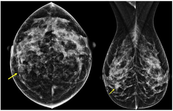
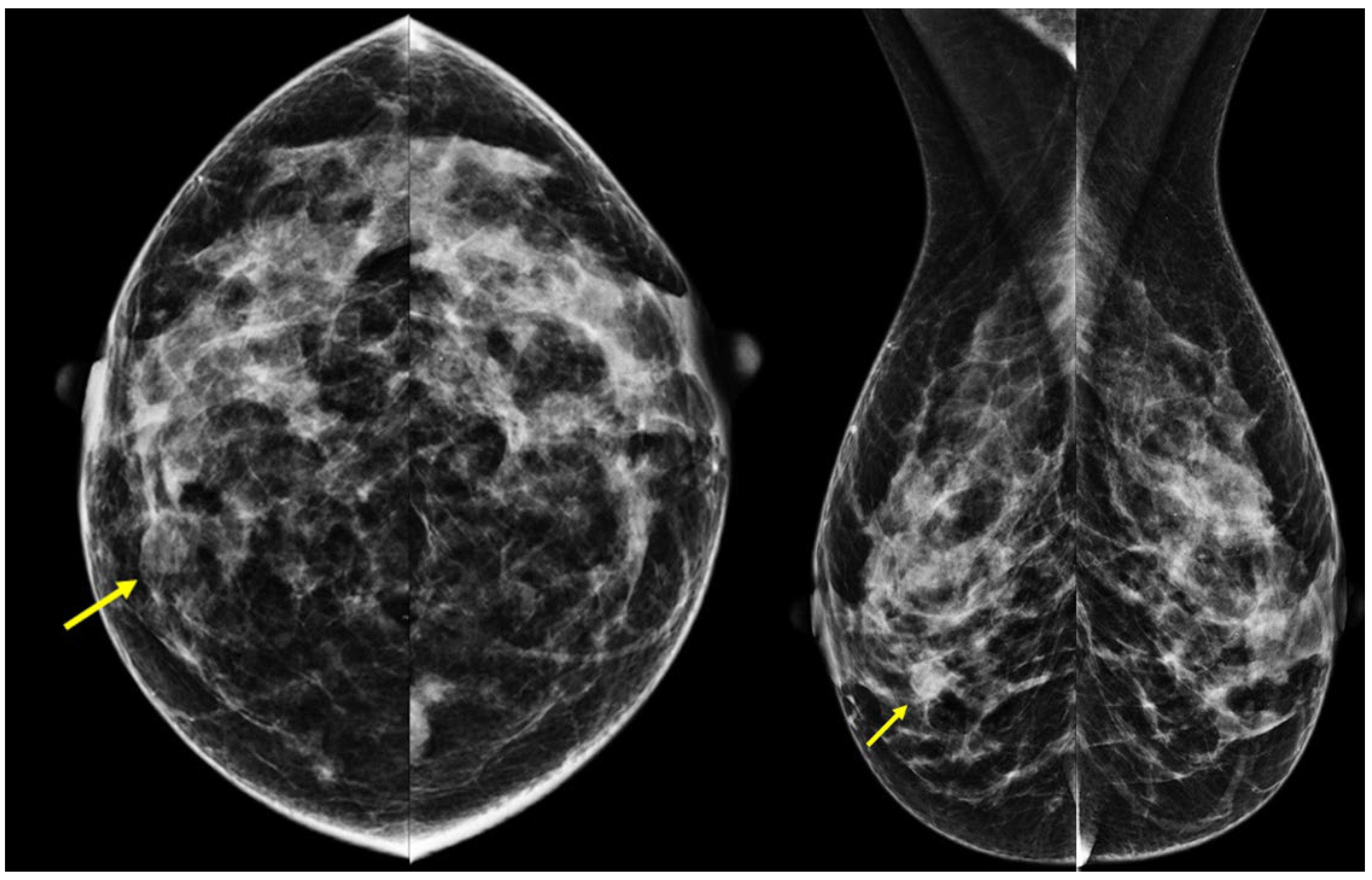
MMG demonstrated an ACR breast density category B. A round mass measuring 1.1 × 0.8 cm, with partially obscured margins and a density similar to the surrounding fibroglandular tissue, was identified at the border of the inner quadrants of the right breast (Figure 1). No associated microcalcifications, architectural distortion, or skin thickening were observed. The MMG examination was assigned a BI-RADS 0.
Figure 1.
Ductal adenoma on MMG (CC and MLO views): round mass with partially obscured margins and a density similar to the surrounding fibroglandular tissue, without associated microcalcifications, architectural distortion, or skin thickening (arrow, right breast).
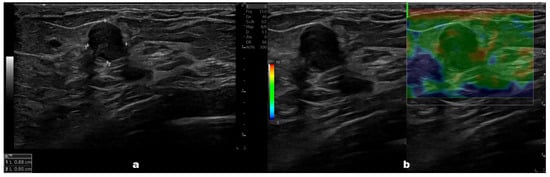
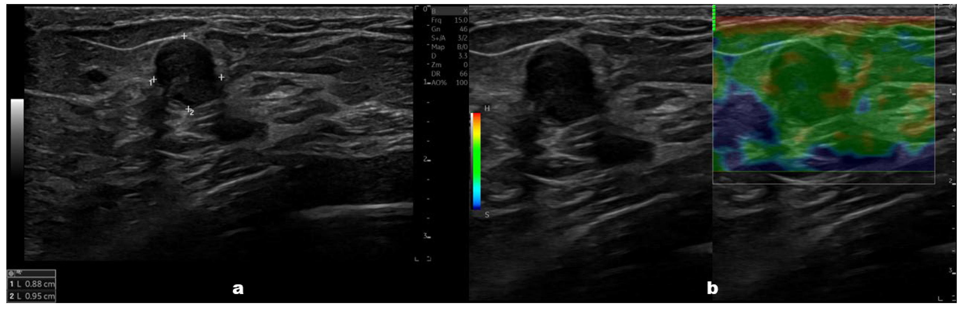
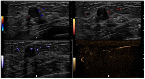
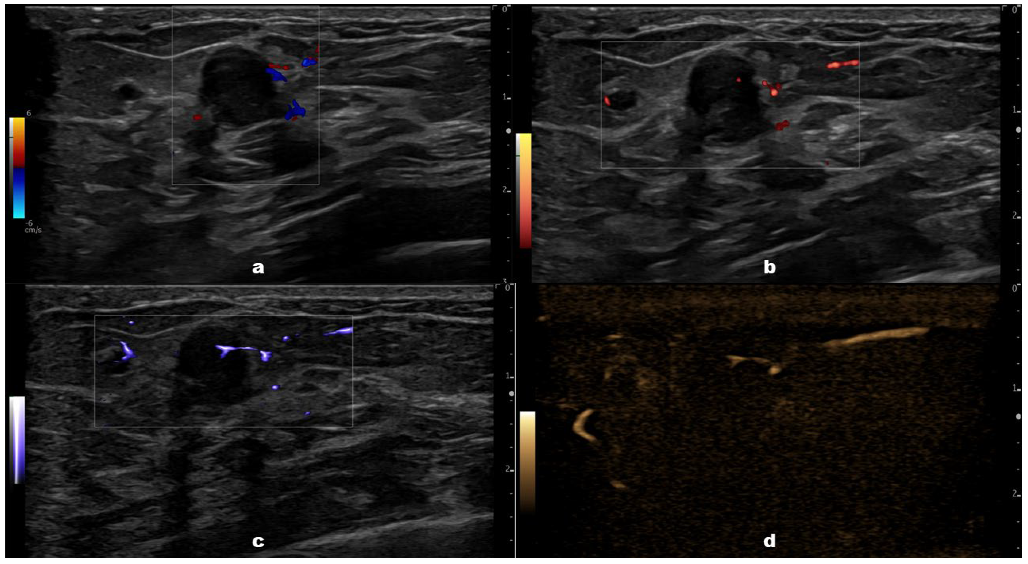
Seven days later, the multiparametric breast US was performed using a Logiq Fortis system (GE Healthcare, Chicago, IL, USA) with a 6–15 MHz linear transducer. It revealed an oval hypoechoic mass measuring 1.1 × 0.8 cm, located at the 3 o’clock position, approximately 2 cm from the nipple. The lesion exhibited a non-parallel orientation, microlobulated to angular margins, and mixed posterior acoustic features, comprising both shadowing and enhancement (Figure 2a). No suspicious axillary lymph nodes were found. Strain elastography demonstrated a soft mass, corresponding to a Tsukuba elasticity score of 1; however, quantitative assessment (strain ratio and E/B ratio) was not performed (Figure 2b). Color (CDI) and power Doppler imaging (PDI) showed peripheral vascularity (Figure 3a,b), while microvascular imaging (MVI) and B-flow imaging identified a penetrating intralesional vessel (Figure 3c,d). Based on these sonographic features, the lesion was upgraded to BI-RADS 4, and breast MRI was performed after 10 days.
Figure 2.
Ductal adenoma on B-mode US (a) and strain elastography (b): (a) oval, hypoechoic mass with a non-parallel orientation, microlobulated to angular margins, and combined posterior acoustic shadowing and enhancement; (b) soft mass (Tsukuba elasticity score 1).
Figure 3.
Ductal adenoma on CDI (a), PDI (b), MVI (c), and B-flow imaging (d): (a,b) mass with peripheral vascularity; (c,d) mass with a penetrating intralesional vessel.
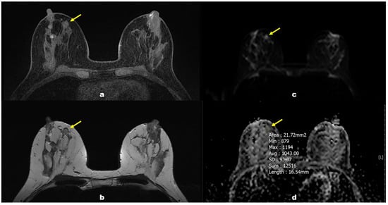
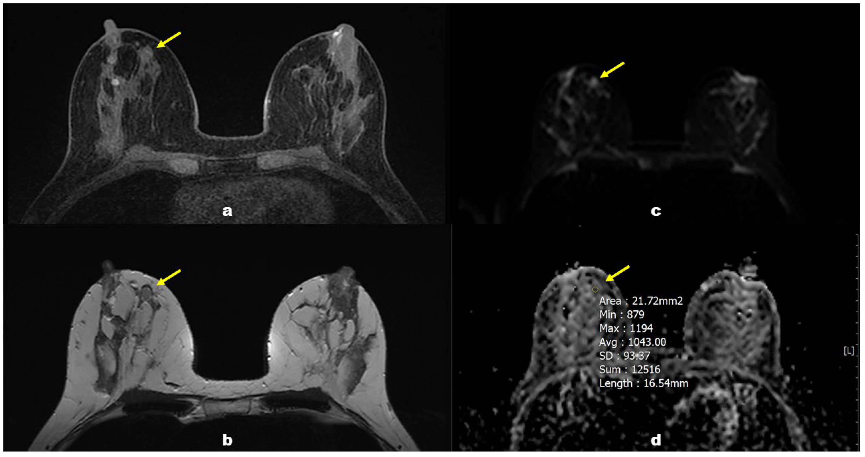
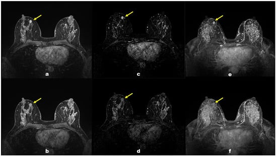
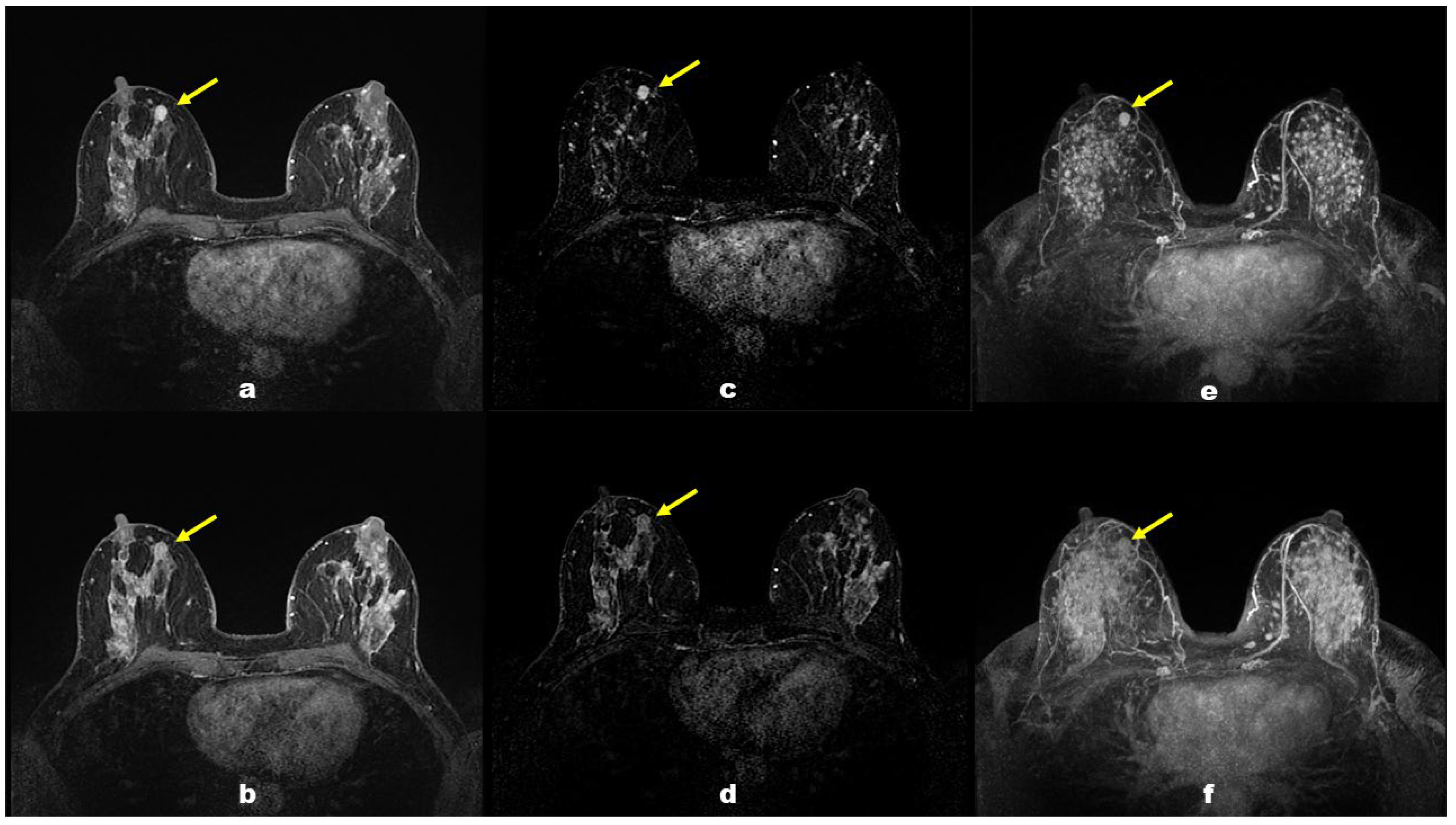
Breast MRI was performed on a 1.5T uMR 570 system (United Imaging Healthcare Co., Shanghai, China). It demonstrated a 1.1 × 0.8 cm oval mass with irregular margins. The mass was isointense relative to fibroglandular tissue on fat-suppressed (FS) T1-weighted (T1W) images and T2-weighted (T2W) images, without signs of perifocal edema (Figure 4a,b). Diffusion-weighted imaging (DWI) demonstrated restricted diffusion (b values 50 and 600), with an apparent diffusion coefficient (ADC) value of 1.04 × 10⁻³ mm2/s (Figure 4c,d). Following gadolinium administration (12 mL of gadoterate meglumine), marked background parenchymal enhancement attributable to fibrocystic changes was present. The mass showed heterogeneous, rapid initial enhancement and delayed washout, consistent with a type III kinetic curve (Figure 5). Application of the Kaiser score algorithm yielded a score of 8 (absent spiculations, delayed phase washout, heterogeneous internal enhancement). No suspicious non-mass enhancement or additional suspicious lesions were identified in either breast. The MRI examination was classified as BI-RADS 5.
Figure 4.
Ductal adenoma on pre-contrast MRI (arrows): (a) FST1W, isointense relative to fibroglandular tissue; (b) T2W, isointense relative to fibroglandular tissue, without signs of perifocal edema; (c) DWI, high-intensity signal; (d) ADC, restricted diffusion with an ADC value of 1.04 × 10⁻³ mm2/s (manual region-of-interest delineation).
Figure 5.
Ductal adenoma on contrast-enhanced MRI (arrows) showing heterogeneous, rapid initial enhancement and delayed washout, consistent with a type III kinetic curve: (a) FST1W, early phase; (b) FST1W, delayed phase; (c) subtraction images, early phase; (d) subtraction images, delayed phase; (e) MIP, early phase; (f) MIP, delayed phase.
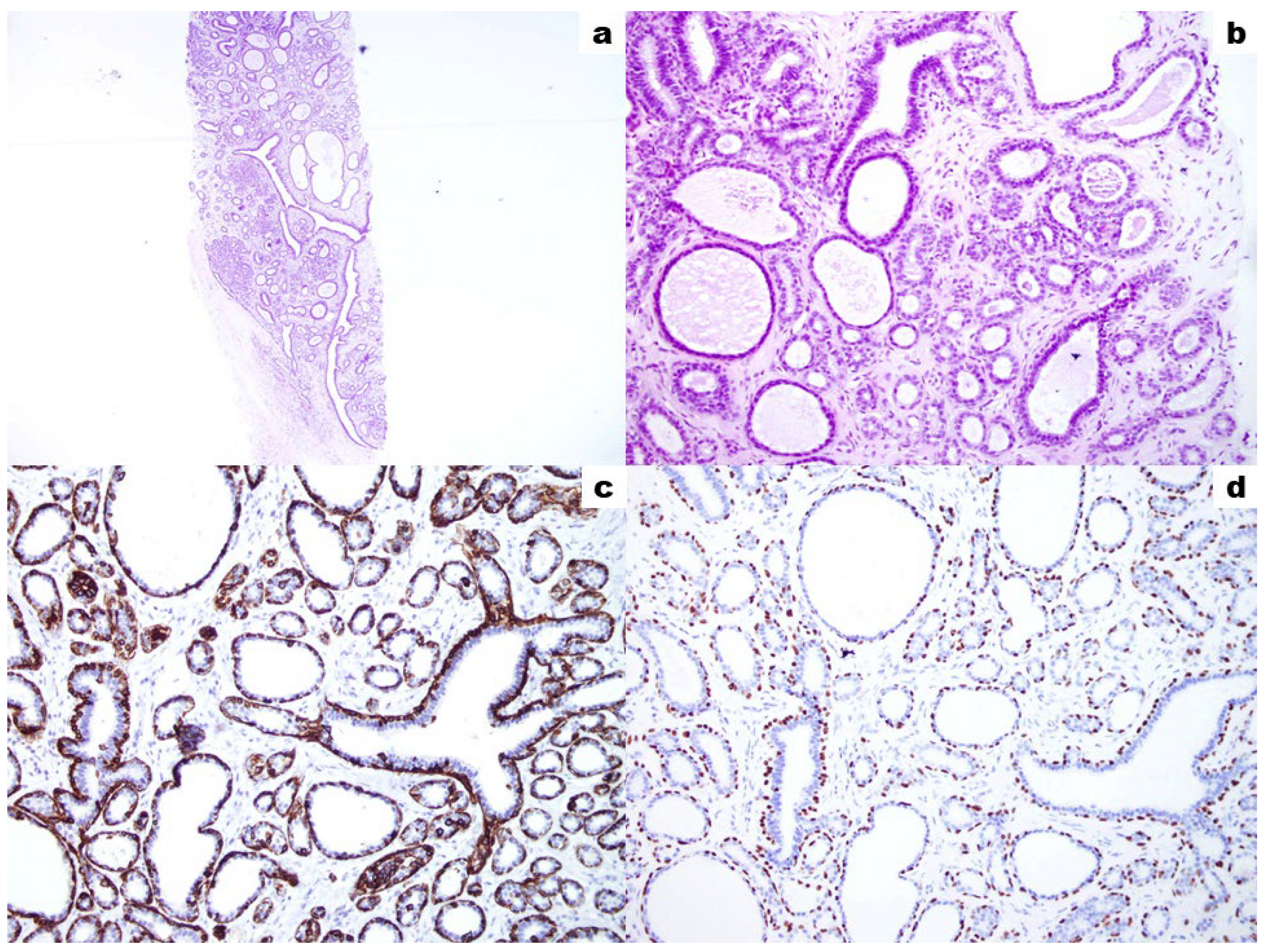
A US-guided core needle biopsy was subsequently performed with a 14-gauge needle, yielding six tissue specimens, clip placement, and no post-biopsy complications. Histopathological examination established the diagnosis of a breast ductal adenoma (B2 lesion) (Figure 6). Following multidisciplinary evaluation, conservative management with routine imaging surveillance was recommended. The mass remained stable at 6-month follow-up.
Figure 6.
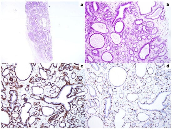
Biopsy specimen of ductal adenoma on histological examination: (a) H&E, magnification ×40; (b) H&E, magnification ×200; (c) immunohistochemistry, magnification ×400, positive staining for Cytokeratin-5; (d) immunohistochemistry, magnification ×400, positive staining for p63. It presented as a circumscribed lesion consisting of a proliferation of densely packed tubules set in a scant fibrous stroma. Focal branching and cystic dilation of the tubules were present. The tubules were lined with a bilayer of myoepithelial cells and overlying cuboidal glandular epithelium. At the lesion margins, parts of a thick-walled fibrous capsule were seen. Immunohistochemistry confirmed the presence of myoepithelial cells by positive staining for Cytokeratin-5 and p63.
3. Discussion
Ductal adenoma of the breast was first described by Azzopardi et al. in 1984 [3]. It predominantly affects women older than 50 years, with only isolated cases reported in patients younger than 40 years [3,4]. The lesion typically presents as a solitary unilateral mass, although bilateral involvement has been described in association with Carney syndrome [1,6]. Ductal adenomas are generally small, rarely exceeding 2 cm in greatest dimension [5]. Although it is regarded as a benign neoplasm, malignant transformation has been reported in a single case [7].
Owing to its rarity, the imaging spectrum of ductal adenoma remains incompletely characterized. We performed a literature review of published cases reporting imaging findings (MMG, US, MRI, and/or positron emission tomography/computed tomography). Two investigators independently searched PubMed/MEDLINE and Google using the terms Ductal adenoma AND breast AND imaging, with no language or publication date restrictions. Eligible studies included original articles, case reports, and case series reporting ductal adenoma with any imaging findings. Studies without imaging features were excluded. Retrieved records were screened independently, disagreements were resolved by consensus, and eligible full-text articles describing imaging features were reviewed. As of 3 November 2026, only nine publications describing imaging findings in 12 patients were identified, and their imaging characteristics are summarized in Table 1.
Table 1.
List of previously reported cases of breast ductal adenoma with imaging features.
On MMG, the ductal adenoma in the present case appeared as a round mass with partially obscured margins and density comparable to the surrounding fibroglandular tissue, without associated microcalcifications. In contrast, one previously reported case demonstrated no mammographic abnormality [11], whereas calcifications (microcalcifications or coarse calcifications) accompanied the lesion in 45% (5/11) of reported cases [5,6,9,10]. In the US, the ductal adenoma presented as a solid hypoechoic mass, while a cystic component was described in 36% (4/11) of previously reported cases [5,6,8]. Strain elastography demonstrated a uniformly soft lesion, a finding that, to our knowledge, has not been well documented in ductal adenoma. It was consistent with a Tsukuba elasticity score of 1, which is common in benign lesions [12,13]. On CDI and PDI, the ductal adenoma showed peripheral vascularity, while MVI and B-flow imaging identified a penetrating intralesional vessel. According to a study by Bartolotta et al. [14], the detection rate of a penetrating vessel was significantly higher with MVI than with either CDI or PDI, and this vascular feature was significantly more common in malignant than in benign breast masses. Breast MRI demonstrated an oval mass with irregular margins and an ADC of 1.04 × 10⁻³ mm2/s. This ADC value falls below the threshold of 1.11 × 10⁻³ mm2/s proposed by Tsvetkova et al. for differentiating benign breast lesions from malignant ones, which yielded a sensitivity of 94.23%, specificity of 94.29%, and diagnostic accuracy of 98% [15]. To date, only one study has reported an ADC value for breast ductal adenoma: 0.8 × 10⁻³ mm2/s [2]. Following gadolinium administration, the ductal adenoma exhibited heterogeneous enhancement with rapid initial enhancement and delayed washout, resulting in a Kaiser score of 8 [16]. To date, MRI findings have been described in only four cases of ductal adenoma, all of which demonstrated persistent or plateau enhancement kinetics [6]. Therefore, the washout enhancement pattern observed in the present case is poorly documented.
In the present case, MMG and strain elastography features favored a benign diagnosis, whereas B-mode US, MVI, and MRI demonstrated several imaging features typically associated with malignancy. In particular, the low ADC value and washout kinetic pattern on MRI were the principal factors prompting US-guided core needle biopsy. Histopathological examination established the diagnosis of a ductal adenoma. The histopathological differential diagnosis of ductal adenoma includes tubular adenoma, sclerosing adenosis, and well-differentiated invasive carcinoma. Ductal adenoma is distinguished from tubular adenoma by its more prominent ductal architecture and from sclerosing adenosis by the absence of the characteristic lobulocentric proliferation of compressed acini set in a sclerotic stroma [17,18]. Although well-differentiated invasive carcinoma may show a similar glandular architecture, it is characterized by infiltrative growth, a desmoplastic stromal reaction, and the loss of the myoepithelial layer [19]. Pseudoinvasion, also described in ductal adenoma, is characterized by entrapment of benign ductal elements within collagenized stroma extending beyond the circumscribed lesion, which may mimic invasive carcinoma [6].
Following multidisciplinary review, conservative management with routine imaging surveillance was recommended, given the rare report of malignant transformation in ductal adenoma [7]. The mass remained stable at 6-month follow-up. Management should be guided by radiological-pathological concordance rather than imaging appearance alone. Imaging surveillance may be considered when adequate tissue sampling enables a definitive benign diagnosis with no associated atypia or malignancy, as in our case.
The main limitation of this report is its single-case design, which precludes generalizability. Furthermore, the lack of comparison with histologically similar lesions limits assessment of the observed imaging features. Accordingly, these findings should be interpreted cautiously and not extrapolated beyond the present case. Further studies involving larger patient cohorts are needed.
4. Conclusions
Ductal adenoma is a rare benign breast lesion that may exhibit multimodality imaging features highly suggestive of malignancy. In the present case, multiparametric US and MRI evaluation demonstrated suspicious features, whereas MMG and strain elastography favored a benign diagnosis. This discordance highlights the diagnostic limitations of imaging alone and underscores the importance of histopathological confirmation in lesions with suspicious imaging features. Recognition of the potential imaging spectrum of ductal adenoma, including strain elastography characteristics, low ADC values, and washout enhancement kinetics, may contribute to a more comprehensive understanding of this rare entity. However, further reports and comparative studies are needed to establish the diagnostic relevance and reproducibility of these findings.
Author Contributions
Conceptualization, T.F.; methodology, T.F.; formal analysis, T.F.; investigation, T.F. and J.S.; writing—original draft preparation, T.F.; writing—review and editing, J.S., H.S.M., L.M. and V.V.; visualization, H.S.M. and L.M.; supervision, V.V. All authors have read and agreed to the published version of the manuscript.
Funding
This research received no external funding.
Institutional Review Board Statement
The study was conducted in accordance with the Declaration of Helsinki and was approved by the Ethics Committee of Merkur University Hospital (protocol code 03/1-4772/9, approved on 25 May 2026).
Informed Consent Statement
Written informed consent has been obtained from the patient for the publication of this paper.
Data Availability Statement
The original contributions presented in this study are included in the article. Further inquiries can be directed to the corresponding author.
Conflicts of Interest
The authors declare no conflicts of interest.
References
- Laraqui, Y.; Boujida, I.; Raissouni, K.; Echcharif, S.; Bekkarsabein, S.; El Khannoussi, B. Breast Ductal Adenoma: Confusion with Carcinoma is Real. J. Pathol. Allied Med. 2023, 1, 1–5. [Google Scholar] [CrossRef] [Scilit]
- Macedo, M.; Bassaganyas, C.; Ganau, S.; Sanfeliu, E.; Ubeda, B.; Bargallo, X. Ultrasound Findings of Breast Adenomas. J. Ultrasound Med. 2020, 39, 2173–2180. [Google Scholar] [CrossRef] [Scilit] [PubMed]
- Azzopardi, J.G.; Salm, R. Ductal adenoma of the breast: A lesion which can mimic carcinoma. J. Pathol. 1984, 144, 15–23. [Google Scholar] [CrossRef] [Scilit] [PubMed]
- Lammie, G.A.; Millis, R.R. Ductal adenoma of the breast—A review of fifteen cases. Hum. Pathol. 1989, 20, 903–908. [Google Scholar] [CrossRef] [Scilit] [PubMed]
- Okada, K.; Suzuki, Y.; Saito, Y.; Umemura, S.; Tokuda, Y. Two cases of ductal adenoma of the breast. Breast Cancer 2006, 13, 354–359. [Google Scholar] [CrossRef] [Scilit] [PubMed]
- Matsubayashi, R.N.; Momosaki, S.; Muranaka, T. Ductal adenoma of breast: Imaging characteristics and radiologic-pathologic correlation of unique findings which reflect “pseudoinvasion”. Breast Cancer 2016, 23, 597–606. [Google Scholar] [CrossRef] [Scilit] [PubMed]
- Hayashi, H.; Ohtani, H.; Yamaguchi, J.; Shimokawa, I. Malignant transformation of breast ductal adenoma: A diagnostic pitfall. Malays. J. Pathol. 2015, 37, 281–285. [Google Scholar] [PubMed]
- Scicolone, G.; Zen, F.; Visonà, A.; Motteran, F.; Contin, F.; Bano, A.; Vicentini, A. Ductal adenoma of the breast: Correlations between clinical-radiological and histopathological aspects (considerations on a case). Chir. Ital. 1986, 38, 543–548. [Google Scholar] [PubMed]
- Moskovic, E.; Ramachandra, S. Ductal adenoma of the breast: Mammographic appearances and pathological correlation. Br. J. Radiol. 1989, 62, 1021–1023. [Google Scholar] [CrossRef] [Scilit] [PubMed]
- Kim, E.K.; Oh, K.K.; Lee, K.S.; Lee, H.H. Ductal adenoma of the breast: A case report. J. Korean Radiol. Soc. 1997, 37, 949–951. [Google Scholar] [CrossRef] [Scilit]
- Yamaguchi, R.; Futamata, Y.; Yoshimura, F.; Murakami, N.; Koufuji, K.; Kutami, R.; Kojima, K.; Ohki, S.; Kurata, S.; Kaida, H.; et al. Mastopathic-type fibroadenoma and ductal adenoma of the breast with false-positive fluorodeoxyglucose positron emission tomography. Jpn. J. Radiol. 2009, 27, 280–284. [Google Scholar] [CrossRef] [Scilit] [PubMed]
- Dietrich, C.F.; Barr, R.G.; Farrokh, A.; Dighe, M.; Hocke, M.; Jenssen, C.; Dong, Y.; Saftoiu, A.; Havre, R.F. Strain elastography—How to do it? Ultrasound Int. Open 2017, 3, E137–E149. [Google Scholar] [CrossRef] [Scilit] [PubMed]
- Itoh, A.; Ueno, E.; Tohno, E.; Kamma, H.; Takahashi, H.; Shiina, T.; Yamakawa, M.; Matsumura, T. Breast disease: Clinical application of US elastography for diagnosis. Radiology 2006, 239, 341–350. [Google Scholar] [CrossRef] [Scilit] [PubMed]
- Bartolotta, T.V.; Orlando, A.A.M.; Schillaci, M.I.; Spatafora, L.; Marco, M.D.; Matranga, D.; Firenze, A.; Cirino, A.; Ienzi, R. Ultrasonographic detection of vascularity of focal breast lesions: Microvascular imaging versus conventional color and power Doppler imaging. Ultrason. Imaging 2021, 43, 273–281. [Google Scholar] [CrossRef] [Scilit] [PubMed]
- Tsvetkova, S.; Doykova, K.; Vasilska, A.; Sapunarova, K.; Doykov, D.; Andonov, V.; Uchikov, P. Differentiation of benign and malignant breast lesions using ADC values and ADC ratio in breast MRI. Diagnostics 2022, 12, 332. [Google Scholar] [CrossRef] [Scilit] [PubMed]
- Dietzel, M.; Baltzer, P.A.T. How to use the Kaiser score as a clinical decision rule for diagnosis in multiparametric breast MRI: A pictorial essay. Insights Imaging 2018, 9, 325–335. [Google Scholar] [CrossRef] [Scilit] [PubMed]
- Turashvili, G. Nonneoplastic and neoplastic sclerosing lesions of the breast. Histopathology 2024, 85, 383–396. [Google Scholar] [CrossRef] [Scilit] [PubMed]
- Ekblom, P.; Miettinen, M.; Forsman, L.; Andersson, L.C. Basement membrane and apocrine epithelial antigens in differential diagnosis between tubular carcinoma and sclerosing adenosis of the breast. J. Clin. Pathol. 1984, 37, 357–363. [Google Scholar] [CrossRef] [Scilit] [PubMed]
- Bachert, S.E.; Chapel, E.; Schnitt, S.J. Breast myoepithelial markers: Problems and pitfalls for the practicing pathologist. Hum. Pathol. 2025, 162, 105909. [Google Scholar] [CrossRef] [Scilit] [PubMed]

Disclaimer/Publisher’s Note: The statements, opinions and data contained in all publications are solely those of the individual author(s) and contributor(s) and not of MDPI and/or the editor(s). MDPI and/or the editor(s) disclaim responsibility for any injury to people or property resulting from any ideas, methods, instructions or products referred to in the content. |
© 2026 by the authors. Licensee MDPI, Basel, Switzerland. This article is an open access article distributed under the terms and conditions of the Creative Commons Attribution (CC BY) license.