


Abstract
Background and Clinical Significance: Amniocentesis is a commonly performed procedure for the detection of genetic abnormalities and may be associated with a higher risk of complications when performed at 24 weeks of gestation or later compared with procedures performed earlier. Case Presentation: We present the case of a 40-year-old gravida 4 para 0211 at 29 1/7 weeks of gestation with a history of simultaneous pancreas and renal transplant who developed severe abdominal pain and a non-reassuring fetal tracing following amniocentesis performed in the setting of newly diagnosed non-immune hydrops fetalis. The diagnosis of placental abruption was suspected, and an emergency cesarean delivery was performed. Intraoperative findings revealed a large hemoperitoneum and active bleeding from a uterine sinus vessel at the amniocentesis needle puncture site. Conclusions: Hemoperitoneum after late amniocentesis is a rare but clinically significant complication that can mimic placental abruption. Dialysis-dependent renal disease may have contributed to bleeding susceptibility; the role of immunosuppressive therapy remains uncertain.
1. Introduction and Clinical Significance
Amniocentesis is a transabdominal, ultrasound-guided procedure that involves withdrawing a small amount of amniotic fluid from the gestational sac, most commonly for genetic testing. The procedure is typically performed between 15 and 20 weeks of gestation, but can be performed at any gestational age later during pregnancy [1]. A recent meta-analysis including 42,000 patients who underwent amniocentesis estimated a procedure-related miscarriage rate of 0.11% [2]. Other potential complications include amniotic fluid leakage in 1–2% of cases, and postprocedural chorioamnionitis in <0.1% [3]. The term ‘late’ amniocentesis refers to procedures performed from 24 weeks’ gestation onwards [4]. We present the case of a pregnant patient at 29 1/7 weeks of gestation with a history of a simultaneous pancreas and renal transplant who developed severe abdominal pain, a non-reassuring fetal heart rate tracing and suspected placental abruption following amniocentesis. Hemoperitoneum and active bleeding from a uterine sinus vessel at the amniocentesis needle puncture site were identified at the time of her emergency cesarean delivery. Other potential causes of hemoperitoneum in pregnancy include abdominal trauma, uterine rupture, invasive placentation, ruptured ovarian cyst, ruptured hepatic subcapsular hematoma associated with hemolysis, elevated liver enzymes and low platelets syndrome or rupture of a splenic artery aneurysm. This unique case suggests that hemoperitoneum after late amniocentesis is a rare but clinically significant complication that can mimic placental abruption.
2. Case Presentation
A 40-year-old gravida 4 para 0211 with suspected fetal growth restriction presented to Labor and Delivery at 29 1/7 weeks’ gestation for evaluation in the setting of a non-reassuring nonstress test and ultrasound findings of newly diagnosed hydrops fetalis. Fetal growth restriction was previously suspected based on a prior ultrasound demonstrating an estimated fetal weight at the 10th percentile with an abnormal umbilical artery Doppler pulsatility index. On the day of hospital presentation, ultrasound findings demonstrated a fetus with an estimated fetal weight of 1258 g (21st percentile), bilateral pleural effusions, ascites, and skin edema. Umbilical artery Doppler evaluation was notable for an increased pulsatility index > 95th percentile with forward flow throughout the cardiac cycle. She had a complex medical and surgical history including a simultaneous pancreas and renal transplant in 2019 for poorly controlled type 1 diabetes mellitus with good pancreas allograft function and poor renal allograft function resulting in kidney failure requiring dialysis six times per week. Additional medical comorbidities included chronic hypertension requiring three antihypertensive medications (nifedipine extended-release 90 mg daily, hydralazine 50 mg three times daily, and labetalol 400 mg three times daily) for control, severe anemia, prior stroke, and several complications secondary to her long-standing diabetes including retinopathy, gastroparesis, and neuropathy. Her obstetric history was significant for two medically indicated preterm cesarean deliveries at 31 and 27 weeks’ gestation in the setting of superimposed preeclampsia with severe features and severe fetal growth restriction, with neonatal demise following the second preterm delivery. Her medications also included tacrolimus and azathioprine. Genetic screening in this pregnancy revealed a low fetal fraction with cell-free DNA testing; she declined prenatal diagnostic genetic testing during early pregnancy. She had a normal detailed fetal anatomy ultrasound. Evaluation of fetal cardiac anatomy was within normal limits at that time.
On presentation, the patient reported increased swelling of the face, abdomen, and bilateral lower extremities. She denied any new-onset headache, visual disturbances, right upper quadrant or epigastric pain, or vaginal bleeding. On exam, she had mildly elevated blood pressures along with significant generalized swelling. Lab evaluation initially performed was notable for the following: antibody screen negative, hemoglobin 6.5 g/dL, platelet count 125,000 cells/µL, partial thromboplastin time (PTT) 29 s, prothrombin time (PT) 8.9 s, international normalized ratio (INR) 0.82, creatinine 5.68 mg/dL, aspartate aminotransferase 25 IU/L, alanine aminotransferase 29 IU/L, and urine protein:creatinine ratio of 1.34. After evaluation, diagnoses such as Mirror Syndrome and superimposed preeclampsia were considered. The patient received betamethasone and underwent serology testing for cytomegalovirus, toxoplasma, and parvovirus to assess for infectious etiologies of hydrops fetalis. All serology testing was negative for acute infection. Kleihauer-Betke testing was also negative. Her hemoglobin increased from 6.5 g/dL to 7.6 g/dL after transfusion of 1 unit of packed red blood cells on hospital day 2. She was also counseled on the option of amniocentesis and its associated risks versus benefits to assess for genetic causes and ultimately decided to proceed with the procedure. The platelet count prior to the procedure was 154,000 cells/µL.
On hospital day 3, amniocentesis was performed under ultrasound guidance by an experienced operator with a single insertion of a 20-gauge 3.5-inch needle. Due to the anterior location of the placenta, transplacental puncture was necessary, and 60 mL of clear yellow amniotic fluid was obtained. The fetus was in cephalic presentation, and there were no immediate post-procedure abnormal ultrasound findings. The amniotic fluid sample was sent for chromosomal microarray and polymerase chain reaction testing for cytomegalovirus, toxoplasma, and parvovirus. Approximately 3 h following the procedure, the patient developed worsening abdominal pain unrelieved by intravenous pain medications. Electronic fetal monitoring was initiated and showed a normal baseline with moderate variability, variable decelerations, and uterine irritability. The patient’s abdomen was soft and diffusely tender on palpation. A cervical exam was performed, and the cervix was noted to be closed. Repeat lab evaluation demonstrated a drop in hemoglobin from 7.6 g/dL to 6.5 g/dL and a fibrinogen level of 380 mg/dL. Additional coagulation studies at that time included a PTT of 25 s, PT of 9.0 s, and INR of 0.83. Given the patient’s worsening pain following the procedure and persistent fetal heart rate decelerations, placental abruption was suspected, and the decision was made to proceed with an emergency repeat cesarean delivery. Approximately 500 cc of hemoperitoneum was noted upon entry into the patient’s abdomen. Once evacuated, a small pinpoint area of active bleeding was noted from a uterine sinus vessel on the anterior uterine serosa. A 1 cm area of uterine dehiscence was also noted adjacent to but separate from the bleeding vessel, which was superior to the prior cesarean scar. There was no evidence of immediate placental separation, retroplacental hematoma, or Couvelaire changes observed intraoperatively. A liveborn male was subsequently delivered, weighing 1120 g, with Apgar scores of 2 and 6 at 1 and 5 min, respectively. Delivery was complicated by a postpartum hemorrhage of 1596 mL due to hemoperitoneum and uterine atony. Her postpartum course was complicated by a prolonged hospitalization for pain management and dialysis care. She received one additional unit of packed red blood cells postpartum. She was discharged by post-operative day 14 in stable condition. Pathological examination of the placenta demonstrated an immature placenta weighing 244 g with marked distal villous hypoplasia (which is associated with maternal vascular malperfusion and fetal growth restriction), fetal membranes with no significant histopathology, and a histologically unremarkable trivascular umbilical cord. These findings were likely unrelated to the source of intraperitoneal hemorrhage or cause of fetal hydrops. The neonate was admitted to the neonatal intensive care unit for 100 days and subsequently discharged in stable condition. Neonatal complications included bilateral pleural effusions, grade 2 bronchopulmonary dysplasia, apnea of prematurity, hyperbilirubinemia, grade 1 intraventricular hemorrhage, and feeding intolerance. Chromosomal microarray analysis of the amniotic fluid obtained via amniocentesis revealed a normal male result. Expanded exome or genome testing was declined at that time.
3. Discussion
Late amniocentesis is most commonly performed for the evaluation of fetal anomalies, suspected intrauterine infection, fetal growth restriction, or late presentation of non-immune hydrops fetalis [4]. Although the risk of culture failure increases with advancing gestational age [3], diagnostic yield has improved with advanced techniques such as chromosomal microarray analysis, which does not require cell culture in contrast to conventional karyotyping [5,6]. A recent systematic review and meta-analysis involving 911 singleton pregnancies evaluated obstetrical outcomes following amniocentesis performed after 24 weeks of gestation and reported an incidence of preterm birth (PTB) < 37 weeks of 4.85% and PTB within 1 week of amniocentesis of 1.42% [7]. Preterm prelabor rupture of membranes occurred in 2.85% of cases and placental abruption in 0.91% [7]. Hemoperitoneum was not reported as a complication after late amniocentesis in this systematic review.
Although uncommon, intraperitoneal hemorrhage following amniocentesis has been described previously. Di Francesco [8] reported major intraperitoneal bleeding complicating amniocentesis, while Jung and Künzig [9] described hemorrhagic shock resulting from injury to a small subserosal artery in the uterine fundus. These reports demonstrate that maternal uterine vessel injury can cause clinically significant intra-abdominal bleeding after amniocentesis. The prior reports establish this complication as a recognized possibility, but their isolated nature precludes reliable estimates of its incidence or associated risk factors. Our case adds to this literature by describing hemoperitoneum following late amniocentesis at 29 weeks’ gestation in a medically complex pregnancy. Based on the patient’s severe abdominal pain after the procedure, placental abruption was initially considered; however, hemoperitoneum was instead found to be secondary to bleeding from the amniocentesis needle puncture site. Although an area of uterine dehiscence was identified intraoperatively, it was separate from the bleeding site and likely did not contribute to this patient’s clinical presentation. The dehiscence was likely related to her two prior cesarean deliveries and may reflect localized structural weakness of the uterine wall.
In this case, the onset of symptoms shortly after amniocentesis and the intraoperative finding of active bleeding support an association with the procedure. However, a single case cannot establish a causal relationship between late amniocentesis and adverse outcomes, particularly in a pregnancy with substantial pre-existing maternal disease, including a history of transplant, immunosuppressive therapy, and dialysis dependence. This clinical presentation may have been related to the bleeding diathesis due to her underlying chronic kidney disease requiring dialysis. Patients with end-stage renal disease have an increased bleeding tendency that is mainly due to abnormalities of primary hemostasis, in particular, platelet dysfunction and impaired platelet-vessel wall interaction [10]. Intrinsic platelet abnormalities with reduced thromboxane A2 synthesis in response to adenosine diphosphate, collagen and thrombin are seen in end-stage renal disease patients [11]. Binding of both von Willebrand factor and fibrinogen to glycoprotein IIb/IIIa is believed to be decreased in uremic patients, leading to impaired platelet adhesion to the subendothelium [12]. Hemodialysis typically improves the platelet-dependent fibrin clot formation and contributes to improved platelet function after dialysis [13]. However, it may also itself contribute to bleeding as the artificial surfaces of the dialyzer and the blood tubing can cause platelet activation, induction, degranulation, and loss of glycoprotein receptors [14].
The patient’s long-standing type 1 diabetes mellitus was complicated by substantial end-organ disease, including nephropathy and retinopathy. Studies of patients with type 1 diabetes and microangiopathy have demonstrated endothelial dysfunction and platelet hyperactivity. These findings do not establish an increased susceptibility to uterine vessel rupture or post-amniocentesis hemoperitoneum. In this patient, the potential relevance of diabetes may have been indirect, through its contribution to advanced renal disease.
Apart from the inherent bleeding diathesis seen in patients with end-stage renal disease requiring dialysis, this type of complication might have been related to increased tissue fragility with prolonged exposure to immunosuppression due to her underlying pancreas and kidney transplant. However, evidence to support this association is lacking, and further studies are needed to determine whether an association exists between prolonged immunosuppression and increased tissue fragility in pregnancy.
4. Conclusions
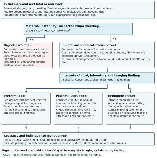
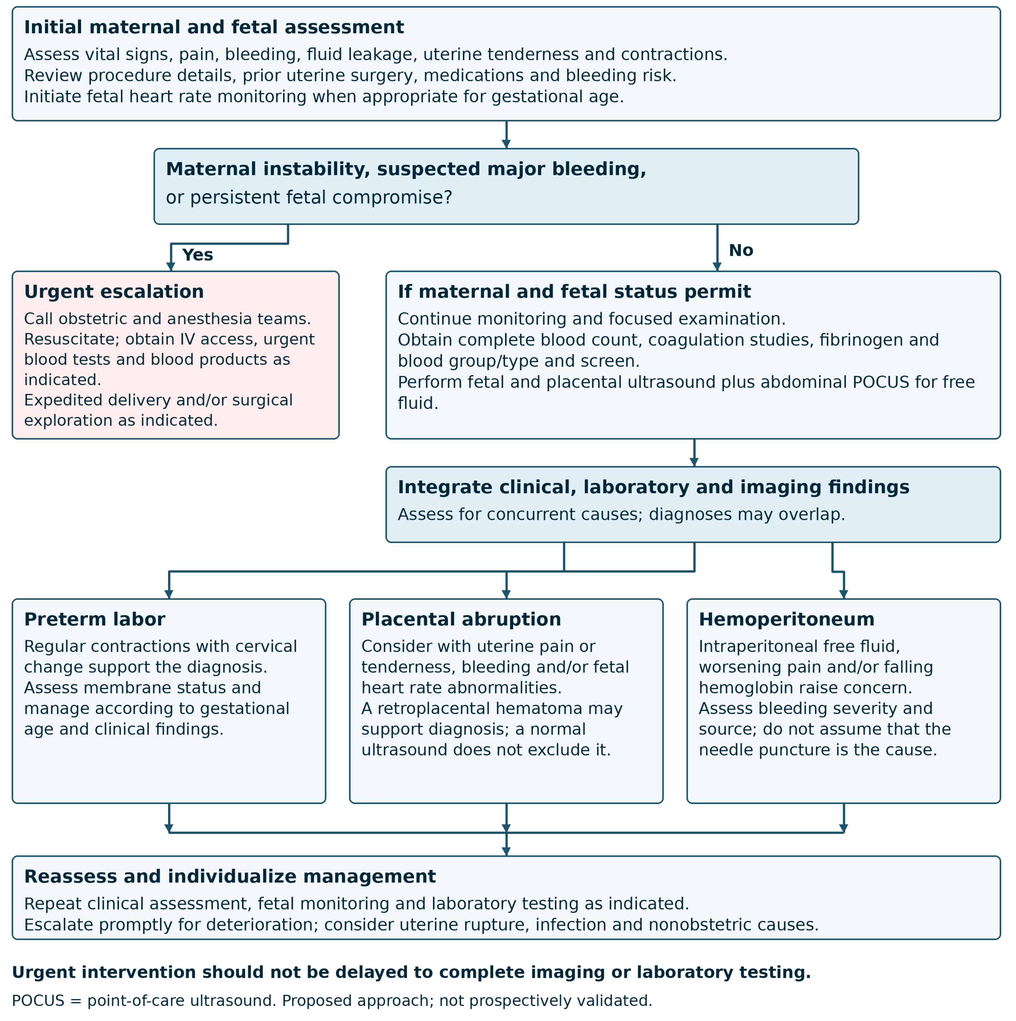
Our case highlights the complex interplay between uremic platelet dysfunction and pregnancy-induced vascular changes. Pregnant patients with a history of a solid organ transplant or those requiring dialysis therapy who are considering amniocentesis should be counseled regarding the potential risk of postprocedural bleeding complications; however, the limited evidence and single case report cannot establish or quantify increased procedural bleeding risk in transplant or dialysis populations. Coexisting medical conditions, relevant medication exposure, and prior uterine surgery should also be emphasized during pre-procedural counseling. Proposed evaluation strategies and management algorithms for pregnant patients with abdominal pain after late amniocentesis are displayed in Figure 1 and Table 1.
Figure 1.
Proposed clinical assessment approach for abdominal pain after late amniocentesis.
Table 1.
Proposed evaluation for abdominal pain after late amniocentesis.
Author Contributions
Conceptualization, S.K.D.C., M.G.; validation, S.K.D.C., M.G.; writing—original draft preparation, S.K.D.C.; writing—review and editing, M.G.; supervision, M.G. All authors have read and agreed to the published version of the manuscript.
Funding
This research received no external funding.
Institutional Review Board Statement
Ethical review and approval were not required by the Institutional Review Board at Thomas Jefferson University because case reports are not considered research. The patient’s information has been de-identified.
Informed Consent Statement
Written patient consent was obtained for publication of this case report.
Data Availability Statement
The original data presented in the study are included in the article; further inquiries can be directed to the corresponding author.
Conflicts of Interest
The authors declare no conflicts of interest.
References
- American College of Obstetricians and Gynecologists. Practice Bulletin No. 162: Prenatal Diagnostic Testing for Genetic Disorders. Obstet. Gynecol. 2016, 127, e108–e122. [Google Scholar] [CrossRef] [Scilit] [PubMed]
- Akolekar, R.; Beta, J.; Picciarelli, G.; Ogilvie, C.; D’Antonio, F. Procedure-related risk of miscarriage following amniocentesis and chorionic villus sampling: A systematic review and meta-analysis. Ultrasound Obstet. Gynecol. 2015, 45, 16–26. [Google Scholar] [CrossRef] [Scilit] [PubMed]
- Ghi, T.; Sotiriadis, A.; Calda, P.; Costa, F.D.S.; Raine-Fenning, N.; Alfirevic, Z.; McGillivray, G. ISUOG Practice Guidelines: Invasive procedures for prenatal diagnosis. Ultrasound Obstet. Gynecol. 2016, 48, 256–268. [Google Scholar] [CrossRef] [Scilit] [PubMed]
- Zemet, R.; Maktabi, M.A.; Tinfow, A.; Giordano, J.L.; Heisler, T.M.; Yan, Q.; Plaschkes, R.; Stokes, J.; Walsh, J.M.; Corcoran, S.; et al. Amniocentesis in pregnancies at or beyond 24 weeks: An international multicenter study. Am. J. Obstet. Gynecol. 2025, 232, 402.e1–402.e16. [Google Scholar] [CrossRef] [Scilit] [PubMed]
- Li, Y.; Yan, H.; Chen, J.; Chen, F.; Jian, W.; Wang, J.; Ye, X.; Li, Y.; Li, N.; Chiu, P.C.N.; et al. The application of late amniocentesis: A retrospective study in a tertiary fetal medicine center in China. BMC Pregnancy Childbirth 2021, 21, 266. [Google Scholar] [CrossRef] [Scilit] [PubMed]
- Monier, I.; Receuver, A.; Houfflin-Debarge, V.; Goua, V.; Castaigne, V.; Jouannic, J.-M.; Mousty, E.; Saliou, A.-H.; Bouchghoul, H.; Rousseau, T.; et al. Should prenatal chromosomal microarray analysis be offered for isolated fetal growth restriction? A French multicenter study. Am. J. Obstet. Gynecol. 2021, 225, 676.e1–676.e15. [Google Scholar] [CrossRef] [Scilit] [PubMed]
- Nassr, A.A.; Hessami, K.; D’Alberti, E.; Giancotti, A.; Meshinchiasl, N.; Evans, M.I.; Di Mascio, D.; Shamshirsaz, A.A. Obstetrical outcomes following amniocentesis performed after 24 weeks of gestation: A systematic review and meta-analysis. Prenat. Diagn. 2023, 43, 1425–1432. [Google Scholar] [CrossRef] [Scilit] [PubMed]
- Di Francesco, A. Major intraperitoneal bleeding complicating amniocentesis—Case report. Aust. N. Z. J. Obstet. Gynaecol. 1972, 12, 255–256. [Google Scholar] [CrossRef] [Scilit] [PubMed]
- Jung, V.; Künzig, H.J. Hemorrhagic shock caused by intraperitoneal hemorrhage after amniocentesis. Geburtshilfe Frauenheilkd. 1995, 55, 411–413. [Google Scholar] [CrossRef] [Scilit] [PubMed]
- Boccardo, P.; Remuzzi, G.; Galbusera, M. Platelet dysfunction in renal failure. Semin. Thromb. Hemost. 2024, 5, 579–589. [Google Scholar]
- Macconi, D.; Vigano, G.; Bisogno, G.; Galbusera, M.; Orisio, S.; Remuzzi, G. Defective platelet aggregation in response to platelet activation factor in uremia associated with low platelet thromboxane A2 generation. Am. J. Kidney Dis. 1992, 19, 318–325. [Google Scholar] [CrossRef] [Scilit] [PubMed]
- Benigni, A.; Boccardo, P.; Galbusera, M. Reversible activation defect of the platelet glycoprotein IIb-IIIa complex in patients with uremia. Am. J. Kidney Dis. 1993, 22, 668–676. [Google Scholar] [CrossRef] [Scilit] [PubMed]
- Murakonda, V.B.; Mohapatra, A.; Geevar, T.; Vijayan, R.; Kakde, S.; Jacob, S.; Alexander, S.; David, V.G.; Nair, S.C.; Varughese, S.; et al. Does Hemodialysis Need to be Initiated to Improve Platelet Function in CKD G5 Patients? A Pilot Prospective, Observational Cohort Study. Indian J. Nephrol. 2021, 31, 43–49. [Google Scholar] [CrossRef] [Scilit] [PubMed]
- Sloand, J.A.; Sloand, E.M. Studies on platelet membrane glycoproteins and platelet function during hemodialysis. J. Am. Soc. Nephrol. 1997, 8, 799–803. [Google Scholar] [CrossRef] [Scilit] [PubMed]

Disclaimer/Publisher’s Note: The statements, opinions and data contained in all publications are solely those of the individual author(s) and contributor(s) and not of MDPI and/or the editor(s). MDPI and/or the editor(s) disclaim responsibility for any injury to people or property resulting from any ideas, methods, instructions or products referred to in the content. |
© 2026 by the authors. Licensee MDPI, Basel, Switzerland. This article is an open access article distributed under the terms and conditions of the Creative Commons Attribution (CC BY) license.