


Abstract
Background: Machine learning may facilitate the integration of imaging findings and hematological inflammatory indices for clinical classification. This study evaluated whether these variables could discriminate women with established thrombophilia receiving low-molecular-weight heparin (LMWH) from matched women without thrombophilia. Methods: This prospective, single-center, matched case–control study included 160 women undergoing term cesarean delivery: 80 with established thrombophilia receiving prophylactic LMWH and 80 age- and parity-matched controls without thrombophilia. The Postpartum Uterine Ultrasonographic Scale (PUUS) was assessed 24–48 h after delivery, and 11 hematological inflammatory indices were derived from complete blood counts obtained within 24 h before delivery. Five supervised machine-learning algorithms were evaluated: elastic-net logistic regression, random forest, XGBoost, support vector machine with a radial basis function kernel, and multilayer perceptron. Model discrimination was assessed using the area under the receiver operating characteristic curve (AUC), with internal bootstrap-based assessment of optimism and model-selection effects. Results: PUUS scores differed between groups, with PUUS ≥ 1 observed in 32.5% of women with thrombophilia and 13.8% of controls (odds ratio [OR] 3.02, 95% confidence interval [CI] 1.38–6.61; p = 0.005). After adjustment for multiple comparisons, NLPR, IIC, NLR, PLR, SII, SIRI, and AISI differed significantly between groups. Among the five evaluated algorithms, XGBoost achieved the highest observed AUC in the 32-participant hold-out evaluation set (0.938, 95% CI 0.826–0.989), with 90.6% accuracy, 93.8% sensitivity, and 87.5% specificity. Five machine-learning algorithms were evaluated to explore the discrimination between women with LMWH-treated thrombophilia and matched controls without thrombophilia. However, the original analytical dataset and computational outputs were unavailable for independent verification. Consequently, the comparative performance of the algorithms and the previously reported AUC estimates could not be independently confirmed. Conclusions: Machine learning integrating postpartum uterine ultrasonography and hematological inflammatory indices showed promising discrimination between women with LMWH-treated, established thrombophilia and matched controls in this single-center cohort. However, thrombophilia status and LMWH exposure were completely confounded, and the small hold-out evaluation set and lack of external validation limit the interpretation of model performance. The findings should be considered a methodological proof-of-concept rather than evidence of a validated diagnostic or screening tool for untreated or previously undiagnosed thrombophilia. Independent multicenter validation in populations that permit separation of disease-related and treatment-related effects is required before clinical application can be considered.
1. Introduction
Thrombophilia comprises inherited and acquired conditions associated with an increased predisposition to thrombosis. Pregnancy is itself a physiologically hypercoagulable state, characterized by coordinated changes in coagulation, anticoagulant, and fibrinolytic pathways that become particularly pronounced toward delivery. In women with thrombophilia, these physiological adaptations may further increase the risk of venous thromboembolism and may be associated with selected adverse pregnancy outcomes [1,2,3].
Because thrombophilia testing has important implications for clinical management but is not appropriate as universal screening in the general obstetric population, contemporary recommendations favor testing in selected clinical circumstances in which the result is expected to influence management [4]. This selective approach reflects, among other considerations, the variable clinical significance of individual thrombophilic abnormalities and the challenges associated with interpreting thrombotic risk in pregnancy [1,4].
The search for accessible biomarkers that can characterize women at increased thrombotic or obstetric risk has therefore included hematological inflammatory indices derived from the complete blood count. Such indices have been investigated in pregnant populations and in women with thrombophilia, reflecting the interaction between inflammatory, hematological, and thrombotic processes [5]. Multivariable approaches have also been explored for the prediction of thrombophilia in specific clinical populations, including women with unexplained recurrent pregnancy loss, supporting the potential value of combining multiple routinely available variables rather than relying on a single biomarker [6].
Postpartum uterine ultrasonography represents another potentially informative source of clinical information. The Postpartum Uterine Ultrasonographic Scale (PUUS) was developed as a semi-quantitative method for describing postpartum uterine cavity contents and uterine involution [7]. Subsequent work evaluated the PUUS after cesarean delivery in women with treated thrombophilia and controls without thrombophilia, providing clinical data on the potential relationship between sonographic findings and hematological parameters in this setting [8]. Importantly, these studies do not establish PUUS as an independent diagnostic test for thrombophilia; rather, they provide a rationale for evaluating postpartum sonographic characteristics as one component of a broader multimodal phenotype [7,8].
Machine-learning approaches provide a framework for integrating multiple heterogeneous predictors and for identifying nonlinear relationships or interactions that may be difficult to capture using conventional statistical models [9,10,11,12]. Nevertheless, predictive performance is intrinsically dependent on the population used for model development, the definition of the target condition, the quality of predictor measurements, and the extent to which the development population corresponds to the intended clinical-use population [9,10,11,12]. These considerations are particularly important in the present setting because all women with established thrombophilia included in the study were receiving low-molecular-weight heparin (LMWH), whereas the comparison group was not receiving LMWH. Consequently, inflammatory and sonographic characteristics measured in the study may reflect a combination of disease-related and treatment-related effects rather than the biological phenotype of untreated thrombophilia alone.
Accordingly, the present study was designed as a single-center methodological proof-of-concept to evaluate whether machine-learning algorithms can discriminate women with established thrombophilia receiving LMWH from matched women without thrombophilia using postpartum uterine ultrasonography and pre-delivery hematological inflammatory indices. The study was not designed to identify previously undiagnosed or treatment-naïve thrombophilia. Rather, its purpose was to assess the feasibility of multimodal machine-learning classification in a defined, treated clinical population and to identify candidate predictors that may warrant evaluation in future treatment-naïve cohorts.
Study Aim and Objectives
Aim: The primary aim of this study was to evaluate the feasibility of machine-learning-based classification of women with established thrombophilia receiving low-molecular-weight heparin (LMWH) versus matched women without thrombophilia by integrating postpartum uterine ultrasonographic findings with pre-delivery hematological inflammatory indices.
Specific objectives: The study objectives were:
To evaluate the discriminatory performance of different supervised machine-learning algorithms for distinguishing women with LMWH-treated thrombophilia from women without thrombophilia, using postpartum uterine ultrasonography and hematological inflammatory indices.
To compare the predictive performance of five complementary modeling approaches, including elastic-net logistic regression, random forest, XGBoost, support vector machine with a radial basis function kernel, and multilayer perceptron.
To identify the predictors contributing most strongly to model discrimination, with particular attention to the Postpartum Uterine Ultrasonographic Scale (PUUS) and the hematological inflammatory indices.
To quantify model discrimination and predictive performance using the area under the receiver operating characteristic curve (AUC), sensitivity, specificity, and classification accuracy, together with bootstrap-based assessment of optimism.
To assess the potential effect of model-selection optimism arising from comparison of multiple candidate algorithms and to provide a more conservative estimate of model performance.
To perform a prespecified sensitivity analysis excluding participants with isolated MTHFR polymorphisms, in order to determine whether their inclusion materially affected model performance.
To define the limitations of the current model with respect to clinical applicability, particularly the confounding introduced by LMWH exposure, the single-center design, and the use of postpartum measurements that cannot be assumed to represent treatment-naïve thrombophilia.
2. Materials and Methods
2.1. Study Design and Participants
This was a prospective, single-center, matched case–control observational study conducted at the Elena Doamna Obstetrics and Gynecology University Hospital, Iași, Romania, between October 2017 and December 2021. Participants were recruited prospectively and underwent standardized clinical, laboratory, and postpartum ultrasonographic assessment. The machine-learning analyses were subsequently performed using the prospectively collected dataset.
The study protocol was approved by the Institutional Ethics Committee of the Elena Doamna Obstetrics and Gynecology University Hospital (approval no. 9, 15 September 2017) and was conducted in accordance with the Declaration of Helsinki. Written informed consent was obtained from all participants before enrollment.
The present study included the same 160 participants recruited at the same institution between October 2017 and December 2021, as previously reported by Filip et al. [8]. Therefore, the current analysis represents a secondary analysis of the previously reported cohort rather than an independent replication. However, the original postpartum ultrasound examinations were reassessed for the present study. All 160 examinations were independently reviewed by two experienced ultrasonographers who were blinded to participants’ thrombophilia status and study-group allocation. PUUS scores were recalculated according to the original five-grade scoring system (grades 0–4), and disagreements between the two assessors were resolved by consensus. Consequently, the PUUS values used in the present analysis reflect the reassessment of the original ultrasound examinations rather than the scores reported in the previous publication. The current study extends the previously reported clinical assessment by integrating the reassessed PUUS scores with hematological inflammatory indices in a machine-learning framework.
PUUS data provenance and reassessment. The participant-level PUUS scores used in the present analysis were obtained through a new assessment of the original ultrasound examinations. All 160 examinations were independently reassessed by two experienced ultrasonographers blinded to thrombophilia status and group allocation. Each examination was scored using the original five-grade PUUS scale (grades 0–4). Disagreements were resolved by consensus. Thus, the scores used in the current analysis were newly assigned following image reassessment and were not simply transferred from the previously published dataset [8]. For secondary analyses, PUUS was dichotomized into PUUS = 0 and PUUS ≥ 1, with grades 1–4 grouped into a single category. This binary variable represented an analytical recoding of the newly reassessed original five-grade scores.
The examinations were reassessed specifically for the present study by two experienced ultrasonographers who were blinded to participants’ thrombophilia status and study-group allocation. The reassessment was performed using the standardized PUUS scoring system, with disagreements resolved by consensus. However, no formal inter-operator reliability analysis was performed. Accordingly, although the reassessment was blinded to clinical group allocation, the reproducibility of PUUS scoring between independent operators and across institutions remains to be established.
Control participants were recruited during the same study period from women undergoing cesarean delivery and were individually matched to cases according to age (±2 years) and parity. Thrombophilia status in controls was assessed by specialized reference laboratories before inclusion.
The thrombophilia group included women with Factor V Leiden heterozygosity or homozygosity, prothrombin G20210A heterozygosity, protein C deficiency, protein S deficiency, antithrombin deficiency, and isolated or combined abnormalities. Six women with isolated MTHFR (methylenetetrahydrofolate reductase) polymorphisms were retained in the primary analysis and subsequently excluded in a prespecified sensitivity analysis. Participants with combined abnormalities involving MTHFR and another established thrombophilic defect remained in the primary cohort.
All participants underwent term cesarean delivery. Eligibility criteria included age 18–45 years, singleton pregnancy, delivery at ≥37 weeks of gestation, availability of a complete blood count obtained within 24 h before delivery, and availability of a postpartum uterine ultrasound examination performed within 24–48 h after delivery. Exclusion criteria included pre-existing thrombocytopenia (platelet count < 100,000/μL), active deep vein thrombosis or pulmonary embolism at delivery, previous cerebral thrombosis, multiple gestation, known bleeding disorders, active malignancy, acute infection at delivery, and incomplete clinical or laboratory information.
Because all women with established thrombophilia were receiving LMWH at the time of biomarker assessment, the study was designed to distinguish LMWH-treated thrombophilia from women without thrombophilia, rather than untreated thrombophilia from non-thrombophilic women. No untreated thrombophilia subgroup was available for comparison. This distinction is fundamental to the interpretation of all subsequent analyses.
2.2. Perioperative Anticoagulation Management
Women in the thrombophilia group were receiving prophylactic low-molecular-weight heparin (LMWH) according to the institutional protocol. However, the specific LMWH preparation, dose, dosing frequency, and exact timing of the last administration relative to pre-delivery blood sampling and postpartum uterine ultrasonography could not be verified from the available study documentation. The timing of LMWH resumption after cesarean delivery could likewise not be established. Consequently, the interval between LMWH administration and the assessment of hematological indices or postpartum ultrasonographic findings cannot be determined reliably. This limitation is important because treatment exposure may have influenced the measured parameters and should be considered when interpreting the observed differences between the study groups.
Thrombophilia status and LMWH exposure. All participants in the thrombophilia group received LMWH, whereas participants in the control group did not receive LMWH. Consequently, thrombophilia status and LMWH exposure were perfectly aligned in the study sample. The study design therefore does not allow the independent effects of thrombophilia and LMWH exposure to be distinguished. Accordingly, the classification task was defined as discrimination between women with treated thrombophilia and untreated women without diagnosed thrombophilia, rather than identification of thrombophilia-specific effects independent of anticoagulant treatment.
2.3. Predictor Variables and Data Collection
The predictor set consisted of postpartum uterine ultrasonographic findings and pre-delivery hematological inflammatory indices.
2.3.1. Postpartum Uterine Ultrasonographic Scale
Transabdominal pelvic ultrasonography was performed 24–48 h after cesarean delivery using a Siemens Acuson X300 ultrasound system (Siemens Healthineers, Erlangen, Germany). The uterus was examined in longitudinal and transverse planes, and postpartum uterine cavity contents were evaluated according to the previously described Postpartum Uterine Ultrasonographic Scale (PUUS) [7,8].
PUUS grades were defined from 0 to 4 according to the extent of intrauterine fluid, blood, or debris. For the machine-learning analyses, PUUS was considered as an ordinal/continuous variable (grades 0–4) and, in secondary analyses, as a binary variable distinguishing absence of detectable intrauterine debris (PUUS = 0) from any detectable debris (PUUS ≥ 1).
PUUS data provenance and coding. The PUUS variable was analyzed using the original five-level scale (grades 0–4) and a binary classification distinguishing PUUS = 0 from PUUS ≥ 1. The binary variable was derived by grouping grades 1–4 into a single category. The source of the participant-level PUUS values and their correspondence with the values reported by Filip et al. [8] must be distinguished from this analytical recoding. Any reassessment of ultrasound examinations, modification of original scores, or revision of the underlying database should be reported only if supported by the study records.
All postpartum ultrasound examinations were assessed by two experienced ultrasonographers using the standardized Postpartum Uterine Ultrasonographic Scale (PUUS) protocol. Disagreements between the assessors were resolved by consensus. However, no formal assessment of inter-rater agreement was performed, and therefore the reproducibility of PUUS scoring between the two assessors could not be quantified. Furthermore, the available study documentation does not allow confirmation of whether the ultrasonographers were blinded to participants’ group assignments (thrombophilia versus control). Consequently, potential observer-related assessment bias cannot be excluded. The examinations were conducted at the institution where the PUUS was developed and previously evaluated, and the operators had been involved in its development. Therefore, the present assessment should not be considered an independent external validation of the scale. In addition, perioperative factors potentially relevant to postpartum uterine cavity findings, including blood loss, labor onset, uterotonic administration, concomitant antiplatelet therapy, and blood transfusion, were not collected systematically. Their potential influence on PUUS assessment could therefore not be adequately evaluated.
2.3.2. Hematological Inflammatory Indices
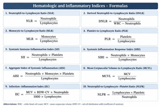
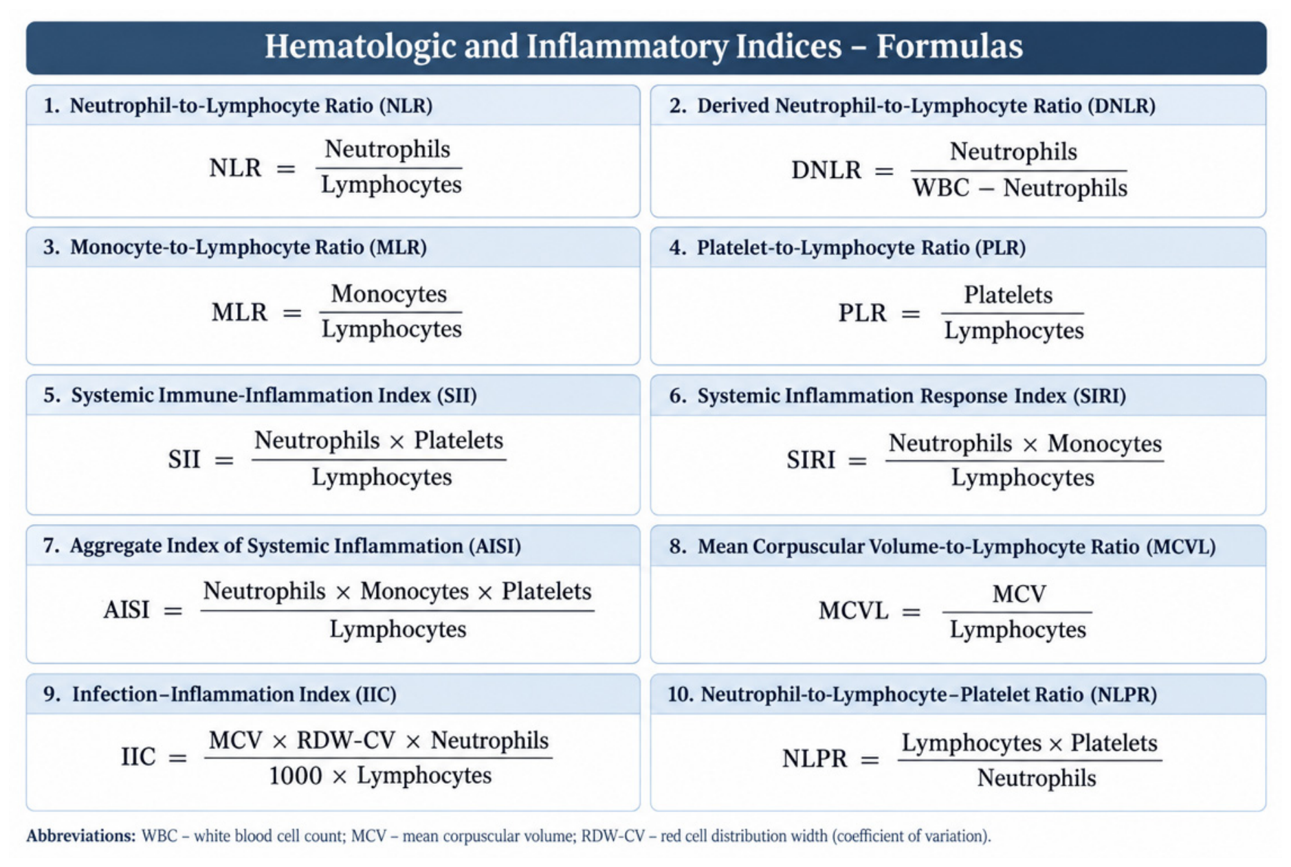
Peripheral blood samples were obtained within 24 h before cesarean delivery. Complete blood counts were analyzed using an automated hematology analyzer, and the following 11 variables were derived from the available hematological parameters: neutrophil-to-lymphocyte ratio (NLR), derived neutrophil-to-lymphocyte ratio (DNLR), monocyte-to-lymphocyte ratio (MLR), platelet-to-lymphocyte ratio (PLR), mean platelet volume (MPV), systemic immune-inflammation index (SII), systemic inflammation response index (SIRI), aggregate index of systemic inflammation (AISI), mean corpuscular volume-to-lymphocyte ratio (MCVL), cumulative inflammatory index (IIC), and neutrophil-to-lymphocyte and platelet ratio (NLPR).
The formulas used were, Figure 1:
Figure 1.
Hematological and Systemic Inflammatory Indices Derived from Complete Blood Count Parameters.
The hematological indices were calculated using the formulas presented in Figure 1. For indices based on ratios of cell counts expressed in the same measurement units, the resulting values are dimensionless. For composite indices involving multiplication or division of hematological parameters, the resulting units depend on the measurement units of the individual components. MPV was retained as an individual platelet-related parameter and expressed in femtoliters (fL). To facilitate interpretation and comparison with previous studies, the measurement units or scaling conventions of the derived indices are reported.
MPV was retained as an individual platelet-related variable rather than a composite inflammatory ratio. The detailed formulas and units can be provided in Supplementary Materials to reduce the length of the main manuscript.
Additional demographic and clinical variables extracted from the medical records included maternal age, gestational age, parity, pre-pregnancy body mass index, neonatal birth weight, Apgar scores, and neonatal sex. These variables were primarily used to characterize the study population and matching procedure rather than as components of the primary machine-learning predictor set.
2.4. Machine-Learning Framework
The analytical dataset comprised 160 participants and 12 candidate predictors, including the Postpartum Uterine Ultrasonographic Scale (PUUS) score and 11 hematological inflammatory indices. No predictor had more than 5% missing values; therefore, complete-case analysis was applied. The dataset was divided using a stratified 80:20 split into a development set (n = 128) and a hold-out evaluation set (n = 32). Given the balanced case–control design, no additional class-balancing procedures were required.
Data preprocessing and prevention of information leakage: All data-dependent preprocessing procedures were performed using training data only. Univariate extreme values were identified using a standardized-score threshold of |z| > 3, while multivariate outliers were assessed using Mahalanobis distance. Winsorization limits were determined from the 1st and 99th percentiles of the relevant training data. Similarly, means and standard deviations for z-score standardization were calculated exclusively from the training data. The resulting preprocessing parameters were subsequently applied unchanged to the corresponding validation or evaluation data. Within cross-validation, all preprocessing steps were refitted independently in each training fold and then applied to the corresponding validation fold. The hold-out evaluation set was not used to estimate preprocessing parameters or optimize hyperparameters.
Model development and hyperparameter optimization: Five supervised machine-learning algorithms were evaluated: elastic-net logistic regression, random forest, XGBoost, support vector machine with a radial basis function kernel, and multilayer perceptron. These approaches represented regularized linear, bagging-based ensemble, boosting-based ensemble, kernel-based, and neural-network modeling strategies, respectively [9,10,11,12]. Hyperparameter optimization was performed within the development set using stratified 10-fold cross-validation repeated five times with different random seeds. Mean cross-validated area under the receiver operating characteristic curve (AUC) was used as the optimization criterion.
Model comparison and selection: Following hyperparameter optimization, all five candidate algorithms were evaluated using the same 32-participant hold-out evaluation set. The algorithm achieving the highest observed AUC was selected as the best-performing model. Although the hold-out set was not used for preprocessing parameter estimation or hyperparameter optimization, its use for comparing candidate algorithms and selecting the final model introduces a potential source of model-selection optimism. Consequently, this dataset cannot be considered an entirely independent test set, and the resulting performance estimates should be interpreted as exploratory. Nested cross-validation was not performed.
The hyperparameter grids and learning-curve analyses are provided in the Supplementary Materials (Tables S1–S8 and Figures S1 and S2).
2.5. Model Evaluation and Interpretability
Model discrimination was assessed using the area under the receiver operating characteristic curve (AUC). Sensitivity, specificity, and classification accuracy were additionally reported for the selected model.
Internal optimism correction was performed using 1000 bootstrap resamples of the model-development dataset. Within each resample, the model-development procedure was repeated, including hyperparameter optimization and cross-validation. Optimism was estimated by comparing model performance in the bootstrap sample with performance when the fitted model was evaluated on the corresponding original development data, and the mean estimated optimism was subtracted from the apparent AUC. A separate procedure using 500 bootstrap resamples was undertaken to estimate optimism associated with selecting the best-performing algorithm from the five candidate models. This procedure was intended to account for the additional bias introduced by model selection. The resulting estimates represent internal performance corrections and do not constitute external validation.
Because the final algorithm was selected after comparison of five candidate models using the hold-out evaluation set, a separate bootstrap procedure with 500 resamples was used to approximate the magnitude of model-selection optimism. Within each resample, model development, hyperparameter tuning, comparison of the five algorithms, and selection of the highest-AUC model were repeated. The resulting optimism estimate was used to derive a selection-corrected AUC.
Model calibration was specified as an additional component of the planned model-evaluation framework. The intended calibration assessment included the Brier score and graphical evaluation using calibration plots. Decision-curve analysis was also specified as a secondary exploratory component of the analytical framework. However, because the original participant-level predicted probabilities and the corresponding computational outputs were unavailable for independent verification, the implementation and results of these analyses could not be confirmed.
Feature importance and predictor interpretation: Predictor importance was explored using model-specific interpretability methods. For tree-based models, permutation importance and impurity-based measures were considered; standardized coefficients were examined for elastic-net logistic regression, while permutation-based or model-specific measures were considered for the support vector machine and multilayer perceptron. These approaches were used to explore the contribution of individual predictors to model discrimination. Feature-importance estimates were interpreted as descriptive, model-dependent measures rather than tests of statistical significance or evidence of independent biological effects. Given the mathematical interdependence of several hematological indices, individual importance estimates may also reflect shared information among correlated predictors.
A prespecified sensitivity analysis excluded the six participants with isolated MTHFR polymorphisms. Their exclusion was evaluated in terms of model discrimination and feature-importance ranking.
2.6. Statistical Analysis
Continuous variables were summarized as mean ± standard deviation (SD) when approximately normally distributed and as median with interquartile range (IQR) when distributions were non-normal. Categorical variables were summarized as frequencies and percentages. Distributional assumptions were assessed using the Shapiro–Wilk test. Between-group comparisons were performed using statistical tests appropriate to the distribution and measurement scale of each variable. All hypothesis tests were two-sided, with a nominal significance threshold of p < 0.05.
For analyses involving multiple hematological inflammatory indices, false-discovery-rate-adjusted q-values were used where applicable. Model discrimination was primarily expressed using AUC with corresponding 95% confidence intervals, while bootstrap procedures were used for optimism-adjusted estimates.
All analyses were performed using Python 3.9, with scikit-learn 1.0.2, XGBoost 1.5.0, pandas 1.4.0, NumPy 1.21.0, Matplotlib 3.5.0, and seaborn 0.11.2. The development and validation workflow was designed to separate model fitting and hyperparameter optimization from performance evaluation, with all data-dependent preprocessing parameters estimated using training data only. Internal bootstrap correction was used to quantify potential optimism, whereas no independent external validation cohort was available.
3. Results
3.1. Study Population
The analytical cohort comprised 160 women, including 80 women with established thrombophilia receiving LMWH and 80 matched women without thrombophilia. The groups were comparable with respect to age, gestational age, parity, pre-pregnancy BMI, neonatal birth weight, Apgar score, and neonatal sex (all p > 0.05) (Table 1).
Table 1.
Baseline demographic and clinical characteristics.
The thrombophilia group included 32 women with heterozygous Factor V Leiden, 2 with homozygous Factor V Leiden, 21 with heterozygous prothrombin G20210A, 7 with protein C deficiency, 9 with protein S deficiency, 3 with antithrombin deficiency, and 6 with isolated MTHFR polymorphisms. Fifteen participants carried combined or compound thrombophilic abnormalities. All women in the thrombophilia group were receiving LMWH at the time of biomarker assessment.
The perioperative factors potentially relevant to postpartum uterine ultrasonographic findings, including blood loss, labor onset, uterotonic administration, concomitant antiplatelet therapy, and blood transfusion, were not collected systematically. Consequently, their distributions across the study groups and their potential associations with PUUS findings could not be assessed reliably.
3.2. Postpartum Uterine Ultrasonography
The distribution of PUUS scores differed between groups. A PUUS score of ≥1, corresponding to detectable intrauterine blood or debris, was present in 26/80 (32.5%) women with thrombophilia and 11/80 (13.8%) controls, whereas PUUS = 0 was observed in 54/80 (67.5%) and 69/80 (86.2%), respectively (Table 2).
Table 2.
Distribution of PUUS scores.
The odds of having PUUS ≥ 1 were higher in the thrombophilia group (OR 3.02, 95% CI 1.38–6.61; p = 0.005). The association was evaluated by comparing women with PUUS ≥ 1 against those with PUUS = 0 (OR 3.02, 95% CI 1.38–6.61; p = 0.005).
3.3. Hematological Inflammatory Indices
Several inflammatory indices differed between women with thrombophilia and controls (Table 3). The largest numerical differences were observed for NLPR and IIC. NLPR was 0.085 ± 0.042 in the thrombophilia group versus 0.042 ± 0.021 in controls, while IIC was 12.47 ± 5.82 versus 6.84 ± 3.15, respectively (both q < 0.001).
Table 3.
Hematological inflammatory indices in the thrombophilia and control groups.
After Benjamini–Hochberg adjustment, NLPR, IIC, NLR, PLR, SII, SIRI, and AISI remained significant at q < 0.05. MLR had a nominal p value of 0.042 but an adjusted q value of 0.058 and therefore did not meet the prespecified 0.05 threshold for statistical significance after correction. DNLR, MCVL, and MPV were not significant after adjustment.
3.4. Machine-Learning Model Performance
Five supervised machine-learning algorithms were specified for the classification task: elastic-net logistic regression, random forest, XGBoost, support vector machine with a radial basis function kernel, and multilayer perceptron (Table 4).
Table 4.
Performance of the evaluated machine-learning algorithms in the hold-out evaluation set.
However, the original analytical dataset and the computational outputs required to independently reproduce the model-development and evaluation procedures were unavailable for verification. Therefore, the previously reported discrimination metrics, including AUC, sensitivity, specificity, and classification accuracy, cannot be considered independently verified.
The comparative performance of the five algorithms and the selection of XGBoost as the best-performing model should consequently be regarded as exploratory. No definitive conclusion regarding the superiority of one algorithm over another can be drawn from the available evidence.
The observed discrimination of XGBoost should be interpreted in the context of the study design. The hold-out evaluation set was used both to compare the five candidate algorithms and to select the final model. Therefore, the reported AUC does not represent an independent estimate of the performance of a prespecified model. The findings should be considered exploratory and require confirmation in an independent cohort.
The XGBoost sensitivity corresponded to 15 of 16 thrombophilia cases correctly classified, while its specificity corresponded to 14 of 16 controls correctly classified. Given the small evaluation set, the corresponding confidence intervals were wide and the point estimates should be considered imprecise.
3.5. Optimism-Corrected and Selection-Corrected Performance
The reported model-performance estimates require cautious interpretation because the original participant-level analytical dataset and the computational outputs underlying the machine-learning analyses were not available for independent verification. Consequently, the reported apparent AUC, bootstrap-based optimism-corrected AUC, and selection-corrected AUC could not be independently reproduced.
The previously reported performance estimates are therefore considered exploratory and should not be interpreted as independently verified measures of model discrimination. In particular, the bootstrap-based corrections cannot be regarded as confirmed estimates of the generalizability of the selected model.
Independent verification of the analytical dataset, preprocessing procedures, model-fitting workflow, and resampling procedures is required before these estimates can support conclusions regarding predictive performance. The corrected estimates should be regarded as internal, resampling-based estimates rather than external validation results.
3.6. Comparison with Standard Logistic Regression
On the hold-out evaluation set, XGBoost yielded an AUC of 0.938 (95% CI 0.826–0.989) compared with 0.845 (95% CI 0.698–0.939) for an unregularized logistic regression model using the same predictors. DeLong’s test yielded p = 0.041.
Given the small evaluation sample and the fact that the algorithm-selection procedure was performed on this dataset, this comparison should be interpreted as an exploratory comparison rather than evidence of definitive superiority of XGBoost.
3.7. Calibration
Model calibration was included in the planned analytical framework and was intended to be evaluated using the Brier score and calibration plots. However, the original participant-level predicted probabilities and the corresponding computational outputs were unavailable for independent verification. Consequently, neither the implementation of the calibration procedures nor the previously reported calibration results could be independently confirmed.
Accordingly, no numerical Brier score, calibration curve, calibration slope, calibration intercept, or other quantitative calibration estimate is presented as a verified result. The calibration performance of the evaluated machine-learning models therefore remains undetermined based on the available documentation.
Further assessment of calibration would require access to the original analytical dataset, the fitted model specifications, and the participant-level predicted probabilities. Moreover, because the cohort was deliberately balanced between cases and controls, calibration estimates from this sample would not establish the accuracy of absolute thrombophilia risk predictions in an unselected obstetric population.
3.8. Feature Importance
Permutation importance was used in the original analysis to explore the relative contribution of individual predictors to model discrimination. The previously reported analysis suggested that NLPR, IIC, and PUUS might contribute to the classification performance of the XGBoost model. However, the original analytical dataset and the corresponding computational outputs were unavailable for independent verification. Consequently, the reported permutation-importance values and the relative ordering of individual predictors could not be independently reproduced.
Therefore, no definitive ranking of predictor importance is presented. NLPR, IIC, and PUUS are retained as exploratory candidate predictors identified in the previously reported analysis, rather than as confirmed leading predictors. Furthermore, several hematological inflammatory indices are mathematically interdependent and share underlying complete blood count components. Their estimated importance may therefore reflect overlapping predictive information rather than independent contributions to model discrimination. These findings should be interpreted descriptively and require independent verification in future studies.
3.9. Sensitivity Analysis Excluding Isolated MTHFR Polymorphisms
A sensitivity analysis excluding participants with isolated MTHFR polymorphisms was specified to assess the potential influence of their inclusion on model performance. However, the original analytical dataset and the corresponding computational outputs were unavailable for independent verification. Consequently, the performance estimates previously attributed to this analysis could not be confirmed and are not reported here. The findings of the primary analysis should therefore be interpreted without assuming that the sensitivity analysis established the robustness of model performance to the exclusion of these participants.
4. Discussion
4.1. Main Findings
The present study evaluated the feasibility of machine-learning-based classification of women with established thrombophilia receiving low-molecular-weight heparin (LMWH) versus matched women without thrombophilia using a combination of postpartum uterine ultrasonography and pre-delivery hematological inflammatory indices. The original analysis explored the feasibility of integrating postpartum uterine ultrasonographic findings with pre-delivery hematological inflammatory indices for classification of the two study groups. However, the comparative performance of the evaluated algorithms could not be independently confirmed. Accordingly, the machine-learning component should be regarded as exploratory rather than as evidence of an established diagnostic or predictive approach.
The relatively wide 95% confidence interval of the observed AUC (0.826–0.989) further emphasizes the uncertainty associated with the small hold-out evaluation set. Therefore, the corrected AUC of 0.920 should not be interpreted as external validation or as an estimate of clinical performance in an independent population. Rather, it represents a more conservative internal estimate of discrimination after accounting for identified sources of optimism.
4.2. Hematological Inflammatory Indices
Several hematological inflammatory indices differed between the two study groups, with the largest numerical differences observed for NLPR and IIC. After Benjamini–Hochberg adjustment, NLPR, IIC, NLR, PLR, SII, SIRI, and AISI remained statistically significant, whereas MLR did not reach the prespecified adjusted significance threshold (q = 0.058). These findings indicate that the studied groups differed with respect to several CBC-derived inflammatory measures, but they do not establish any individual index as a specific biomarker of thrombophilia.
The use of composite hematological indices in pregnancy has been increasingly explored in different clinical settings. Pantea et al. reported that NLPR and other CBC-derived inflammatory measures were associated with systemic inflammatory response syndrome in premature newborns [13]. Yalcin et al. subsequently evaluated several inflammatory indices in pregnancies complicated by pregestational diabetes and demonstrated associations between these indices and adverse perinatal outcomes [14]. Similarly, Dereli et al. reported associations of systemic immune-inflammation and systemic inflammatory response indices with late-onset fetal growth restriction [15]. These studies and others [16,17] support the broader concept that hematological indices may capture clinically relevant inflammatory information, but their populations and outcomes differ substantially from those of the present study. Accordingly, these data provide biological context rather than direct validation of these indices for thrombophilia classification.
The exploratory feature-importance analysis suggested that NLPR and IIC may contribute to the discrimination achieved by the XGBoost model. However, because the original analytical dataset and computational outputs were unavailable for independent verification, their relative importance and ranking cannot be confirmed. Accordingly, these findings should be regarded as hypothesis-generating observations rather than evidence of independently established predictor contributions. This distinction is particularly relevant because several hematological indices share underlying blood count components, making it difficult to separate their individual predictive contributions. Future studies using independently verifiable datasets should assess the stability of feature-importance estimates and determine whether these indices provide complementary or overlapping predictive information.
4.3. Postpartum Uterine Ultrasonography
PUUS was identified as a potentially relevant predictor in the exploratory feature-importance analysis of the XGBoost model. However, its relative ranking could not be independently verified because the original analytical dataset and computational outputs were unavailable.
This finding suggests that PUUS may contribute to discrimination within the current dataset when combined with hematological variables. However, it should be interpreted as exploratory rather than as evidence of independent validation of the scale. The examinations were performed at the institution where PUUS was developed and previously evaluated, by operators involved in its development. No independent blinded reassessment or formal inter-operator validation was performed in the present cohort. Therefore, the reproducibility and independent diagnostic value of PUUS remain unconfirmed.
This finding must, however, be interpreted cautiously in light of the previous literature. The original PUUS study established the scale as a method for quantitative assessment of postpartum uterine involution [7]. In a subsequent prospective study, Filip et al. evaluated 80 women with treated thrombophilia and 80 matched controls after term cesarean delivery during the period from October 2017 to December 2021 and reported no significant difference in postpartum uterine involution between the two groups [8].
The comparison with Filip et al. [8] requires consideration of the PUUS reassessment performed for the present study. Although both publications include the same 160 participants, the PUUS distributions differ because the ultrasound examinations were reassessed for the current analysis by two experienced ultrasonographers blinded to thrombophilia status and study-group allocation. In particular, PUUS = 0 was identified in 54/80 women with thrombophilia and 69/80 controls in the present reassessment, compared with 65/80 and 69/80, respectively, in the previous publication. Thus, the difference in the reported distributions reflects the use of newly reassigned PUUS scores rather than the direct reuse of the previously published scores. The current analysis therefore represents a secondary analysis of the same clinical cohort incorporating a new assessment of the original ultrasound examinations, rather than an independent replication or a direct reuse of the previously reported PUUS dataset. Nevertheless, the absence of formal inter-operator reliability statistics warrants caution when interpreting the reproducibility of the reassessed scores.
These differences require verification against the original participant-level records and the scoring and analytical procedures used in both studies. The current results cannot be attributed to reassessment of the ultrasound examinations, recoding of the original scores, or database revision unless the relevant procedures are documented. Until this verification is completed, the PUUS findings should be interpreted cautiously, and the present analysis should not be described as an independent replication of the previously published cohort.
The appropriate interpretation is therefore that PUUS contributed predictive information within the current machine-learning framework, while its independent and disease-specific diagnostic value remains uncertain.
4.4. Machine-Learning Performance and Model Selection
The machine-learning component was designed to explore whether the combination of postpartum uterine ultrasonographic findings and hematological inflammatory indices could distinguish the two clinical groups. Five supervised algorithms were specified for this purpose. However, the original analytical dataset and the corresponding computational outputs were unavailable for independent verification. Consequently, the previously reported discrimination estimates, bootstrap-based corrections, and comparative performance results cannot be considered independently confirmed.
An additional methodological consideration is that the hold-out evaluation set was used both to compare the candidate algorithms and to select the final model. This approach may introduce model-selection optimism, as the selected algorithm is evaluated on the same sample that informed its selection. Therefore, the reported model comparisons should not be interpreted as definitive evidence of algorithmic superiority or generalizability.
The machine-learning findings should consequently be regarded as exploratory and hypothesis-generating. Independent evaluation using a larger cohort, a prespecified modeling strategy, and an appropriate validation framework is necessary before the predictive performance of these approaches can be established.
An important limitation is that the hold-out evaluation set was used both to compare the five candidate algorithms and to select the final model. This procedure introduces the possibility of selection-related optimism because the algorithm with the highest observed AUC was selected on the basis of its performance in the same sample. Although a separate bootstrap procedure was used to estimate selection-related optimism, this internal correction does not replace nested cross-validation or independent external validation. Accordingly, the observed and corrected AUC estimates should be interpreted as exploratory findings and should not be considered definitive evidence of the model’s performance in new patients.
The comparison between XGBoost and standard logistic regression was part of the exploratory model-evaluation procedure. However, because the original analytical dataset and the corresponding computational outputs were unavailable for independent verification, the reported AUC estimates and the DeLong test result could not be independently reproduced. Therefore, no reliable conclusion can be drawn regarding the comparative performance of XGBoost and standard logistic regression. Any apparent differences between the algorithms should be considered unverified and hypothesis-generating.
The interpretation of model performance must also account for the complete confounding between thrombophilia status and LMWH exposure. All women in the thrombophilia group received LMWH, whereas none of the controls received this treatment. Consequently, the observed discrimination cannot be attributed specifically to thrombophilia and may reflect the underlying clinical condition, treatment-associated effects, or a combination of both. The models should therefore be interpreted as distinguishing women with established thrombophilia receiving LMWH from women without thrombophilia who were not receiving LMWH, rather than as identifying thrombophilia-specific features independently of treatment.
Although calibration was included in the planned analytical framework, its implementation and results could not be independently verified because the original predicted probabilities and computational outputs were unavailable. Therefore, no conclusions can be drawn regarding the calibration performance of the evaluated models. Furthermore, the deliberately balanced case–control design limits the interpretation of predicted probabilities, even if calibration were subsequently confirmed. In particular, probabilities estimated in this sample cannot be assumed to represent absolute thrombophilia risk in an unselected obstetric population. Accordingly, the findings should be interpreted primarily in terms of exploratory discrimination rather than individual risk prediction or clinical probability estimation.
The reliability of the reported performance estimates is limited by the small hold-out evaluation set, which included only 32 participants (16 women with thrombophilia and 16 controls). In a sample of this size, the classification of only a few individuals can materially change estimates of sensitivity, specificity, accuracy, and other performance metrics. Although XGBoost achieved an observed AUC of 0.938 (95% CI 0.826–0.989), the confidence interval indicates substantial uncertainty around the point estimate. Furthermore, the same hold-out set was used to compare the five candidate algorithms and select the best-performing model, introducing potential model-selection optimism. Bootstrap-based optimism correction provides an internal assessment of this uncertainty but does not substitute for evaluation in an independent external cohort. Accordingly, the reported performance should be regarded as exploratory and hypothesis-generating rather than as evidence of established clinical validity or generalizability.
4.5. The Central Role of LMWH Exposure
A central consideration in interpreting the observed differences between the study groups is the complete confounding between thrombophilia status and LMWH exposure. All women in the thrombophilia group received LMWH, whereas none of the controls received this treatment. Consequently, the study design does not allow the independent contributions of the underlying condition and anticoagulant treatment to be distinguished.
Furthermore, the specific LMWH preparation, dose, dosing frequency, and timing of administration relative to blood sampling and postpartum ultrasonography could not be established from the available study documentation. Therefore, the potential influence of anticoagulant exposure on the measured hematological and ultrasonographic parameters remains uncertain.
This limitation is particularly relevant to the interpretation of the inflammatory indices and postpartum uterine findings. Although these variables may reflect biologically relevant inflammatory, hematological, or hemostatic processes, the present study does not permit attribution of their observed differences specifically to thrombophilia or to treatment-related effects.
Accordingly, the findings should be interpreted as differences between women with established thrombophilia receiving LMWH and women without diagnosed thrombophilia who were not receiving LMWH. They should not be interpreted as evidence of thrombophilia-specific effects independent of treatment.
4.6. Sensitivity Analysis Excluding Isolated MTHFR Polymorphisms
The robustness of the primary model to the exclusion of participants with isolated MTHFR polymorphisms could not be established because the original analytical dataset and the corresponding model outputs were unavailable for verification. Accordingly, no conclusion can be drawn regarding the effect of this exclusion on discrimination or feature-importance rankings. This limitation should be considered when interpreting the results of the primary analysis.
4.7. Limitations
Several limitations should be considered when interpreting the findings of this study.
First, this was a single-center study involving 160 participants, with a hold-out evaluation set comprising only 32 participants. The relatively small sample size limits the precision of performance estimates and may affect the stability of predictive models.
Second, the hold-out evaluation set was used both to compare the five candidate algorithms and to select the final model. This procedure may introduce model-selection optimism. Nested cross-validation was not performed, and internal resampling procedures cannot replace independent external validation. Accordingly, the generalizability of the machine-learning findings remains undetermined.
Third, the original participant-level analytical dataset and the computational outputs required to reproduce the machine-learning analyses were unavailable for independent verification. Consequently, the reported discrimination estimates, bootstrap-based corrections, calibration results, sensitivity analyses, and feature-importance rankings could not be independently confirmed. This limitation restricts the reproducibility of the computational findings and requires that the machine-learning results be interpreted as exploratory.
Fourth, all participants underwent term cesarean delivery. Therefore, the findings cannot be assumed to apply to women with vaginal delivery, preterm delivery, or other obstetric populations.
Fifth, PUUS assessments were performed at the institution where the scale was developed and previously evaluated. The available documentation does not establish whether the assessors were blinded to group allocation, and no formal inter-operator reliability analysis was performed. Consequently, observer-related assessment bias cannot be excluded, and the reproducibility of PUUS scoring across independent operators and institutions remains uncertain.
Sixth, perioperative factors potentially affecting postpartum uterine findings, including blood loss, labor onset, uterotonic administration, concomitant antiplatelet therapy, and blood transfusion, were not collected systematically. Their potential influence on the observed ultrasonographic findings could therefore not be adequately evaluated.
Seventh, the 11 hematological inflammatory indices were derived from overlapping complete blood count components, particularly neutrophil, lymphocyte, monocyte, and platelet counts. This mathematical interdependence may complicate the interpretation of individual predictor contributions and affect the stability of feature-importance estimates. The independent contribution of individual indices therefore remains uncertain.
Finally, the complete confounding between thrombophilia status and LMWH exposure prevents the independent effects of the underlying condition and anticoagulant treatment from being distinguished. In addition, the unavailable details regarding LMWH preparation, dosage, dosing frequency, and timing of administration further restrict the interpretation of the observed hematological and ultrasonographic differences.
4.8. Implications for Future Research
Future studies should focus on independent multicenter validation using larger and more diverse obstetric populations. A prespecified modeling strategy, transparent analytical workflows, and access to reproducible computational outputs will be essential to establish the reliability of machine-learning performance estimates.
Further research should also include study groups that allow the effects of thrombophilia and LMWH exposure to be evaluated separately. Independent and blinded PUUS assessment, together with formal inter- and intra-observer reliability analyses, would help establish the reproducibility of the ultrasonographic scale.
Finally, the stability and independent contribution of hematological inflammatory indices should be investigated using approaches that account for correlations between their underlying components. These steps are necessary before the potential diagnostic or predictive value of the combined imaging and hematological approach can be established.
5. Conclusions
This single-center, matched case–control study examined the differences in postpartum uterine ultrasonographic findings and pre-delivery hematological inflammatory indices between women with established thrombophilia receiving low-molecular-weight heparin (LMWH) and matched controls without thrombophilia.
The observed differences in PUUS scores and several hematological inflammatory indices suggest potential associations between the two clinical groups. However, these findings must be interpreted in the context of the study design and the complete confounding between thrombophilia status and LMWH exposure. Consequently, the observed differences cannot be attributed specifically to thrombophilia or independently distinguished from potential treatment-associated effects.
Although machine-learning approaches were specified and evaluated in the original analytical framework, the underlying participant-level dataset and computational outputs were unavailable for independent verification. Therefore, the reported model-performance estimates, comparative algorithm rankings, calibration results, and individual predictor-importance rankings cannot be considered independently confirmed. No conclusion regarding the superiority of XGBoost or the independent predictive importance of NLPR, IIC, or PUUS can be established from the verifiable evidence currently available.
The findings should therefore be considered exploratory and hypothesis-generating rather than evidence of an established predictive or diagnostic tool. Further research involving larger independent multicenter cohorts, transparent analytical workflows, reproducible computational outputs, and study designs capable of separating thrombophilia-related effects from LMWH exposure is required before the clinical relevance and predictive performance of these approaches can be established.
Supplementary Materials
The following supporting information can be downloaded at https://www.mdpi.com/article/10.3390/reports9040398/s1, Table S1: PUUS grading definitions; Table S2: Definitions of the hematological inflammatory indices; Table S3: Hyperparameter grids and selected settings; Table S4: Extended performance metrics on the hold-out evaluation set; Table S5: XGBoost performance stratified by PUUS category; Table S6: XGBoost performance by maternal age category; Table S7: Sensitivity analysis after exclusion of isolated MTHFR polymorphisms; Table S8: Permutation-importance ranking for the XGBoost model; Figure S1: Exploratory decision curve analysis for the XGBoost model. The plot is retained as a secondary analysis and should be interpreted only within the limitations of the study design; Figure S2: Confusion matrices for the five machine-learning models in the hold-out evaluation set (n = 32; 16 thrombophilia cases and 16 controls).
Author Contributions
Conceptualization, L.T. and C.F.; methodology, M.C.T.; software, R.C.; validation, D.S., A.C. and L.R.; formal analysis, A.G.; investigation, D.L. and S.S.; resources, F.C.; data curation, R.F.P.; writing—original draft preparation, C.F. and M.M.; writing—review and editing, L.T.; visualization, R.C.; supervision, D.L.; project administration, F.C.; funding acquisition, L.R. All authors have read and agreed to the published version of the manuscript.
Funding
This research received no external funding.
Institutional Review Board Statement
The study was conducted in accordance with the Declaration of Helsinki and was approved by the Ethics Committee of Elena Doamna Obstetrics and Gynecology University Hospital (approval number 9 from 15 September 2017).
Informed Consent Statement
Informed consent was obtained from all subjects involved in the study.
Data Availability Statement
The original contributions presented in this study are included in the article/Supplementary Materials. Further inquiries can be directed to the corresponding author. The participant-level dataset and the computational outputs supporting the machine-learning analyses are not currently available for independent verification. The authors acknowledge that this limits the reproducibility of the reported results. Access to the original analytical dataset and computational materials will be considered subject to institutional policies, ethical approval, and applicable data-protection requirements.
Conflicts of Interest
The authors declare no conflicts of interest.
Abbreviations
| AISI | Aggregate Index of Systemic Inflammation |
| AUC | Area Under the Receiver Operating Characteristic Curve |
| CBC | Complete Blood Count |
| CI | Confidence Interval |
| DNLR | Derived Neutrophil-to-Lymphocyte Ratio |
| IIC | Cumulative Inflammatory Index |
| LMWH | Low-Molecular-Weight Heparin |
| MCVL | Mean Corpuscular Volume-to-Lymphocyte Ratio |
| MLR | Monocyte-to-Lymphocyte Ratio |
| MPV | Mean Platelet Volume |
| NLR | Neutrophil-to-Lymphocyte Ratio |
| NLPR | Neutrophil-to-Lymphocyte and Platelet Ratio |
| NPV | Negative Predictive Value |
| PLR | Platelet-to-Lymphocyte Ratio |
| PPV | Positive Predictive Value |
| PUUS | Postpartum Uterine Ultrasonographic Scale |
| ROC | Receiver Operating Characteristic |
| SHAP | SHapley Additive exPlanations |
| SII | Systemic Immune-Inflammation Index |
| SIRI | Systemic Inflammation Response Index |
| VTE | Venous Thromboembolism |
References
- Samfireag, M.; Potre, C.; Potre, O.; Tudor, R.; Hoinoiu, T.; Anghel, A. Approach to Thrombophilia in Pregnancy—A Narrative Review. Medicina 2022, 58, 692. [Google Scholar] [CrossRef] [Scilit] [PubMed]
- Greer, I.A. Pregnancy Complicated by Venous Thrombosis. N. Engl. J. Med. 2015, 373, 540–547. [Google Scholar] [CrossRef] [Scilit] [PubMed]
- Hilali, C.; Aboulaghras, S.; Lamalmi, N. Pathophysiological, Immunogenetic, Anatomopathological Profile of Thrombophilia in Pregnancy. Transfus. Clin. Biol. 2023, 30, 360–367. [Google Scholar] [CrossRef] [Scilit] [PubMed]
- Middeldorp, S.; Nieuwlaat, R.; Kreuziger, L.B.; Coppens, M.; Houghton, D.; James, A.H.; Lang, E.; Moll, S.; Myers, T.; Bhatt, M.; et al. American Society of Hematology 2023 Guidelines for Management of Venous Thromboembolism: Thrombophilia Testing. Blood Adv. 2023, 7, 7101–7138. [Google Scholar] [CrossRef] [Scilit] [PubMed]
- Zheng, L.; Ge, R.; Weng, X.; Lin, L. Predictive Value of Serum Immune-Inflammatory Markers for Adverse Pregnancy Outcomes in Pregnant Women with Thrombophilia: A Retrospective Cohort Study. J. Inflamm. Res. 2024, 17, 6083–6091. [Google Scholar] [CrossRef] [Scilit] [PubMed]
- Wang, T.; Kang, X.; He, L.; Liu, Z.; Xu, H.; Zhao, A. Prediction of Thrombophilia in Patients with Unexplained Recurrent Pregnancy Loss Using a Statistical Model. Int. J. Gynecol. Obstet. 2017, 138, 283–287. [Google Scholar] [CrossRef] [Scilit] [PubMed]
- Covali, R.; Socolov, D.; Carauleanu, A.; Pavaleanu, I.; Akad, M.; Boiculese, L.V.; Socolov, R.V. The Importance of the Novel Postpartum Uterine Ultrasonographic Scale in Numerical Assessments of Uterine Involution Regarding Perinatal Maternal and Fetal Outcomes. Diagnostics 2021, 11, 1731. [Google Scholar] [CrossRef] [Scilit] [PubMed]
- Filip, C.; Covali, R.; Socolov, D.; Carauleanu, A.; Tanasa, I.A.; Scripcariu, I.S.; Ciuhodaru, M.; Butureanu, T.; Pavaleanu, I.; Akad, M.; et al. The Postpartum Uterine Ultrasonographic Scale in Assessment of Uterine Involution after Cesarean Section in Treated Thrombophilia Pregnant Patients at Term. J. Clin. Lab. Anal. 2022, 36, e24645. [Google Scholar] [CrossRef] [Scilit] [PubMed]
- Sidey-Gibbons, J.A.M.; Sidey-Gibbons, C.J. Machine Learning in Medicine: A Practical Introduction. BMC Med. Res. Methodol. 2019, 19, 64. [Google Scholar] [CrossRef] [Scilit] [PubMed]
- Obermeyer, Z.; Emanuel, E.J. Predicting the Future—Big Data, Machine Learning, and Clinical Medicine. N. Engl. J. Med. 2016, 375, 1216–1219. [Google Scholar] [CrossRef] [Scilit] [PubMed]
- Rajkomar, A.; Dean, J.; Kohane, I. Machine Learning in Medicine. N. Engl. J. Med. 2019, 380, 1347–1358. [Google Scholar] [CrossRef] [Scilit] [PubMed]
- Beam, A.L.; Kohane, I.S. Big Data and Machine Learning in Health Care. JAMA 2018, 319, 1317–1318. [Google Scholar] [CrossRef] [Scilit]
- Pantea, M.; Iacob, D.; Bortea, C.I.; Enatescu, I.; Barbos, V.; Prodan, M.; Tudor, R.; Cozma, G.V. Predictive Role of NLR, dNLR, PLR, NLPR, and Other Laboratory Markers in Diagnosing SIRS in Premature Newborns. Clin. Pract. 2024, 14, 1065–1075. [Google Scholar] [CrossRef] [Scilit] [PubMed]
- Yalcin, S.E.; Ozler, M.R.; Yalcin, Y.; Saglam, E.; Yilmaz, E.S.; Nerez, N. Association of Early Pregnancy Inflammatory Indices with Preterm Birth and Perinatal Outcomes in Pregnancies with Pregestational Diabetes. J. Clin. Med. 2025, 14, 6834. [Google Scholar] [CrossRef] [Scilit] [PubMed]
- Dereli, M.L.; Sucu, S.; Kurt, D.S.; Kurt, A.; Fıratlıgil, F.B.; Ağbal, T.; Çağlar, A.T. Systemic Immune Inflammation Index, Systemic Inflammatory Response Index and Pan-Immune Inflammation Value in the Prediction of Idiopathic Late-Onset Fetal Growth Restriction. Cytokine 2026, 198, 157081. [Google Scholar] [CrossRef] [Scilit] [PubMed]
- Alamin, A.A. Neutrophil Extracellular Traps (NETs): Relevance to Thrombosis and Hemostasis. A Narrative Review. Rev. Romana Med. Lab. 2025, 33, 79–86. [Google Scholar] [CrossRef] [Scilit]
- Varjú, I.; Tanka-Salamon, A.; Kolev, K. Neutrophil Extracellular Traps: At the Interface of Thrombosis and Comorbidities. Semin. Thromb. Hemost. 2025, 51, 724–735. [Google Scholar] [CrossRef] [Scilit] [PubMed]

Disclaimer/Publisher’s Note: The statements, opinions and data contained in all publications are solely those of the individual author(s) and contributor(s) and not of MDPI and/or the editor(s). MDPI and/or the editor(s) disclaim responsibility for any injury to people or property resulting from any ideas, methods, instructions or products referred to in the content. |
© 2026 by the authors. Licensee MDPI, Basel, Switzerland. This article is an open access article distributed under the terms and conditions of the Creative Commons Attribution (CC BY) license.