


Abstract
Proximate composition, lipid classes, and fatty acid profile were comparatively analyzed in different body parts of carp (Cyprinus carpio) cultivated in Madagascar. Higher (p < 0.05) protein values (163.3–200.1 g·kg−1 range) were detected in muscle tissue (epaxial, hypaxial, and tail) than in the liver tissue (118.4 g·kg−1). The average lipid content showed the following decreasing tissue sequence: liver > hypaxial > tail > epaxial. The highest levels (p < 0.05) of phospholipids and sterols were obtained in the epaxial muscle part (94.0 and 24.0 g·kg−1 of lipids, respectively). A high presence of free fatty acids (183.2 g·kg−1 of lipids) was detected in the liver. In all tissues, α-tocopherol provided higher values (p < 0.05) than γ- and δ-tocopherol compounds; among body parts, the liver showed the highest levels (p < 0.05) for all tocopherols. Fatty acid (FA) groups revealed a higher range value (g·100 g−1 of total FAs) for monounsaturated FAs (48.19–52.57) than for saturated FAs (SFAs; 28.50–29.41) and polyunsaturated FAs (PUFAs; 17.96–22.57). The following increasing sequence was observed for ω3-PUFA/ω6-PUFA ratios in body parts: liver < hypaxial < tail < epaxial. All parts of muscle tissue contained higher (p < 0.05) PUFA/SFA values (0.72–0.77) than liver (0.61). Comparison of body parts revealed an inhomogeneous distribution of bioactive compounds. The liver tissue was found a good source of valuable constituents.
Key Contribution:
A comparative chemical study of muscle (epaxial, hypaxial, and tail) and liver tissues of common carp was carried out. The work provides valuable information regarding the lipid class composition and the presence of bioactive compounds (phospholipids, tocopherols, and ω3 polyunsaturated fatty acids) in the liver tissue of this cultivated freshwater species.
1. Introduction
Aquatic foods are considered a remarkable source of high amounts of important nutritional components to human health and nutrition such as digestible proteins, ω3 fatty acids (FAs), lipophilic antioxidants, and essential macro- and microelements [1,2]. However, such constituents are inhomogeneously distributed through the specimen body according to different anatomical and physiological factors [3,4]. Remarkably, the content and composition of the lipid fraction are the most highly influenced and lead to important composition differences among different parts of the muscle tissue in wild fatty [5,6] and lean [7,8] species as well as in farmed species [9,10].
Common carp (Cyprinus carpio), also known as Eurasian carp or European carp, belongs to the order Cypriniformes and the family Cyprinidae. It is a freshwater species widely distributed in almost all countries of the world but is especially common in Asia and some European countries [11,12]. On the basis of its great adaptive capability [13,14], common carp is currently one of the most widely cultivated freshwater fish species in the world and therefore, highly important from a commercial point of view [15]. As a consequence, great efforts have been carried out for the development of its aquaculture production. Previous research includes the comparative proximate and FA profile study of farmed and wild specimens [16,17,18,19] and the effect of experimental farming conditions [20,21,22] and the diet provided [23,24,25]. However, previous research regarding the lipid class analysis can be considered as very scarce or even negligible in common carp. From a technological point of view, the use of hot-smoked carp [26] and preparation of fingers [27] have been reported, as well as the autolysis changes in white and dark muscles during the refrigerated storage [28]. Related to carp by-products, the proximate composition of cooked and dried discarded parts (i.e., mixture of digestive gland, liver, pancreas, and gonads) [29], the FA profile of viscera tissue [30] and the extraction of oils from viscera [31], and the extraction of gelatine from skin [32] have been described.
The present research focused on the proximate analysis and the chemical study of the lipid fraction of common carp cultivated in Madagascar. A comparative study of different body parts was carried out. Human consumption of fish has been especially focused on muscle zones. On the contrary, other parts of the fish body have been discarded or considered as by-products and employed for minor purposes [33,34]. In the present research, three different parts of the muscle tissue (epaxial, hypaxial, and tail muscles) and the liver tissue were studied. Although considered in most cases as a non-edible part of the carp body, the liver tissue was found interesting as a possible source of valuable constituents. Therefore, the presence of bioactive compounds such as proteins, phospholipids (PLs), tocopherols, and ω3-FAs was studied in all body parts in order to assess the nutritional and functional value of this farmed freshwater species.
2. Materials and Methods
2.1. Sampling Procedure
Thirty carp specimens were obtained from an aquaculture facility (Coopération des Éleveurs de Poissons, Maison du Petit Élevage, Antananarivo, Madagascar) in November 2024. The fish were received dead under frozen condition. For all specimens, the length and weight were within the 61–93 cm and 165–233 g ranges, respectively.
The fish were randomly distributed into three groups (10 specimens per group). Each group was considered and analyzed independently throughout the study (n = 3). In each individual fish, four different body parts were separated, i.e., epaxial muscle (from head till anal fin), hypaxial muscle (from head till anal fin), and tail (epaxial and hypaxial part of the muscle from anal fin till the end of the fish body), and liver. For each tissue, tissue samples belonging to the same 10-specimen group were pooled together and analyzed independently. In each chemical analysis, tissue samples corresponding to each group were analyzed in triplicate. Before carrying out the different chemical analyses, tissue samples were stored at −40 °C for 1–2 weeks; before analysis, samples were thawed overnight at 4 °C. All tissues were processed under identical conditions and within the same timeframe.
Solvents and chemical reagents used in this study were of reagent grade (Merck, Darmstadt, Germany); otherwise, the supplier is identified.
2.2. Proximate Composition
Moisture was assessed as the weight difference (1–2 g) before and after 4 h at 105 °C in agreement with the official method 950.46B [35]. Results are expressed as g·kg−1 of tissue.
Protein content was assessed by the Kjeldahl method according to procedure 928.08 (alternative II) [35]; a conversion factor of 6.25 was used. Results are expressed as g·kg−1 of wet tissue.
Ash was assessed by heating at 550 °C in agreement with the official method 920.153 [35]. Results are expressed as g·kg−1 of wet tissue.
Extraction of the lipid fraction was carried out according to the Bligh and Dyer [36] procedure. As a first step, a chloroform/methanol/water (1.0/2.0/0.8, v/v/v) mixture was used as the extracting solvent system. Then, chloroform and water were added to the mixture so that a 2.0/2.0/1.8 (chloroform/methanol/water, v/v/v) solvent ratio was obtained. As a result, two phases were derived. The one at the bottom was carefully taken as corresponding to the lipid fraction of the fish tissue. Lipid extracts were maintained at –40 °C in a nitrogen atmosphere before being employed for analysis. Results on crude lipid yield are expressed as g lipids·kg−1 of wet tissue.
2.3. Lipid Class Analysis
The PL value in the lipid fraction was determined according to the procedure proposed by Raheja et al. [37]. This method is based on the formation of a complex with ammonium molybdate. To carry out the quantification, several quantities (0, 5, 10, 20, 40, 60, 80, 100, 130, and 150 μL) of a 1,2-dipalmitoyl-rac-glycero-3-phosphocholine (DPPC) solution in chloroform (15.3 mg/5 mL) were used. The validity range was 16.1–483.0 μg of DPPC and the R2 value of the analytical procedure was 0.9995. Results are expressed as g PLs·kg−1 of crude lipid extracts and g PLs·kg−1 of tissue.
The total sterol (TS) value in the lipid extract was determined using the procedure proposed by Huang et al. [38]. This method is based on the Liebermann–Buchardt reaction. To carry out the quantification, several quantities (0, 5, 10, 20, 40, 60, 80, 100, 130, and 150 μL) of a cholesterol solution in acetic acid (12.2 mg/5 mL) were used. The validity range was 11.5–345.0 μg of cholesterol and the R2 value of the analytical procedure was 0.9998. Results are expressed as g TSs·kg−1 of crude lipid extracts and as mg TSs·kg−1 of tissue.
The free fatty acid (FFA) value in the lipid extract was assessed using the procedure proposed by Lowry and Tinsley [39]. This method is based on the formation of a complex with cupric acetate–pyridine. However, benzene was replaced by toluene as the organic solvent. To carry out the quantification, several quantities (0, 5, 10, 20, 40, 60, 80, 100, 130, and 150 μL) of an oleic acid solution in toluene (705.3 mg/25 mL) were used. The validity range was 0.5–15.0 μmol of oleic acid and the R2 value of the analytical procedure was 0.9998. Results are expressed as g FFAs·kg−1 of crude lipid extracts.
The tocopherol compound profile was determined using the method proposed by Cabrini et al. [40]. Thus, carp tissues were extracted with hexane, which was then eliminated under nitrogen flux; then, the resulting extracts were dissolved in methanol. The column was fluxed with methanol for 2 min; a gradient from 0 to 60% of isopropanol in 10 min was applied. Next, 10 μL samples were injected into the HPLC system (ODS column, 15 cm × 0.46 cm i.d.; Agilent, Santa Clara, CA, USA). The column temperature was set at 35 °C and the flow rate was 1.5 mL·min−1. A fluorescence detector (Waters 2475, Water Corporation, Milford, MA, USA) and a photodiode array (Waters 2996, Waters Corporation, Milford, MA, USA) were employed. Fluorescence detection (272 nm excitation/328 nm emission) was employed for quantification and photodiode array detection was employed for identification of tocopherol molecules by analyzing the resulting spectra between 210 nm and 400 nm. The presence of different tocopherol compounds (α, β, γ, and δ) was checked. Once identified, their concentration was determined using calibration curves prepared from the corresponding commercial tocopherol compound. For it, commercial tocopherol molecules (10–100 μL from a 1000 ppm of tocopherol/methanol solution) were employed. Contents are expressed as mg·kg−1 of lipids and as mg·kg−1 of tissue.
2.4. Fatty Acid Analysis
The FA profile was analyzed taking into account previous research [41]. For it, FA methyl esters (FAMEs) were obtained from lipid extracts by using acetyl chloride in methanol. Then, FAMEs were analyzed by gas–liquid chromatography (Perkin-Elmer 8700 chromatograph, Madrid, Spain). A fused silica capillary column SP-2330 (0.25 mm i.d. × 30 m, Supelco, Inc., Bellefonte, PA, USA) was employed. A temperature program was developed, i.e., increase from 145 to 190 °C at 1.0 °C·min−1 and from 190 °C to 210 °C at 5.0 °C·min−1, and held for 13.5 min at 210 °C. As carrier gas, nitrogen at 10 psig was employed and a flame ionization detector at 250 °C was used. A programmed temperature vaporizer injector was employed in the split mode (150:1); for it, heating from 45 to 275 °C at 15 °C·min−1 was carried out.
For the qualitative analysis of FAMEs, retention times of peaks obtained were compared to those of standard mixtures (Qualmix Fish, Larodan, Malmo, Sweden; Supelco 37 Component FAME Mix, Sigma-Aldrich, Laramie, WY, USA). Areas of the resulting peaks were automatically integrated. For quantitative purposes, C19:0 was used as the internal standard; for it, 100 μL (i.e., 40 μg of C19:0) of a 0.4 mg·mL−1 solution in toluene was added to each sample before the methylation reaction with acetyl chloride in methanol. Detection and quantification limits were 500 and 1500 area units, respectively. For it, individual response factors were used for each of the different FAMEs according to the abovementioned Supelco 37 FAME Mix. Peaks below the quantification limits were not considered. All reported FAMEs were above the quantitative threshold in all tissues.
The value of each FA was calculated as g·100 g−1 of total FAs. Results regarding FA groups (saturated FAs, SFAs; monounsaturated FAs, MUFAs; polyunsaturated FAs, PUFAs; total ω3-PUFAs; total ω6-PUFAs) and FA ratios (total ω3-PUFAs/total ω6-PUFAs; total PUFAs/total SFAs) were determined on the basis of the results obtained from individual FAs.
2.5. Statistical Analysis
Once values obtained for the different chemical parameters verified a normal distribution (Kolmogorov–Smirnov test) and homogeneity of variances (Bartlett’s test), parametric analysis was carried out. As expressed in the sampling procedure, three independent batches were considered and analyzed separately for each carp tissue (n = 3). Data obtained from the proximate analysis, lipid class content, and FA profile (individual FAs and FA groups and ratios) were subjected to the one-way ANOVA method in order to establish composition differences among carp tissues. Statistical comparisons were conducted via the PASW Statistics 18 Software for Windows (SPSS Inc., Chicago, IL, USA). Comparison of means was performed using the Fisher’s least-squares difference (LSD) method.
3. Results and Discussion
3.1. Proximate Composition
In all carp body parts, moisture was the major constituent (Table 1). The liver tissue had significantly (p < 0.05) lower values than the remaining tissues. Among muscle parts, higher (p < 0.05) moisture levels were observed in the epaxial and tail muscles than in the hypaxial one.
Table 1.
Proximate composition (g·kg−1 of tissue) * of different body parts of common carp cultivated in Madagascar **.
A valuable presence of proteins was detected in the muscle parts; i.e., all were in the range of 163.3–200.1 g·kg−1 of muscle (Table 1). On the contrary, the liver tissue revealed a lower protein presence (p < 0.05) than the muscle parts. Notably, epaxial and hypaxial muscles showed a higher (p < 0.05) protein presence than the tail muscle.
Regarding the ash content, all tissues depicted values in the range of 8.3–12.1 g·kg−1 of tissue (Table 1). The lowest content (p < 0.05) was detected in the liver tissue and the highest one (p < 0.05) was observed in the hypaxial muscle.
A very high lipid content (294.3 g·kg−1 of tissue) was observed in the liver tissue (Table 1), which agrees with the inverse ratio trend reported for values of lipid and moisture constituents [3,9]. Regarding the muscle parts, the following decreasing sequence was detected for the average value of lipid contents: hypaxial > tail > epaxial. Comparison of current lipid values of muscle parts (36.7–58.2 g·kg−1 of tissue) to those corresponding to aquatic species in general indicates that the lipid content of the current farmed fish would agree with the lipid value of a medium-fat species [3,42]; on the contrary, the liver tissue would correspond to a high-fat substrate, according to previous studies regarding viscera tissue of aquatic species [43,44].
Regarding the proximate composition, no previous research has been carried out on common carp cultivated in Madagascar. However, a seasonal study of the lipid content in the wild carp muscle obtained from two different locations in Madagascar (reserves of Sisaony Station and Itasy Lake) was carried out by Rasoarahona et al. [45]. As a result, ranges of 0.93–1.73 and 0.95–1.65 g·100 g−1 of muscle were obtained, respectively, which can be considered as notably lower than the values observed in the current study, in agreement with previous research focused on the comparative composition of wild and farmed aquatic species [4,10].
The proximate composition of muscle parts of common carp cultivated in different countries has been reported previously [46,47,48,49,50]. As detailed in the following two paragraphs, the different muscle parts were pooled and analyzed together in most of these studies. Remarkably, in the different muscle tissues taken into account, values can be considered relatively similar to those obtained in the current study. However, no previous information is available regarding the proximate composition of liver and any other viscera of cultivated common carp.
Thus, Afkhami et al. [46] detected values of 75.5, 15.2, 1.5, and 3.5 g·100 g−1 of flesh for moisture, proteins, ash, and lipids, respectively, in carp cultured in Noshahr, Iran. Specimens cultivated in Serbian ponds provided values of 75.1, 15.6, 0.9, and 6.9 g·100 g−1 of flesh for moisture, proteins, ash, and lipids, respectively [47]. Specimens farmed in Poland showed levels of 77.8, 19.3, 1.2, and 1.5 g·100 g−1 of flesh for moisture, proteins, ash, and lipids, respectively [48]. Recently, carp developed in Egyptian farming provided values of 74.5, 17.7, 2.1, and 4.8 g·100 g−1 of flesh for moisture, proteins, ash, and lipids, respectively [49].
The composition of different muscle parts of common carp cultured in China was studied by Dong et al. [50]. Moisture values were in the range of 77.8–78.9 g·100 g−1 of flesh, as detected for the upper back, lower back, and tail body parts, but less in the belly part (i.e., 68.1 g·100 g−1 of flesh). Further, values of 16.3–17.3 (upper back, lower back, and tail) and 16.5 (belly) g·100 g−1 of flesh were detected for proteins, 3.6–4.8 (upper back, lower back, and tail) and 14.8 (belly) g·100 g−1 of flesh for lipids, and 0.9–1.1 (upper back, lower back, and tail) and 1.0 (belly) g·100 g−1 of flesh.
3.2. Lipid Class Composition
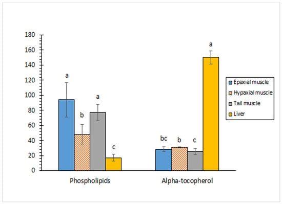
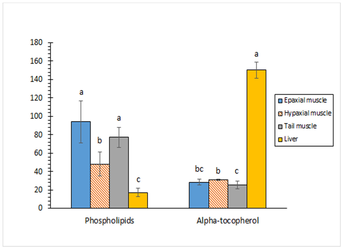
The PL content indicated a highly inhomogeneous distribution among the different body parts considered (Figure 1). Thus, the epaxial and tail muscle parts provided the highest (p < 0.05) values (94.0 and 77.2 g·kg−1 of lipids, respectively) and the liver tissue showed the lowest levels (p < 0.05) (17.2 g·kg−1 of lipids). Remarkably, an inverse pattern for the PL presence in the crude lipid extract was observed, in contrast to the total lipid value in tissue. This inverse relationship between both lipid parameters (i.e., presence of total lipids and total PLs) has already been described in most aquatic species [3,4,8]. As an explanation for this opposite trend, PL classes are reported to be structural lipids, their content being hardly dependent on external factors, as in other lipid classes such as those corresponding to depot or accumulation functions like triacylglycerols (TAGs), waxes, sterol esters, etc. [3].
Figure 1.
Determination of phospholipid (PL; g·kg−1 of lipids) and α-tocopherol (mg·kg−1 of lipids) values in different body parts of common carp cultivated in Madagascar. Average values of three replicates (n = 3); standard deviations are expressed by bars. Different lowercase letters indicate significant differences (p < 0.05) among body parts.
Present PL levels, expressed on muscle basis, were 3.45, 2.81, and 3.29 g PLs·kg−1 of muscle for epaxial, hypaxial, and tail body parts, respectively. Such values can be considered similar or even higher than those reported for the muscle tissue corresponding to other fish species like turbot (Psetta maxima) [4], angler (Lophius piscatorius) [8], or blackspot seabream (Pagellus bogaraveo) [10]. Additionally, PL presence in the liver tissue was found higher than in muscle parts (i.e., 5.06 g PL·kg−1 of tissue).
The PL group is present in cell membranes and plays decisive roles in living bodies. Based on the amphiphilic character [51], PL compounds have shown to be better vectors of ω3-PUFA compounds than other lipid classes like TAGs, methyl esters, or waxes. Therefore, the extraction of PL compounds from natural sources has received much interest from nutritional and pharmaceutical industries [52,53]. The higher bioavailability and absorption properties of PLs have shown to be especially relevant regarding the triad of oxidative stress, inflammation, and immune cell aging, closely related to chronic disease and aging [54,55]. Additionally, valuable functions of PL compounds have been related to food production and pharmaceutical industries [52,56], these including antioxidant properties during processing of fish and food in general [57,58]. Therefore, present tissues, and especially the liver tissue, can be considered a valuable source of this functional lipid group.
Regarding the TS presence in current body parts, an inhomogeneous distribution was detected (Table 2). Thus, the hypaxial muscle showed the lowest (p < 0.05) content (11.9 g·kg−1 of lipids), while the epaxial muscle provided the highest (p < 0.05) level (24.0 g·kg−1 of lipids). Additionally, a ca. 17 g·kg−1 of lipid value was obtained for tail and liver body parts. An inverse ratio between the TS presence in crude lipid extracts and the total lipid value has been detected in muscle parts of aquatic species [4,8,10]. As previously mentioned for the PL fraction, this inverse ratio has been reported according to the fact that sterol compounds are known to provide functional properties and play structural roles in living bodies; consequently, their content would not depend on external factors as do lipid classes related to accumulation roles [3].
Table 2.
Lipid composition * of different body parts of common carp cultivated in Madagascar **.
A notably higher presence (p < 0.05) of FFAs was observed in the liver tissue than in the muscle parts (Table 2). Furthermore, epaxial and hypaxial muscles showed a higher FFA value (p < 0.05) than the tail part. FFAs are produced during metabolic processes in living bodies [59,60] and in food during processing and storage [61,62], resulting from the hydrolysis of high-molecular weight lipid classes (namely, TAGs and PLs). Viscera, and especially the liver tissue, are reported to include a high content of hydrolytic enzymes (i.e., lipases and phospholipases) [63,64]. The current high FFA level in liver can be justified according to the presence of such hydrolytic enzymes. Such enzymes may have been active, releasing FFA compounds during the post-mortem period and the handling procedure [42,62].
Results on the presence of tocopherol compounds are indicated in Figure 1 and Table 2. Thus, α-tocopherol was the most abundant tocopherol compound in all body parts and notably lower values were obtained for γ- and δ-tocopherol. Remarkably, the presence of the β-tocopherol compound was not detected in any of the body parts considered in the current work.
A notably higher (p < 0.05) value of α-tocopherol was observed in the liver when compared to the remaining body parts (Figure 1); among muscle parts, a higher content (p < 0.05) was obtained in the hypaxial muscle than in the tail muscle. Regarding γ-tocopherol, a higher presence (p < 0.05) was observed in the liver tissue when compared to the muscle body parts (Table 2); additionally, no differences (p > 0.05) in this tocopherol compound were detected among muscle parts. A similar distribution was observed for both δ-tocopherol and γ-tocopherol (Table 2); i.e., the highest content (p < 0.05) was detected in the liver tissue and no differences (p > 0.05) could be detected among muscle parts.
Vitamin E is a natural and highly effective lipophilic antioxidant that refers to a group of eight chemically related compounds, i.e., four tocopherols (α, β, γ, and δ) and four tocotrienols (α, β, γ, and δ). Such compounds possess a 6-chromanol ring structure attached to a carbon side chain [65,66]. In agreement with the important role of this molecule group as chain-breaking antioxidants, the main role in living bodies is to protect the unsaturated FAs from oxidation [67,68]. Among aquatic animals, especially in vertebrate ones, α-tocopherol has been reported as the most abundant [3]. In agreement with the lipophilic antioxidant properties, it has shown an important role in vivo [69] and in the stability of food fat and oils [70]. According to this protective effect on unsaturated FAs, α-tocopherol content has shown to decrease with quality loss in processed fish during processing and storage [71]. In the present study, values of 1.05, 1.82, and 1.09 mg α-tocopherol·kg−1 of muscle were detected for α-tocopherol in epaxial, hypaxial, and tail body parts, respectively; such values are found lower than those reported for other farmed [4,10] and wild [8,65] fish species. However, and in agreement with its higher lipid content, the liver tissue showed a notably higher α-tocopherol value (i.e., 44.26 mg·kg−1 of tissue), which indicates that this tissue can be considered a good source of α-tocopherol.
Previous related research regarding the content of lipid classes in cultivated common carp tissues can be considered scarce. Remarkably, research has been focused on the assessment of the cholesterol presence in muscle body parts. On the contrary, the cholesterol content in liver and any other viscera of farmed common carp has not been addressed, to the best of our knowledge.
Thus, cholesterol values have shown to vary in common carp muscle depending on different factors such as breeding, age, husbandry system, rearing time, and harvest season [20,21]. Values of 69.4–77.6 [72] and 47 [73], expressed as mg·100 g−1 of muscle, were detected for common carp farmed in Czech ponds. Further, levels of 50.0 [26], 57.8 [21], and 55.8 [47], expressed as mg·100 g−1 of muscle, were observed in common carp cultivated in Serbian ponds. For wild carp captured in the Danube River, a value of 45.4 mg·100 g−1 of muscle was obtained [74].
In the present study, values of 880.8, 692.6, and 762.5 mg·kg−1 of muscle were detected for TSs in epaxial, hypaxial, and tail body parts, respectively; according to its higher lipid content, the liver tissue showed a notably higher (p < 0.05) value (i.e., 5150.3 mg·kg−1 of tissue). On the basis that cholesterol has been reported to be the major sterol compound in fish species [3,75], the present results can be considered in agreement with the abovementioned cholesterol values for farmed common carp.
3.3. Fatty Acid Profile
Table 3 includes the FA composition of the different carp body parts. All parts provided a similar profile regarding the individual FA composition. As a general behavior, the most abundant FA was C18:1ω9 (35.86–40.31 g·100 g−1 of total FAs). Furthermore, a remarkable presence in all body parts was obtained for C16:0 (20.44–21.35 g·100 g−1 of total FAs). Other relatively abundant FAs were C18:2ω6 (14.85–17.30 g·100 g−1 of total FAs), C18:0 (6.43–7.01 g·100 g−1 of total FAs), and C16:1ω7 (5.20–5.62 g·100 g−1 of total FAs). However, some content differences (p < 0.05) were detected.
Table 3.
Fatty acid (FA) profile (g·100 g−1 of total FAs) * of different body parts of common carp cultivated in Madagascar **.
To better analyze differences among the different body parts regarding the FA profile, the content of FA groups and values obtained for FA ratios are described and discussed in the following paragraphs.
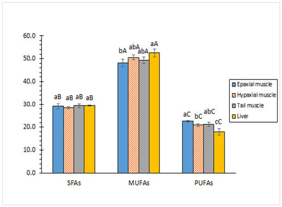
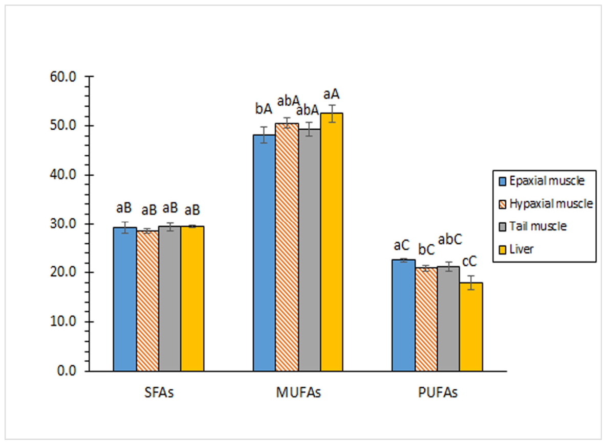
The analysis of the FA groups (SFAs, MUFAs, and PUFAs) indicated that the MUFA group was the major one (p < 0.05) in all body parts; on the contrary, PUFAs showed the lowest presence (p < 0.05) (Figure 2). For the SFA content, no differences (p > 0.05) could be concluded among the body parts considered. Inversely, differences could be observed for MUFA and PUFA values according to the body part taken into account. Thus, a higher MUFA content (p < 0.05) was detected in the liver tissue than in the epaxial muscle; additionally, a higher content (p < 0.05) of PUFAs was observed in the epaxial muscle than in the liver and hypaxial tissues.
Figure 2.
Determination of fatty acid (FA) group (saturated FAs, SFAs; monounsaturated FAs, MUFAs; polyunsaturated FAs, PUFAs) values (g·100 g−1 of total FAs) in different body parts of common carp cultivated in Madagascar. Average values of three replicates (n = 3); standard deviations are indicated by bars. Different lowercase letters indicate significant differences (p < 0.05) among body parts. Different capital letters denote significant differences (p < 0.05) among FA groups.
Unsaturated FAs belonging to the ω3- and ω6-PUFA series have been reported to be strongly linked to the development of certain human diseases [67,76]. For ω3-PUFAs, the present study revealed higher values (p < 0.05) for the epaxial and tail muscles (3.32 and 2.83 g·100 g−1 of total FAs, respectively) than in the case of the hypaxial and liver body parts (1.73 and 1.48 g·100 g−1 of total FAs, respectively) (Table 4). Regarding the ω6-PUFA presence, values were within the range of 16.48–19.18 g·100 g−1 of total FA (Table 4). A higher content (p < 0.05) was observed in the epaxial and hypaxial muscles than in the liver tissue.
Table 4.
Determination * of fatty acid (FA) group and ratio values in different body parts of common carp cultivated in Madagascar **.
Among ω3-PUFAs, EPA and DHA have shown beneficial effects on certain health properties [77,78]. Thus, EPA consumption has been associated with low prevalence of circulatory, coronary, and inflammatory diseases [77]. Additionally, DHA has been reported to be responsible for the prevention of neurodegenerative diseases and correct fetal development and functioning of the nervous system [78]. In spite of its generally lower content than EPA and DHA in aquatic food, docosapentaenoic acid (DPA) has shown a remarkable presence in the human brain and a high level in human milk, which implies a potential impact during pregnancy and early development [79]. Moreover, DPA has been reported to be related to the improvement of cardiovascular and metabolic diseases [80].
In agreement with the results depicted in Table 3, DHA was the most abundant ω3-PUFA in the present study; its content was found higher (p < 0.05) in the tail and epaxial muscles (2.18 and 2.63 g·100 g−1 of total FAs, respectively) than in the hypaxial and liver tissues (1.27 and 1.18 g·100 g−1 of total FAs, respectively). For EPA, the liver tissue showed lower (p < 0.05) values than the remaining body parts; among muscle parts, no differences (p > 0.05) could be concluded. Regarding DPA, the liver tissue showed the lowest (p < 0.05) presence; additionally, epaxial and tail muscles provided higher values (p < 0.05) than the hypaxial muscle.
Recent research has established certain FA ratios to valuable properties related to human health and nutrition. It has been mentioned that Western countries do not consume necessary levels of ω3-PUFAs, so that a relatively low ω3/ω6 ratio value results in the corresponding human diet [72,81]. To prevent health disorders such as cardiovascular, neurological, and inflammatory diseases, the World Health Organization (WHC) has recommended a higher ratio than 1:10 for the ω3/ω6 ratio [76]. Additionally, notable health benefits have been mentioned by the European Nutritional Society if an ω3/ω6 ratio of 1.5 or higher was maintained in the human diet [82].
On the basis of the ω3- and ω6-PUFA values obtained in the present research, a higher ω3/ω6 ratio value (p < 0.05) was detected in the epaxial and tail muscles than in the hypaxial and liver tissues (Table 4). However, ω3/ω6 ratio values obtained in current body parts can be considered lower than those recommended by the abovementioned institutions. Thus, present results on carp body parts have shown lower levels for EPA, DHA, and ω3/ω6 ratio than those reported for the muscle of marine fish and invertebrate species [4,8,10], and in wild freshwater species generally [3,83]. However, the muscle of common carp captured in the Tigris River revealed lower values for EPA (0.11 g·100 g−1 total FAs) and DHA (0.11 g·100 g−1 total FAs) than in the present study [84]. Additionally, current results are found higher than those corresponding to non-aquatic food such as poultry and egg [85], milk [86], and meat [87]. To increase the EPA and DHA content and the ω3/ω6 ratio, a diet including an FA composition as similar as possible to the one obtained in the wild condition is recommended.
Another remarkable FA ratio is total PUFA/total SFA. This ratio has also been found valuable for the human diet, in agreement with its direct relationship with digestibility and nutritional values and with antioxidant properties [88,89]. Additionally, aquatic food technologists found this ratio as a valuable index for the lipid damage assessment, and accordingly, for the quality loss of aquatic foods during different steps of processing and storage [43,90]; remarkably, this index should decrease as long as the quality of the product decreases. The present results indicate a range of 0.72–0.77 for this ratio in muscle body parts; this value was found higher (p < 0.05) than the one detected in the liver tissue (i.e., 0.61) (Table 4). Current PUFA/SFA values are lower than those corresponding to marine fish species (turbot, angler, and blackspot seabream) [4,8,10] or wild freshwater species [3,83]. However, the muscle of common carp captured in the Tigris River revealed a lower PUFA/SFA ratio (0.16) than for muscle body parts described in the present study [84]. Additionally, higher values were detected in the present study than in other kinds of foods such as poultry and egg [85], milk [86], and meat [87].
Regarding the FA profile, no previous research has been addressed to common carp cultivated in Madagascar. However, a seasonal study was carried out by Rasoarahona et al. [40] focused on the FA composition of wild carp muscle obtained from two different locations in Madagascar (reserves of Sisaony Station and Itasy Lake). In agreement with the present results, the same three major FAs were detected, although quantitative values were relatively modified, i.e., C18:1ω9 (17.0–21.2 and 16.9–21.5), C16:0 (16.1–13.2 and 13.1–16.4), and C18:2ω6 (10.3–12.9 and 10.3–13.2) g·100 g−1 of total FAs, respectively. In agreement with the reported differences for farmed and wild aquatic fish [4,10], carp specimens corresponding to both locations revealed higher ω3/ω6 ratios (i.e., 0.48–0.78 and 0.50–0.75, respectively) than in the present study. Furthermore, values (expressed as g·100 g−1 of total FAs) for DHA (3.15–6.69 and 2.86–6.53), EPA (2.13–2.84 and 2.14–3.43), and DPA (1.76–4.02 and 1.62–4.28) were also higher than in the current research.
The FA profile of the muscle of common carp cultivated in different parts of the world has been reported [26,46,47,91]. In agreement with the present research, C18:1ω9, C16:0, and C18:2ω6 were detected in most cases as the most abundant FAs; additionally, the same decreasing FA group sequence, i.e., MUFA > SFA > PUFA, was observed. Concerning the presence of ω3-PUFAs and FA ratios, differences with the present results could be detected and justified as being influenced by the set diet provided in each case.
Thus, Afkhami et al. [46] detected values of 0.7 and 0.8 for ω3/ω6 and PUFA/SFA ratios in farmed carp from Noshahr, Iran. In carp farmed in a Serbian pond [47], the following FAs were found as the most abundant (values expressed as g·100 g−1 of total FAs): C18:1ω9 (51.4), C16:0 (17.3), C18:2ω6 (8.8), and C16:1 (6.2); additionally, values of 0.19, 0.18, 0.25, 0.14, and 0.45 were obtained for EPA, DPA, DHA, ω3/ω6 ratio, and PUFA/SFA ratio, respectively. In farmed carp from Poland [91], C18:1ω9 (37.3), C16:0 (17.7), and 18:2ω6 (16.3) were detected as the most abundant FAs (values expressed as g·100 g−1 of total FAs); additionally, EPA, DPA, DHA, ω3/ω6 ratio, and PUFA/SFA ratio values of 1.48, 0.46, 1.34, 0.37, and 0.98, respectively, were detected. The main FAs in carp cultivated in a Serbian facility were (values expressed as g·100 g−1 of total FAs): C18:1ω9 (32.4), C18:2ω6 (19.7), and C16:0 (18.8) [26]; regarding DHA, EPA, DPA, ω3/ω6 ratio, and PUFA/SFA ratio, values of 1.2, 0.6, 1.2, 0.26, and 1.12, respectively, were detected.
Regarding the study of the FA profile of liver and any other viscera of farmed common carp, previous studies can be considered very scarce. Thus, Lu et al. [30] analyzed comparatively the FA composition of viscera and muscle corresponding to carp farmed in China during a seasonal study. In agreement with the current work, the main FAs for viscera and muscle, respectively, expressed as g·100 g−1 of total FAs, were C18:1ω9 (21–34 and 18–34), C16:0 (16–20 and 15–18), and C18:2ω6 (21–28 and 23–26). Additionally, great variability in the DHA (1.2–4.9 and 1.5–2.5) and EPA (0.16–1.15 and 0.28–1.09) presence was detected in both kinds of tissues. Furthermore, similar values were obtained in both tissues for the ω3/ω6 (0.12–0.27 and 0.15–0.24, respectively) and PUFA/SFA (1.2–1.5 and 1.2–1.5, respectively) ratios, respectively.
The seasonal variation in the FA composition in common carp liver was studied in specimens captured in the Atatürk Dam Lake [92]. As a result, C:16:0, C18:1ω9, and C20:4ω6 were found to be the major FAs. FA group contents revealed a different relative composition when compared with the present results; thus, value ranges (g·100 g−1 of total FAs) of 32.0–41.4, 24.8–37.8, and 30.0–40.2 were obtained for SFA, MUFA, and PUFA groups, respectively. Additionally, higher ω3/ω6 (i.e., 1.3–2.6) and PUFA/SFA (i.e., 0.5–1.4) ratios were observed when compared to the present study.
4. Conclusions
The different body parts taken into account showed an inhomogeneous distribution of bioactive compounds (proteins, PLs, tocopherols, and ω3-PUFAs). In general, differences were found more important by comparing the different parts of the muscle tissue and the liver. This study provides valuable information regarding the lipid class (i.e., PLs, TSs, FFAs, and tocopherols) composition of different muscle parts from the current cultivated freshwater species and novel information regarding the liver composition, a by-product commonly discarded or employed for minor purposes.
The study agrees with the general need to provide a comprehensive knowledge of the composition of aquatic species included in the human diet. Further research that takes into account a wider sample size, specimens corresponding to different aquaculture facilities, the feed composition, and environmental feeding factors ought to be addressed. From a nutritional point of view, an FA composition in diet that imitates the one received during the wild condition is recommended. Since PLs are recognized as valuable vectors of ω3-PUFAs, FA composition of the different PL classes deserve detailed analysis in upcoming research. According to the valuable composition of the liver tissue detected in the present work, studying the composition of other viscera and underutilized body parts (skin, head, tails, etc.) of cultivated common carp as possible sources of bioactive compounds is recommended.
Author Contributions
Conceptualization, S.P.A., D.V., and F.M.R.; methodology, M.T., L.P., D.M.A., N.E.R., and F.M.R.; data curation, M.T., L.P., D.M.A., and N.E.R.; writing—original draft preparation, S.P.A.; writing—review and editing, S.P.A., D.V., and F.M.R. All authors have read and agreed to the published version of the manuscript.
Funding
This research was funded by the ACE South Africa, Ramón Areces Foundation and Spanish Embassies in South Africa by providing a visiting grant to author S.P.A. in Antananarivo, Madagascar (November 2024).
Institutional Review Board Statement
The fish samples used in the experimental study were obtained from an aquaculture facility (Coopération des Éleveurs de Poissons, Maison du Petit Élevage, Antananarivo, Madagascar) in November 2024. The fish were already dead before being obtained.
Informed Consent Statement
Not applicable.
Data Availability Statement
The original contributions presented in the study are included in the article; further inquiries can be directed to the corresponding author.
Acknowledgments
ACE South Africa, Ramón Areces Foundation and Spanish Embassies in South Africa are greatly acknowledged for allowing and facilitating the development of the current collaboration study between our both institutions.
Conflicts of Interest
The authors declare no conflicts of interest.
References
- Tilami, S.K.; Sampels, S. Nutritional value of fish: Lipids, proteins, vitamins, and minerals. Rev. Fish. Sci. Aquac. 2018, 26, 242–253. [Google Scholar]
- Xu, H.; Turchini, G.M.; Francis, D.S.; Liang, M.; Mock, T.S.; Rombenso, A.; Ai, Q. Are fish what they eat? A fatty acid’s perspective. Prog. Lipid Res. 2020, 80, 101064. [Google Scholar] [CrossRef] [Scilit]
- Piclet, G. Le poisson aliment. Composition-Intérêt nutritionnel. Cah. Nutr. Diét. 1987, XXII, 317–335. [Google Scholar]
- Aubourg, S.P.; Losada, V.; Prego, R. Distribution of lipids and trace minerals in different muscle sites of farmed and wild turbot (Psetta maxima). Int. J. Food Sci. Technol. 2007, 42, 1456–1464. [Google Scholar] [CrossRef] [Scilit]
- Body, D.; Vlieg, P. Distribution of the lipid classes and eicosapentaenoic (20:5) and docosahexaenoic (22:6) acids in differnet sites in blue mackerel (Scomber australasicus) fillets. J. Food Sci. 1989, 54, 569–572. [Google Scholar] [CrossRef] [Scilit]
- Nakamura, Y.N.; Ando, M.; Seoka, M.; Kawasaki, K.I.; Tsukamasa, Y. Changes of proximate and fatty acid compositions of the dorsal and ventral ordinary muscles of the full-cycle cultured Pacific bluefin tuna Thunnus orientalis with the growth. Food Chem. 2007, 103, 234–241. [Google Scholar] [CrossRef] [Scilit]
- Aubourg, S.P.; Rey-Mansilla, M.; Sotelo, C. Differential lipid damage in various muscle zones of frozen hake (Merluccius merluccius). Z. Lebensm. Unters. Forsch. 1999, 208, 189–193. [Google Scholar] [CrossRef] [Scilit]
- Prego, R.; Pazos, M.; Medina, I.; Aubourg, S.P. Comparative chemical composition of different muscle zones in angler (Lophius piscatorius). J. Food Comp. Anal. 2012, 28, 81–87. [Google Scholar] [CrossRef] [Scilit]
- Testi, S.; Bonaldo, A.; Gatta, P.P.; Badiani, A. Nutritional traits of dorsal and ventral fillets from three farmed fish species. Food Chem. 2006, 98, 104–111. [Google Scholar] [CrossRef] [Scilit]
- Álvarez, V.; Medina, I.; Prego, R.; Aubourg, S.P. Lipid and mineral distribution in different zones of farmed and wild blackspot seabream (Pagellus bogaraveo). Eur. J. Lipid Sci. Technol. 2009, 111, 957–966. [Google Scholar] [CrossRef] [Scilit]
- Kottelat, M.; Freyhof, J. Handbook of European Freshwater Fishes; Maurice Kottelat (Privately Published): Cornol, Switzerland, 2007; 646p. [Google Scholar]
- Sahtout, F.; Boualleg, C.; Kaouachi, N.; Khelifi, N.; Menasria, A.; Bensouilah, M. Feeding habits of Cyprinus carpio in Foum El-Khanga Dam, Souk-Ahras, Algeria. AACL Bioflux 2018, 11, 554–564. [Google Scholar]
- Weber, M.J.; Brown, M.L. Effects of common carp on aquatic ecosystems 80 years after carp as a dominant: Ecological insights for fisheries management. Rev. Fish. Sci. 2009, 17, 524–537. [Google Scholar] [CrossRef] [Scilit]
- Tessema, A.; Getahun, A.; Mengistou, S.; Fetahi, T.; Dejen, E. Reproductive biology of common carp (Cyprinus carpio Linnaeus, 1758) in Lake Hayq, Ethiopia. Fish. Aquat. Sci. 2020, 23, 16. [Google Scholar] [CrossRef] [Scilit]
- FAO. El Estado Mundial de la Pesca y la Acuicultura: Hacia la Transformación Azul; Organización de las Naciones Unidas para la Alimentación y la Agricultura: Rome, Italy, 2024; p. 28. [Google Scholar]
- Yeganeh, S.; Shabanpour, B.; Hosseini, H.; Imanpour, M.R.; Shabani, A. Comparison of farmed and wild common carp (Cyprinus carpio): Seasonal variations in chemical composition and fatty acid profile. Czech J. Food Sci. 2012, 30, 503–511. [Google Scholar] [CrossRef] [Scilit]
- Choudhary, M.A.; Fatima, S.; Yasin, R.; Samiullah, K.; Mubarik, M.S.; Malik, I.U.; Fazal, R.M.; Rauf, A.; Draz, O.; Tabassum, M. Comparative study of proximate composition and mineral contents in Cyprinus carpio and Hypothalmichthys molitrix collected from River Sutluj. J. Bio. Environ. Sci. 2018, 13, 185–193. [Google Scholar]
- Blazhekovikj–Dimovska, D.; Ahmed, S. Some quality properties of common carp (Cyprinus carpio, L. 1758) from different aquatic environment in N. Macedonia. Carpathian J. Food Sci. Technol. 2020, 12, 31–40. [Google Scholar]
- Wang, L.; Jia, S.P.; Zhang, L.; Ma, F.R.; Zhang, M.; Yu, M.; Jiang, H.X.; Qiao, Z.G.; Li, X.J. Comparative study on the nutritional quality and volatile flavour compounds of muscle in Cyprinus carpio haematoreptus under wild, traditional pond and in-pond raceway system culture. Aquac. Rep. 2022, 25, 101194. [Google Scholar] [CrossRef] [Scilit]
- Bieniarz, K.; Koldras, M.; Kaminski, J.; Mejza, T. Fatty acids, fat and cholesterol in some lines of carp (Cyprinus carpio) in Poland. Arch. Pol. Fish. 2001, 9, 5–24. [Google Scholar]
- Ćirković, M.; Trbović, D.; Ljubojević, D.; Đorđević, V. Meat quality of fish farmed in polyculture in carp ponds in Republic of Serbia. Tehnol. Mesa 2011, 52, 106–121. [Google Scholar]
- He, Z.; Wang, J.; Wei, Y.; Yan, X.; Li, Y.; Xie, D.; Nie, G. Optimizing muscle quality in common carp (Cyprinus carpio L.): Impacts of body size on nutrient composition, texture and volatile profile. Foods 2025, 14, 2794. [Google Scholar] [CrossRef] [Scilit] [PubMed]
- Eljasik, P.; Panicz, R.; Sobczak, M.; Sadowski, J.; Tórz, A.; Barbosa, V.; Marques, A.; Dias, J. Structural and molecular indices in common carp (Cyprinus carpio L.) fed n-3 PUFA enriched diet. Food Chem. Toxicol. 2021, 151, 112146. [Google Scholar] [CrossRef] [Scilit]
- He, Z.; Xu, C.; Chen, F.; Lou, Y.; Nie, G.; Xia, D. Dietary DHA enhanced the textural firmness of common carp (Cyprinus carpio L.) fed plant-derived diets through restraining FoxO1 pathways. Foods 2022, 11, 3600. [Google Scholar] [CrossRef] [Scilit]
- Dey, S.K.; Nayeen, J.; Dey, P.; Tuser, A.H.; Himel, I.A.; Sultana, R.; Khan, M.N.A.; Khatoon, H. Data on grow performance and nutritional composition of common carp (Cyprinus carpio) fry fed with Chlamydomonas sp. Data Brief 2025, 54, 110494. [Google Scholar] [CrossRef] [Scilit]
- Ljubojević, D.; Radosavljević, V.; Pelić, M.; Đorđević, V.; Živkov Baloš, M.; Ćirković, M. Fatty acid composition, chemical composition and processing yield of traditional hot smoked common carp (Cyprinus carpio, L). Iran. J. Fish. Sci. 2016, 15, 1293–1306. [Google Scholar]
- Tokur, B.; Ozkütük, S.; Atici, E.; Ozyurt, G.; Ozyurt, C.E. Chemical and sensory quality changes of fish fingers, made from mirror carp (Cyprinus carpio L., 1758), during frozen storage (–18 °C). Food Chem. 2006, 99, 335–341. [Google Scholar] [CrossRef] [Scilit]
- Li, Q.; Zhang, L.; Lu, H.; Song, S.; Luo, Y. Comparison of post-mortem changes in ATP-related, protein degradation and endogenous enzyme activity of white muscle and dark muscle from common carp (Cyprinus carpio) stored at 4 °C. LWT-Food Sci. Technol. 2017, 78, 317–324. [Google Scholar] [CrossRef] [Scilit]
- Hailu, D.; Tuma, N. Analysis of nutrient content in common carp (Cyprinus carpio) and Crucian carp (Carassius carassius) fish offal. J. Fish. Livest. Prod. 2022, 10, 359. [Google Scholar]
- Lu, H.; Hong, H.; Luo, Y. The seasonal fatty acids composition in different tissues of farmed common carp (Cyprinus carpio). SDRP J. Food Sci. Technol. 2015, 1, 11–18. [Google Scholar]
- Crexi, V.T.; Souza-Soares, L.A.; Pinto, L.A.A. Carp (Cyprinus carpio) oils obtained by fishmeal and ensilage processes: Characteristics and lipid profiles. Int. J. Food Sci. Technol. 2009, 44, 1642–1648. [Google Scholar] [CrossRef] [Scilit]
- Tkaczewska, J.; Morawska, M.; Kulawik, P.; Zaja, M. Characterization of carp (Cyprinus carpio) skin gelatin extracted using different pretreatments method. Food Hydrocoll. 2018, 81, 169–179. [Google Scholar] [CrossRef] [Scilit]
- Olsen, R.L.; Toppe, J.; Karunasagar, I. Challenges and realistic opportunities in the use of by-products from processing of fish and shellfish. Trends Food Sci. Technol. 2014, 36, 144–152. [Google Scholar] [CrossRef] [Scilit]
- Atef, M.; Ojagh, M. Health benefits and food applications of bioactive compounds from fish byproducts: A Review. J. Funct. Foods 2017, 35, 673–681. [Google Scholar] [CrossRef] [Scilit]
- AOAC. Official Methods for Analysis of the Association of Analytical Chemistry, 15th ed.; Association of Official Chemists, Inc.: Arlington, VA, USA, 1990; pp. 931–937. [Google Scholar]
- Bligh, E.; Dyer, W. A rapid method of total extraction and purification. Can. J. Biochem. Physiol. 1959, 37, 911–917. [Google Scholar] [CrossRef] [Scilit]
- Raheja, R.; Kaur, C.; Singh, A.; Bhatia, A. New colorimetric method for the quantitative determination of phospholipids without acid digestion. J. Lipid Res. 1973, 14, 695–697. [Google Scholar] [CrossRef] [Scilit]
- Huang, T.; Chen, C.; Wefler, V.; Raftery, A. A stable reagent for the Liebermann-Buchardt reaction. Anal. Chem. 1961, 33, 1405–1407. [Google Scholar] [CrossRef] [Scilit]
- Lowry, R.; Tinsley, I. Rapid colorimetric determination of free fatty acids. J. Am. Oil Chem. Soc. 1976, 53, 470–472. [Google Scholar] [CrossRef] [Scilit]
- Cabrini, L.; Landi, L.; Stefanelli, C.; Barzanti, V.; Sechi, A. Extraction of lipid and lipophilic antioxidants from fish tissues: A comparison among different methods. Comp. Biochem. Physiol. Part B Comp. Biochem. 1992, 101, 383–386. [Google Scholar] [CrossRef] [Scilit]
- Aubourg, S.P.; Medina, I.; Pérez-Martín, R. Polyunsaturated fatty acids in tuna phospholipids: Distribution in the sn-2 location and changes during cooking. J. Agric. Food Chem. 1996, 44, 585–589. [Google Scholar] [CrossRef] [Scilit]
- Aubourg, S.P.; Piñeiro, C.; González, M.J. Quality loss related to rancidity development during frozen storage of horse mackerel (Trachurus trachurus). J. Amer. Oil Chem. Soc. 2004, 81, 671–678. [Google Scholar] [CrossRef] [Scilit]
- Rodríguez, A.; Trigo, M.; Aubourg, S.P.; Medina, I. Optimisation of healthy-lipid content and oxidative stability during oil extraction from squid (Illex argentinus) viscera by green processing. Mar. Drugs 2021, 19, 616. [Google Scholar] [CrossRef] [Scilit] [PubMed]
- Méndez, L.; Rodríguez, A.; Aubourg, S.P.; Medina, I. Low-toxicity solvents for the extraction of valuable lipid compounds from octopus (Octopus vulgaris) waste. Foods 2023, 12, 3631. [Google Scholar] [CrossRef] [Scilit]
- Rasoarahona, J.R.E.; Barnathan, G.; Bianchino, J.P.; Gaydou, E.M. Annual evolution of fatty acid profile from muscle lipids of the common carp (Cyprinus carpio) in Madagascar Inland Waters. J. Agric. Food Chem. 2004, 52, 7339–7344. [Google Scholar] [CrossRef] [Scilit] [PubMed]
- Afkhami, M.; Mokhlesi, A.; Bastami, K.D.; Khoshnood, R.; Eshaghi, N.; Ehsanpour, M. Survey of some chemical compositions and fatty acids in cultured common carp (Cyprinus carpio) and grass carp (Ctenopharyngodon idella), Noshahr, Iran. World J. Fish Mar. Sci. 2011, 3, 533–538. [Google Scholar]
- Ćirković, M.; Ljubojević, D.; Đorđević, V.; Novakov, N.; Petronijević, R. Chemical composition of body including fatty acids of four cyprinids fish species cultured at the same conditions. Arch. Zootech. 2012, 15, 37–50. [Google Scholar]
- Skałecki, P.; Florek, M.; Pyć, A.; Kaliniak, A.; Staszowska, A. Comparison of physicochemical properties, fatty acid composition and mineral contents in common carp (Cyprinus carpio L.) fillet and the native traditional product carp ham. Pol. J. Food Nutr. Sci. 2016, 66, 311–319. [Google Scholar] [CrossRef] [Scilit]
- Talab, A.S.; Khallaf, M.M.M.; El-Kalyoubi, M.H.O.; Abdel-Fattah, A.A.; Abou-Taleb, S.M. Chemical composition, physicochemical and microbiological quality properties of Cyprinus carpio and Euthynnus alletteratus. Egypt. J. Aq. Biol. Fish. 2024, 28, 1059–1066. [Google Scholar] [CrossRef] [Scilit]
- Dong, X.P.; Wu, Q.; Li, D.Y.; Wang, T.; Pan, J.F.; Zheng, J.J.; Fu, X.X.; Qi, L.B.; Chen, G.B. Physicochemical, micro-structural, and textural properties of different parts from farmed common carp (Cyprinus carpio). J. Food Prop. 2017, 20, 946–955. [Google Scholar] [CrossRef] [Scilit]
- Küllenberg, D.; Taylor, L.A.; Schneider, M.; Massing, U. Health effects of dietary phospholipids. Lipids Health Dis. 2012, 11, 3. [Google Scholar] [CrossRef] [Scilit]
- Köhler, A.; Sarkinnen, E.; Tapola, N.; Niskanen, T.; Bruheim, I. Bioavailability of fatty acids from krill oil, krill meal and fish oil in healthy subjects–a randomized, single-dose, cross-over trial. Lipids Health Dis. 2015, 14, 19. [Google Scholar] [CrossRef] [Scilit] [PubMed]
- Li, J.; Wang, X.; Zhang, T.; Huang, Z.; Luo, X.; Deng, Y. A review on phospholipids and their main applications in drug delivery systems. Asian J. Pharm. Sci. 2015, 10, 81–98. [Google Scholar] [CrossRef] [Scilit]
- Grey, A.; Bolland, M. Clinical trial evidence and use of fish oil supplements. JAMA Intern. Med. 2014, 174, 460–462. [Google Scholar] [CrossRef] [Scilit] [PubMed]
- Magalhães, J.P.; Müller, M.; Rainger, G.; Steegenga, W. Fish oil supplements, longevity and aging. Aging 2016, 8, 1578–1582. [Google Scholar] [CrossRef] [Scilit]
- Takahashi, K.; Inoue, Y. Marine by-product phospholipids as booster of medicinal compounds. Adv. Food Nutr. Res. 2012, 65, 31–46. [Google Scholar] [PubMed]
- Bandarra, N.M.; Campos, R.M.; Batista, I.; Nunes, M.L.; Empis, J.M. Antioxidant synergy of alpha-tocopherol and phospholipids. J. Am. Oil Chem. Soc. 1999, 76, 905–913. [Google Scholar] [CrossRef] [Scilit]
- Hidalgo, F.J.; León, M.M.; Zamora, R. Antioxidative activity of amino phospholipids and phospholipid/amino acid mixtures in edible oils as determined by the rancimat method. J. Agric. Food Chem. 2006, 54, 5461–5467. [Google Scholar] [CrossRef] [Scilit]
- Henderson, R.J. Fatty acid metabolism in freshwater fish with particular reference to polyunsaturated fatty acids. Arch. Tierernaehrung 1996, 49, 5–22. [Google Scholar] [CrossRef] [Scilit]
- Tocher, D.R. Metabolism and functions of lipids and fatty acids in teleost fish. Rev. Fish. Sci. 2003, 11, 107–184. [Google Scholar] [CrossRef] [Scilit]
- Sikorski, Z.E.; Kolakowski, E. Endogenous enzyme activity and seafood quality: Influence of chilling, freezing, and other environmental factors. In Seafood Enzymes: Utilization and Influence on Postharvest Seafood Quality; Haard, N.F., Simpson, B.K., Eds.; Marcel Dekker: New York, NY, USA, 2000; pp. 451–487. [Google Scholar]
- Aubourg, S.P.; Quitral, V.; Larraín, M.A.; Rodríguez, A.; Gómez, J.; Maier, L.; Vinagre, J. Autolytic degradation and microbiological activity in farmed Coho salmon (Oncorhynchus kisutch) during chilled storage. Food Chem. 2007, 104, 369–375. [Google Scholar] [CrossRef] [Scilit]
- Wu, T.H.; Bechtel, P.J. Salmon by-product storage and oil extraction. Food Chem. 2008, 111, 868–871. [Google Scholar] [CrossRef] [Scilit]
- Fan, L.; Xian, C.; Tang, S.; Ding, W.; Xu, C.H.; Wang, X.C. Effect of frozen storage emperature on lipid stability of hepatopancreas of Eriocheir sinensis. LWT-Food Sci. Technol. 2022, 154, 112513. [Google Scholar] [CrossRef] [Scilit]
- Kalogeropoulos, N.; Chiou, A. Antioxidants. In Handbook of Seafood and Seafood Products Analysis; Nollet, L.M., Toldrá, F., Eds.; CRC Press: Boca Raton, FL, USA; Francis and Taylor Group: Boca Raton, FL, USA, 2010; pp. 309–326. [Google Scholar]
- Kim, Y.N. Vitamins. In Handbook of Seafood and Seafood Products Analysis; Nollet, L.M., Toldrá, F., Eds.; CRC Press: Boca Raton, FL, USA; Francis and Taylor Group: Boca Raton, FL, USA, 2010; pp. 327–350. [Google Scholar]
- Komprda, T. Eicosapentaenoic and docosahexaenoic acids as inflammation-modulating and lipid homeostasis influencing nutraceuticals: A review. J. Funct. Foods 2012, 4, 25–38. [Google Scholar] [CrossRef] [Scilit]
- Kamal-Eldin, A.; Appelqvist, L. The chemistry and antioxidant properties of tocopherols and tocotrienols. Lipids 1996, 31, 671–701. [Google Scholar] [CrossRef] [Scilit]
- Pope, S.A.S.; Burtin, G.E.; Clayton, P.T.; Madge, D.J.; Muller, D.P.R. Synthesis and analysis of conjugates of the major vitamin E metabolite, α-CEHC. Free. Radic. Biol. Med. 2002, 33, 807–817. [Google Scholar] [CrossRef] [Scilit] [PubMed]
- Frankel, E.N. Antioxidants. In Lipid Oxidation; The Oily Press: Dundee, Scotland, UK, 1998. [Google Scholar]
- Pazos, M.; Sánchez, L.; Medina, I. α-tocopherol oxidation in fish muscle during chilling and frozen storage. J. Agric. Food Chem. 2005, 53, 4000–4005. [Google Scholar] [CrossRef] [Scilit]
- Komprda, T.; Zelenka, J.; Fajmonova, E.; Bakaj, P.; Pechova, P. Cholesterol content in meat of some poultry and fish species as influenced by live weight and total lipid content. J. Agric. Food Chem. 2003, 51, 7692–7697. [Google Scholar] [CrossRef] [Scilit]
- Kopicova, Z.; Vavreinova, S. Occurrence of squalene and cholesterol in various species of Czech freshwater fish. Czech J. Food Sci. 2007, 25, 195–201. [Google Scholar] [CrossRef] [Scilit]
- Ljubojevic, D.; Trbovic, D.; Lujic, J.; Bjelic-Cabrilo, O.; Kostic, D.; Novakov, N.; Cirkovic, M. Fatty acid composition of fishes from inland waters. Bulg. J. Agric. Sci. 2013, 19, 62–71. [Google Scholar]
- Özoğul, F.; Kuley, E.; Özoğul, Y. Sterol content of fish, crustacea and mollusc: Effects of cooking methods. Int. J. Food Prop. 2015, 18, 2026–2041. [Google Scholar] [CrossRef] [Scilit]
- Kumari, P.; Kumar, M.; Reddy, C.R.; Jha, B. Algal lipids, fatty acids and sterols. In Functional Ingredients from Algae for Foods and Nutraceuticals; Domínguez, H., Ed.; Woodhead Publishing: Cambridge, UK, 2013; pp. 87–134. [Google Scholar]
- Swanson, S.; Block, R.; Mousa, S. Omega-3 fatty acids EPA and DHA: Health benefits throughout life. Adv. Nutr. 2012, 3, 1–7. [Google Scholar] [CrossRef] [Scilit]
- Ofosu, F.K.; Daliri, E.B.M.; Lee, B.H.; Yu, X. Current trends and future perspectives on omega-3 fatty acids. Res. J. Biol. 2017, 5, 11–20. [Google Scholar]
- Li, J.; Yin, H.; Bibus, D.M.; Byelashov, O.A. The role of Omega-3 docosapentaenoic acid in pregnancy and early development. Eur. J. Lipid Sci. Technol. 2016, 118, 1692–1701. [Google Scholar] [CrossRef] [Scilit]
- Drouin, G.; Rioux, V.; Legrand, P. The n-3 docosapentaenoic acid (DPA): A new player in the n-3 long chain polyunsaturated fatty acid family. Biochimie 2019, 159, 36–48. [Google Scholar] [CrossRef] [Scilit]
- Uauy, R.; Valenzuela, A. Marine oils: The health benefits of n-3 fatty acids. Nutrition 2000, 16, 680–684. [Google Scholar] [CrossRef] [Scilit]
- Simopoulos, A.P. The importance of the omega-6/omega-3 fatty acid ratio in cardiovascular disease and other chronic diseases. Exp. Biol. Med. 2008, 233, 674–688. [Google Scholar] [CrossRef] [Scilit]
- Bote, A.; Trigo, M.; Martínez, S.; Aubourg, S.P. Presence of bioactive compounds in European eel (Anguilla anguilla) skin: A comparative study with edible tissue. Mar. Drugs 2024, 22, 105. [Google Scholar] [CrossRef] [Scilit]
- Ekin, I. Nutritional quality, proximate and fatty acid composition of commercially important fish from different rivers in SE Türkiye: A comparative research. Aquat. Sci. Eng. 2023, 38, 168–179. [Google Scholar] [CrossRef] [Scilit]
- Cantor, A.H.; Decker, E.A.; Collins, V.P. Fatty acids in poultry and egg products. In Fatty Acids in Foods and Their Health Implications, 3rd ed.; Chow, C.K., Ed.; CRC Press: Boca Raton, FL, USA, 2008; pp. 127–154. [Google Scholar]
- Palmquist, D.L.; Jensen, R.G. Fatty acids in milk fat. In Fatty Acids in Foods and Their Health Implications, 3rd ed.; Chow, C.K., Ed.; CRC Press: Boca Raton, FL, USA, 2008; pp. 109–125. [Google Scholar]
- Wood, J.D.; Enser, M.; Richardson, R.I.; Whittington, F.M. Fatty acids in meat and meat products. In Fatty Acids in Foods and Their Health Implications, 3rd ed.; Chow, C.K., Ed.; CRC Press: Boca Raton, FL, USA, 2008; pp. 87–107. [Google Scholar]
- Augimeri, G.; Galluccio, A.; Caparello, G.; Avolio, E.; la Russa, D.; de Rose, D.; Morelli, C.; Barone, I.; Catalano, S.; Ando, S.; et al. Potential antioxidant and anti-inflammatory properties of serum from healthy adolescents with optimal Mediterranean diet adherence: Findings from DIMENU cross-sectional study. Antioxidants 2021, 10, 1172. [Google Scholar] [CrossRef] [Scilit]
- Tibbetts, S.M.; Patelakis, S.J.J. Apparent digestibility coefficients (ADCs) of intact-cell marine microalgae meal (Pavlova sp. 459) for juvenile Atlantic salmon (Salmo salar L.). Aquaculture 2022, 546, 737236. [Google Scholar] [CrossRef] [Scilit]
- Özoğul, Y. Methods for freshness quality and deterioration. In Handbook of Seafood and Seafood Products Analysis; Nollet, L., Toldrá, F., Eds.; CRC Press: Boca Raton, FL, USA; Taylor & Francis Group: Boca Raton, FL, USA, 2010; pp. 189–214. [Google Scholar]
- Skałecki, P.; Florek, M.; Litwińczuk, A.; Staszowska, A.; Kaliniak, A. The nutritional value and chemical composition of muscle tissue of carp (Cyprinus carpio L.) and rainbow trout (Oncorhynchus mykiss Walb.) obtained from fish farms in the Lublin region. Sci. Ann. Pol. Soc. Anim. Prod. 2013, 9, 57–621. [Google Scholar]
- Kaçar, S.; Başhan, M. Seasonal variations on the fatty acid composition in liver tissue of Cyprinus carpio. Black Sea J. Sci. 2022, 12, 66–81. [Google Scholar]

Disclaimer/Publisher’s Note: The statements, opinions and data contained in all publications are solely those of the individual author(s) and contributor(s) and not of MDPI and/or the editor(s). MDPI and/or the editor(s) disclaim responsibility for any injury to people or property resulting from any ideas, methods, instructions or products referred to in the content. |
© 2026 by the authors. Licensee MDPI, Basel, Switzerland. This article is an open access article distributed under the terms and conditions of the Creative Commons Attribution (CC BY) license.