Next Issue
Volume 12, May
Previous Issue
Volume 12, March


Bioengineering, Volume 12, Issue 4 (April 2025) – 109 articles

Cover Story (view full-size image): 
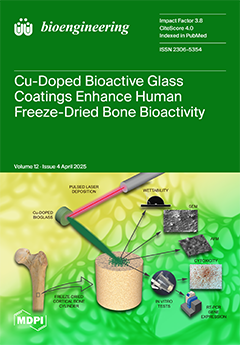
Critical-sized bone defects require innovative grafts combining mechanical support and biological activity. Here, human cortical bone was coated with copper-doped bioactive glass (Cu-BG) using pulsed laser deposition. The coating enhanced surface wettability and displayed a microstructured morphology favorable for cell adhesion. In vitro tests confirmed biocompatibility with osteoblasts and demonstrated Cu-BG’s dual regenerative potential, upregulating early osteogenic markers (SP7, SPP1, BGLAP) in mesenchymal stem cells and enhancing VEGF-mediated angiogenesis. Cu-BG-coated allografts emerge as promising “off-the-shelf” solutions for bone regeneration, merging human bone strength with bioactive healing properties. View this paper
  • Issues are regarded as officially published after their release is announced to the table of contents alert mailing list.
  • You may sign up for e-mail alerts to receive table of contents of newly released issues.
  • PDF is the official format for papers published in both, html and pdf forms. To view the papers in pdf format, click on the "PDF Full-text" link, and use the free Adobe Reader to open them.
Order results
Result details
Section
Select all
Export citation of selected articles as:
12 pages, 1779 KiB  
Article
Deep Learning-Based Estimation of Myocardial Material Parameters from Cardiac MRI
by Yunhe Chen, Xiwen Zhang, Yongzhong Huo and Shuo Wang
Bioengineering 2025, 12(4), 433; https://doi.org/10.3390/bioengineering12040433 - 21 Apr 2025
Viewed by 118
Abstract
Background: Accurate estimation of myocardial material parameters is crucial to understand cardiac biomechanics and plays a key role in advancing computational modeling and clinical applications. Traditional inverse finite element (FE) methods rely on iterative optimization to infer these parameters, which is computationally expensive [...] Read more.
Background: Accurate estimation of myocardial material parameters is crucial to understand cardiac biomechanics and plays a key role in advancing computational modeling and clinical applications. Traditional inverse finite element (FE) methods rely on iterative optimization to infer these parameters, which is computationally expensive and time-consuming, limiting their clinical applicability. Methods: This study proposes a deep learning-based approach to rapidly and accurately estimate the left ventricular myocardial material parameters directly from routine cardiac magnetic resonance imaging (CMRI) data. A ResNet18-based model was trained on FEM-derived parameters from a dataset of 1288 healthy subjects. Results: The proposed model demonstrated high predictive accuracy on healthy subjects, achieving mean absolute errors of 0.0146 for Ca and 0.0139 for Cb, with mean relative errors below 5.00%. Additionally, we evaluated the model on a small pathological subset (including ARV and HCM cases). The results revealed that while the model maintained strong performance on healthy data, the prediction errors in the pathological samples were higher, indicating increased challenges in modeling diseased myocardial tissue. Conclusion: This study establishes a computationally efficient and accurate deep learning framework for estimating myocardial material parameters, eliminating the need for time-consuming iterative FE optimization. While the model shows promising performance on healthy subjects, further validation and refinement are required to address its limitations in pathological conditions, thereby paving the way for personalized cardiac modeling and improved clinical decision-making. Full article
Show Figures

Figure 1

14 pages, 4027 KiB  
Article
Introducing the Stool Stomper: A Device Designed to Enable Accelerated and Standardized Stool Sample Preparation Using the Kato–Katz Technique
by Asher C. Altman, Andrew D. Bohner, Victoria J. Brown, Lauren Barger, Rudolph L. Gleason, James K. Rains, James B. Stubbs, Kelsey P. Kubelick and Mariana Stephens
Bioengineering 2025, 12(4), 432; https://doi.org/10.3390/bioengineering12040432 - 19 Apr 2025
Viewed by 191
Abstract
Soil-transmitted helminths (STHs) are parasitic worms that impact over 1.5 billion people globally. The Kato–Katz technique analyzes stool samples for STHs, allowing for individual diagnoses of STH infection and the estimation of community-level prevalence. One challenge that arises with the procedure is that [...] Read more.
Soil-transmitted helminths (STHs) are parasitic worms that impact over 1.5 billion people globally. The Kato–Katz technique analyzes stool samples for STHs, allowing for individual diagnoses of STH infection and the estimation of community-level prevalence. One challenge that arises with the procedure is that lab technicians often struggle to prepare microscope slides of sufficient quality for analysis after one attempt. As a result, Kato–Katz procedures are repeated, wasting time and resources. To aid technicians during in-field slide preparation, we created the Stool Stomper. The Stool Stomper is a user-friendly, handheld mechanical device that applies constant, uniform pressure to stool samples to ensure standardized sample preparation onto microscope slides to improve egg counts. The Stool Stomper was assessed using artificial eggs during in-country testing in a lab setting in Dodoma, Tanzania, by lab technicians with various experience levels, from beginner to advanced. Compared to the traditional method, we found that the Stool Stomper reduced slide preparation time, reduced artificial egg counting time, and standardized artificial egg counts with more consistent and accurate readings. The current pilot study highlights the potential for future development and integration of the Stool Stomper device into the Kato–Katz technique to improve community-based STH treatment. Full article
(This article belongs to the Section Biomedical Engineering and Biomaterials)
Show Figures

Graphical abstract

10 pages, 2960 KiB  
Article
Comparing the Efficiency of Valved Trocar Cannulas for Pars Plana Vitrectomy
by Tommaso Rossi, Giorgio Querzoli, Giov Battista Angelini, Camilla Pellizzaro, Veronica Santoro, Giulia Rosari, Mariacristina Parravano, David H. Steel and Mario R. Romano
Bioengineering 2025, 12(4), 431; https://doi.org/10.3390/bioengineering12040431 - 19 Apr 2025
Viewed by 153
Abstract
Purpose: To compare the efficiency of different manufacturers’ valved cannulas (23G, 25G and 27G) (Alcon, Bausch & Lomb, BVI, DORC, Optikon) in maintaining intraocular pressure during vitrectomy by measuring leak pressure and the difference between set and actual intraocular pressure, under BSS and [...] Read more.
Purpose: To compare the efficiency of different manufacturers’ valved cannulas (23G, 25G and 27G) (Alcon, Bausch & Lomb, BVI, DORC, Optikon) in maintaining intraocular pressure during vitrectomy by measuring leak pressure and the difference between set and actual intraocular pressure, under BSS and air infusion. Methods: A BSS-filled reservoir was connected to a model eye allowing placement of leak-proof valved cannulas. A pressure sensor was interposed and the bottle height increased until leakage occurred. Air leakage was measured by connecting an air pump to different manufacturers’ valved cannulas, inserted upside down to blow air against the valve with inside-out direction and immersed in soapy water to detect air bubbling. Results: The average BSS leaking pressure was 7.69 ± 0.77 mmHg for 23G, 9.92 ± 0.57 mmHg for 25G and 7.57 ± 0.80 mmHg for 27G. The 25G valved cannulas opened at higher pressure (p < 0.05). The difference between set and actual pressure in BSS never exceeded 4 mmHg. Leakage pressure under air ranged between 10 and 55 mmHg. The 27G valves opened at an average 47.2 ± 3.9 mmHg vs. 29.4 ± 7.2 for 25G and 24.1 ± 16.5 for 23G (27G vs. other gauges p < 0.05). The difference between set and actual pressure under air infusion never exceeded 2 mmHg. Conclusion: Despite significant differences, all tested valved cannulas satisfy safety criteria by keeping a surgically negligible difference between the set and actual intraocular pressure. The minimal leakage measure may act as a safety pressure damper under critical conditions. Full article
(This article belongs to the Section Biomedical Engineering and Biomaterials)
Show Figures

Figure 1

17 pages, 2572 KiB  
Review
Advances in Targeted Delivery of Doxorubicin for Cancer Chemotherapy
by Wenhui Xia and Martin W. King
Bioengineering 2025, 12(4), 430; https://doi.org/10.3390/bioengineering12040430 - 19 Apr 2025
Viewed by 315
Abstract
Doxorubicin (DOX) is one of the most powerful chemotherapy drugs used to treat different kinds of cancer. However, its usage has been limited by typical side effects and drug resistance, particularly cardiotoxicity. According to studies, a more effective and promising method is to [...] Read more.
Doxorubicin (DOX) is one of the most powerful chemotherapy drugs used to treat different kinds of cancer. However, its usage has been limited by typical side effects and drug resistance, particularly cardiotoxicity. According to studies, a more effective and promising method is to conjugate it or entrap it in biocompatible nanoparticles. Compared to free DOX and traditional formulations, nanoparticles using specific processes or techniques can improve drug stability, minimize premature release at untargeted locations, and lower systemic toxicity. This review explains how various nanocarriers target the tumor to improve therapeutic efficacy while reducing the negative effects of DOX. Full article
(This article belongs to the Section Nanobiotechnology and Biofabrication)
Show Figures

Graphical abstract

17 pages, 9341 KiB  
Article
Simulation of the Diffusion Characteristics of Multifunctional Nanocarriers in Tumor Tissues Using Lattice Gas Automata and the Lattice Boltzmann Method
by Yuming Qin, Kai Yue, Xiaoling Yu, Yu You, Chao Yang and Xinxin Zhang
Bioengineering 2025, 12(4), 429; https://doi.org/10.3390/bioengineering12040429 - 18 Apr 2025
Viewed by 153
Abstract
Understanding the diffusion mechanisms of nanocarriers in tumor tissues is crucial for enhancing drug delivery to target areas. This study developed a simulation method combining lattice gas automata and the lattice Boltzmann method to explore the diffusion behaviors of ligand-coated nanoparticles (NPs) in [...] Read more.
Understanding the diffusion mechanisms of nanocarriers in tumor tissues is crucial for enhancing drug delivery to target areas. This study developed a simulation method combining lattice gas automata and the lattice Boltzmann method to explore the diffusion behaviors of ligand-coated nanoparticles (NPs) in the extracellular matrix (ECM) and tumor tissues under the influence of external fields. We propose mathematical models to describe how the movement of NPs is affected by thermomagnetic effects and by their interactions with ECM fiber walls and cells, and to calculate the flow field and temperature distribution in tumor tissues containing interstitial fluids. The results show that reduced tissue porosity and increased ECM fiber and cell densities hinder NP transport. Conversely, degrading ECM collagen fibers with thermal or other energy forms significantly improved NP diffusion in treated tissues. Modifying the surface zeta potential of NPs allowed for the regulation of NP adhesion to ECM fibers and cell membranes based on their charged components. However, altering the charge on the NP surface did not further enhance diffusion once a certain charge level was reached. Increased temperatures from NP heat generation under external fields improved interstitial fluid flow, thereby enhancing NP diffusion. Additionally, a static magnetic field gradient considerably increased the penetration depth of magnetic NPs in the direction of the field, with minimal effects on diffusion in other directions and, in some cases, reducing diffusion. Full article
(This article belongs to the Section Nanobiotechnology and Biofabrication)
Show Figures

Graphical abstract

16 pages, 581 KiB  
Review
Review on Current Advancements in Facilitation of Burn Wound Healing
by Wing Sum Siu, Hui Ma and Ping Chung Leung
Bioengineering 2025, 12(4), 428; https://doi.org/10.3390/bioengineering12040428 - 18 Apr 2025
Viewed by 167
Abstract
Burns are common injuries, but their treatment remains challenging due to the complex nature of the wound healing process. Burn wounds are classified into different categories based on their size and depth. Treatment modalities vary significantly across these categories, primarily focusing on the [...] Read more.
Burns are common injuries, but their treatment remains challenging due to the complex nature of the wound healing process. Burn wounds are classified into different categories based on their size and depth. Treatment modalities vary significantly across these categories, primarily focusing on the inflammation, proliferation, and remodeling phases of burn wound healing. This review summarizes current research on various approaches to enhance burn wound recovery, including advancements in wound dressings, the use of platelet-rich plasma, stem cells, their soluble factors primarily in the form of secretomes or extracellular vesicles, and nano-technologies. Additionally, advancements in modernized traditional medicine are discussed to give a new aspect for burn wound healing. This review also summarizes the barriers in translating bench research to clinical practice in burn wound treatment methods. For an effective translation, researchers and industrial partners should work more closely, while regulatory bodies should streamline the approval procedure. Full article
(This article belongs to the Special Issue Recent Advancements in Wound Healing and Repair)
Show Figures

Graphical abstract

21 pages, 15451 KiB  
Article
Self-Healing Polymeric Puerarin Hydrogel Dressing Promotes Diabetic Wound Healing Through Synergistic Immunomodulation and Tissue-Regenerative Remodeling
by Shaohui Geng, Li Liu, Mureziya Yimingjiang, Zhimin Lin, Jingyuan Fu, Shasha Yu, Xinxin Li, Aimin Yan, Kai Yuan, Guangrui Huang and Anlong Xu
Bioengineering 2025, 12(4), 427; https://doi.org/10.3390/bioengineering12040427 - 18 Apr 2025
Viewed by 229
Abstract
Chronic wound healing is a significant challenge in diabetes. Puerarin is an active compound extracted from the traditional Chinese medicine Pueraria lobata. Puerarin has been used in the treatment of diabetes and derives benefits from its antioxidant, anti-inflammatory, antibacterial, and pro-angiogenesis properties, [...] Read more.
Chronic wound healing is a significant challenge in diabetes. Puerarin is an active compound extracted from the traditional Chinese medicine Pueraria lobata. Puerarin has been used in the treatment of diabetes and derives benefits from its antioxidant, anti-inflammatory, antibacterial, and pro-angiogenesis properties, but its efficacy is hampered by poor water solubility and bioavailability. In this study, we designed a polyvinyl alcohol (PVA)–borax–puerarin (BP) hydrogel system that self-assembled via boronic ester bonds. The BP hydrogel exhibited exceptional physical characteristics, including adaptability, injectability, plasticity, self-healing capabilities, and robust compressive strength, as well as good biocompatibility. In the chronic wound diabetic rats model, the BP hydrogel significantly accelerated wound healing, as evidenced by hematoxylin and eosin (HE) staining, as well as Masson and picrosirius red (PSR) staining. RNA–sequencing and multiple immunohistochemistry (mIHC) analyses revealed that the BP hydrogel exerts a therapeutic effect by modulating macrophage polarization, promoting angiogenesis, and regulating collagen remodeling. Our findings suggest that the BP hydrogel represents a promising wound dressing and holds great potential for clinical applications in acute and chronic wound management. Full article
Show Figures

Graphical abstract

18 pages, 13599 KiB  
Article
Comprehensive Analysis of Oncogenic Somatic Alterations of Mismatch Repair Gene in Breast Cancer Patients
by Yin Yan, Yang Wang, Junjie Tang, Xiaoran Liu, Jichuan Wang, Guohong Song and Huiping Li
Bioengineering 2025, 12(4), 426; https://doi.org/10.3390/bioengineering12040426 - 18 Apr 2025
Viewed by 206
Abstract
Recent clinical trials have suggested that solid cancers with mismatch repair (MMR) deficiency are highly responsive to immunotherapy, regardless of cancer types. Previous MMR-related studies on breast cancer have predominantly focused on germline variants. However, the somatic MMR alterations have not been comprehensively [...] Read more.
Recent clinical trials have suggested that solid cancers with mismatch repair (MMR) deficiency are highly responsive to immunotherapy, regardless of cancer types. Previous MMR-related studies on breast cancer have predominantly focused on germline variants. However, the somatic MMR alterations have not been comprehensively characterized in breast cancer. In this study, we integrated genomic, transcriptomic, and clinical data from over 3000 breast cancer cases across six public cohorts. Our findings revealed that 1.2% of breast cancers harbored oncogenic somatic MMR alterations, with triple-negative breast cancer (TNBC) demonstrating the highest mutation rate at 3.1%. Additionally, somatic MMR alterations were significantly associated with microsatellite instability-high (MSI-H) and MMR-related mutational signatures, indicating that somatic MMR alterations led to impaired function of the MMR system. Biallelic inactivation of MMR genes resulted in a more pronounced loss of MMR function compared to monoallelic inactivation. Importantly, these MMR alterations significantly increased the tumor mutational burden (TMB) and neoantigen load in breast cancer, regardless of MSI-H status. These findings indicate that the frequency of MMR alterations is highest in TNBC and that MMR alterations in breast cancer can lead to MMR functional deficiencies, suggesting that some patients harboring such alterations may benefit from immunotherapy. Full article
(This article belongs to the Section Cellular and Molecular Bioengineering)
Show Figures

Graphical abstract

9 pages, 2524 KiB  
Communication
Expression of Tailored α-N-Acetylglucosaminidase in Escherichia coli for Synthesizing Mannose-6-Phosphate on N-Linked Oligosaccharides of Lysosomal Enzymes
by Yunsong Cao and Wei Wang
Bioengineering 2025, 12(4), 425; https://doi.org/10.3390/bioengineering12040425 - 17 Apr 2025
Viewed by 188
Abstract
Lysosomal enzymes are synthesized as N-glycosylated glycoproteins with mannose-6-phosphate (M6P) moieties, which are responsible for their binding to M6P receptors and transporting to the lysosome. In the M6P biosynthetic pathway, a Man8GlcNAc2 glycoform is converted to M6P groups through two [...] Read more.
Lysosomal enzymes are synthesized as N-glycosylated glycoproteins with mannose-6-phosphate (M6P) moieties, which are responsible for their binding to M6P receptors and transporting to the lysosome. In the M6P biosynthetic pathway, a Man8GlcNAc2 glycoform is converted to M6P groups through two consecutive enzymatic reactions, including N-acetylglucosamine (GlcNAc)-1-phosphotransferase (GNPT), transferring GlcNAc-1-phosphate from UDP-GlcNAc to the C6 hydroxyl groups of mannose residues, and then, removal of the covering GlcNAc moiety from the GlcNAc-P-mannose phosphodiester was carried out using an α-N-acetylglucosaminidase (referred to as ‘uncovering enzyme’, UCE) in the trans-Golgi network (TGN). Here, we expressed differently tailored versions of the UCE, including four truncated variants, in Escherichia coli. The four variants with the signal peptide, transmembrane domain, propiece and cytoplasmic tail truncated, respectively, were purified by affinity chromatography, and their enzymatic activities were assayed using a UDP-Glo kit. By fusing a maltose-binding protein (MBP) in the N-terminus of the UCE variants, the fusion proteins could be soluble when expressed in E. coli. The highest concentration of the purified enzyme was 80.5 mg/L of fermentation broth. Furthermore, the UCE with the core catalytic domain exhibited the highest uncovering activity. Full article
(This article belongs to the Section Biochemical Engineering)
Show Figures

Graphical abstract

18 pages, 7406 KiB  
Article
Comparing the Accuracy of Markerless Motion Analysis and Optoelectronic System for Measuring Gait Kinematics of Lower Limb
by Luca Emanuele Molteni and Giuseppe Andreoni
Bioengineering 2025, 12(4), 424; https://doi.org/10.3390/bioengineering12040424 - 16 Apr 2025
Viewed by 201
Abstract
(1) Background: Marker-based optical motion tracking is the gold standard in gait analysis; however, markerless solutions are rapidly emerging today. Algorithms like Openpose can track human movement from a video. Few studies have assessed the validity of this method. This study aimed to [...] Read more.
(1) Background: Marker-based optical motion tracking is the gold standard in gait analysis; however, markerless solutions are rapidly emerging today. Algorithms like Openpose can track human movement from a video. Few studies have assessed the validity of this method. This study aimed to assess the reliability of Openpose in measuring the kinematics and spatiotemporal gait parameters. (2) Methods: This analysis used simultaneously recorded video and optoelectronic motion capture data. We assessed 20 subjects with different gait impairments (healthy, right hemiplegia, left hemiplegia, paraparesis). The two methods were compared using computing absolute errors (AEs), intraclass correlation coefficients (ICCs), and cross-correlation coefficients (CCs) for normalized gait cycle joint angles. (3) Results: The spatiotemporal parameters showed an ICC between good to excellent, and the absolute error was very small: cadence AE = 1.63 step/min, Mean Velocity AE = 0.16 m/s. The Range of Motion (ROM) showed a good to excellent agreement in the sagittal plane. Furthermore, the normalized gait cycle CCC values indicated moderate to strong coupling in the sagittal plane. (4) Conclusions: We found Openpose to be accurate for sagittal plane gait kinematics and for spatiotemporal gait parameters in the healthy and pathological subjects assessed. Full article
(This article belongs to the Special Issue Technological Advances for Gait and Balance Assessment)
Show Figures

Figure 1

14 pages, 6047 KiB  
Article
A Novel Approach to Predict the Location and Fatigue Life of Intervertebral Disc Degeneration
by Zanni Zhang, Taoxi Wang, Huwei Bian, Xing Shen, Minjun Liang, Ee-Chon Teo and Tao Jiang
Bioengineering 2025, 12(4), 423; https://doi.org/10.3390/bioengineering12040423 - 16 Apr 2025
Viewed by 176
Abstract
This study presents a novel approach for predicting the location and fatigue life of degenerative intervertebral discs (IVDs) under cyclic loading conditions, aiming to improve the understanding of disc degeneration mechanisms. Based on mechanical theories linking IVD degeneration to stress imbalance and water [...] Read more.
This study presents a novel approach for predicting the location and fatigue life of degenerative intervertebral discs (IVDs) under cyclic loading conditions, aiming to improve the understanding of disc degeneration mechanisms. Based on mechanical theories linking IVD degeneration to stress imbalance and water loss, a finite element (FE) model of the L4–L5 lumbar spine was developed, combining probability-weighted anatomical structures, inverse dynamics, and cumulative fatigue mechanics. By quantifying stress variations and calculating cumulative damage across disc regions, stress-concentration areas prone to degeneration were identified, and validation via a case study of a retired weightlifter diagnosed with intervertebral disc disease (IVDD) demonstrated that the predicted degeneration location correlated well with affected areas observed in CT scan images. These findings suggest that prolonged, abnormal stress imbalances within the disc may contribute significantly to degeneration, offering potential clinical applications in preventive assessment and targeted treatment for spine health. Full article
Show Figures

Figure 1

13 pages, 4011 KiB  
Article
Imaging Peripheral Nerves In Vivo with CT Neurogram Using Novel 2,4,6-Tri-Iodinated Lidocaine Contrast Agent
by Rui Tang, Ron Perez, David M. Brogan, Mikhail Y. Berezin and James E. McCarthy
Bioengineering 2025, 12(4), 422; https://doi.org/10.3390/bioengineering12040422 - 16 Apr 2025
Viewed by 202
Abstract
Peripheral nerve injuries are a significant concern in surgical procedures, often leading to chronic pain and functional impairment. Despite advancements in imaging, preoperative and intraoperative visualization of peripheral nerves remains a challenge. This study introduces and evaluates a novel tri-iodinated lidocaine-based contrast agent [...] Read more.
Peripheral nerve injuries are a significant concern in surgical procedures, often leading to chronic pain and functional impairment. Despite advancements in imaging, preoperative and intraoperative visualization of peripheral nerves remains a challenge. This study introduces and evaluates a novel tri-iodinated lidocaine-based contrast agent for computed tomography neurography, aiming to enhance the intraoperative visibility of peripheral nerves in vivo. A tri-iodinated lidocaine analogue was synthesized and characterized for its radiodensity, sodium channel binding and nerve affinity. Sodium channel affinity was performed using molecular docking. In vitro contrast enhancement was assessed by comparing the agent’s Hounsfield unit (HU) values with those of Omnipaque, a clinically approved contrast medium. In vivo imaging was conducted on rat sciatic nerves using micro-CT, followed by ex vivo validation. Nerve conduction blockade was assessed via electrical stimulation and histological analysis was performed to evaluate neurotoxicity. Experimental results revealed the tri-iodinated lidocaine analogue to have similar or higher affinity toward voltage-gated sodium channels than the parent lidocaine and a radiodensity comparable to the commercial CT contrast agent Omnipaque in vitro. In vivo, the contrast agent provided CT visualization of the sciatic nerve, with a significant increase in HU values compared to untreated nerves. Electrical stimulation confirmed transient nerve conduction blockade without observable histological damage, supporting its dual role as an imaging and nerve-blocking agent. This study presents a novel tri-iodinated lidocaine-based contrast agent that enables clear CT visualization of peripheral nerves while maintaining reversible nerve inhibition. These findings support its potential application in preoperative planning and intraoperative nerve protection to reduce surgical nerve injuries. Further studies are warranted to optimize imaging conditions and evaluate its clinical feasibility. Full article
Show Figures

Graphical abstract

17 pages, 41392 KiB  
Article
DermViT: Diagnosis-Guided Vision Transformer for Robust and Efficient Skin Lesion Classification
by Xuejun Zhang, Yehui Liu, Ganxin Ouyang, Wenkang Chen, Aobo Xu, Takeshi Hara, Xiangrong Zhou and Dongbo Wu
Bioengineering 2025, 12(4), 421; https://doi.org/10.3390/bioengineering12040421 - 16 Apr 2025
Viewed by 261
Abstract
Early diagnosis of skin cancer can significantly improve patient survival. Currently, skin lesion classification faces challenges such as lesion–background semantic entanglement, high intra-class variability, artifactual interference, and more, while existing classification models lack modeling of physicians’ diagnostic paradigms. To this end, we propose [...] Read more.
Early diagnosis of skin cancer can significantly improve patient survival. Currently, skin lesion classification faces challenges such as lesion–background semantic entanglement, high intra-class variability, artifactual interference, and more, while existing classification models lack modeling of physicians’ diagnostic paradigms. To this end, we propose DermViT, a medically driven deep learning architecture that addresses the above issues through a medically-inspired modular design. DermViT consists of three main modules: (1) Dermoscopic Context Pyramid (DCP), which mimics the multi-scale observation process of pathological diagnosis to adapt to the high intraclass variability of lesions such as melanoma, then extract stable and consistent data at different scales; (2) Dermoscopic Hierarchical Attention (DHA), which can reduce computational complexity while realizing intelligent focusing on lesion areas through a coarse screening–fine inspection mechanism; (3). Dermoscopic Feature Gate (DFG), which simulates the observation–verification operation of doctors through a convolutional gating mechanism and effectively suppresses semantic leakage of artifact regions. Our experimental results show that DermViT significantly outperforms existing methods in terms of classification accuracy (86.12%, a 7.8% improvement over ViT-Base) and number of parameters (40% less than ViT-Base) on the ISIC2018 and ISIC2019 datasets. Our visualization results further validate DermViT’s ability to locate lesions under interference conditions. By introducing a modular design that mimics a physician’s observation mode, DermViT achieves more logical feature extraction and decision-making processes for medical diagnosis, providing an efficient and reliable solution for dermoscopic image analysis. Full article
Show Figures

Graphical abstract

20 pages, 16427 KiB  
Article
Leveraging Diverse Cell-Death Patterns to Decipher the Interactive Relation of Unfavorable Outcome and Tumor Microenvironment in Breast Cancer
by Yue Li, Ting Ding, Tong Zhang, Shuangyu Liu, Jinhua Wang, Xiaoyan Zhou, Zeqi Guo, Qian He and Shuqun Zhang
Bioengineering 2025, 12(4), 420; https://doi.org/10.3390/bioengineering12040420 - 15 Apr 2025
Viewed by 255
Abstract
Background: Programmed cell death (PCD) dynamically influences breast cancer (BC) prognosis through interactions with the tumor microenvironment (TME). We investigated 13 PCD patterns to decipher their prognostic impact and mechanistic links to TME-driven outcomes. Our study aimed to explore the complex mechanisms underlying [...] Read more.
Background: Programmed cell death (PCD) dynamically influences breast cancer (BC) prognosis through interactions with the tumor microenvironment (TME). We investigated 13 PCD patterns to decipher their prognostic impact and mechanistic links to TME-driven outcomes. Our study aimed to explore the complex mechanisms underlying these interactions and establish a prognostic prediction model for breast cancer. Methods: Using TCGA and METABRIC datasets, we integrated single-sample gene set enrichment analysis (ssGSEA), weighted gene co-expression network analysis (WGCNA), and Least Absolute Shrinkage and Selection Operator (LASSO) to explore PCD-TME interactions. Multi-dimensional analyses included immune infiltration, genomic heterogeneity, and functional pathway enrichment. Results: Our results indicated that high apoptosis and pyroptosis activity, along with low autophagy, correlated with favorable prognosis, which was driven by enhanced anti-tumor immunity, including more M1 macrophage polarization and activated CD8+ T cells in TME. PCD-related genes could promote tumor metastasis and poor prognosis via VEGF/HIF-1/MAPK signaling and immune response, including Th1/Th2 cell differentiation, while new tumor event occurrences (metastasis/secondary cancers) were linked to specific clinical features and gene mutation spectrums, including TP53/CDH1 mutations and genomic instability. We constructed a six-gene LASSO model (BCAP31, BMF, GLUL, NFKBIA, PARP3, PROM2) to predict prognosis and identify high-risk BC patients (for five-year survival, AUC = 0.76 in TCGA; 0.74 in METABRIC). Therein, the high-risk subtype patients demonstrated a poorer prognosis, also characterized by lower microenvironment matrix and downregulated immunocyte infiltration. These six gene signatures also showed prognostic value with significant differential expression in gene and protein levels of BC samples. Conclusion: Our study provided a comprehensive landscape of the cancer survival difference and related PCD-TME interaction axis and highlighted that high-apoptosis/pyroptosis states caused favorable prognosis, underlying mechanisms closely related with the TME where anti-tumor immunity would be beneficial for patient prognosis. These findings highlighted the model’s potential for risk stratification in BC. Full article
(This article belongs to the Special Issue Recent Advances in Cancer Bioinformatics)
Show Figures

Figure 1

17 pages, 4482 KiB  
Article
The Effect of Dental Implant Restoration on the Biomechanics of the Temporomandibular Joint in Patients with Posterior Tooth Loss: A Pilot Study
by Yuanli Zhang, Chongzhi Yin, Fei Chen, Guizhi Zhang, Po Hao, Yongli Pu, Haidong Teng, Hong Huang and Zhan Liu
Bioengineering 2025, 12(4), 419; https://doi.org/10.3390/bioengineering12040419 - 14 Apr 2025
Viewed by 203
Abstract
Currently, controversy persists over whether dental implant restoration exacerbates or alleviates temporomandibular disorders (TMDs). This study aimed to analyze the impact of dental implant restoration on the biomechanics of the temporomandibular joint (TMJ) in patients with posterior tooth loss. Ten healthy volunteers (Control [...] Read more.
Currently, controversy persists over whether dental implant restoration exacerbates or alleviates temporomandibular disorders (TMDs). This study aimed to analyze the impact of dental implant restoration on the biomechanics of the temporomandibular joint (TMJ) in patients with posterior tooth loss. Ten healthy volunteers (Control group) and twenty patients with posterior tooth loss (preoperative in the Pre group and postoperative in the Post group) were recruited. Three-dimensional maxillofacial models of the maxilla, mandible, dentition, and articular discs were reconstructed. The von Mises, contact, and tensile stresses of the TMJ were analyzed. Before implant restoration, the stresses of the TMJ in the Pre group were considerably higher than those in the Control group, especially on the missing tooth side. After restoration, the stresses in the Post group decreased significantly, with a near-symmetrical distribution. Additionally, before restoration, the patients with TMD had the highest stresses of the TMJ, followed by those without TMD, and the Control group had the lowest. After restoration, the stress magnitudes in the patients with or without TMD returned to the normal range. In summary, dental implant restoration can significantly improve the asymmetric stress distribution of the TMJs, substantially reduce excessive stress caused by tooth loss, and alleviate or eliminate the symptoms related to TMDs. Full article
(This article belongs to the Special Issue Advanced Engineering Technologies in Orthopaedic Research)
Show Figures

Graphical abstract

19 pages, 18176 KiB  
Article
Dual Transverse Arch Foot Orthosis Improves Gait Biomechanics in Females with Flexible Flatfoot
by Linjie Zhang, Qiaolin Zhang, Qian Liu, Xinyan Jiang, János Simon, Tibor Hortobágyi and Yaodong Gu
Bioengineering 2025, 12(4), 418; https://doi.org/10.3390/bioengineering12040418 - 14 Apr 2025
Viewed by 300
Abstract
(1) Background: Flexible flatfoot is characterized by medial arch collapse, leading to musculoskeletal impairments. We examined the effects of single-arch foot orthosis (SFO) and dual-arch foot orthosis (DFO) on arch height, kinematics, and kinetics in young females during walking and jogging. (2) Methods: [...] Read more.
(1) Background: Flexible flatfoot is characterized by medial arch collapse, leading to musculoskeletal impairments. We examined the effects of single-arch foot orthosis (SFO) and dual-arch foot orthosis (DFO) on arch height, kinematics, and kinetics in young females during walking and jogging. (2) Methods: Healthy females (n = 19) with flexible flatfoot were tested under three conditions: regular shoes, SFO, and DFO. Motion capture and a 3D force plate gathered biomechanical data. We also used a high-speed dual fluoroscopic imaging system (DFIS) to assess dynamic foot morphology. Outcomes included normalized truncated navicular height, medial arch angle, angles and moments at the metatarsophalangeal, subtalar, ankle, knee, and hip joints. (3) Results: Both types of orthoses improved the normalized navicular height and reduced the medial arch angle, with DFO vs. SFO showing greater effects (p < 0.001). DFO vs. SFO was also more effective in limiting the range of motion (ROM) of the metatarsophalangeal joint and dorsiflexion (p < 0.001). Additionally, DFO reduced the ankle range of motion and the maximum knee flexion during walking. Both orthoses reduced subtalar plantarflexion moments during stance (p < 0.001) and modulated ankle plantarflexion moments throughout different phases of gait. DFO uniquely enhanced metatarsophalangeal plantarflexion moments during jogging (p < 0.001). (4) Conclusions: Dual vs. single transverse arch foot orthosis is more effective in improving gait biomechanics in females with flexible flatfoot. Longitudinal studies are needed to confirm these benefits. Full article
(This article belongs to the Special Issue Mechanobiology in Biomedical Engineering)
Show Figures

Figure 1

18 pages, 1862 KiB  
Study Protocol
Epidemiology and Risk Prediction Model of Multidrug-Resistant Organism Infections After Liver Transplant Recipients: A Single-Center Cohort Study
by Chuanlin Chen, Desheng Li, Zhengdon Zhou, Qinghua Guan, Bo Sheng, Yongfang Hu and Zhenyu Zhang
Bioengineering 2025, 12(4), 417; https://doi.org/10.3390/bioengineering12040417 - 14 Apr 2025
Viewed by 244
Abstract
Objective: Accurate risk stratification at an early stage may reduce the incidence of infection and improve the survival rate of recipients by adopting targeted interventions. This study aimed to develop a nomogram to predict the risk of multidrug-resistant organism (MDRO) infections in liver [...] Read more.
Objective: Accurate risk stratification at an early stage may reduce the incidence of infection and improve the survival rate of recipients by adopting targeted interventions. This study aimed to develop a nomogram to predict the risk of multidrug-resistant organism (MDRO) infections in liver transplant (LT) recipients. Methods: We retrospectively collected clinical data from 301 LT recipients and randomly divided them into a training set (210 cases) and validation set (91 cases) using a 7:3 split ratio. Factors related to the risk of MDRO infection after LT were determined using univariate and multivariate bidirectional stepwise logistic regression. The model’s predictive performance and discrimination ability were evaluated using receiver operating characteristic (ROC) curves, calibration curves, and decision curve analysis (DCA). Results: 56 (18.60%) patients developed a MDRO infection, including 37 (17.62%) in the training cohort and 19 (20.88%) in the validation cohort. Ultimately, five factors related to MDRO infection after LT surgery were established: ascites (OR = 3.48, 95% CI [1.33–9.14], p = 0.011), total bilirubin (OR = 1.01, 95% CI [1.01–1.01], p < 0.001), albumin (OR = 0.85, 95% CI [0.75–0.96], p = 0.010), history of preoperative ICU stay (OR = 1.09, 95% CI [1.01–1.17], p = 0.009), and length of ICU stay (OR = 3.70, 95% CI [1.39–9.84], p = 0.019). The model demonstrated strong discrimination, and the area under the curve (AUC), sensitivity, and specificity of the training set were 0.88 (95% CI [0.81–0.94]), 0.82 (95% CI [0.76–0.87]), and 0.86 (95% CI [0.75–0.98]), respectively, while for the validation set, they were 0.77 (95% CI [0.65–0.90]), 0.76 (95% CI [0.67–0.86]), and 0.68 (95% CI [0.48–0.89]). The mean absolute error (MAE) in the validation cohort was 0.029, indicating a high accuracy. DCA showed a clinical benefit within a threshold probability range of 0.1 to 0.7. Conclusions: This study developed a clinically accessible nomogram to predict the risk of MDRO infection in LT recipients, enabling early risk stratification and the real-time assessment of infection risk based on the length of postoperative ICU stay. The model incorporates five easily obtainable clinical parameters (ascites, total bilirubin, albumin, preoperative ICU stay history, and length of ICU stay) and demonstrates strong predictive performance, facilitating the early identification of high-risk patients. Future research should focus on refining the model by incorporating additional clinical factors (e.g., immunosuppressive therapy adherence) and validating its generalizability in multicenter, large-sample cohorts to enhance its clinical utility. Full article
(This article belongs to the Section Biomedical Engineering and Biomaterials)
Show Figures

Figure 1

20 pages, 5694 KiB  
Article
Mechanical Characterization of Porous Bone-like Scaffolds with Complex Microstructures for Bone Regeneration
by Brandon Coburn and Roozbeh Ross Salary
Bioengineering 2025, 12(4), 416; https://doi.org/10.3390/bioengineering12040416 - 14 Apr 2025
Viewed by 249
Abstract
The patient-specific treatment of bone fractures using porous osteoconductive scaffolds has faced significant clinical challenges due to insufficient mechanical strength and bioactivity. These properties are essential for osteogenesis, bone bridging, and bone regeneration. Therefore, it is crucial to develop and characterize biocompatible, biodegradable, [...] Read more.
The patient-specific treatment of bone fractures using porous osteoconductive scaffolds has faced significant clinical challenges due to insufficient mechanical strength and bioactivity. These properties are essential for osteogenesis, bone bridging, and bone regeneration. Therefore, it is crucial to develop and characterize biocompatible, biodegradable, and mechanically robust scaffolds for effective bone regeneration. The objective of this study is to systematically investigate the mechanical performance of SimuBone, a medical-grade biocompatible and biodegradable material, using 10 distinct triply periodic minimal surface (TPMS) designs with various internal structures. To assess the material’s tensile properties, tensile structures based on ASTM D638-14 (Design IV) were fabricated, while standard torsion structures were designed and fabricated to evaluate torsional properties. Additionally, this work examined the compressive properties of the 10 TPMS scaffold designs, parametrically designed in the Rhinoceros 3D environment and subsequently fabricated using fused deposition modeling (FDM) additive manufacturing. The FDM fabrication process utilized a microcapillary nozzle (heated to 240 °C) with a diameter of 400 µm and a print speed of 10 mm/s, depositing material on a heated surface maintained at 60 °C. It was observed that SimuBone had a shear modulus of 714.79 ± 11.97 MPa as well as an average yield strength of 44 ± 1.31 MPa. Scaffolds fabricated with horizontal material deposition exhibited the highest tensile modulus (5404.20 ± 192.30 MPa), making them ideal for load-bearing applications. Also, scaffolds with large voids required thicker walls to prevent collapse. The P.W. Hybrid scaffold design demonstrated high vertical stiffness but moderate horizontal stiffness, indicating anisotropic mechanical behavior. The Neovius scaffold design balanced mechanical stiffness and porosity, making it a promising candidate for bone tissue engineering. Overall, the outcomes of this study pave the way for the design and fabrication of scaffolds with optimal properties for the treatment of bone fractures. Full article
Show Figures

Figure 1

11 pages, 2099 KiB  
Article
ACM-Assessor: An Artificial Intelligence System for Assessing Angle Closure Mechanisms in Ultrasound Biomicroscopy
by Yuyu Cong, Weiyan Jiang, Zehua Dong, Jian Zhu, Yuanhao Yang, Yujin Wang, Qian Deng, Yulin Yan, Jiewen Mao, Xiaoshuo Shi, Jiali Pan, Zixian Yang, Yingli Wang, Juntao Fang, Biqing Zheng and Yanning Yang
Bioengineering 2025, 12(4), 415; https://doi.org/10.3390/bioengineering12040415 - 14 Apr 2025
Viewed by 224
Abstract
Primary angle-closure glaucoma (PACG), characterized by angle closure (AC) with insidious and irreversible progression, requires precise assessment of AC mechanisms for accurate diagnosis and treatment. This study developed an artificial intelligence system, ACM-Assessor, to evaluate AC mechanisms in ultrasound biomicroscopy (UBM) images. A [...] Read more.
Primary angle-closure glaucoma (PACG), characterized by angle closure (AC) with insidious and irreversible progression, requires precise assessment of AC mechanisms for accurate diagnosis and treatment. This study developed an artificial intelligence system, ACM-Assessor, to evaluate AC mechanisms in ultrasound biomicroscopy (UBM) images. A dataset of 8482 UBM images from 1160 patients was retrospectively collected. ACM-Assessor comprises models for pixel-to-physical spacing conversion, anterior chamber angle boundary segmentation, and scleral spur localization, along with three binary classification models to assess pupillary block (PB), thick peripheral iris (TPI), and anteriorly located ciliary body (ALCB). The integrated assessment model classifies AC mechanisms into pure PB, pure non-PB, multiple mechanisms (MM), and others. ACM-Assessor’s evaluation encompassed external testing (2266 images), human–machine competition and assisting beginners’ assessment (an independent test set of 436 images). ACM-Assessor achieved accuracies of 0.924 (PB), 0.925 (TPI), 0.947 (ALCB), and 0.839 (integrated assessment). In man–machine comparisons, the system’s accuracy was comparable to experts (p > 0.05). With model assistance, beginners’ accuracy improved by 0.117 for binary classification and 0.219 for integrated assessment. ACM-Assessor demonstrates expert-level accuracy and enhances beginners’ learning in UBM analysis. Full article
Show Figures

Figure 1

10 pages, 2073 KiB  
Article
In Vitro Biomechanical Experiment on the Effect of Unilateral Partial Facetectomy Performed by Percutaneous Endoscopy on the Stability of Lumbar Spine
by Tao Ma, Xiaoshuang Tu, Junyang Li, Jingwei Wu and Luming Nong
Bioengineering 2025, 12(4), 414; https://doi.org/10.3390/bioengineering12040414 - 14 Apr 2025
Viewed by 231
Abstract
Objectives: This study’s purpose is to investigate the lumbar biomechanical effects of unilateral partial facetectomy (UPF) of different facet joint (FJ) portions under percutaneous endoscopy. Methods: Forty fresh calf spine models were used to simulate UPF under a physiological load performed through three [...] Read more.
Objectives: This study’s purpose is to investigate the lumbar biomechanical effects of unilateral partial facetectomy (UPF) of different facet joint (FJ) portions under percutaneous endoscopy. Methods: Forty fresh calf spine models were used to simulate UPF under a physiological load performed through three commonly used needle insertion points (IPs): (1) The apex of the superior FJ (as the first IP); (2) The midpoint of the ventral side of the superior FJ (as the second IP); (3) The lowest point of the ventral side of the superior FJ (as the third IP). The range of motion (ROM) and the L4/5 intradiscal maximum pressure (IMP) were measured and analyzed under a physiological load in all models during flexion, extension, left–right lateral flexion, and left–right axial rotation. Results: When UPF was performed through the second IP, the ROM of the lumbar spine and the L4/5 IMP in the calf spine models were not statistically different from the intact calf spine model. Conclusions: UPF through the second IP resulted in a minimal impact on the biomechanics of the lumbar spine. Thus, it might be considered the most appropriate IP for UPF. Full article
(This article belongs to the Special Issue Biomechanics and Motion Analysis)
Show Figures

Figure 1

15 pages, 7166 KiB  
Article
Comparative Analysis of Deep Neural Networks for Automated Ulcerative Colitis Severity Assessment
by Andreas Vezakis, Ioannis Vezakis, Ourania Petropoulou, Stavros T. Miloulis, Athanasios Anastasiou, Ioannis Kakkos and George K. Matsopoulos
Bioengineering 2025, 12(4), 413; https://doi.org/10.3390/bioengineering12040413 - 13 Apr 2025
Viewed by 246
Abstract
Background: Ulcerative colitis (UC) is a chronic inflammatory bowel disease characterized by continuous inflammation of the colon and rectum. Accurate disease assessment is essential for effective treatment, with endoscopic evaluation, particularly the Mayo Endoscopic Score (MES), serving as a key diagnostic tool. However, [...] Read more.
Background: Ulcerative colitis (UC) is a chronic inflammatory bowel disease characterized by continuous inflammation of the colon and rectum. Accurate disease assessment is essential for effective treatment, with endoscopic evaluation, particularly the Mayo Endoscopic Score (MES), serving as a key diagnostic tool. However, MES measurement can be subjective and inconsistent, leading to variability in treatment decisions. Deep learning approaches have shown promise in providing more objective and standardized assessments of UC severity. Methods: This study utilized publicly available endoscopic images of UC patients to analyze and compare the performance of state-of-the-art deep neural networks for automated MES classification. Several state-of-the-art architectures were tested to determine the most effective model for grading disease severity. The F1 score, accuracy, recall, and precision were calculated for all models, and statistical analysis was conducted to verify statistically significant differences between the networks. Results: VGG19 was found to be the best-performing network, achieving a QWK score of 0.876 and a macro-averaged F1 score of 0.7528 across all classes. However, the performance differences among the top-performing models were very small suggesting that selection should depend on specific deployment requirements. Conclusions: This study demonstrates that multiple state-of-the-art deep neural network architectures could automate UC severity classification. Simpler architectures were found to achieve competitive results with larger models, challenging the assumption that larger networks necessarily provide better clinical outcomes. Full article
Show Figures

Figure 1

16 pages, 3892 KiB  
Article
Causal Links Between Corneal Biomechanics and Myopia: Evidence from Bidirectional Mendelian Randomization in the UK Biobank
by Xuefei Li, Shenglong Luo, Kuangching Lin, Hera Soha, Meixiao Shen, Fan Lu and Junjie Wang
Bioengineering 2025, 12(4), 412; https://doi.org/10.3390/bioengineering12040412 - 13 Apr 2025
Viewed by 261
Abstract
Background: Myopia is a leading cause of visual impairment worldwide, and accumulating evidence suggests that biomechanics may be closely linked to its development. Understanding this relationship may help clarify the underlying mechanisms of myopia and guide treatment strategies. The aim of the study [...] Read more.
Background: Myopia is a leading cause of visual impairment worldwide, and accumulating evidence suggests that biomechanics may be closely linked to its development. Understanding this relationship may help clarify the underlying mechanisms of myopia and guide treatment strategies. The aim of the study is to investigate the causal relationship between myopia and corneal biomechanics using the UK Biobank (UKB) database. Methods: Data from 11,064 eyes in the UKB, including refraction results and Ocular Response Analyzer (ORA) measurements, were analyzed. Eyes were categorized by spherical equivalent (SE) into emmetropia, mild myopia, moderate myopia, and high myopia. One-way ANOVA assessed differences in corneal biomechanical parameters across the varying myopia groups, while Quantile Regression (QR) explored the relationship between these parameters and myopia severity across the different quantiles. A Mendelian randomization (MR) analysis was employed to explore the causal relationships. Results: Significant differences in corneal biomechanical parameters and intraocular pressure (IOP) were observed across the myopia levels (p < 0.001). High myopia was associated with lower corneal hysteresis (CH), a lower corneal resistance factor (CRF), and increased IOP. The QR analysis demonstrated that lower corneal biomechanics were associated with higher degrees of myopia, with the impact of corneal biomechanics becoming more pronounced as the myopia severity increased. The MR analysis indicated that low CH (OR = 0.9943, p = 0.004) and CRF (OR = 0.9946, p = 0.002) values were risk factors for myopia, while no causal effect was found when the myopia was treated as the exposure and corneal biomechanics as the outcome. Conclusions: This study establishes a causal relationship where reduced corneal biomechanics contribute to myopia, while myopia itself does not directly affect biomechanics. Corneal biomechanics could serve as a biomarker for assessing high myopia risk. These findings offer new insights into high myopia’s pathological mechanisms and targeted prevention. Full article
(This article belongs to the Section Biomedical Engineering and Biomaterials)
Show Figures

Figure 1

15 pages, 1538 KiB  
Review
Breathomics: A Non-Invasive Approach for the Diagnosis of Breast Cancer
by Hélène Yockell-Lelièvre, Romy Philip, Palash Kaushik, Ashok Prabhu Masilamani and Sarkis H. Meterissian
Bioengineering 2025, 12(4), 411; https://doi.org/10.3390/bioengineering12040411 - 12 Apr 2025
Viewed by 363
Abstract
Breast cancer is the most commonly diagnosed cancer worldwide, underscoring the critical need for effective early detection methods to reduce mortality. Traditional detection techniques, such as mammography, present significant limitations, particularly in women with dense breast tissue, highlighting the need for alternative screening [...] Read more.
Breast cancer is the most commonly diagnosed cancer worldwide, underscoring the critical need for effective early detection methods to reduce mortality. Traditional detection techniques, such as mammography, present significant limitations, particularly in women with dense breast tissue, highlighting the need for alternative screening approaches. Breathomics, based on the analysis of Volatile Organic Compounds (VOCs) present in exhaled breath, offers a non-invasive, potentially transformative diagnostic tool. These VOCs are metabolic byproducts from various organs of the human body whose presence and varying concentrations in breath are reflective of different health conditions. This review explores the potential of breathomics, highlighting its promise as a rapid, cost-effective screening approach for breast cancer, facilitated through the integration of portable solutions like electronic noses (e-noses). Key considerations for clinical translation—including patient selection, environmental confounders, and different breath collection methods—will be examined in terms of how each of them affects the breath profile. However, there are also challenges such as patient variability in VOC signatures, and the need for standardization in breath sampling protocols. Future research should prioritize standardizing sampling and analytical procedures and validating their clinical utility through large-scale clinical trials. Full article
(This article belongs to the Special Issue Breast Cancer: From Precision Medicine to Diagnostics)
Show Figures

Figure 1

18 pages, 2833 KiB  
Article
Optimizing Transcutaneous Spinal Cord Stimulation: An Exploratory Study on the Role of Electrode Montages and Stimulation Intensity on Reflex Pathway Modulation
by Shirin Madarshahian, Michael Trakhtorchuck, Tatiana Guerrero-David, Kristin Gustafson, James S. Harrop, Caio M. Matias, M. J. Mulcahey, Alessandro Napoli, Alexander Vaccaro and Mijail Serruya
Bioengineering 2025, 12(4), 410; https://doi.org/10.3390/bioengineering12040410 - 12 Apr 2025
Viewed by 356
Abstract
Transcutaneous spinal cord stimulation (tSCS) is a promising non-invasive method to improve motor function in individuals with spinal cord injury (SCI) by enhancing spinal reflex pathways. This study aimed to investigate the effects of different tSCS electrode placement montages and targeted spinal levels [...] Read more.
Transcutaneous spinal cord stimulation (tSCS) is a promising non-invasive method to improve motor function in individuals with spinal cord injury (SCI) by enhancing spinal reflex pathways. This study aimed to investigate the effects of different tSCS electrode placement montages and targeted spinal levels on neurophysiological responses such as spinally evoked motor responses (sEMRs), dorsal root reflex activation, and muscle recruitment in individuals with SCI and healthy controls to optimize stimulation strategies for motor recovery. Five participants (three individuals with SCI and two controls) underwent transcutaneous spinal cord stimulation using various electrode montages, target spinal level stimulation, and single- and paired-pulse paradigms. Electromyographic responses were analyzed to determine sEMR threshold, amplitudes, and paired-pulse attenuation. Different spinal levels and spatial configurations of electrode placements influenced the sEMR threshold and incidence of sEMR across all participants. Paired-pulse analysis showed more pronounced second-pulse attenuation in SCI participants (48 ± 36%) than in controls (12 ± 20%, p = 0.0425), with distinct trends observed across montages and muscle groups. These findings suggest that spinal level, electrode configuration, and paired-pulse effects are key factors in personalizing tSCS, informing the development of patient-centered therapeutic strategies. Future studies with larger and more diverse cohorts are needed to validate and expand these findings. Full article
(This article belongs to the Special Issue Bioengineering Approaches for Brain and Spinal Cord Injuries)
Show Figures

Figure 1

20 pages, 6681 KiB  
Article
CRISPR-Cas9-Mediated ATF6B Gene Editing Enhances Membrane Protein Production in HEK293T Cells
by Ho Joong Choi, Ba Reum Kim, Ok-Hee Kim and Say-June Kim
Bioengineering 2025, 12(4), 409; https://doi.org/10.3390/bioengineering12040409 - 11 Apr 2025
Viewed by 190
Abstract
This study aims to enhance membrane protein production in HEK293T cells through genetic modification. HEK293T cells are used for recombinant protein and viral vector production due to their human origin and post-translational modification capabilities. This study explores enhancing membrane protein production in these [...] Read more.
This study aims to enhance membrane protein production in HEK293T cells through genetic modification. HEK293T cells are used for recombinant protein and viral vector production due to their human origin and post-translational modification capabilities. This study explores enhancing membrane protein production in these cells by deleting the C-terminal of the ATF6B gene using CRISPR-Cas9 technology. The objective of this research is to investigate the effect of C-terminal deletion of the ATF6B gene on membrane protein production in HEK293T cells using CRISPR-Cas9 technology. To identify effective gene targets, sgRNAs were initially designed against multiple UPR-related genes, including ATF6A, IRE1A, IRE1B, PERK, and ATF6B. Among them, ATF6B was selected as the primary target for further investigation due to its superior editing efficiency. The efficiency of sgRNAs was evaluated using the T7E1 assay, and sequencing was performed to verify gene editing patterns. Membrane proteins were extracted from both ATF6B C-terminally deleted (ATF6B-ΔC) and wild-type (WT) cell lines for comparison. Flow cytometry was employed to assess membrane protein production by analyzing GFP expression in Membrane-GFP-expressing cells. HEK293T cells with C-terminally deleted ATF6B (ATF6B-ΔC) significantly increased membrane protein production by approximately 40 ± 17.6% compared to WT cells (p < 0.05). Sequencing revealed 11, 14, 1, and 10 bp deletions in the ATF6B-ΔC edited cells, which disrupted exon sequences, induced exon skipping, and introduced premature stop codons, suppressing normal protein expression. Flow cytometry confirmed a 23.9 ± 4.2% increase in GFP intensity in ATF6B-ΔC cells, corroborating the enhanced membrane protein production. These findings suggest that CRISPR-Cas9-mediated C-terminal deletion of the ATF6B gene can effectively enhance membrane protein production in HEK293T cells by activating the unfolded protein response pathway and improving the cell’s capacity to manage misfolded proteins. This strategy presents significant potential for the biotechnology and pharmaceutical industries, where efficient membrane protein production is essential for drug development and various applications. Full article
(This article belongs to the Section Cellular and Molecular Bioengineering)
Show Figures

Figure 1

14 pages, 1138 KiB  
Article
Utility of Clustering in Mortality Risk Stratification in Pulmonary Hypertension
by Pasquale Tondo, Lucia Tricarico, Giuseppe Galgano, Maria Pia C. Varlese, Daphne Aruanno, Crescenzio Gallo, Giulia Scioscia, Natale D. Brunetti, Michele Correale and Donato Lacedonia
Bioengineering 2025, 12(4), 408; https://doi.org/10.3390/bioengineering12040408 - 11 Apr 2025
Viewed by 186
Abstract
Background: Pulmonary hypertension (PH) is a condition characterized by increased pressure in the pulmonary arteries with poor prognosis and, therefore, an optimal management is necessary. The study’s aim was to search for PH phenotypes and develop a predictive model of five-year mortality [...] Read more.
Background: Pulmonary hypertension (PH) is a condition characterized by increased pressure in the pulmonary arteries with poor prognosis and, therefore, an optimal management is necessary. The study’s aim was to search for PH phenotypes and develop a predictive model of five-year mortality using machine learning (ML) algorithms. Methods: This multicenter study was conducted on 122 PH patients. Clinical and demographic data were collected and then used to identify phenotypes through clustering. Subsequently, a predictive model was performed by different ML algorithms. Results: Three PH clusters were identified: Cluster 1 (mean age 68.57 ± 10.54) includes 57% females, 69% from non-respiratory PH groups, and better cardiac (NYHA class 2.61 ± 0.84) and respiratory function (FEV1% 78.78 ± 21.54); Cluster 2 includes 50% females, mean age of 71.36 ± 8.32 years, 44% from PH group 3, worse respiratory function (FEV 1% 68.12 ± 10.20); intermediate cardiac function (NYHA class 3.18 ± 0.49) and significantly higher mortality (75%); Cluster 3 represents the youngest cluster (mean age 61.11 ± 13.50) with 65% males, 81% from non-respiratory PH groups, intermediate respiratory function (FEV1% 70.51 ± 17.91) and worse cardiac performance (NYHA class 3.22 ± 0.58). After testing ML models, logistic regression showed the best predictive performance (AUC = 0.835 and accuracy = 0.744) and identified three mortality-risk factors: age, NYHA class, and number of medications taken. Conclusions: The results suggest that the integration of ML into clinical practice can improve risk stratification to optimize treatment strategies and improve outcomes for PH patients. Full article
Show Figures

Figure 1

15 pages, 686 KiB  
Article
IDNet: A Diffusion Model-Enhanced Framework for Accurate Cranio-Maxillofacial Bone Defect Repair
by Xueqin Ji, Wensheng Wang, Xiaobiao Zhang and Xinrong Chen
Bioengineering 2025, 12(4), 407; https://doi.org/10.3390/bioengineering12040407 - 11 Apr 2025
Viewed by 259
Abstract
Cranio-maxillofacial bone defect repair poses significant challenges in oral and maxillofacial surgery due to the complex anatomy of the region and its substantial impact on patients’ physiological function, aesthetic appearance, and quality of life. Inaccurate reconstruction can result in serious complications, including functional [...] Read more.
Cranio-maxillofacial bone defect repair poses significant challenges in oral and maxillofacial surgery due to the complex anatomy of the region and its substantial impact on patients’ physiological function, aesthetic appearance, and quality of life. Inaccurate reconstruction can result in serious complications, including functional impairment and psychological trauma. Traditional methods have notable limitations for complex defects, underscoring the need for advanced computational approaches to achieve high-precision personalized reconstruction. This study presents the Internal Diffusion Network (IDNet), a novel framework that integrates a diffusion model into a standard U-shaped network to extract valuable information from input data and produce high-resolution representations for 3D medical segmentation. A Step-Uncertainty Fusion module was designed to enhance prediction robustness by combining diffusion model outputs at each inference step. The model was evaluated on a dataset consisting of 125 normal human skull 3D reconstructions and 2625 simulated cranio-maxillofacial bone defects. Quantitative evaluation revealed that IDNet outperformed mainstream methods, including UNETR and 3D U-Net, across key metrics: Dice Similarity Coefficient (DSC), True Positive Rate (RECALL), and 95th percentile Hausdorff Distance (HD95). The approach achieved an average DSC of 0.8140, RECALL of 0.8554, and HD95 of 4.35 mm across seven defect types, substantially surpassing comparison methods. This study demonstrates the significant performance advantages of diffusion model-based approaches in cranio-maxillofacial bone defect repair, with potential implications for increasing repair surgery success rates and patient satisfaction in clinical applications. Full article
Show Figures

Figure 1

22 pages, 2872 KiB  
Article
Wavelet-Guided Multi-Scale ConvNeXt for Unsupervised Medical Image Registration
by Xuejun Zhang, Aobo Xu, Ganxin Ouyang, Zhengrong Xu, Shaofei Shen, Wenkang Chen, Mingxian Liang, Guiqi Zhang, Jiashun Wei, Xiangrong Zhou and Dongbo Wu
Bioengineering 2025, 12(4), 406; https://doi.org/10.3390/bioengineering12040406 - 11 Apr 2025
Viewed by 306
Abstract
Medical image registration is essential in clinical practices such as surgical navigation and image-guided diagnosis. The Transformer architecture of TransMorph demonstrates better accuracy in non-rigid registration tasks. However, its weaker spatial locality priors necessitate large-scale training datasets and a heavy number of parameters, [...] Read more.
Medical image registration is essential in clinical practices such as surgical navigation and image-guided diagnosis. The Transformer architecture of TransMorph demonstrates better accuracy in non-rigid registration tasks. However, its weaker spatial locality priors necessitate large-scale training datasets and a heavy number of parameters, which conflict with the limited annotated data and real-time demands of clinical workflows. Moreover, traditional downsampling and upsampling always degrade high-frequency anatomical features such as tissue boundaries or small lesions. We proposed WaveMorph, a wavelet-guided multi-scale ConvNeXt method for unsupervised medical image registration. A novel multi-scale wavelet feature fusion downsampling module is proposed by integrating the ConvNeXt architecture with Haar wavelet lossless decomposition to extract and fuse features from eight frequency sub-images using multi-scale convolution kernels. Additionally, a lightweight dynamic upsampling module is introduced in the decoder to reconstruct fine-grained anatomical structures. WaveMorph integrates the inductive bias of CNNs with the advantages of Transformers, effectively mitigating topological distortions caused by spatial information loss while supporting real-time inference. In both atlas-to-patient (IXI) and inter-patient (OASIS) registration tasks, WaveMorph demonstrates state-of-the-art performance, achieving Dice scores of 0.779 ± 0.015 and 0.824 ± 0.021, respectively, and real-time inference (0.072 s/image), validating the effectiveness of our model in medical image registration. Full article
Show Figures

Figure 1

4 pages, 139 KiB  
Editorial
Advancing Natural Bioactive Formulations: Innovations in Agri-Food-Pharma for Enhanced Health and Sustainability
by Kandi Sridhar, Baskaran Stephen Inbaraj and Minaxi Sharma
Bioengineering 2025, 12(4), 405; https://doi.org/10.3390/bioengineering12040405 - 11 Apr 2025
Viewed by 216
Abstract
Over the years, it has been confirmed that bioactive compounds play a pivotal role in human health, particularly in the prevention of chronic diseases [...] Full article
Show Figures

Graphical abstract

27 pages, 3778 KiB  
Article
Patch-Based Texture Feature Extraction Towards Improved Clinical Task Performance
by Tao Lian, Chunyan Deng and Qianjin Feng
Bioengineering 2025, 12(4), 404; https://doi.org/10.3390/bioengineering12040404 - 10 Apr 2025
Viewed by 243
Abstract
Texture features can capture microstructural patterns and tissue heterogeneity, playing a pivotal role in medical image analysis. Compared to deep learning-based features, texture features offer superior interpretability in clinical applications. However, as conventional texture features focus strictly on voxel-level statistical information, they fail [...] Read more.
Texture features can capture microstructural patterns and tissue heterogeneity, playing a pivotal role in medical image analysis. Compared to deep learning-based features, texture features offer superior interpretability in clinical applications. However, as conventional texture features focus strictly on voxel-level statistical information, they fail to account for critical spatial heterogeneity between small tissue volumes, which may hold significant importance. To overcome this limitation, we propose novel 3D patch-based texture features and develop a radiomics analysis framework to validate the efficacy of our proposed features. Specifically, multi-scale 3D patches were created to construct patch patterns via k-means clustering. The multi-resolution images were discretized based on labels of the patterns, and then texture features were extracted to quantify the spatial heterogeneity between patches. Twenty-five cross-combination models of five feature selection methods and five classifiers were constructed. Our methodology was evaluated using two independent MRI datasets. Specifically, 145 breast cancer patients were included for axillary lymph node metastasis prediction, and 63 cervical cancer patients were enrolled for histological subtype prediction. Experimental results demonstrated that the proposed 3D patch-based texture features achieved an AUC of 0.76 in the breast cancer lymph node metastasis prediction task and an AUC of 0.94 in cervical cancer histological subtype prediction, outperforming conventional texture features (0.74 and 0.83, respectively). Our proposed features have successfully captured multi-scale patch-level texture representations, which could enhance the application of imaging biomarkers in the precise prediction of cancers and personalized therapeutic interventions. Full article
Show Figures

Figure 1

Previous Issue
Next Issue
Back to TopTop