Next Issue
Volume 10, August
Previous Issue
Volume 10, June


Bioengineering, Volume 10, Issue 7 (July 2023) – 136 articles

Cover Story (view full-size image): 
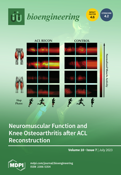
We coupled electromyography wavelet analysis—a frequency-based form of signal analysis and visualization (cover image)—with a machine learning approach to show that muscle activation patterns are bilaterally symmetrical in patients who had undergone anterior cruciate ligament reconstruction (ACL Recon) over a decade earlier, and that the patterns are different from controls. Because muscle size responds to function, we further investigated its association with magnetic resonance image-based structural features associated with posttraumatic osteoarthritis (PTOA) and showed that thigh muscle girth is a significant predictor of degeneration consistent with PTOA. Our results suggest that neuromuscular abnormalities in ACL Recon patients have a potential role in exacerbating PTOA risk. View this paper
  • Issues are regarded as officially published after their release is announced to the table of contents alert mailing list.
  • You may sign up for e-mail alerts to receive table of contents of newly released issues.
  • PDF is the official format for papers published in both, html and pdf forms. To view the papers in pdf format, click on the "PDF Full-text" link, and use the free Adobe Reader to open them.
Order results
Result details
Section
Select all
Export citation of selected articles as:
24 pages, 2696 KiB  
Article
Evaluation of Mutual Information and Feature Selection for SARS-CoV-2 Respiratory Infection
by Sekar Kidambi Raju, Seethalakshmi Ramaswamy, Marwa M. Eid, Sathiamoorthy Gopalan, Faten Khalid Karim, Raja Marappan and Doaa Sami Khafaga
Bioengineering 2023, 10(7), 880; https://doi.org/10.3390/bioengineering10070880 - 24 Jul 2023
Viewed by 1591
Abstract
This study aims to develop a predictive model for SARS-CoV-2 using machine-learning techniques and to explore various feature selection methods to enhance the accuracy of predictions. A precise forecast of the SARS-CoV-2 respiratory infections spread can help with efficient planning and resource allocation. [...] Read more.
This study aims to develop a predictive model for SARS-CoV-2 using machine-learning techniques and to explore various feature selection methods to enhance the accuracy of predictions. A precise forecast of the SARS-CoV-2 respiratory infections spread can help with efficient planning and resource allocation. The proposed model utilizes stochastic regression to capture the virus transmission’s stochastic nature, considering data uncertainties. Feature selection techniques are employed to identify the most relevant and informative features contributing to prediction accuracy. Furthermore, the study explores the use of neighbor embedding and Sammon mapping algorithms to visualize high-dimensional SARS-CoV-2 respiratory infection data in a lower-dimensional space, enabling better interpretation and understanding of the underlying patterns. The application of machine-learning techniques for predicting SARS-CoV-2 respiratory infections, the use of statistical measures in healthcare, including confirmed cases, deaths, and recoveries, and an analysis of country-wise dynamics of the pandemic using machine-learning models are used. Our analysis involves the performance of various algorithms, including neural networks (NN), decision trees (DT), random forests (RF), the Adam optimizer (AD), hyperparameters (HP), stochastic regression (SR), neighbor embedding (NE), and Sammon mapping (SM). A pre-processed and feature-extracted SARS-CoV-2 respiratory infection dataset is combined with ADHPSRNESM to form a new orchestration in the proposed model for a perfect prediction to increase the precision of accuracy. The findings of this research can contribute to public health efforts by enabling policymakers and healthcare professionals to make informed decisions based on accurate predictions, ultimately aiding in managing and controlling the SARS-CoV-2 pandemic. Full article
Show Figures

Figure 1

13 pages, 1254 KiB  
Article
Prompt-Based Tuning of Transformer Models for Multi-Center Medical Image Segmentation of Head and Neck Cancer
by Numan Saeed, Muhammad Ridzuan, Roba Al Majzoub and Mohammad Yaqub
Bioengineering 2023, 10(7), 879; https://doi.org/10.3390/bioengineering10070879 - 24 Jul 2023
Cited by 1 | Viewed by 1868
Abstract
Medical image segmentation is a vital healthcare endeavor requiring precise and efficient models for appropriate diagnosis and treatment. Vision transformer (ViT)-based segmentation models have shown great performance in accomplishing this task. However, to build a powerful backbone, the self-attention block of ViT requires [...] Read more.
Medical image segmentation is a vital healthcare endeavor requiring precise and efficient models for appropriate diagnosis and treatment. Vision transformer (ViT)-based segmentation models have shown great performance in accomplishing this task. However, to build a powerful backbone, the self-attention block of ViT requires large-scale pre-training data. The present method of modifying pre-trained models entails updating all or some of the backbone parameters. This paper proposes a novel fine-tuning strategy for adapting a pretrained transformer-based segmentation model on data from a new medical center. This method introduces a small number of learnable parameters, termed prompts, into the input space (less than 1% of model parameters) while keeping the rest of the model parameters frozen. Extensive studies employing data from new unseen medical centers show that the prompt-based fine-tuning of medical segmentation models provides excellent performance regarding the new-center data with a negligible drop regarding the old centers. Additionally, our strategy delivers great accuracy with minimum re-training on new-center data, significantly decreasing the computational and time costs of fine-tuning pre-trained models. Our source code will be made publicly available. Full article
(This article belongs to the Special Issue Artificial Intelligence in Biomedical Imaging)
Show Figures

Figure 1

13 pages, 3754 KiB  
Article
An Aquaporin Gene (KoPIP2;1) Isolated from Mangrove Plant Kandelia obovata Had Enhanced Cold Tolerance of Transgenic Arabidopsis thaliana
by Jiao Fei, Youshao Wang, Hao Cheng, Hui Wang, Meilin Wu, Fulin Sun and Cuici Sun
Bioengineering 2023, 10(7), 878; https://doi.org/10.3390/bioengineering10070878 - 24 Jul 2023
Viewed by 1221
Abstract
Aquaporins (AQPs) are essential channel proteins that play central roles in maintaining water homeostasis. Here, a novel aquaporin gene, named KoPIP2;1, was cloned from the mangrove plant Kandelia obovata by RACE technology. The KoPIP2;1 gene was 1404 bp in length with an [...] Read more.
Aquaporins (AQPs) are essential channel proteins that play central roles in maintaining water homeostasis. Here, a novel aquaporin gene, named KoPIP2;1, was cloned from the mangrove plant Kandelia obovata by RACE technology. The KoPIP2;1 gene was 1404 bp in length with an open reading frame (ORF) of 852 bp, encoded with 283 amino acids. Database comparisons revealed that KoPIP2;1 protein shared the highest identity (91.26%) with the aquaporin HbPIP2;2, which was isolated from Hevea brasiliensis. Gene expression analysis revealed that the KoPIP2;1 gene was induced higher in leaves than in stems and roots of K. obovata under cold stress. Transient expression of KoPIP2;1 in Nicotiana benthamiana epidermal cells revealed that the KoPIP2;1 protein was localized to the plasma membrane. Overexpressing KoPIP2;1 in Arabidopsis significantly enhanced the lateral root number of the transgenic lines. KoPIP2;1 transgenic Arabidopsis demonstrated better growth, elevated proline content, increased superoxide dismutase (SOD) and peroxidase (POD) activities, and reduced malondialdehyde (MDA) content compared with the wild-type Arabidopsis when exposed to cold stress. The findings suggest that overexpression of KoPIP2;1 probably conferred cold tolerance of transgenic Arabidopsis by enhancing osmoregulation and antioxidant capacity. This present data presents a valuable gene resource that contributes to the advancement of our understanding of aquaporins and their potential application in enhancing plant stress tolerance. Full article
(This article belongs to the Section Cellular and Molecular Bioengineering)
Show Figures

Graphical abstract

15 pages, 3450 KiB  
Article
In Vivo Assessment of High-Strength and Corrosion-Controlled Magnesium-Based Bone Implants
by Hamdy Ibrahim, Caroline Billings, Moataz Abdalla, Ahmed Korra and David Edger Anderson
Bioengineering 2023, 10(7), 877; https://doi.org/10.3390/bioengineering10070877 - 24 Jul 2023
Viewed by 1181
Abstract
The biodegradable nature of magnesium in aqueous mediums makes it an attractive material for various biomedical applications when it is not recommended that the material stay permanently in the body. Some of the main challenges that hinder the use of magnesium for bone [...] Read more.
The biodegradable nature of magnesium in aqueous mediums makes it an attractive material for various biomedical applications when it is not recommended that the material stay permanently in the body. Some of the main challenges that hinder the use of magnesium for bone fracture repair are its limited mechanical strength and fast corrosion rates. To this end, we developed a novel Mg-Zn-Ca-Mn-based alloy and post-fabrication methods that can deliver high-strength and corrosion-controlled implant materials to address these challenges. This study is focused on assessing the in vitro corrosion and in vivo biocompatibility of the developed magnesium-based alloy and post-fabrication processes. The developed heat treatment process resulted in an increase in the microhardness from 71.9 ± 5.4 HV for the as-cast Mg alloy to as high as 98.1 ± 6.5 HV for the heat-treated Mg alloy, and the ceramic coating resulted in a significant reduction in the corrosion rate from 10.37 mm/yr for the uncoated alloy to 0.03 mm/yr after coating. The in vivo assessments showed positive levels of biocompatibility in terms of degradation rates and integration of the implants in a rabbit model. In the rabbit studies, the implants became integrated into the bone defect and showed minimal evidence of an immune response. The results of this study show that it is possible to produce biocompatible Mg-based implants with stronger and more corrosion-controlled properties based on the developed Mg-Zn-Ca-Mn-based alloy and post-fabrication methods. Full article
(This article belongs to the Special Issue Engineering Biodegradable-Implant Materials, Volume II)
Show Figures

Figure 1

19 pages, 6435 KiB  
Article
PCA of Running Biomechanics after 5 km between Novice and Experienced Runners
by Xinyan Jiang, Datao Xu, Yufei Fang, István Bíró, Julien S. Baker and Yaodong Gu
Bioengineering 2023, 10(7), 876; https://doi.org/10.3390/bioengineering10070876 - 24 Jul 2023
Cited by 1 | Viewed by 1426
Abstract
Increased running experience appears to lower the risk of running-related injuries, but the mechanisms underlying this are unknown. Studying the biomechanics of runners with different running experiences before and after long-distance running can improve our understanding of the relationship between faulty running mechanics [...] Read more.
Increased running experience appears to lower the risk of running-related injuries, but the mechanisms underlying this are unknown. Studying the biomechanics of runners with different running experiences before and after long-distance running can improve our understanding of the relationship between faulty running mechanics and injury. The purpose of the present study was to investigate if there were any differences in lower-limb biomechanics between runners after a 5 km run. Biomechanical data were collected from 15 novice and 15 experienced runners. Principal component analysis (PCA) with single-component reconstruction was used to identify variations in running biomechanics across the gait waveforms. A two-way repeated-measures ANOVA was conducted to explore the effects of runner and a 5 km run. Significant runner group differences were found for the kinematics and kinetics of lower-limb joints and ground reaction force (GRF) with respect to the magnitude across the stance phase. We found that novice runners exhibited greater changes in joint angles, joint moments, and GRFs than experienced runners regardless of the prolonged running session, and those patterns may relate to lower-limb injuries. The results of this study suggest that the PCA approach can provide unique insight into running biomechanics and injury mechanisms. The findings from the study could potentially guide training program developments and injury prevention protocols for runners with different running experiences. Full article
Show Figures

Figure 1

10 pages, 2083 KiB  
Article
Effect of Fabrication Technology on the Accuracy of Surgical Guides for Dental-Implant Surgery
by Lucio Lo Russo, Laura Guida, Pierluigi Mariani, Vincenzo Ronsivalle, Crescenzio Gallo, Marco Cicciù and Luigi Laino
Bioengineering 2023, 10(7), 875; https://doi.org/10.3390/bioengineering10070875 - 24 Jul 2023
Cited by 9 | Viewed by 1368
Abstract
Background: The accuracy of surgical guides is a relevant factor in both surgical safety and prosthetic implications. The impact of widespread fabrication technologies (milling and 3D printing) was investigated. Methods: Surgical guides manufactured by means of two specific milling and 3D-printing systems were [...] Read more.
Background: The accuracy of surgical guides is a relevant factor in both surgical safety and prosthetic implications. The impact of widespread fabrication technologies (milling and 3D printing) was investigated. Methods: Surgical guides manufactured by means of two specific milling and 3D-printing systems were digitized and compared in a 3D analysis with the digital file of the designed guides. The surface mean 3D distance (at the surface where the teeth and mucosa made contact) and the axial and linear deviations of the sleeves’ housings were measured by means of a metrological software program. Univariate and multivariate statistical analyses were used to investigate the effects of the fabrication technology, type of support, and arch type on the surgical guides’ accuracy. Results: The median deviations of the intaglio surface in contact with the mucosa were significantly (p < 0.001) lower for the milled surgical guides (0.05 mm) than for the 3D-printed guides (−0.07 mm), in comparison with the reference STL file. The generalized estimated equation models showed that the axial deviations of the sleeves’ housings (a median of 0.82 degrees for the milling, and 1.37 degrees for the 3D printing) were significantly affected by the fabrication technology (p = 0.011) (the milling exhibited better results), the type of support (p < 0.001), and the combined effect of the fabrication technology and the sleeve-to-crest angle (p = 0.003). The linear deviation (medians of 0.12 mm for the milling and 0.21 mm for the 3D printing) of their center points was significantly affected by the type of support (p = 0.001), with the milling performing slightly better than the 3D printing. Conclusions: The magnitude of the difference might account for a limited clinical significance. Full article
(This article belongs to the Section Biomedical Engineering and Biomaterials)
Show Figures

Figure 1

11 pages, 1798 KiB  
Article
Multi-Modal Data Correspondence for the 4D Analysis of the Spine with Adolescent Idiopathic Scoliosis
by Nicolas Comte, Sergi Pujades, Aurélien Courvoisier, Olivier Daniel, Jean-Sébastien Franco, François Faure and Edmond Boyer
Bioengineering 2023, 10(7), 874; https://doi.org/10.3390/bioengineering10070874 - 24 Jul 2023
Viewed by 1097
Abstract
Adolescent idiopathic scoliosis is a three-dimensional spinal deformity that evolves during adolescence. Combined with static 3D X-ray acquisitions, novel approaches using motion capture allow for the analysis of the patient dynamics. However, as of today, they cannot provide an internal analysis of the [...] Read more.
Adolescent idiopathic scoliosis is a three-dimensional spinal deformity that evolves during adolescence. Combined with static 3D X-ray acquisitions, novel approaches using motion capture allow for the analysis of the patient dynamics. However, as of today, they cannot provide an internal analysis of the spine in motion. In this study, we investigated the use of personalized kinematic avatars, created with observations of the outer (skin) and internal shape (3D spine) to infer the actual anatomic dynamics of the spine when driven by motion capture markers. Towards that end, we propose an approach to create a subject-specific digital twin from multi-modal data, namely, a surface scan of the back of the patient and a reconstruction of the 3D spine (EOS). We use radio-opaque markers to register the inner and outer observations. With respect to the previous work, our method does not rely on a precise palpation for the placement of the markers. We present the preliminary results on two cases, for which we acquired a second biplanar X-ray in a bending position. Our model can infer the spine motion from mocap markers with an accuracy below 1 cm on each anatomical axis and near 5 degrees in orientations. Full article
(This article belongs to the Special Issue Recent Advances of Spine Biomechanics)
Show Figures

Figure 1

18 pages, 3238 KiB  
Article
An Innovative Three-Stage Model for Prenatal Genetic Disorder Detection Based on Region-of-Interest in Fetal Ultrasound
by Jiajie Tang, Jin Han, Yuxuan Jiang, Jiaxin Xue, Hang Zhou, Lianting Hu, Caiyuan Chen and Long Lu
Bioengineering 2023, 10(7), 873; https://doi.org/10.3390/bioengineering10070873 - 23 Jul 2023
Viewed by 1348
Abstract
A global survey has revealed that genetic syndromes affect approximately 8% of the population, but most genetic diagnoses are typically made after birth. Facial deformities are commonly associated with chromosomal disorders. Prenatal diagnosis through ultrasound imaging is vital for identifying abnormal fetal facial [...] Read more.
A global survey has revealed that genetic syndromes affect approximately 8% of the population, but most genetic diagnoses are typically made after birth. Facial deformities are commonly associated with chromosomal disorders. Prenatal diagnosis through ultrasound imaging is vital for identifying abnormal fetal facial features. However, this approach faces challenges such as inconsistent diagnostic criteria and limited coverage. To address this gap, we have developed FGDS, a three-stage model that utilizes fetal ultrasound images to detect genetic disorders. Our model was trained on a dataset of 2554 images. Specifically, FGDS employs object detection technology to extract key regions and integrates disease information from each region through ensemble learning. Experimental results demonstrate that FGDS accurately recognizes the anatomical structure of the fetal face, achieving an average precision of 0.988 across all classes. In the internal test set, FGDS achieves a sensitivity of 0.753 and a specificity of 0.889. Moreover, in the external test set, FGDS outperforms mainstream deep learning models with a sensitivity of 0.768 and a specificity of 0.837. This study highlights the potential of our proposed three-stage ensemble learning model for screening fetal genetic disorders. It showcases the model’s ability to enhance detection rates in clinical practice and alleviate the burden on medical professionals. Full article
(This article belongs to the Special Issue Artificial Intelligence (AI) for Medical Image Processing)
Show Figures

Figure 1

12 pages, 4222 KiB  
Article
Carbon Fiber-Reinforced PolyEtherEtherKetone (CFR-PEEK) Instrumentation in Degenerative Disease of Lumbar Spine: A Pilot Study
by Riccardo Ghermandi, Giovanni Tosini, Alberto Lorenzi, Cristiana Griffoni, Luigi La Barbera, Marco Girolami, Valerio Pipola, Giovanni Barbanti Brodano, Stefano Bandiera, Silvia Terzi, Giuseppe Tedesco, Gisberto Evangelisti, Annalisa Monetta, Luigi Emanuele Noli, Luigi Falzetti and Alessandro Gasbarrini
Bioengineering 2023, 10(7), 872; https://doi.org/10.3390/bioengineering10070872 - 23 Jul 2023
Cited by 1 | Viewed by 1526
Abstract
CFR-PEEK is gaining popularity in spinal oncological applications due to its reduction of imaging artifacts and radiation scattering compared with titanium, which allows for better oncological follow-up and efficacy of radiotherapy. We evaluated the use of these materials for the treatment of lumbar [...] Read more.
CFR-PEEK is gaining popularity in spinal oncological applications due to its reduction of imaging artifacts and radiation scattering compared with titanium, which allows for better oncological follow-up and efficacy of radiotherapy. We evaluated the use of these materials for the treatment of lumbar degenerative diseases (DDs) and considered the biomechanical potential of the carbon fiber in relation to its modulus of elasticity being similar to that of bone. Twenty-eight patients with DDs were treated using CRF-PEEK instrumentation. The clinical and radiographic outcomes were collected at a 12-month FU. Spinal fusion was evaluated in the CT scans using Brantigan scores, while the clinical outcomes were evaluated using VAS, SF-12, and EQ-5D scores. Out of the patients evaluated at the 12-month FU, 89% showed complete or almost certain fusion (Brantigan score D and E) and presented a significant improvement in all clinical parameters; the patients also presented VAS scores ranging from 6.81 ± 2.01 to 0.85 ± 1.32, EQ-5D scores ranging from 53.4 ± 19.3 to 85.0 ± 13.7, SF-12 physical component scores (PCSs) ranging from 29.35 ± 7.04 to 51.36 ± 9.75, and SF-12 mental component scores (MCSs) ranging from 39.89 ± 11.70 to 53.24 ± 9.24. No mechanical complications related to the implant were detected, and the patients reported a better tolerance of the instrumentation compared with titanium. No other series of patients affected by DD that was stabilized using carbon fiber implants have been reported in the literature. The results of this pilot study indicate the efficacy and safety of these implants and support their use also for spinal degenerative diseases. Full article
(This article belongs to the Special Issue Recent Advances of Spine Biomechanics)
Show Figures

Figure 1

32 pages, 805 KiB  
Review
Cognitive Impairment in Multiple Sclerosis
by Kenneth Maiese
Bioengineering 2023, 10(7), 871; https://doi.org/10.3390/bioengineering10070871 - 23 Jul 2023
Cited by 3 | Viewed by 3028
Abstract
Almost three million individuals suffer from multiple sclerosis (MS) throughout the world, a demyelinating disease in the nervous system with increased prevalence over the last five decades, and is now being recognized as one significant etiology of cognitive loss and dementia. Presently, disease [...] Read more.
Almost three million individuals suffer from multiple sclerosis (MS) throughout the world, a demyelinating disease in the nervous system with increased prevalence over the last five decades, and is now being recognized as one significant etiology of cognitive loss and dementia. Presently, disease modifying therapies can limit the rate of relapse and potentially reduce brain volume loss in patients with MS, but unfortunately cannot prevent disease progression or the onset of cognitive disability. Innovative strategies are therefore required to address areas of inflammation, immune cell activation, and cell survival that involve novel pathways of programmed cell death, mammalian forkhead transcription factors (FoxOs), the mechanistic target of rapamycin (mTOR), AMP activated protein kinase (AMPK), the silent mating type information regulation 2 homolog 1 (Saccharomyces cerevisiae) (SIRT1), and associated pathways with the apolipoprotein E (APOE-ε4) gene and severe acute respiratory syndrome coronavirus (SARS-CoV-2). These pathways are intertwined at multiple levels and can involve metabolic oversight with cellular metabolism dependent upon nicotinamide adenine dinucleotide (NAD+). Insight into the mechanisms of these pathways can provide new avenues of discovery for the therapeutic treatment of dementia and loss in cognition that occurs during MS. Full article
(This article belongs to the Special Issue Cognitive Impairment in Multiple Sclerosis)
Show Figures

Figure 1

16 pages, 4032 KiB  
Article
Recognizing Pediatric Tuberous Sclerosis Complex Based on Multi-Contrast MRI and Deep Weighted Fusion Network
by Dian Jiang, Jianxiang Liao, Cailei Zhao, Xia Zhao, Rongbo Lin, Jun Yang, Zhi-Cheng Li, Yihang Zhou, Yanjie Zhu, Dong Liang, Zhanqi Hu and Haifeng Wang
Bioengineering 2023, 10(7), 870; https://doi.org/10.3390/bioengineering10070870 - 22 Jul 2023
Cited by 2 | Viewed by 1544
Abstract
Multi-contrast magnetic resonance imaging (MRI) is wildly applied to identify tuberous sclerosis complex (TSC) children in a clinic. In this work, a deep convolutional neural network with multi-contrast MRI is proposed to diagnose pediatric TSC. Firstly, by combining T2W and FLAIR images, a [...] Read more.
Multi-contrast magnetic resonance imaging (MRI) is wildly applied to identify tuberous sclerosis complex (TSC) children in a clinic. In this work, a deep convolutional neural network with multi-contrast MRI is proposed to diagnose pediatric TSC. Firstly, by combining T2W and FLAIR images, a new synthesis modality named FLAIR3 was created to enhance the contrast between TSC lesions and normal brain tissues. After that, a deep weighted fusion network (DWF-net) using a late fusion strategy is proposed to diagnose TSC children. In experiments, a total of 680 children were enrolled, including 331 healthy children and 349 TSC children. The experimental results indicate that FLAIR3 successfully enhances the visibility of TSC lesions and improves the classification performance. Additionally, the proposed DWF-net delivers a superior classification performance compared to previous methods, achieving an AUC of 0.998 and an accuracy of 0.985. The proposed method has the potential to be a reliable computer-aided diagnostic tool for assisting radiologists in diagnosing TSC children. Full article
(This article belongs to the Special Issue Artificial Intelligence in Medical Image Processing and Segmentation)
Show Figures

Figure 1

20 pages, 2322 KiB  
Article
Semi-Supervised Medical Image Segmentation with Co-Distribution Alignment
by Tao Wang, Zhongzheng Huang, Jiawei Wu, Yuanzheng Cai and Zuoyong Li
Bioengineering 2023, 10(7), 869; https://doi.org/10.3390/bioengineering10070869 - 21 Jul 2023
Viewed by 1555
Abstract
Medical image segmentation has made significant progress when a large amount of labeled data are available. However, annotating medical image segmentation datasets is expensive due to the requirement of professional skills. Additionally, classes are often unevenly distributed in medical images, which severely affects [...] Read more.
Medical image segmentation has made significant progress when a large amount of labeled data are available. However, annotating medical image segmentation datasets is expensive due to the requirement of professional skills. Additionally, classes are often unevenly distributed in medical images, which severely affects the classification performance on minority classes. To address these problems, this paper proposes Co-Distribution Alignment (Co-DA) for semi-supervised medical image segmentation. Specifically, Co-DA aligns marginal predictions on unlabeled data to marginal predictions on labeled data in a class-wise manner with two differently initialized models before using the pseudo-labels generated by one model to supervise the other. Besides, we design an over-expectation cross-entropy loss for filtering the unlabeled pixels to reduce noise in their pseudo-labels. Quantitative and qualitative experiments on three public datasets demonstrate that the proposed approach outperforms existing state-of-the-art semi-supervised medical image segmentation methods on both the 2D CaDIS dataset and the 3D LGE-MRI and ACDC datasets, achieving an mIoU of 0.8515 with only 24% labeled data on CaDIS, and a Dice score of 0.8824 and 0.8773 with only 20% data on LGE-MRI and ACDC, respectively. Full article
(This article belongs to the Special Issue Recent Advance of Machine Learning in Biomedical Image Analysis)
Show Figures

Figure 1

17 pages, 8215 KiB  
Article
Multifunctional Sodium Hyaluronate/Chitosan Foam Used as an Absorbable Hemostatic Material
by Ran Chen, Fanglin Du and Qipeng Yuan
Bioengineering 2023, 10(7), 868; https://doi.org/10.3390/bioengineering10070868 - 21 Jul 2023
Cited by 1 | Viewed by 1018
Abstract
Absorbable hemostatic materials have great potential in clinical hemostasis. However, their single coagulation mechanism, long degradation cycles, and limited functionality mean that they have restricted applications. Here, we prepared a sodium hyaluronate/carboxymethyl chitosan absorbable hemostatic foam (SHCF) by combining high-molecular-weight polysaccharide sodium hyaluronate [...] Read more.
Absorbable hemostatic materials have great potential in clinical hemostasis. However, their single coagulation mechanism, long degradation cycles, and limited functionality mean that they have restricted applications. Here, we prepared a sodium hyaluronate/carboxymethyl chitosan absorbable hemostatic foam (SHCF) by combining high-molecular-weight polysaccharide sodium hyaluronate with carboxymethyl chitosan via hydrogen bonding. SHCFs have rapid liquid absorption performance and can enrich blood cells. They transform into a gel when it they come into contact with blood, and are more easily degraded in this state. Meanwhile, SHCFs have multiple coagulation effects and promote hemostasis. In a rabbit liver bleeding model, SHCFs reduced the hemostatic time by 85% and blood loss by 80%. In three severe and complex bleeding models of porcine liver injury, uterine wall injury, and bone injury, bleeding was well-controlled and anti-tissue adhesion effects were observed. In addition, degradation metabolism studies show that SHCFs are 93% degraded within one day and almost completely metabolized within three weeks. The absorbable hemostatic foam developed in this study is multifunctional; with rapid hemostasis, anti-adhesion, and rapid degradation properties, it has great clinical potential for in vivo hemostasis. Full article
(This article belongs to the Special Issue Advances in Biomimetic Materials and Biomedical Devices)
Show Figures

Figure 1

16 pages, 1621 KiB  
Article
Correlations between Ratings and Technical Measurements in Hand-Intensive Work
by Gunilla Dahlgren, Per Liv, Fredrik Öhberg, Lisbeth Slunga Järvholm, Mikael Forsman and Börje Rehn
Bioengineering 2023, 10(7), 867; https://doi.org/10.3390/bioengineering10070867 - 21 Jul 2023
Viewed by 1439
Abstract
An accurate rating of hand activity and force is essential in risk assessment and for the effective prevention of work-related musculoskeletal disorders. However, it is unclear whether the subjective ratings of workers and observers correlate to corresponding objective technical measures of exposure. Fifty-nine [...] Read more.
An accurate rating of hand activity and force is essential in risk assessment and for the effective prevention of work-related musculoskeletal disorders. However, it is unclear whether the subjective ratings of workers and observers correlate to corresponding objective technical measures of exposure. Fifty-nine workers were video recorded while performing a hand-intensive work task at their workplace. Self-ratings of hand activity level (HAL) and force (Borg CR10) using the Hand Activity Threshold Limit Value® were assessed. Four ergonomist observers, in two pairs, also rated the hand activity and force level for each worker from video recordings. Wrist angular velocity was measured using inertial movement units. Muscle activity in the forearm muscles flexor carpi radialis (FCR) and extensor carpi radialis (ECR) was measured with electromyography root mean square values (RMS) and normalized to maximal voluntary electrical activation (MVE). Kendall’s tau-b correlations were statistically significant between self-rated hand activity and wrist angular velocity at the 10th, 50th, and 90th percentiles (0.26, 0.31, and 0.23) and for the ratings of observers (0.32, 0.41, and 0.34). Significant correlations for force measures were found only for observer-ratings in five of eight measures (FCR 50th percentile 0.29, time > 10%MVE 0.43, time > 30%MVE 0.44, time < 5% −0.47) and ECR (time > 30%MVE 0.26). The higher magnitude of correlation for observer-ratings suggests that they may be preferred to the self-ratings of workers. When possible, objective technical measures of wrist angular velocity and muscle activity should be preferred to subjective ratings when assessing risks of work-related musculoskeletal disorders. Full article
(This article belongs to the Special Issue Human Movement and Ergonomics)
Show Figures

Figure 1

21 pages, 3626 KiB  
Article
Empirical Myoelectric Feature Extraction and Pattern Recognition in Hemiplegic Distal Movement Decoding
by Alexey Anastasiev, Hideki Kadone, Aiki Marushima, Hiroki Watanabe, Alexander Zaboronok, Shinya Watanabe, Akira Matsumura, Kenji Suzuki, Yuji Matsumaru and Eiichi Ishikawa
Bioengineering 2023, 10(7), 866; https://doi.org/10.3390/bioengineering10070866 - 21 Jul 2023
Viewed by 1704
Abstract
In myoelectrical pattern recognition (PR), the feature extraction methods for stroke-oriented applications are challenging and remain discordant due to a lack of hemiplegic data and limited knowledge of skeletomuscular function. Additionally, technical and clinical barriers create the need for robust, subject-independent feature generation [...] Read more.
In myoelectrical pattern recognition (PR), the feature extraction methods for stroke-oriented applications are challenging and remain discordant due to a lack of hemiplegic data and limited knowledge of skeletomuscular function. Additionally, technical and clinical barriers create the need for robust, subject-independent feature generation while using supervised learning (SL). To the best of our knowledge, we are the first study to investigate the brute-force analysis of individual and combinational feature vectors for acute stroke gesture recognition using surface electromyography (EMG) of 19 patients. Moreover, post-brute-force singular vectors were concatenated via a Fibonacci-like spiral net ranking as a novel, broadly applicable concept for feature selection. This semi-brute-force navigated amalgamation in linkage (SNAiL) of EMG features revealed an explicit classification rate performance advantage of 10–17% compared to canonical feature sets, which can drastically extend PR capabilities in biosignal processing. Full article
(This article belongs to the Special Issue Machine Learning for Biomedical Applications, Volume II)
Show Figures

Figure 1

12 pages, 2002 KiB  
Article
First Insights in the Relationship between Lower Limb Anatomy and Back Squat Performance in Resistance-Trained Males and Females
by Céline Knopfli, Basil Achermann, Katja Oberhofer and Silvio R. Lorenzetti
Bioengineering 2023, 10(7), 865; https://doi.org/10.3390/bioengineering10070865 - 21 Jul 2023
Cited by 3 | Viewed by 1394
Abstract
Identifying key criteria of squat performance is essential to avoiding injuries and optimizing strength training outcomes. To work towards this goal, this study aimed to assess the correlation between lower limb anatomy and back squat performance during a set-to-exhaustion in resistance-trained males and [...] Read more.
Identifying key criteria of squat performance is essential to avoiding injuries and optimizing strength training outcomes. To work towards this goal, this study aimed to assess the correlation between lower limb anatomy and back squat performance during a set-to-exhaustion in resistance-trained males and females. Optical motion captures of squat performance and data from magnetic resonance imaging (MRI) of the lower limbs were acquired in eight healthy participants (average: 28.4 years, four men, four women). It was hypothesized that there is a correlation between subject-specific musculoskeletal and squat-specific parameters. The results of our study indicate a high correlation between the summed volume of the hamstrings and quadriceps and squat depth normalized to thigh length (r = −0.86), and a high correlation between leg size and one-repetition maximum load (r = 0.81), respectively. Thereby, a marked difference was found in muscle volume and one-repetition maximum load between males and females, with a trend of females squatting deeper. The present study offers new insights for trainers and athletes for targeted musculoskeletal conditioning using the squat exercise. It can be inferred that greater muscle volume is essential to achieving enhanced power potential, and, consequently, a higher 1RM value, especially for female athletes that tend to squat deeper than their male counterparts. Full article
(This article belongs to the Special Issue Biomechanics, Health, Disease and Rehabilitation)
Show Figures

Figure 1

10 pages, 4525 KiB  
Communication
Accelerating High b-Value Diffusion-Weighted MRI Using a Convolutional Recurrent Neural Network (CRNN-DWI)
by Zheng Zhong, Kanghyun Ryu, Jonathan Mao, Kaibao Sun, Guangyu Dan, Shreyas S. Vasanawala and Xiaohong Joe Zhou
Bioengineering 2023, 10(7), 864; https://doi.org/10.3390/bioengineering10070864 - 21 Jul 2023
Viewed by 1299
Abstract
Purpose: To develop a novel convolutional recurrent neural network (CRNN-DWI) and apply it to reconstruct a highly undersampled (up to six-fold) multi-b-value, multi-direction diffusion-weighted imaging (DWI) dataset. Methods: A deep neural network that combines a convolutional neural network (CNN) and recurrent neural network [...] Read more.
Purpose: To develop a novel convolutional recurrent neural network (CRNN-DWI) and apply it to reconstruct a highly undersampled (up to six-fold) multi-b-value, multi-direction diffusion-weighted imaging (DWI) dataset. Methods: A deep neural network that combines a convolutional neural network (CNN) and recurrent neural network (RNN) was first developed by using a set of diffusion images as input. The network was then used to reconstruct a DWI dataset consisting of 14 b-values, each with three diffusion directions. For comparison, the dataset was also reconstructed with zero-padding and 3D-CNN. The experiments were performed with undersampling rates (R) of 4 and 6. Standard image quality metrics (SSIM and PSNR) were employed to provide quantitative assessments of the reconstructed image quality. Additionally, an advanced non-Gaussian diffusion model was employed to fit the reconstructed images from the different approaches, thereby generating a set of diffusion parameter maps. These diffusion parameter maps from the different approaches were then compared using SSIM as a metric. Results: Both the reconstructed diffusion images and diffusion parameter maps from CRNN-DWI were better than those from zero-padding or 3D-CNN. Specifically, the average SSIM and PSNR of CRNN-DWI were 0.750 ± 0.016 and 28.32 ± 0.69 (R = 4), and 0.675 ± 0.023 and 24.16 ± 0.77 (R = 6), respectively, both of which were substantially higher than those of zero-padding or 3D-CNN reconstructions. The diffusion parameter maps from CRNN-DWI also yielded higher SSIM values for R = 4 (>0.8) and for R = 6 (>0.7) than the other two approaches (for R = 4, <0.7, and for R = 6, <0.65). Conclusions: CRNN-DWI is a viable approach for reconstructing highly undersampled DWI data, providing opportunities to reduce the data acquisition burden. Full article
(This article belongs to the Special Issue Advanced Diffusion MRI and Its Clinical Applications)
Show Figures

Graphical abstract

19 pages, 6483 KiB  
Article
Sixty-Month Follow Up of Clinical MRONJ Cases Treated with CGF and Piezosurgery
by Gianna Dipalma, Angelo Michele Inchingolo, Giuseppina Malcangi, Irene Ferrara, Fabio Viapiano, Anna Netti, Assunta Patano, Ciro Gargiulo Isacco, Alessio Danilo Inchingolo and Francesco Inchingolo
Bioengineering 2023, 10(7), 863; https://doi.org/10.3390/bioengineering10070863 - 20 Jul 2023
Cited by 1 | Viewed by 1056
Abstract
Aims: Medication-related osteonecrosis of the jaw (MRONJ) is a drug-related adverse reaction characterized by bone destruction and necrosis in the jaw. This case series aims to evaluate the treatment approaches and outcomes in MRONJ patients. Materials and methods: The retrospective study was conducted [...] Read more.
Aims: Medication-related osteonecrosis of the jaw (MRONJ) is a drug-related adverse reaction characterized by bone destruction and necrosis in the jaw. This case series aims to evaluate the treatment approaches and outcomes in MRONJ patients. Materials and methods: The retrospective study was conducted at the Dental Unit of the University of Bari, Italy. Patients with MRONJ were treated and followed up for 60 months. The treatment approach involved piezosurgery and concentrated growth factor (CGF). Six clinical cases from this group are described in detail. Results: None of the patients showed recurrence of necrotic MRONJ lesions during the follow-up period. The surgical interventions, including bone resections and the application of CGF, resulted in successful mucosal healing and the prevention of disease progression. Conclusions: This study highlights the complexity of managing MRONJ and the importance of a multidisciplinary approach. Conservative treatment options and minimally invasive surgery have shown efficacy in controlling symptoms and improving patients’ quality of life. However, the optimal treatment approach remains a challenge, and further studies are needed to evaluate alternative therapies and resective surgery. A comprehensive preoperative evaluation and collaboration among dental, endocrinology, and oncology specialists are crucial for personalized and multidisciplinary management. Ongoing research efforts are necessary to explore new therapeutic modalities and improve our understanding of MRONJ management, providing better support to patients dealing with this complex condition. Full article
Show Figures

Figure 1

18 pages, 397 KiB  
Article
Detecting Dementia from Face-Related Features with Automated Computational Methods
by Chuheng Zheng, Mondher Bouazizi, Tomoaki Ohtsuki, Momoko Kitazawa, Toshiro Horigome and Taishiro Kishimoto
Bioengineering 2023, 10(7), 862; https://doi.org/10.3390/bioengineering10070862 - 20 Jul 2023
Cited by 1 | Viewed by 2196
Abstract
Alzheimer’s disease (AD) is a type of dementia that is more likely to occur as people age. It currently has no known cure. As the world’s population is aging quickly, early screening for AD has become increasingly important. Traditional screening methods such as [...] Read more.
Alzheimer’s disease (AD) is a type of dementia that is more likely to occur as people age. It currently has no known cure. As the world’s population is aging quickly, early screening for AD has become increasingly important. Traditional screening methods such as brain scans or psychiatric tests are stressful and costly. The patients are likely to feel reluctant to such screenings and fail to receive timely intervention. While researchers have been exploring the use of language in dementia detection, less attention has been given to face-related features. The paper focuses on investigating how face-related features can aid in detecting dementia by exploring the PROMPT dataset that contains video data collected from patients with dementia during interviews. In this work, we extracted three types of features from the videos, including face mesh, Histogram of Oriented Gradients (HOG) features, and Action Units (AU). We trained traditional machine learning models and deep learning models on the extracted features and investigated their effectiveness in dementia detection. Our experiments show that the use of HOG features achieved the highest accuracy of 79% in dementia detection, followed by AU features with 71% accuracy, and face mesh features with 66% accuracy. Our results show that face-related features have the potential to be a crucial indicator in automated computational dementia detection. Full article
Show Figures

Figure 1

16 pages, 3994 KiB  
Article
Laser Micropatterning Promotes Rete Ridge Formation and Enhanced Engineered Skin Strength without Increased Inflammation
by Britani N. Blackstone, Megan M. Malara, Molly E. Baumann, Kevin L. McFarland, Dorothy M. Supp and Heather M. Powell
Bioengineering 2023, 10(7), 861; https://doi.org/10.3390/bioengineering10070861 - 20 Jul 2023
Viewed by 1365
Abstract
Rete ridges play multiple important roles in native skin tissue function, including enhancing skin strength, but they are largely absent from engineered tissue models and skin substitutes. Laser micropatterning of fibroblast-containing dermal templates prior to seeding of keratinocytes was shown to facilitate rete [...] Read more.
Rete ridges play multiple important roles in native skin tissue function, including enhancing skin strength, but they are largely absent from engineered tissue models and skin substitutes. Laser micropatterning of fibroblast-containing dermal templates prior to seeding of keratinocytes was shown to facilitate rete ridge development in engineered skin (ES) both in vitro and in vivo. However, it is unknown whether rete ridge development results exclusively from the microarchitectural features formed by ablative processing or whether laser treatment causes an inflammatory response that contributes to rete ridge formation. In this study, laser-micropatterned and non-laser- treated ES grafts were developed and assessed during culture and for four weeks post grafting onto full-thickness wounds in immunodeficient mice. Decreases in inflammatory cytokine secretion were initially observed in vitro in laser-treated grafts compared to non-treated controls, although cytokine levels were similar in both groups five days after laser treatment. Post grafting, rete ridge-containing ES showed a significant increase in vascularization at week 2, and in collagen deposition and biomechanics at weeks 2 and 4, compared with controls. No differences in inflammatory cytokine expression after grafting were observed between groups. The results suggest that laser micropatterning of ES to create rete ridges improves the mechanical properties of healed skin grafts without increasing inflammation. Full article
(This article belongs to the Section Biofabrication and Biomanufacturing)
Show Figures

Graphical abstract

13 pages, 1159 KiB  
Article
Effects of Alterations in Resting-State Neural Networks on the Severity of Neuropathic Pain after Spinal Cord Injury
by Eunhee Park, Jang Woo Park, Eunji Kim, Yu-Sun Min, Hui Joong Lee, Tae-Du Jung and Yongmin Chang
Bioengineering 2023, 10(7), 860; https://doi.org/10.3390/bioengineering10070860 - 20 Jul 2023
Viewed by 1114
Abstract
Neuropathic pain (NP) following spinal cord injury (SCI) is refractory to pain control strategies, and the underlying neuronal mechanisms remain poorly understood. This study aimed to determine the brain regions engaged in maintaining a spontaneous resting state and the link between those regions [...] Read more.
Neuropathic pain (NP) following spinal cord injury (SCI) is refractory to pain control strategies, and the underlying neuronal mechanisms remain poorly understood. This study aimed to determine the brain regions engaged in maintaining a spontaneous resting state and the link between those regions and the severity of NP in patients with incomplete SCI. Seventy-three subjects (41 patients and 32 age- and sex-matched healthy controls) participated in this retrospective study. Regarding the neurological level of injury, patients with incomplete SCI experienced at-level or below-level NP. The severity of NP was evaluated using a visual analog scale (VAS), and patients were divided into mild and moderate–severe NP groups based on VAS scores. Graph theory and fractional amplitude of low-frequency fluctuation (fALFF) analyses were performed to compare resting-state functional magnetic resonance imaging (fMRI) analysis results among the three groups. Graph theory analysis was performed through a region of interest (ROI)-to-ROI analysis and then fALFF analysis was performed in the brain regions demonstrating significant differences among the three groups analyzed using the graph theory. We evaluated whether the brain regions showing significant differences using graph theory and fALFF correlated with the VAS scores. Patients with moderate–severe NP showed reduced node degree and fALFF in the left middle frontal gyrus compared with those with mild NP and healthy controls. Furthermore, patients with severe NP demonstrated increased average path lengths and reduced fALFF values in the posterior cingulate gyrus. This study found that changes in intrinsic oscillations of fMRI signals in the middle frontal gyrus and posterior cingulate gyrus were significant considering the severity of NP. Full article
(This article belongs to the Special Issue Recent Advances in Biomedical Imaging)
Show Figures

Figure 1

19 pages, 9511 KiB  
Article
Biofunctionalized Decellularized Tissue-Engineered Heart Valve with Mesoporous Silica Nanoparticles for Controlled Release of VEGF and RunX2-siRNA against Calcification
by Wenpeng Yu, Xiaowei Zhu, Jichun Liu and Jianliang Zhou
Bioengineering 2023, 10(7), 859; https://doi.org/10.3390/bioengineering10070859 - 20 Jul 2023
Cited by 2 | Viewed by 1346
Abstract
The goal of tissue-engineered heart valves (TEHV) is to replace normal heart valves and overcome the shortcomings of heart valve replacement commonly used in clinical practice. However, calcification of TEHV is the major bottleneck to break for both clinical workers and researchers. Endothelialization [...] Read more.
The goal of tissue-engineered heart valves (TEHV) is to replace normal heart valves and overcome the shortcomings of heart valve replacement commonly used in clinical practice. However, calcification of TEHV is the major bottleneck to break for both clinical workers and researchers. Endothelialization of TEHV plays a crucial role in delaying valve calcification by reducing platelet adhesion and covering the calcified spots. In the present study, we loaded RunX2-siRNA and VEGF into mesoporous silica nanoparticles and investigated the properties of anti-calcification and endothelialization in vitro. Then, the mesoporous silica nanoparticle was immobilized on the decellularized porcine aortic valve (DPAV) by layer self-assembly and investigated the anti-calcification and endothelialization. Our results demonstrated that the mesoporous silica nanoparticles delivery vehicle demonstrated good biocompatibility, and a stable release of RunX2-siRNA and VEGF. The hybrid decellularized valve exhibited a low hemolysis rate and promoted endothelial cell proliferation and adhesion while silencing RunX2 gene expression in valve interstitial cells, and the hybrid decellularized valve showed good mechanical properties. Finally, the in vivo experiment showed that the mesoporous silica nanoparticles delivery vehicle could enhance the endothelialization of the hybrid valve. In summary, we constructed a delivery system based on mesoporous silica to biofunctionalized TEHV scaffold for endothelialization and anti-calcification. Full article
Show Figures

Graphical abstract

19 pages, 2809 KiB  
Article
StackDPP: Stacking-Based Explainable Classifier for Depression Prediction and Finding the Risk Factors among Clinicians
by Fahad Ahmed Al-Zahrani, Lway Faisal Abdulrazak, Md Mamun Ali, Md Nazrul Islam and Kawsar Ahmed
Bioengineering 2023, 10(7), 858; https://doi.org/10.3390/bioengineering10070858 - 20 Jul 2023
Viewed by 1060
Abstract
Mental health is a major concern for all classes of people, but especially physicians in the present world. A challenging task is to identify the significant risk factors that are responsible for depression among physicians. To address this issue, the study aimed to [...] Read more.
Mental health is a major concern for all classes of people, but especially physicians in the present world. A challenging task is to identify the significant risk factors that are responsible for depression among physicians. To address this issue, the study aimed to build a machine learning-based predictive model that will be capable of predicting depression levels and finding associated risk factors. A raw dataset was collected to conduct this study and preprocessed as necessary. Then, the dataset was divided into 10 sub-datasets to determine the best possible set of attributes to predict depression. Seven different classification algorithms, KNN, DT, LGBM, GB, RF, ETC, and StackDPP, were applied to all the sub-datasets. StackDPP is a stacking-based ensemble classifier, which is proposed in this study. It was found that StackDPP outperformed on all the datasets. The findings indicate that the StackDPP with the sub-dataset with all the attributes gained the highest accuracy (0.962581), and the top 20 attributes were enough to gain 0.96129 accuracy by StackDPP, which was close to the performance of the dataset with all the attributes. In addition, risk factors were analyzed in this study to reveal the most significant risk factors that are responsible for depression among physicians. The findings of the study indicate that the proposed model is highly capable of predicting the level of depression, along with finding the most significant risk factors. The study will enable mental health professionals and psychiatrists to decide on treatment and therapy for physicians by analyzing the depression level and finding the most significant risk factors. Full article
Show Figures

Figure 1

16 pages, 3143 KiB  
Review
Assessing Tumorigenicity in Stem Cell-Derived Therapeutic Products: A Critical Step in Safeguarding Regenerative Medicine
by Zongjie Wang
Bioengineering 2023, 10(7), 857; https://doi.org/10.3390/bioengineering10070857 - 19 Jul 2023
Cited by 2 | Viewed by 1793
Abstract
Stem cells hold promise in regenerative medicine due to their ability to proliferate and differentiate into various cell types. However, their self-renewal and multipotency also raise concerns about their tumorigenicity during and post-therapy. Indeed, multiple studies have reported the presence of stem cell-derived [...] Read more.
Stem cells hold promise in regenerative medicine due to their ability to proliferate and differentiate into various cell types. However, their self-renewal and multipotency also raise concerns about their tumorigenicity during and post-therapy. Indeed, multiple studies have reported the presence of stem cell-derived tumors in animal models and clinical administrations. Therefore, the assessment of tumorigenicity is crucial in evaluating the safety of stem cell-derived therapeutic products. Ideally, the assessment needs to be performed rapidly, sensitively, cost-effectively, and scalable. This article reviews various approaches for assessing tumorigenicity, including animal models, soft agar culture, PCR, flow cytometry, and microfluidics. Each method has its advantages and limitations. The selection of the assay depends on the specific needs of the study and the stage of development of the stem cell-derived therapeutic product. Combining multiple assays may provide a more comprehensive evaluation of tumorigenicity. Future developments should focus on the optimization and standardization of microfluidics-based methods, as well as the integration of multiple assays into a single platform for efficient and comprehensive evaluation of tumorigenicity. Full article
(This article belongs to the Special Issue Bioanalysis Systems: Materials, Methods, Designs and Applications)
Show Figures

Figure 1

14 pages, 6760 KiB  
Article
Information-Rich Multi-Functional OCT for Adult Zebrafish Intra- and Extracranial Imaging
by Di Yang, Weike Wang, Zhuoqun Yuan and Yanmei Liang
Bioengineering 2023, 10(7), 856; https://doi.org/10.3390/bioengineering10070856 - 19 Jul 2023
Viewed by 1106
Abstract
The zebrafish serves as a valuable animal model for both intra- and extracranial research, particularly in relation to the brain and skull. To effectively investigate the development and regeneration of adult zebrafish, a versatile in vivo imaging technique capable of showing both intra- [...] Read more.
The zebrafish serves as a valuable animal model for both intra- and extracranial research, particularly in relation to the brain and skull. To effectively investigate the development and regeneration of adult zebrafish, a versatile in vivo imaging technique capable of showing both intra- and extracranial conditions is essential. In this paper, we utilized a high-resolution multi-functional optical coherence tomography (OCT) to obtain rich intra- and extracranial imaging outcomes of adult zebrafish, encompassing pigmentation distribution, tissue-specific information, cranial vascular imaging, and the monitoring of traumatic brain injury (TBI). Notably, it is the first that the channels through the zebrafish cranial suture, which may have a crucial function in maintaining the patency of the cranial sutures, have been observed. Rich imaging results demonstrated that a high-resolution multi-functional OCT system can provide a wealth of novel and interpretable biological information for intra- and extracranial studies of adult zebrafish. Full article
(This article belongs to the Special Issue Machine-Learning-Driven Medical Image Analysis)
Show Figures

Graphical abstract

16 pages, 1005 KiB  
Review
Microbial PolyHydroxyAlkanoate (PHA) Biopolymers—Intrinsically Natural
by Anindya Mukherjee and Martin Koller
Bioengineering 2023, 10(7), 855; https://doi.org/10.3390/bioengineering10070855 - 19 Jul 2023
Cited by 9 | Viewed by 4077
Abstract
Global pollution from fossil plastics is one of the top environmental threats of our time. At their end-of-life phase, fossil plastics, through recycling, incineration, and disposal result in microplastic formation, elevated atmospheric CO2 levels, and the pollution of terrestrial and aquatic environments. [...] Read more.
Global pollution from fossil plastics is one of the top environmental threats of our time. At their end-of-life phase, fossil plastics, through recycling, incineration, and disposal result in microplastic formation, elevated atmospheric CO2 levels, and the pollution of terrestrial and aquatic environments. Current regional, national, and global regulations are centered around banning plastic production and use and/or increasing recycling while ignoring efforts to rapidly replace fossil plastics through the use of alternatives, including those that occur in nature. In particular, this review demonstrates how microbial polyhydroxyalkanoates (PHAs), a class of intrinsically natural polymers, can successfully remedy the fossil and persistent plastic dilemma. PHAs are bio-based, biosynthesized, biocompatible, and biodegradable, and thus, domestically and industrially compostable. Therefore, they are an ideal replacement for the fossil plastics pollution dilemma, providing us with the benefits of fossil plastics and meeting all the requirements of a truly circular economy. PHA biopolyesters are natural and green materials in all stages of their life cycle. This review elaborates how the production, consumption, and end-of-life profile of PHAs are embedded in the current and topical, 12 Principles of Green Chemistry, which constitute the basis for sustainable product manufacturing. The time is right for a paradigm shift in plastic manufacturing, use, and disposal. Humankind needs alternatives to fossil plastics, which, as recalcitrant xenobiotics, contribute to the increasing deterioration of our planet. Natural PHA biopolyesters represent that paradigm shift. Full article
(This article belongs to the Special Issue Advances in Polyhydroxyalkanoate (PHA) Production, Volume 4)
Show Figures

Figure 1

19 pages, 6368 KiB  
Article
Hydrogel-Impregnated Self-Oxygenating Electrospun Scaffolds for Bone Tissue Engineering
by Robin Augustine, Vasilios K. Nikolopoulos and Gulden Camci-Unal
Bioengineering 2023, 10(7), 854; https://doi.org/10.3390/bioengineering10070854 - 19 Jul 2023
Viewed by 1619
Abstract
Bone defects resulting from trauma, disease, or aging present significant challenges in the clinic. Although biomaterial scaffolds for bone-tissue engineering have shown promising results, challenges remain, including the need for adequate mechanical strength and suitable bioactive agents within scaffolds to promote bone formation. [...] Read more.
Bone defects resulting from trauma, disease, or aging present significant challenges in the clinic. Although biomaterial scaffolds for bone-tissue engineering have shown promising results, challenges remain, including the need for adequate mechanical strength and suitable bioactive agents within scaffolds to promote bone formation. Oxygen is a critical factor for successful bone formation, and low oxygen tension inhibits it. In this study, we developed gelatin methacryloyl (GelMA) hydrogel-impregnated electrospun polycaprolactone (PCL) scaffolds that can release oxygen over 3 weeks. We investigated the potential of composite scaffolds for cell survival in bone-tissue engineering. Our results showed that the addition of an increased amount of CaO2 nanoparticles to the PCL scaffolds significantly increased oxygen generation, which was modulated by GelMA impregnation. Moreover, the resulting scaffolds showed improved cytocompatibility, pre-osteoblast adhesion, and proliferation under hypoxic conditions. This finding is particularly relevant since hypoxia is a prevalent feature in various bone diseases. In addition to providing oxygen, CaO2 nanoparticles also act as reinforcing agents improving the mechanical property of the scaffolds, while the incorporation of GelMA enhances cell adhesion and proliferation properties. Overall, our newly developed self-oxygenating composite biomaterials are promising scaffolds for bone-tissue engineering applications. Full article
(This article belongs to the Section Regenerative Engineering)
Show Figures

Figure 1

11 pages, 1946 KiB  
Article
Pressure-Volume Loop Analysis of Voiding Workload: An Application in Trans-Vaginal Mesh-Repaired Pelvic Organ Prolapse Patients
by Hui-Hsuan Lau, Cheng-Yuan Lai, Ming-Chun Hsieh, Hsien-Yu Peng, Dylan Chou, Tsung-Hsien Su, Jie-Jen Lee and Tzer-Bin Lin
Bioengineering 2023, 10(7), 853; https://doi.org/10.3390/bioengineering10070853 - 19 Jul 2023
Viewed by 988
Abstract
Although trans-vaginal mesh (TVM) offers a successful anatomical reconstruction and can subjectively relieve symptoms/signs in pelvic organ prolapse (POP) patients, its objective benefits to the voiding function of the bladder have not been well established. In this study, we investigated the therapeutic advantage [...] Read more.
Although trans-vaginal mesh (TVM) offers a successful anatomical reconstruction and can subjectively relieve symptoms/signs in pelvic organ prolapse (POP) patients, its objective benefits to the voiding function of the bladder have not been well established. In this study, we investigated the therapeutic advantage of TVM on bladder function by focusing on the thermodynamic workload of voiding. The histories of 31 POP patients who underwent TVM repair were retrospectively reviewed. Cystometry and pressure volume analysis (PVA) of the patients performed before and after the operation were analyzed. TVM postoperatively decreased the mean voiding resistance (mRv, p < 0.05, N = 31), reduced the mean and peak voiding pressure (mPv, p < 0.05 and pPv, p < 0.01, both N = 31), and elevated the mean flow rate (mFv, p < 0.05, N = 31) of voiding. While displaying an insignificant effect on the voided volume (Vv, p < 0.05, N = 31), TVM significantly shortened the voiding time (Tv, p < 0.05, N = 31). TVM postoperatively decreased the loop-enclosed area (Apv, p < 0.05, N = 31) in the PVA, indicating that TVM lessened the workload of voiding. Moreover, in 21 patients who displayed postvoiding urine retention before the operation, TVM decreased the residual volume (Vr, p < 0.01, N = 21). Collectively, our results reveal that TVM postoperatively lessened the workload of bladder voiding by diminishing voiding resistance, which reduced the pressure gradient required for driving urine flow. Full article
(This article belongs to the Special Issue Biomechanics, Health, Disease and Rehabilitation)
Show Figures

Graphical abstract

13 pages, 5810 KiB  
Article
Allogenic Stem Cells Carried by Porous Silicon Scaffolds for Active Bone Regeneration In Vivo
by Matthieu Renaud, Philippe Bousquet, Gerard Macias, Gael Y. Rochefort, Jean-Olivier Durand, Lluis F. Marsal, Frédéric Cuisinier, Frédérique Cunin and Pierre-Yves Collart-Dutilleul
Bioengineering 2023, 10(7), 852; https://doi.org/10.3390/bioengineering10070852 - 19 Jul 2023
Cited by 2 | Viewed by 1135
Abstract
To date, bone regeneration techniques use many biomaterials for bone grafting with limited efficiencies. For this purpose, tissue engineering combining biomaterials and stem cells is an important avenue of development to improve bone regeneration. Among potentially usable non-toxic and bioresorbable scaffolds, porous silicon [...] Read more.
To date, bone regeneration techniques use many biomaterials for bone grafting with limited efficiencies. For this purpose, tissue engineering combining biomaterials and stem cells is an important avenue of development to improve bone regeneration. Among potentially usable non-toxic and bioresorbable scaffolds, porous silicon (pSi) is an interesting biomaterial for bone engineering. The possibility of modifying its surface can allow a better cellular adhesion as well as a control of its rate of resorption. Moreover, release of silicic acid upon resorption of its nanostructure has been previously proved to enhance stem cell osteodifferentiation by inducing calcium phosphate formation. In the present study, we used a rat tail model to experiment bone tissue engineering with a critical size defect. Two groups with five rats per group of male Wistar rats were used. In each rat, four vertebrae were used for biomaterial implantation. Randomized bone defects were filled with pSi particles alone or pSi particles carrying dental pulp stem cells (DPSC). Regeneration was evaluated in comparison to empty defect and defects filled with xenogenic bone substitute (Bio-Oss®). Fluorescence microscopy and SEM evaluations showed adhesion of DPSCs on pSi particles with cells exhibiting distribution throughout the biomaterial. Histological analyzes revealed the formation of a collagen network when the defects were filled with pSi, unlike the positive control using Bio-Oss®. Overall bone formation was objectivated with µCT analysis and showed a higher bone mineral density with pSi particles combining DPSC. Immunohistochemical assays confirmed the increased expression of bone markers (osteocalcin) when pSi particles carried DPSC. Surprisingly, no grafted cells remained in the regenerated area after one month of healing, even though the grafting of DPSC clearly increased bone regeneration for both bone marker expression and overall bone formation objectivated with µCT. In conclusion, our results show that the association of pSi with DPSCs in vivo leads to greater bone formation, compared to a pSi graft without DPSCs. Our results highlight the paracrine role of grafted stem cells by recruitment and stimulation of endogenous cells. Full article
Show Figures

Figure 1

15 pages, 5195 KiB  
Article
Robust Heart Rate Variability Measurement from Facial Videos
by Ismoil Odinaev, Kwan Long Wong, Jing Wei Chin, Raghav Goyal, Tsz Tai Chan and Richard H. Y. So
Bioengineering 2023, 10(7), 851; https://doi.org/10.3390/bioengineering10070851 - 18 Jul 2023
Cited by 2 | Viewed by 1921
Abstract
Remote Photoplethysmography (rPPG) is a contactless method that enables the detection of various physiological signals from facial videos. rPPG utilizes a digital camera to detect subtle changes in skin color to measure vital signs such as heart rate variability (HRV), an important biomarker [...] Read more.
Remote Photoplethysmography (rPPG) is a contactless method that enables the detection of various physiological signals from facial videos. rPPG utilizes a digital camera to detect subtle changes in skin color to measure vital signs such as heart rate variability (HRV), an important biomarker related to the autonomous nervous system. This paper presents a novel contactless HRV extraction algorithm, WaveHRV, based on the Wavelet Scattering Transform technique, followed by adaptive bandpass filtering and inter-beat-interval (IBI) analysis. Furthermore, a novel method is introduced to preprocess noisy contact-based PPG signals. WaveHRV is bench-marked against existing algorithms and public datasets. Our results show that WaveHRV is promising and achieves the lowest mean absolute error (MAE) of 10.5 ms and 6.15 ms for RMSSD and SDNN on the UBFCrPPG dataset. Full article
(This article belongs to the Special Issue Contactless Technologies for Human Vital Signs Monitoring)
Show Figures

Figure 1

Previous Issue
Next Issue
Back to TopTop