Next Issue
Volume 12, January
Previous Issue
Volume 11, November


Dent. J., Volume 11, Issue 12 (December 2023) – 25 articles

Cover Story (view full-size image): 
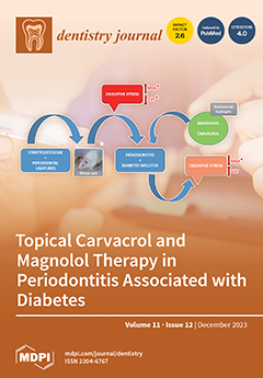
The bidirectional relationship between periodontitis and diabetes has raised the interest of researchers worldwide in improving the management of these diseases in terms of prophylaxis and treatment. Since oxidative stress has been implicated in the pathology of both periodontitis and diabetes, natural antioxidant extracts are considered and studied as potential beneficial agents. In the present study, periodontal hydrogels containing carvacrol and magnolol, administered single or combined, were tested in periodontitis associated with diabetes in Wistar rats. The combined carvacrol and magnolol treatment had the most important effect in reducing oxidative stress, followed by the administration of magnolol alone.View this paper
  • Issues are regarded as officially published after their release is announced to the table of contents alert mailing list.
  • You may sign up for e-mail alerts to receive table of contents of newly released issues.
  • PDF is the official format for papers published in both, html and pdf forms. To view the papers in pdf format, click on the "PDF Full-text" link, and use the free Adobe Reader to open them.
Order results
Result details
Section
Select all
Export citation of selected articles as:
10 pages, 247 KiB  
Article
Profile of Orthodontic Use across Demographics
by Man Hung, Golnoush Zakeri, Sharon Su and Amir Mohajeri
Dent. J. 2023, 11(12), 291; https://doi.org/10.3390/dj11120291 - 15 Dec 2023
Viewed by 2337
Abstract
Objectives: Population-based studies have focused on patients’ rendering of orthodontic treatment due to their malocclusion and medical needs. However, there is a scarcity of data from national sources on the prevalence of orthodontic visits and associated patient characteristics, as well as their effect [...] Read more.
Objectives: Population-based studies have focused on patients’ rendering of orthodontic treatment due to their malocclusion and medical needs. However, there is a scarcity of data from national sources on the prevalence of orthodontic visits and associated patient characteristics, as well as their effect on dental health. This study evaluated the demographic differences of orthodontic patients and examined the association between orthodontics use and risk of periodontal disease and oral surgical intervention. Methods: This study used data collected from the 2019 Medical Expenditure Panel Survey. Descriptive statistics were used to evaluate socio-demographics and covariates by the use of orthodontics. Chi-square tests were used to assess demographic differences among respondents who stated whether or not they used orthodontics. Logistic regression models were then used to examine the association of orthodontics and oral health outcomes. Results: The sample size was 12,422, of which 491 respondents indicated a usage of orthodontics. There were significant differences among demographic variables (p < 0.05) that included 61.1% females, 60.8% Whites, 67.6% participants under the age of 18 years old, and a family total income of $100,000 or more (52.7%). After controlling for socio-demographics and covariates, individuals who used orthodontics were less likely to have periodontal disease (AOR = 0.623, 95% CI = 0.610 to 0.637) and tooth extractions (AOR = 0.071, 95% CI = 0.070 to 0.073) than those who did not. Conclusions: Our findings indicate orthodontics usage was higher in females, younger patients, and Whites, highlighting the possible demographic disparities in orthodontics use. Additionally, those who used orthodontics were less likely to have oral health issues. Full article
13 pages, 8983 KiB  
Article
The Effect of Laser Irradiation to Surfaces of Computer-Aided Design/Computer-Aided Fabrication Resin Blocks Coated with a Silane Coupling Agent on Bond Strength between the Resin Blocks and Composite Resin
by Hiroshi Ohno, Masaya Suzuki and Koichi Shinkai
Dent. J. 2023, 11(12), 290; https://doi.org/10.3390/dj11120290 - 13 Dec 2023
Viewed by 1364
Abstract
The aim of this study was to investigate the effect of laser irradiation to computer-aided design/computer-aided fabrication (CAD/CAM) resin blocks coated with a silane coupling agent on the bond strength between resin blocks and composite resin. The CAD/CAM resin blocks used in this [...] Read more.
The aim of this study was to investigate the effect of laser irradiation to computer-aided design/computer-aided fabrication (CAD/CAM) resin blocks coated with a silane coupling agent on the bond strength between resin blocks and composite resin. The CAD/CAM resin blocks used in this study were Cerasmart 300 (GC) and Vita Enamic (Vita); they were cut into plates and then subjected to a series of treatments. After processing with a silane coupling agent, treatment with a semiconductor laser was performed at 3.0, 5.0, and 7.0 W, followed by bonding procedures. The control group included those exposed to silane and bonded without laser application. After bonding, a mold with a simulated cavity was formed on the specimen and filled with flowable composite resin, and they were stored for 24 h or stressed by thermal cycling for subsequent testing that assessed the shear bond strength (n = 10). The results revealed that the bond strength was significantly enhanced by laser irradiation after applying a silane coupling agent (p < 0.03), whereas significant increase was not detected between the materials (p > 0.05). Particularly, 7 W laser irradiation had a significant increase on the bond strength between the composite resin and Cerasmart block after thermal cycling (p = 0.009). The SBS of the composite resin to CAD/CAM resin blocks was significantly enhanced by laser irradiation after silane coupling agent application. Full article
Show Figures

Figure 1

11 pages, 870 KiB  
Article
Prosthetic Oral Rehabilitation with CAD/CAM Suprastructures in Patients with Severe Tissue Deficits: A Case Series
by Marisa Laurila, Pilvi Mäntynen, Jari Mauno and Juho Suojanen
Dent. J. 2023, 11(12), 289; https://doi.org/10.3390/dj11120289 - 13 Dec 2023
Viewed by 1586
Abstract
This article presents the outcomes of prosthetic oral rehabilitation using CAD/CAM telescopic bar overdentures in patients with oral cancer (n = 3), severe facial trauma (n = 2), or various syndromes (n = 1), all suffering from severe tissue deficits [...] Read more.
This article presents the outcomes of prosthetic oral rehabilitation using CAD/CAM telescopic bar overdentures in patients with oral cancer (n = 3), severe facial trauma (n = 2), or various syndromes (n = 1), all suffering from severe tissue deficits and requiring complex and comprehensive oral rehabilitation. The aim was to assess the durability and functionality of implant-retained prosthetic structures, ensuring easy oral hygiene and minimizing specialized follow-up needs. The data for this study were sourced from a retrospective cohort at Helsinki University Hospital. The prosthetic reconstruction encompassed the Atlantis 2in1 and the Createch removable telescopic systems. Thus, 40 implants were placed (4 to 7 per patient), with prosthetic structures in the maxilla (n = 4 patients), in the mandible (n = 1), and in both jaws (n = 1). Two patients experienced no complications, two patients had part of the acrylic resin break, and one patient experienced loosening of the bar structure. All complications associated with prosthetic structures were successfully managed, and none of the implants were lost. The follow-up time ranged from 7 to 126 months. This rehabilitation is proved to be an effective solution for patients with complex oral conditions, facilitating both functional restoration and ease of maintenance. These findings underscore the importance of individualized treatment approaches in cases of tissue deficits. Full article
(This article belongs to the Special Issue Advances in Oral Implant Health: Volume II)
Show Figures

Graphical abstract

10 pages, 730 KiB  
Article
Accuracy of Age Estimation Using Three Dental Age Estimation Methods in a Young, Large, and Multiethnic Patient Sample
by Claire Willmann, Gabriel Fernandez De Grado, Céline Kolb, Jean-Sébastien Raul, Anne-Marie Musset, Catherine-Isabelle Gros and Damien Offner
Dent. J. 2023, 11(12), 288; https://doi.org/10.3390/dj11120288 - 13 Dec 2023
Cited by 1 | Viewed by 1480
Abstract
European countries have become host countries for migrants and unaccompanied minors. However, many migrants arrive without identity documents. Many methods exist to estimate age; among them, several methods using dental age have been proposed. Our objective was to evaluate the accuracy of biological [...] Read more.
European countries have become host countries for migrants and unaccompanied minors. However, many migrants arrive without identity documents. Many methods exist to estimate age; among them, several methods using dental age have been proposed. Our objective was to evaluate the accuracy of biological age determination in a multiethnic sample using dental age estimated using three methods: Nolla, Demirjian, and the London Atlas. Orthopantomograms collected for 324 patients of various ethnicities aged from 4 to 20 years old were included. Then, for each orthopantomogram, a blind trained examiner used the three methods of age estimation. For each method, the estimated mean age was greater than the real mean age (p < 0.0001). The accuracy after 18 years old with a 1-year margin was under 50%. Demirjian’s method gave a less accurate estimated age than Nolla’s method (p < 0.0001) or the London Atlas (p < 0.001). The most accurate methods were those of Nolla and the London Atlas, with average absolute deviations of 1.3 and 1.2 years, respectively. Demirjian’s method was much less accurate, with a deviation of around 2 years. The evaluated methods are unable to provide reliable information to determine if an individual is a minor. Full article
Show Figures

Graphical abstract

20 pages, 2755 KiB  
Article
Effect of Commercial Children’s Mouthrinses and Toothpastes on the Viability of Neonatal Human Melanocytes: An In Vitro Study
by Shilpi Goenka and Hsi-Ming Lee
Dent. J. 2023, 11(12), 287; https://doi.org/10.3390/dj11120287 - 12 Dec 2023
Cited by 1 | Viewed by 1604
Abstract
In this study, we examined the cytotoxic effects of six commercial children’s mouthrinses (designated as #1, #2, #3, #4, #5, and #6) and four commercial children’s toothpastes (designated as #1, #2, #3, and #4) on primary human neonatal melanocytes that were used as [...] Read more.
In this study, we examined the cytotoxic effects of six commercial children’s mouthrinses (designated as #1, #2, #3, #4, #5, and #6) and four commercial children’s toothpastes (designated as #1, #2, #3, and #4) on primary human neonatal melanocytes that were used as a representative model for oral melanocytes. Mouthrinses diluted directly with culture medium (1:2, 1:5, 1:10, 1:100, and 1:1000) were added to monolayers of melanocytes for 2 min, followed by 24 h recovery, after which MTS cytotoxicity assay was conducted. The extracts of each toothpaste were prepared (50% w/v), diluted in culture medium (1:2, 1:5, 1:10, 1:50, 1:100, and 1:1000), and added to cell monolayers for 2 min (standard brushing time), followed by an analysis of cell viability after 24 h. Results showed that all mouthrinses except mouthrinse #4 showed significantly greater loss of cell viability, ascribed to cetylpyridinium chloride (CPC) that induced significant cytotoxicity to melanocytes (IC50 = 54.33 µM). In the case of toothpastes, the examination of cellular morphology showed that a 2 min exposure to all toothpaste extracts induced a concentration-dependent decline in cell viability, pronounced in toothpaste containing sodium lauryl sulfate (SLS) detergent. Further results suggested SLS to be the critical driver of cytotoxicity (IC50 = 317.73 µM). It is noteworthy that toothpaste #1 exhibited much lower levels of cytotoxicity compared to the other three toothpastes containing SLS. Taken together, these findings suggest that the melanocytotoxicity of children’s mouthrinse (#4) and toothpaste (#1) is comparatively low. To the best of our knowledge, this is the first study to examine the impact of children’s toothpastes and mouthrinses on neonatal primary human melanocytes. Future studies to investigate these findings in a realistic scenario replicating oral cavity conditions of the presence of microbiota, pellicle layer and saliva, and other cell types are warranted. Full article
(This article belongs to the Special Issue Oral Health Care in Paediatric Dentistry Volume 2)
Show Figures

Graphical abstract

13 pages, 4055 KiB  
Article
2D and 3D Erosion Landscape Analysis of Endodontic-Treated Teeth Using EDTA and HEDP as Chelating Agents: A High-Resolution Micro-Computed Tomographic Study
by Parichehr Zarean, Michael Göllner, Paridokht Zarean and Klaus W. Neuhaus
Dent. J. 2023, 11(12), 286; https://doi.org/10.3390/dj11120286 - 12 Dec 2023
Viewed by 1464
Abstract
This study aimed to assess the amount of erosion during activated endodontic irrigation with either HEDP or EDTA via high-resolution micro-computed tomography. Two root canals of twenty premolars were prepared with ProTaper Next and irrigated with sodium hypochlorite. Palatal canals, which served as [...] Read more.
This study aimed to assess the amount of erosion during activated endodontic irrigation with either HEDP or EDTA via high-resolution micro-computed tomography. Two root canals of twenty premolars were prepared with ProTaper Next and irrigated with sodium hypochlorite. Palatal canals, which served as control groups, were sealed, while buccal canals were further irrigated with either EDTA (n = 10) or HEDP (n = 10), which served as test groups. Micro-CT was performed to measure erosion depth. For 2D and 3D measurements, non-parametric repeated ANOVA measurements and post hoc tests were performed. 2D analysis showed highly significant differences between the case groups at each position of the root (p ≤ 0.01). The cervical and apical positions showed significant differences in the EDTA group (p = 0.03). The 3D analysis also showed significant differences between both chelating agents (p < 0.01) and the case and control groups (p = 0.01). The mean erosion depths in the cervical, middle, and apical thirds of the EDTA group were 45.75, 41.79, and 32.25 µm, and for the HEDP group were 20.25, 16.40, and 15.96 µm, respectively. HEDP seems to have a significantly less erosive effect. Different irrigation protocols with harsher conditions, as might be the case during endodontic retreatment, could be assessed with micro-CT. Full article
Show Figures

Figure 1

13 pages, 472 KiB  
Review
Graphene-Based Coatings for Surface Modification and Their Applications in Fixed Orthodontics: A Scoping Review
by Arturo Garrocho-Rangel, Juan Carlos Flores-Arriaga, Cristina Zamora-Soberón, Alan Martínez-Zumarán, Socorro Ruiz-Rodríguez and Amaury Pozos-Guillén
Dent. J. 2023, 11(12), 285; https://doi.org/10.3390/dj11120285 - 11 Dec 2023
Viewed by 1480
Abstract
Background: Surface coating technology can assist fixed appliances by reducing friction, improving antibacterial characteristics, and increasing corrosion resistance. The application of functional coatings composed of graphene onto the surfaces of orthodontic brackets and archwires has been shown to enhance their mechanical qualities. The [...] Read more.
Background: Surface coating technology can assist fixed appliances by reducing friction, improving antibacterial characteristics, and increasing corrosion resistance. The application of functional coatings composed of graphene onto the surfaces of orthodontic brackets and archwires has been shown to enhance their mechanical qualities. The objective of the current study was to carry out a scoping analysis of published recent evidence on the utilization of graphene as a covering material in metallic orthodontic accessories, such as brackets and archwires; Methods: A scoping review was undertaken following the PRISMA-ScR guidelines. PubMed, Embase, the Cochrane Library, Dentistry and Oral Science Source, and Google Scholar were searched between 2003 and 2023; Results: In total, 38 potential references were detected, from which 10 were selected for this review. These articles addressed the benefits of the application of graphene-oxide functional coatings onto the surface of archwires and brackets during fixed orthodontic treatment. Orthodontic graphene-oxide-based coatings provide improved surface characteristics (e.g., reduced friction and anticorrosive effects), antibacterial capabilities, and biocompatibility. These characteristics can increase the effectiveness of orthodontic therapy, improve patient comfort, and lower the likelihood of problems; Conclusion: Orthodontists should be aware of and comprehend the prerequisites for using graphene-oxide-coated archwires and brackets to fulfill needs throughout their clinical practice. Full article
(This article belongs to the Special Issue Current Research Topics in Orthodontics)
Show Figures

Graphical abstract

17 pages, 2631 KiB  
Article
Assessing the Antioxidant Benefits of Topical Carvacrol and Magnolol Periodontal Hydrogel Therapy in Periodontitis Associated with Diabetes in Wistar Rats
by Georgiana Ioana Potra Cicalău, Gabriela Ciavoi, Ioana Scrobotă, Andreea Olivia Marcu, Ioana Romanul, Eleonora Marian, Laura Grațiela Vicaș and Mariana Ganea
Dent. J. 2023, 11(12), 284; https://doi.org/10.3390/dj11120284 - 8 Dec 2023
Viewed by 1593
Abstract
It is well recognized that oxidative stress contributes to chronic stress-induced cytotoxicity, which is a major factor in the progression of many diseases, including periodontitis and diabetes. Formulas based on natural extracts with antioxidant properties are alternative treatment perspectives in the management of [...] Read more.
It is well recognized that oxidative stress contributes to chronic stress-induced cytotoxicity, which is a major factor in the progression of many diseases, including periodontitis and diabetes. Formulas based on natural extracts with antioxidant properties are alternative treatment perspectives in the management of such diseases. The aim of our study was to assess how carvacrol and magnolol influence periodontitis associated with diabetes in Wistar rats. Ninety Wistar rats were distributed in nine groups: I—control group; II—diabetes group (D); III—periodontitis group (P); IV—periodontitis and diabetes group (PD); V—periodontitis and diabetes with vehicle alone (PDV); VI—periodontitis and diabetes treated with carvacrol (PDC); VII—periodontitis and diabetes treated with magnolol (PDM); VIII—periodontitis and diabetes treated with carvacrol and magnolol (PDCM); IX—healthy group with vehicle alone (CV). Blood malondialdehyde (MDA) levels and catalase activity levels (CAT) were measured as indicators of oxidative stress and antioxidant capacity, respectively. Where diabetes and periodontitis were induced, MDA was augmented and CAT was depleted significantly. Whether given alone (PDM) or in combination with carvacrol (PDCM), magnolol significantly decreased MDA. Between the PDM group and the PDCM group, there were no notable differences. In Wistar rats with periodontitis related to diabetes, topical use of hydrogels containing magnolol, either alone or in combination with carvacrol, may reduce oxidative stress. Full article
(This article belongs to the Special Issue Periodontal Health: Disease Prevention and Treatment)
Show Figures

Figure 1

13 pages, 9288 KiB  
Article
The Effects of Antimicrobial Photodynamic Therapy Used to Sterilize Carious Dentin on Rat Dental Pulp Tissue
by Tenyu Takahashi, Fumiaki Sato and Koichi Shinkai
Dent. J. 2023, 11(12), 283; https://doi.org/10.3390/dj11120283 - 6 Dec 2023
Viewed by 1468
Abstract
Antimicrobial photodynamic therapy (aPDT) used to sterilize carious dentin may irritate pulp tissues because of tissue-penetrating laser and singlet oxygen generation. This study aimed to assess the effects of aPDT on rat pulp tissues. A cavity formed in a rat maxillary first molar [...] Read more.
Antimicrobial photodynamic therapy (aPDT) used to sterilize carious dentin may irritate pulp tissues because of tissue-penetrating laser and singlet oxygen generation. This study aimed to assess the effects of aPDT on rat pulp tissues. A cavity formed in a rat maxillary first molar was treated with aPDT. The combined photosensitizer and laser irradiation conditions in the aPDT groups were as follows: methylene blue and 100 mW for 60 s, brilliant blue (BB) and 100 mW for 60 s, BB and 50 mW for 120 s, and BB and 200 mW for 30 s. Each cavity was treated with an all-in-one adhesive and filled with flowable resin. aPDT was not applied for the control. In each group, the rats were sacrificed on postoperative days 1 and 14, and thin sections of the treated teeth were prepared. Pulp tissue disorganization (PTD), inflammatory cell infiltration (ICI), and tertiary dentin formation (TDF) were evaluated. At 1-day evaluation, there were significant differences between the aPDT group and controls with respect to PTD and ICI (p < 0.01); 14 days later, almost all specimens showed tertiary dentin formation. The application of aPDT caused reversible damage to the rat pulp, while in the long term, healing occurred with the formation of tertiary dentin. Full article
Show Figures

Figure 1

12 pages, 257 KiB  
Article
Comparison of the Knowledge, Attitudes, and Practices Regarding Silver Diamine Fluoride (SDF) between Japanese Dental Students with Experience Using SDF and Those with No Experience Using SDF: A Cross-Sectional Study
by Yuko Fujita
Dent. J. 2023, 11(12), 282; https://doi.org/10.3390/dj11120282 - 5 Dec 2023
Viewed by 1415
Abstract
Background: The aim of this study was to evaluate the differences in the knowledge and attitude regarding silver diamine fluoride (SDF) between two groups, differentiated by whether they had experience in SDF use, of dental students and clinical trainee dentists in Japan. Methods: [...] Read more.
Background: The aim of this study was to evaluate the differences in the knowledge and attitude regarding silver diamine fluoride (SDF) between two groups, differentiated by whether they had experience in SDF use, of dental students and clinical trainee dentists in Japan. Methods: A survey was designed consisting of three dental classes (fourth, fifth, and sixth years) and clinical trainees at Kyushu Dental University. A survey was designed consisting of 32 questions about the knowledge, attitudes, esthetic acceptability, and potential barriers regarding the use of SDF. Results: A total of 286 surveys (response rate of 85.4%) were collected. Among all respondents, 21.7% had experience with SDF use in their clinical practice. Regarding the knowledge score for SDF (0 to 12 points), in the respondents with no experience of using SDF, the mean score was 3.06, and that of respondents with experience of using SDF was 1.66, which was a significant difference (p < 0.001). The mean esthetic acceptability score for SDF use (−8 to 8 points) of the clinical trainees was −1.00 and that of the fourth-year students was 0.74, which was a significant difference (p < 0.05). Conclusions: the results indicate that dental students and clinical trainees need to increase their clinical experience with SDF. Full article
18 pages, 3875 KiB  
Review
Current Perspectives on Paradental Cyst: A Literature Review
by Pei Li, Yifan Zhao, Yuehua You, Ling Lin, Dongsheng Yu and Wei Zhao
Dent. J. 2023, 11(12), 281; https://doi.org/10.3390/dj11120281 - 5 Dec 2023
Viewed by 1971
Abstract
Paradental cyst (PC) is an uncommon type of odontogenic cyst of inflammatory origin, which develops near the cervical margin of the outside of the root of a vital tooth. The category of paradental cyst includes the buccal bifurcation cyst, which is found in [...] Read more.
Paradental cyst (PC) is an uncommon type of odontogenic cyst of inflammatory origin, which develops near the cervical margin of the outside of the root of a vital tooth. The category of paradental cyst includes the buccal bifurcation cyst, which is found in the buccal area adjacent to the mandibular first or second molars in children. A conclusive diagnosis of a PC needs to correlate the surgical, radiographic, and histologic findings. When strict diagnosis is neglected, they can be easily misdiagnosed and mistreated. PCs associated with mandibular first and second molars and those associated with the mandibular third molar may have slightly different clinical manifestations but have almost completely different treatment principles due to the distinction in location. For the third molars, removal of both the tooth and the cyst is preferred. However, when the first or second molars are affected, it may be advisable to perform enucleation of the lesion while preserving the associated tooth. There are also more conservative methods to retain vital permanent teeth within the mandibular arch. Additionally, the cyst wall primarily consisted of granulation tissue firmly attached to the periodontal ligament space. The exact origin of these cysts was a subject of ongoing debate, but they were believed to primarily arise from either the reduced enamel epithelium or the inflammatory proliferation of junctional/sulcular epithelium, which originate from the superficial mucosa during tooth eruption. The aim of the present review was to update information on clinical manifestations, diagnosis and treatment strategies of cysts and discuss their pathogenic mechanisms. Raising familiarity with the distinctive features is beneficial for accurately diagnosing these lesions and effectively caring for the patients. Full article
Show Figures

Graphical abstract

21 pages, 659 KiB  
Review
Polydeoxyribonucleotides Pre-Clinical Findings in Bone Healing: A Scoping Review
by Mattia Manfredini, Pier Paolo Poli, Mario Beretta, Matteo Pellegrini, Federica Eugenia Salina and Carlo Maiorana
Dent. J. 2023, 11(12), 280; https://doi.org/10.3390/dj11120280 - 4 Dec 2023
Viewed by 1687
Abstract
Aim: Polydeoxyribonucleotide (PDRN) is a chain-like polymer derived from DNA. Recent in vitro and animal studies have showcased the beneficial impacts of PDRN on the process of bone mending, whether used on its own or in conjunction with other substances that aid in [...] Read more.
Aim: Polydeoxyribonucleotide (PDRN) is a chain-like polymer derived from DNA. Recent in vitro and animal studies have showcased the beneficial impacts of PDRN on the process of bone mending, whether used on its own or in conjunction with other substances that aid in regeneration. This scoping review aims to synthesize the current understanding of how PDRNs influence bone healing. Materials and Methods: The studies included in the screening procedure were randomized controlled clinical trials (RCTs), both retrospective and prospective case–control studies, as well as in vitro and in vivo investigations. Articles were sourced from PubMed (MEDLINE), Scopus, EMBASE, Web of Science, and Google Scholar electronic databases using the following MeSH terms: (polydeoxyribonucleotide) and (bone) and (regeneration). Results: Initially, 228 articles were identified. Following the review process, a total of eight studies were ultimately examined. Among these, two were confined to laboratory studies, five were conducted on living organisms, and one encompassed both evaluations on living organisms and in vitro assessments. A descriptive qualitative approach was employed to present the data extracted from the studies that were included. Conclusions: PDRN has the potential to enhance the process of bone healing and the quantity of newly generated bone when combined with grafting materials. Future clinical studies are warranted to ascertain the appropriate clinical application of PDRN based on the dosage under consideration. Full article
(This article belongs to the Special Issue Bone Augmentation in Dentistry)
Show Figures

Figure 1

21 pages, 1137 KiB  
Systematic Review
Oligodontia in the Clinical Spectrum of Syndromes: A Systematic Review
by Natália Lopes Castilho, Kêmelly Karolliny Moreira Resende, Juliana Amorim dos Santos, Renato Assis Machado, Ricardo D. Coletta, Eliete Neves Silva Guerra, Ana Carolina Acevedo and Hercílio Martelli-Junior
Dent. J. 2023, 11(12), 279; https://doi.org/10.3390/dj11120279 - 4 Dec 2023
Viewed by 1945
Abstract
The aim of this systematic review was to describe the clinical and genetic features of syndromes showing oligodontia as a sign. The review was performed according to the PRISMA 2020 checklist guidelines, and the search was conducted using PubMed, Scopus, Lilacs, Web of [...] Read more.
The aim of this systematic review was to describe the clinical and genetic features of syndromes showing oligodontia as a sign. The review was performed according to the PRISMA 2020 checklist guidelines, and the search was conducted using PubMed, Scopus, Lilacs, Web of science, Livivo, and EMBASE and supplemented by a gray literature search on Google Scholar and ProQuest, applying key terms relevant to the research questions. The systematic review identified 47 types of syndromes in 83 studies, and the most common was hypohidrotic ectodermal dysplasia, which was reported in 24 patients in 22 studies. Other common syndromes that reported oligodontia included Axenfeld–Rieger syndrome, Witkop’s syndrome, Ellis–van Creveld syndrome, blepharocheilodontic syndrome, and oculofaciocardiodental syndrome. The X-linked mode of inheritance was the most reported (n = 13 studies), followed by the autosomal dominant (n = 13 studies). The review describes the main syndromes that may have oligodontia as a clinical sign and reinforces the need for orodental–facial examining for adequate diagnosis and treatment of the affected patients. Molecular analysis in order to better understand the occurrence of oligodontia is imperative. Full article
(This article belongs to the Special Issue Feature Review Papers in Dentistry)
Show Figures

Figure 1

14 pages, 20532 KiB  
Case Report
Biomechanically and Periodontally-Based Orthodontic Treatment of a Patient with Upper Canine Affected by External Cervical Resorption (ECR): A Case Report
by Marino Musilli, Morena Tina Iuorio, Emanuele Vaia, Enzo Vaia, Luca Ramaglia and Vincenzo D’Antò
Dent. J. 2023, 11(12), 278; https://doi.org/10.3390/dj11120278 - 30 Nov 2023
Cited by 1 | Viewed by 1895
Abstract
(1) Background: Orthodontic treatment may be a potential predisposing factor for ECR. The affected tooth goes to ankylosis, which could lead to a malocclusion. Although teeth severely affected by ECR (class IV Heithersay) are usually extracted, this case report aims to present the [...] Read more.
(1) Background: Orthodontic treatment may be a potential predisposing factor for ECR. The affected tooth goes to ankylosis, which could lead to a malocclusion. Although teeth severely affected by ECR (class IV Heithersay) are usually extracted, this case report aims to present the use of an ECR class IV upper canine, both as ankylosed to solve the malocclusion and the occlusal plane canting, as well as not ankylosed to correct its ridge defect with orthodontic extrusion. (2) Methods: A 14-year-old male, complaining of an ugly smile and a failed orthodontic attempt to recover an impacted canine, was referred to the orthodontic clinic. He was diagnosed with class II right subdivision, midline deviation, both upper and lower occlusal plane canting, and an upper left canine, previously impacted, showing ECR class IV. The treatment first included canting resolution with a cantilever and a spring, exploiting the anchorage offered by the ankylosed ECR canine. Then, a coronectomy, endodontic treatment, and orthodontic extrusion of that canine were performed to obtain the implant site development. (3) Results: Clinical and radiographic outcomes showed normocclusion and better bony conditions for safer implant placement in the aesthetic zone. (4) Conclusions: The high aesthetics and the periodontal and bony conditions obtained are probably not achievable by other therapeutic alternatives. Full article
(This article belongs to the Special Issue Orthodontics and New Technologies)
Show Figures

Figure 1

17 pages, 1090 KiB  
Systematic Review
Imaging Evaluation of Platelet-Rich Fibrin in Post-Exodontic Bone Regeneration: A Systematic Review
by Magdalena Molina-Barahona, Bolívar Delgado-Gaete, Denia Morales-Navarro, Joaquín Urbizo-Vélez and Renata Avecillas-Rodas
Dent. J. 2023, 11(12), 277; https://doi.org/10.3390/dj11120277 - 29 Nov 2023
Viewed by 1389
Abstract
Tooth extraction is the most common procedure in dental practice. However, in the long term, it may cause alveolar ridge atrophy. This systematic review aimed to evaluate the role of platelet-rich fibrin (PRF) in post-exodontic alveolar ridge preservation in terms of its effectiveness [...] Read more.
Tooth extraction is the most common procedure in dental practice. However, in the long term, it may cause alveolar ridge atrophy. This systematic review aimed to evaluate the role of platelet-rich fibrin (PRF) in post-exodontic alveolar ridge preservation in terms of its effectiveness in the regeneration of bone tissue as assessed by imaging and its efficacy compared to physiological bone healing. The study is presented in accordance with the preferred reporting items for systematic reviews and meta-analyses (PRISMA) guidelines. This systematic review was conducted using electronic databases such as PubMed, Scopus, Web of Science, and Science Direct. The gray literature search was conducted in the New York Academy of Medicine Grey Literature Report. All the studies in this systematic review were randomized controlled trials (RCTs). The risk of bias was performed according to the Cochrane Handbook for Systematic Reviews of Interventions 6.2 (RevMan 6.2). Considering the inclusion and exclusion criteria, we included 17 randomized clinical trials published up to 2022 investigating the efficacy of PRF in post-exodontic bone regeneration. Based on the results of clinical studies, it can be stated that despite not being statistically significant, PRF promotes neoformation and prevents bone loss between three and four months post-extraction. Full article
Show Figures

Figure 1

11 pages, 1711 KiB  
Article
Clinical Efficacy and Safety of Lidocaine Tape for Topical Anesthesia of the Oral Mucosa: A Preliminary Controlled Trial
by Ryouji Tani, Sachiko Yamasaki, Atsuko Hamada, Mirai Higaki, Yasuyuki Asada and Souichi Yanamoto
Dent. J. 2023, 11(12), 276; https://doi.org/10.3390/dj11120276 - 29 Nov 2023
Viewed by 1627
Abstract
Local anesthesia is administered to reduce pain-induced stress during dental treatment. However, local anesthetic injections are extremely painful; thus, methods to minimize this pain should be developed. Clinical studies on the pain-relieving effects of dental topical anesthetics have shown that few topical anesthetics [...] Read more.
Local anesthesia is administered to reduce pain-induced stress during dental treatment. However, local anesthetic injections are extremely painful; thus, methods to minimize this pain should be developed. Clinical studies on the pain-relieving effects of dental topical anesthetics have shown that few topical anesthetics provide fast and adequate pain relief without harming the oral mucosa. We examined the efficacy and safety of lidocaine tape, which has a potent topical anesthetic effect. Lidocaine tape was applied to the oral mucosa of 14 healthy participants, and its suppression effect was assessed by examining the pain intensity at the non-lidocaine tape-applied site using the visual analog evaluation scale and the verbal evaluation scale. Lidocaine tape application significantly reduced visual analog scale (VAS) scores during mucosal puncture compared to non-application (p < 0.01). Moreover, lidocaine tape application significantly reduced VAS scores during local anesthetic injection compared to non-application (p < 0.001). Adverse events were evaluated using the Common Terminology Criteria for Adverse Events, version 5.0. No adverse events attributed to the application of lidocaine tape were observed in any participant. The findings in this study suggest that the application of lidocaine tape before infiltration anesthesia can reduce patient distress. Full article
(This article belongs to the Section Oral and Maxillofacial Surgery)
Show Figures

Graphical abstract

12 pages, 1102 KiB  
Article
Visual Tooth Color Determination with Different Reference Scales as an Exercise in Dental Students’ Education
by Thomas U. Klinke, Wolfgang B. Hannak, Klaus Böning, Holger A. Jakstat and Elisabeth Prause
Dent. J. 2023, 11(12), 275; https://doi.org/10.3390/dj11120275 - 29 Nov 2023
Cited by 1 | Viewed by 1477
Abstract
Visual color determination (VCD) requires color competence and individual training. The aim of this study was to evaluate the deviations in students’ VCD with two different reference scales. The research hypothesis was that none of the color references would provide a better result. [...] Read more.
Visual color determination (VCD) requires color competence and individual training. The aim of this study was to evaluate the deviations in students’ VCD with two different reference scales. The research hypothesis was that none of the color references would provide a better result. Participants evaluated nine templates randomly using two reference scales (VITA-classical (VC) and 3D-Master-Toothguide (3DM_TG)). The color distance to the chosen color (ΔEab) was calculated in the CIELAB 2000. The sum’s changes in the parameters (LCh°) represented the target variable. Results were evaluated with non-parametric, rank-scaled methods, utilizing the median with a 25%-75% quartile. The significance level (α = 0.05) is determined using the Student’s t-test. The mean ± 95%CI (SD) was −1.27 ± −1.09 (3.18); the median ΔE00 was −1.49 (−1.97; 0.96) for dC3DM_TG. The determination with VC showed noticeable differences (dCVC), with a mean ΔE00 of 0.00 ± 0.00 (2.20) and a median ΔE00 of 0.00 (1.17; 1.71). The standard error was 0.19 for the dCVC and 0.27 for the dC3DM_TG. dC3DM_TG vs. dCVC showed significant differences at p < 0.001. The dental student’s VCD resulted in color deviations, regardless of the reference template used. The color deviations in hue and chroma were comparable, regardless of the reference scale. VCD’s early implementation in dental education is useful to avoid shade misjudgments and potentially expensive remakes of dentures. Full article
(This article belongs to the Special Issue New Trends in Dental Education and Dental Care)
Show Figures

Graphical abstract

20 pages, 1610 KiB  
Systematic Review
Advances in Preventive and Therapeutic Approaches for Dental Erosion: A Systematic Review
by Francesco Inchingolo, Gianna Dipalma, Daniela Azzollini, Irma Trilli, Vincenzo Carpentiere, Denisa Hazballa, Ioana Roxana Bordea, Andrea Palermo, Alessio Danilo Inchingolo and Angelo Michele Inchingolo
Dent. J. 2023, 11(12), 274; https://doi.org/10.3390/dj11120274 - 29 Nov 2023
Cited by 1 | Viewed by 2127
Abstract
This review discusses both preventive measures and clinically implemented therapy procedures that have been developed recently for the prevention and treatment of tooth erosion. Methods: The databases PubMed, Scopus, and Web of Science were used for a thorough search. Studies on the prevention [...] Read more.
This review discusses both preventive measures and clinically implemented therapy procedures that have been developed recently for the prevention and treatment of tooth erosion. Methods: The databases PubMed, Scopus, and Web of Science were used for a thorough search. Studies on the prevention and treatment of dental erosion that were conducted in English and used in vitro were among the inclusion criteria. Results: The search turned up 391 papers in total, with 34 of those publications matching the requirements for inclusion. Varnishes, toothpastes, and solutions containing fluoride and other substances were used as preventive measures. Conclusions: Dental erosion is a significant issue, and taking preventative steps is crucial to lessening the disease’s spread and its effects. Interventions based on fluoride seem to be successful at halting erosion and encouraging remineralization. To effectively address severe tooth erosion, therapeutic methods, including composite restorations, prosthetic crowns, and veneers, are available. Dental erosion causes aesthetic and functional issues that are best addressed with less invasive treatments like direct composite restorations. To improve and broaden the range of available treatments for this common dental issue, additional research and development are required. Full article
(This article belongs to the Special Issue Prevention of Dental Caries and Erosive Tooth Wear)
Show Figures

Figure 1

13 pages, 3402 KiB  
Article
Radiological Study on the Evolution of the Biomaterial’s Image (Shape, Density, Vertical Dimensions) after the Lateral Sinus Lift
by Dragos Epistatu, Octavian-Marius Dinca, Cristian Vladan, Cristina Padurariu, Iulian Damian and Sorin Hostiuc
Dent. J. 2023, 11(12), 273; https://doi.org/10.3390/dj11120273 - 29 Nov 2023
Viewed by 1935
Abstract
The aim of this study is to evaluate the long-term changes of sinus lift material. Materials and methods: We included a total number of 35 patients (20 men and 15 women), between 32 and 80 years old, evaluated on a timeframe of up [...] Read more.
The aim of this study is to evaluate the long-term changes of sinus lift material. Materials and methods: We included a total number of 35 patients (20 men and 15 women), between 32 and 80 years old, evaluated on a timeframe of up to 11.6 years. Diverse biomaterials were used (allograft, xenograft, alloplastic, combinations of them), with autologous bone in some cases. Results: The appearance of the top plane of the bone over time took a large dome shape (36% of cases), a linear shape (32% of cases), an irregular shape (23% of cases), or had micro domes above the implants (7%). No significant differences were found between the groups regarding age. The radiological density of the biomaterial tended to equalize that of the native bone. The final vertical dimensions seemed to be independent of the initial native bone height but seemed to be correlated with the amount of applied biomaterial. For the study group, the biomaterial contracted on average by 10% for the maximum height (H max) and 20% for the minimum height (H min), which can explain the tendency of the upper border of the biomaterial to curve. The annual H max contraction ranged from −0.09 to +0.18 with a mean value of 2.67% (SD = 0.04, CI: [0.011, 0.041]). The median value was 1.8%. The annual H min contraction ranged from −0.24 to +0.24, with a mean value of 4.33% (SD = 0.07, CI: [0.021, 0.065]). The median value was 3.59%. There were no statistically significant gender differences (Mann–Whitney U, p = 0.483, p = 0.642). The additional application of biomaterial together with the implants seemed to have a beneficial effect on the final vertical dimension of the bio-transformed material. Full article
(This article belongs to the Special Issue Oral Implantology and Bone Regeneration)
Show Figures

Figure 1

13 pages, 2033 KiB  
Article
A Novel Ergonomic Curette Design Reduces Dental Prophylaxis-Induced Muscle Work and Fatigue
by Kairong Lin, Cherie Wink, Ben Dolan, Kathryn Osann, Ali A. Habib, Jill Gehrig and Petra Wilder-Smith
Dent. J. 2023, 11(12), 272; https://doi.org/10.3390/dj11120272 - 28 Nov 2023
Cited by 1 | Viewed by 2190
Abstract
Background: To compare fatigue, comfort, and muscle work associated with the use of two periodontal curettes during scaling: one with a novel adaptive design, the other with a conventional non-adaptive design. Methods: Twelve hygienists scaled a typodont using two Universal Barnhart 5/6 curettes: [...] Read more.
Background: To compare fatigue, comfort, and muscle work associated with the use of two periodontal curettes during scaling: one with a novel adaptive design, the other with a conventional non-adaptive design. Methods: Twelve hygienists scaled a typodont using two Universal Barnhart 5/6 curettes: (1) a prototype featuring an adaptive silicone-covered handle (Curette A), and (2) a stainless-steel curette (Curette B). Surface Electromyography (sEMG) traced muscle work. Hand positions, fatigue, comfort, pinch, and grasp strength were recorded. Paired t-tests and a repeated measures ANOVA with covariates were tested for differences. The significance level was set at p < 0.05. Results: Curette A performed significantly better in all categories. Pinch and grasp strength and fatigue were significantly reduced post-instrumentation for Curette B. Curette A required significantly less (i) total muscle work and (ii) work in individual muscles. Comfort, correct grasp, and blade adaptation were significantly better using Curette A. Conclusions: A curette featuring a novel adaptive handle design demonstrated significantly improved ergonomic performance. Additional clinical studies are needed to solidify our understanding of the potential short- and long-term benefits of the novel curette handle design. Practical Implications: A novel adaptive curette handle design that enables the clinician to adapt the instrument across the index finger may reduce musculoskeletal burden and fatigue, as well as improve comfort during periodontal instrumentation. Full article
Show Figures

Figure 1

11 pages, 1042 KiB  
Systematic Review
Fluorescence and Near-Infrared Light for Detection of Secondary Caries: A Systematic Review
by Dimitrios Spagopoulos, Stavroula Michou, Sotiria Gizani, Eftychia Pappa and Christos Rahiotis
Dent. J. 2023, 11(12), 271; https://doi.org/10.3390/dj11120271 - 28 Nov 2023
Viewed by 1525
Abstract
Background: Early detection of secondary caries near dental restorations is essential to prevent further complications. This systematic review seeks to evaluate the sensitivity of fluorescence and near-infrared (NIR) imaging techniques for detecting secondary caries and to provide insight into their clinical utility. Methods: [...] Read more.
Background: Early detection of secondary caries near dental restorations is essential to prevent further complications. This systematic review seeks to evaluate the sensitivity of fluorescence and near-infrared (NIR) imaging techniques for detecting secondary caries and to provide insight into their clinical utility. Methods: A comprehensive search strategy was used to select studies from seven databases, emphasizing diagnostic accuracy studies of secondary caries detection using fluorescence and NIR imaging techniques. The Quality Assessment of Diagnostic Accuracy Studies-2 (QUADAS-2) instrument assessed bias risk and practicality. Two evaluators performed data extraction, screening, and quality assessment independently. Results: From 3110 initial recordings, nine studies were selected for full-text analysis. Wide variations in sensitivity (SE) and specificity (SP) values were reported across the studies. These studies exhibited variable SE and SP values, and the findings highlighted the importance of method selection based on clinical context. This systematic review underlines the potential for fluorescence and NIR imaging to detect secondary caries. However, results from different studies vary, indicating the need to consider additional variables such as restoration materials. Conclusions: Although these technologies exhibit potential for detecting caries, our research underscores the complex procedure of identifying secondary caries lesions. It is a continuous necessity for progress in dental diagnostics to promptly identify secondary caries lesions, particularly those in proximity to tooth-colored ones. Full article
(This article belongs to the Special Issue Feature Review Papers in Dentistry)
Show Figures

Graphical abstract

12 pages, 1701 KiB  
Article
A Potential Intracanal Medicament, 2-Hydroxyisocaproic Acid (HICA): Cytotoxicity, Genotoxicity, and Its Effect on SCAP Differentiation
by Cristine Smoczer, Yun K. Park, James B. Herrington, Mazin A. Askar, Sarah Plecha, Eric Krukonis and Susan B. Paurazas
Dent. J. 2023, 11(12), 270; https://doi.org/10.3390/dj11120270 - 27 Nov 2023
Viewed by 1635
Abstract
Intracanal medicaments with maximal antimicrobial efficacy and minimal damage to resident stem cells are essential for successful regenerative endodontic procedures. 2-Hydroxyisocaproic acid (HICA) could have the attributes of a potential intracanal medicament. This study evaluates its cytotoxicity, genotoxicity, and effects on the odontogenic [...] Read more.
Intracanal medicaments with maximal antimicrobial efficacy and minimal damage to resident stem cells are essential for successful regenerative endodontic procedures. 2-Hydroxyisocaproic acid (HICA) could have the attributes of a potential intracanal medicament. This study evaluates its cytotoxicity, genotoxicity, and effects on the odontogenic and osteogenic differentiation of the stem cells of the apical papilla (SCAP). Cytotoxicity and cell viability assays were performed on cells treated for 24, 48, and 72 h with varying concentrations of HICA and compared to the standard intracanal medicament, calcium hydroxide. The genotoxicity was assessed via immunofluorescence for two markers of DNA double-strand breaks: phosphorylated γH2AX and 53BP1. The SCAP differentiation was evaluated based on the alkaline phosphatase activity, Alizarin Red staining, and expression of odontogenic and osteogenic genes (DSPP1, BSP1, OCN, RUNX2) in the presence of selected HICA concentrations. HICA was not cytotoxic at concentrations up to 10 mg/mL, regardless of the exposure time, although it was cytostatic at all tested concentrations. HICA was not genotoxic at concentrations below 5 mg/mL. No difference in cytotoxicity or genotoxicity was found between HICA and calcium hydroxide at 1 mg/mL. HICA retained about 70% of the osteogenic differentiation potential at 1 mg/mL. Within the limitations of this in vitro study, we show that HICA at 1 mg/mL could be a potential intracanal medicament for REPs. Full article
Show Figures

Graphical abstract

13 pages, 1899 KiB  
Review
In Vitro Models Used in the Formation of Root Caries Lesions—A Review of the Literature
by Zaid Dohan, Lara T. Friedlander, Paul R. Cooper, Kai-Chun Li, Jithendra T. Ratnayake and May L. Mei
Dent. J. 2023, 11(12), 269; https://doi.org/10.3390/dj11120269 - 26 Nov 2023
Viewed by 1512
Abstract
The management of root caries remains a challenge for clinicians due to its unique anatomical location and structure. There is increasing interest in utilising artificial root caries lesions to develop new strategies for remineralisation. An ideal protocol has not yet been agreed upon. [...] Read more.
The management of root caries remains a challenge for clinicians due to its unique anatomical location and structure. There is increasing interest in utilising artificial root caries lesions to develop new strategies for remineralisation. An ideal protocol has not yet been agreed upon. The aim of this review is to provide a structured overview of previously reported in vitro root caries models. The literature was screened and mined for information mainly on substrate selection, model systems utilised, and variables used in the models. Human roots (60%) were the most frequently used substrates, followed by bovine roots (40%). Chemical models (69%) were the most frequently utilised model systems, followed by microbiological models (27%), to form root caries lesions. Acetate buffer solution (80%), pH 5.0 or above (40%), and a demineralisation time of five days (25%) were the common variables used in the chemical systems, while mono-species biofilm was most frequently used (73%) in microbiological models and Streptococcus mutans was the most common bacterial strain utilised in these models (80%). This review highlights the variability amongst the experimental approaches, discusses the advantages and limitations of these approaches, and emphasises that standardisation of experimental conditions along with sustained research will benefit root caries research. Full article
(This article belongs to the Special Issue Feature Review Papers in Dentistry)
Show Figures

Figure 1

13 pages, 1350 KiB  
Article
How Much Do We Know about Oral Cancer?—An Online Survey
by Iva Horvat, Božana Lončar-Brzak, Ana Andabak Rogulj, Livia Cigić, Sonja Pezelj Ribarić, Miroslav Sikora and Danica Vidović-Juras
Dent. J. 2023, 11(12), 268; https://doi.org/10.3390/dj11120268 - 24 Nov 2023
Viewed by 1239
Abstract
Introduction: Oral cancer (OC) is a disease with a high mortality rate due to its late recognition. Since the oral cavity is easily accessible for visual inspection, enabling early diagnosis, the education of healthcare workers about preventive oral examinations is critical. This research [...] Read more.
Introduction: Oral cancer (OC) is a disease with a high mortality rate due to its late recognition. Since the oral cavity is easily accessible for visual inspection, enabling early diagnosis, the education of healthcare workers about preventive oral examinations is critical. This research aimed to assess the level of participants’ OC knowledge, as well as to raise awareness about this diagnosis. Materials and methods: The research was conducted as an online survey among students of dental medicine, students of medicine, doctors of dental medicine and doctors of medicine. The questionnaire was designed solely for the purpose of this study and consisted of 29 questions. The first part of the questionnaire consisted of general questions about the participants, whereas the questions in the second part addressed their knowledge and attitudes towards OC. Results: The surveyed population comprised of 140 dental students, 105 medical students, 159 doctors of dental medicine and 100 medical doctors. The level of knowledge about OC among the participants is not yet satisfactory. The group of dental medicine students scored highest, while medical doctors showed the weakest knowledge. Conclusion: Additional education about OC for doctors of dental medicine and medical doctors is needed. This step will improve prevention and increase chances for early detection. Full article
(This article belongs to the Collection Feature Papers in Dental Education)
Show Figures

Figure 1

11 pages, 1147 KiB  
Article
Sugar Substitute Stevia Inhibits Biofilm Formation, Exopolysaccharide Production, and Downregulates the Expression of Streptococcal Genes Involved in Exopolysaccharide Synthesis
by Sara AlKanderi, Monerah AlFreeh, Radhika G. Bhardwaj and Maribasappa Karched
Dent. J. 2023, 11(12), 267; https://doi.org/10.3390/dj11120267 - 23 Nov 2023
Viewed by 1579
Abstract
Background: Acid production by sucrose fermentation disturbs the balance in dental plaque by lowering the oral pH. As a consequence of the profound effect of sucrose on caries initiation and progression, many studies have been directed towards finding non-cariogenic artificial sweeteners that can [...] Read more.
Background: Acid production by sucrose fermentation disturbs the balance in dental plaque by lowering the oral pH. As a consequence of the profound effect of sucrose on caries initiation and progression, many studies have been directed towards finding non-cariogenic artificial sweeteners that can be used as a substitute to sucrose. Existing literature shows that dietary sucrose upregulates the expression of biofilm associated genes involved in exopolysaccharide (EPS) production. Objective: In this study, we aimed to investigate the effect of the sugar substitute stevia on biofilm formation, EPS secretion, and streptococcal genes encoding glucan-binding proteins (Gbps) and glucosyltransferases (Gtfs), which are essential for the synthesis of EPS. Materials and Methods: Streptococcus mutans and Streptococcus gordonii were grown as biofilm cultures with or without stevia and sucrose. Biomass was quantified for biofilm and EPS production by crystal violet staining and the phenol–sulfuric acid method, respectively. Expression of gtfB and gbpB genes was studied by RT-PCR. Results: The quantities of biofilm were significantly lower when grown in the presence of stevia compared to sucrose in both species (p < 0.05). The proportion of EPS in the biofilm pellet decreased with increasing concentrations of stevia in both species but remained nearly unchanged with sucrose with respect to the control. In both streptococcal species, exposure of stevia decreased the expression of gtfB and gbpB genes compared to sucrose (p < 0.05). In comparison to the untreated control, the expression was decreased in the presence of stevia in both species, while it increased 2.5- to 4-fold in S. mutans and 1.5- to 2.5-fold in S. gordonii in the presence of sucrose. Conclusion: The ability of stevia to inhibit biofilm formation, reduce EPS production, and downregulate the expression of gtfB and gbpB genes in S. mutans and S. gordonii may have potential therapeutic applications in controlling dental plaques and caries. Full article
Show Figures

Figure 1

Previous Issue
Next Issue
Back to TopTop