


Abstract
Antimicrobial resistance is a major global health concern, intensified by the misuse of antibiotics and the lack of new effective treatments. Bacteria capable of forming biofilms exhibit increased resistance, making infections more difficult to treat. This study evaluated the essential oil from Croton pluriglandulosus leaves (OCp) for its antibacterial and antibiofilm properties. The essential oil, obtained by hydrodistillation and analyzed by GC-MS, contained eucalyptol (24.11%), spathulenol (16.90%), α-pinene (11.76%), and caryophyllene oxide (10.07%) as main constituents. Antibacterial activity was determined by Minimum Inhibitory Concentration (MIC) and Minimum Bactericidal Concentration (MBC), with inhibition observed only for Staphylococcus aureus (MIC 10 mg/mL; MBC 5 mg/mL). OCp reduced biofilm biomass and CFUs in most strains, particularly in S. aureus and Escherichia coli. Scanning Electron Microscopy (SEM) and Confocal Laser Scanning Microscopy (CLSM) showed membrane damage in treated cells. Overall, OCp displayed promising antibacterial and antibiofilm potential, representing the first report of such activity for this essential oil.
1. Introduction
The World Health Organization warns that antimicrobial resistance (AMR) poses a critical and escalating threat to global health systems [1]. This natural phenomenon occurs as microorganisms evolve in response to the selective pressure created by antibiotic use. A well-known example is penicillin resistance. After being introduced in 1941, strains of S. aureus resistant to penicillin were already identified by 1942 [2].
AMR is responsible for around 70,000 deaths annually worldwide. Alarmingly, it is predicted that this number could rise significantly, reaching 10 million deaths per year by 2050 [3]. In India, reports indicate that a child dies every 9 min from infections caused by resistant bacteria [2]. An estimated 1.27 million deaths in 2019 were linked directly to bacterial antimicrobial resistance. Among the leading pathogens, Staphylococcus aureus was responsible for over 100,000 deaths, with methicillin-resistant S. aureus (MRSA) identified as one of the major contributors to this global burden [4]. These figures underscore the growing threat of antimicrobial resistance to global health.
Today, around 80% of bacteria can form structures known as biofilms [5]. Biofilms consist of complex biological communities that may or may not be adhered to a surface and produce an extracellular polymeric substance (EPS) composed mainly of exopolysaccharides. These communities have three main mechanisms of drug resistance: physical tolerance, which involves the overproduction of extracellular matrix, hindering the action of antimicrobials; passive tolerance, associated with enzymes in the extracellular polymeric substance that inactivate drugs; and physiological tolerance, where the cells in the deeper layers of the biofilm become metabolically inactive, providing resistance to the drugs [6,7,8].
These microbial communities can trigger infectious processes by adhering to medical devices or patient tissues, making them difficult to treat, often requiring the removal of devices or resulting in chronic infections [9]. Due to the significant impact biofilms have on human health, research aimed at combating and preventing these microbial communities has gained increasing prominence, especially concerning certain species such as S. aureus, Staphylococcus epidermidis and Escherichia coli [10,11].
The increasing number of AMR drives the search for new antimicrobial agents to combat infectious processes caused by resistant microorganisms and their biofilms [12]. In this context, there is a growing interest in exploring the medicinal properties of plants that can offer therapeutic alternatives [13]. Over the centuries, natural products have always been associated with the treatment of various infectious diseases, offering therapeutic alternatives, such as essential oils [14]. Essential oils (EOs) are volatile substances that contain various bioactive components with insecticidal, and antimicrobial properties present in some plant species [15].
Within the group of plants that produce EOs, the Croton genus stands out for its economic value and the production of EOs rich in compounds such as monoterpenes, sesquiterpenes, and phenylpropanoids [16,17]. In Brazil, more than 350 species of Croton have been identified and cataloged, exhibiting a wide variety of components in their EOs. However, few studies have explored the antibacterial potential of Croton pluriglandulosus EO [18].
Despite the recognized antimicrobial potential of Croton species, reports on C. pluriglandulosus EO remain scarce, with no studies addressing its activity against clinically relevant biofilm-forming pathogens. Furthermore, no previous investigation has evaluated its performance across strains with distinct resistance backgrounds (e.g., MRSA, VISA, and methicillin-susceptible strains) or against both Gram-positive and Gram-negative bacteria.
In this context, the present study provides the first evaluation of the antibacterial potential of C. pluriglandulosus EO, encompassing its activity against planktonic bacteria, biofilm formation and mature biofilms, as well as its potential antibacterial mechanism. This approach generates novel evidence supporting the antimicrobial relevance of this species, particularly in the context of biofilm-associated and drug-resistant infections.
2. Materials and Methods
2.1. Collection of Vegetal Material
Fresh mature leaves of C. pluriglandulosus were collected in March 2024 in the Serra do Lima region, municipality of Patu, Rio Grande do Norte, Brazil (06°06′30″ S; 37°37′20″ W). Only fully expanded, non-senescent leaves (third to fifth nodes from the apex) were selected to ensure a consistent developmental stage. Leaves were obtained from healthy adult plants growing under similar environmental and soil conditions, with no visible signs of biotic or abiotic stress.
To reduce chemotypic variability and environmental bias, all samples were collected from plants growing under similar soil and light conditions, during the same time period in the morning (between 07:00 and 09:00). Such measures minimized environmental variability known to influence secondary metabolite production.
The plant material was taxonomically identified by Yuri Rossine de Lima Bezerra (UFPE) following the APG IV system [19], and a voucher specimen (MOSS 15058) was deposited at the Dárdano de Andrade Lima Herbarium (MOSS), Federal Rural Semiarid University (UFERSA).
2.2. Essential Oil Extraction
Leaves were hydrodistilled using a Clevenger-type apparatus as described by Oliveira et al. [20], with modifications. The fresh leaf mass was placed in a 5 L volumetric flask and 2 L of distilled water was added. After the extraction process, the oil was separated from the water and completely dehydrated through filtration and evaporation with anhydrous sodium sulfate and ether. After the extraction process, the oil was packed in amber glass bottles and stored at 4 °C for subsequent analysis.
2.3. Chemical Composition of Croton Pluriglandulosus Essential Oil
The chemical profile of the EO was determined by gas chromatography–mass spectrometry (GC–MS) using a Shimadzu QP-2010 system (Shimadzu Corporation, Kyoto, Japan). Analyses were carried out on a Hewlett-Packard 5971 system equipped with a DB-5 fused-silica capillary column (Milpitas, CA, USA). Helium served as the carrier gas (1 mL/min, split 1:1). The injector and detector were operated at 250 °C and 200 °C, respectively. The oven temperature was programmed in two steps, initially from 35 °C to 180 °C (4 °C/min) and subsequently up to 250 °C (10 °C/min). Compounds were identified by comparing mass spectra with the Wiley library and by evaluating their retention indices.
2.4. Microorganisms and Culture Conditions
The present study used standard strains obtained from the American Type Culture Collection (ATCC), namely Staphylococcus aureus ATCC 25923, Staphylococcus aureus ATCC 700698 (MRSA), Staphylococcus epidermidis ATCC 12228, Staphylococcus epidermidis ATCC 35984 (MRSE) and Escherichia coli ATCC 11303. In addition, the clinical isolate Staphylococcus aureus JKD 6008 (MRSA and VISA) was also used.
Strain selection was based on their clinical relevance as etiological agents of device-associated and hospital infections and on their known biofilm-forming capacity. MRSA, MRSE and the VISA strain were included due to their importance in antimicrobial resistance surveillance, whereas E. coli ATCC 11303 served as a Gram-negative reference strain. To compare different biofilm phenotypes, S. epidermidis ATCC 35984 (strong biofilm former) and ATCC 12228 (poor/non-biofilm former) were also included, enabling evaluation of OCp activity across distinct biofilm backgrounds.
The growth conditions for the bacterial strains followed the protocol of the Clinical and Laboratory Standards Institute [21] with modifications. All ATCC strains were subcultured on standard microbiological media to ensure purity and confirmed by morphology and Gram staining prior to use. The strains were grown in Petri dishes with Mueller Hinton Agar (MHA) (HiMedia Laboratories, Mumbai, India) medium and incubated at 37 °C for 24 h. Isolated colonies were inoculated into Mueller Hinton Broth (MHB) (HiMedia Laboratories, Mumbai, India) and incubated for 24 h. After, the strains were centrifuged at 8500× g for 5 min at 4 °C and resuspended in MHB. The bacterial suspension was adjusted to 1 × 10⁶ CFU/mL using calibration curves for each strain.
2.5. Minimum Inhibitory Concentration (MIC) and Minimum Bactericidal Concentration (MBC) Determination
The antibacterial activity of OCp was evaluated using broth microdilution assays in 96-well plates, following the CLSI guidelines [22] with modifications proposed by Vale et al. [23].
The Minimum Inhibitory Concentration (MIC) was determined by the broth microdilution method. OCp was first solubilized in 8% DMSO (stock solution) (Dinâmica Química Contemporânea Ltd., Indaiatuba, SP, Brazil), and after addition to the assay wells the final DMSO concentration was reduced to 4%. Serial dilutions ranged from 10 to 0.15625 mg/mL, and a bacterial suspension adjusted to 5 × 10⁵ CFU/mL was added to each well. Solvent controls containing 4% DMSO were included in all assays, and the plates were incubated at 37 °C for 24 h.
Each assay contained the following controls: (i) a growth control consisting of medium plus bacterial inoculum; (ii) a sterility control containing medium only; and (iii) a solvent control containing 4% DMSO. These controls ensured medium sterility, solvent compatibility and normal bacterial growth.
In accordance with CLSI criteria, bacterial growth was initially assessed by visual inspection of turbidity in the wells. Optical density at 600 nm was subsequently recorded using a microplate reader (SpectraMax i3; Molecular Devices, San Jose, CA, USA) to quantitatively confirm growth inhibition.
For determination of the Minimum Bactericidal Concentration (MBC), aliquots from wells showing no visible growth were plated onto MHA The lowest concentration yielding no colonies after 24 h was recorded as the MBC.
2.6. Evaluation of Antibiofilm Activity
2.6.1. Activity on Biofilm Formation
The biofilm formation assay was conducted on 96 flat-bottomed polystyrene plates and based on serial microdilution tests, crystal violet (CV) staining, and CFU counting, according to Stepanović et al. [24], with modifications by Vale et al. [23]. The procedure followed a similar setup as the MIC tests, except that the final bacterial concentration was 1 × 10⁶ CFU/mL. The media used for bacterial growth were Tryptone Soy Broth (TSB) and Tryptone Soy Agar (TSA) (HiMedia Laboratories, Mumbai, India).
For all antibiofilm assays, untreated biofilms served as negative controls (NG), and a solvent control containing 4% DMSO was included to exclude solvent-derived effects. These controls ensured medium sterility, solvent compatibility, and normal biofilm development under assay conditions.
2.6.2. Biofilm Biomass Quantification
The biomass of the biofilms was quantified using the crystal violet (CV) staining method. After incubation, the biofilms were washed thrice with PBS (10 mM phosphate; 150 mM NaCl; 2.7 mM KCl; pH 7.2–7.4) (Sigma-Aldrich, St. Louis, MO, USA) to remove the weakly adhered bacteria. The biofilms were then fixed with 200 µL of 99% methanol (Dinâmica Química Contemporânea Ltda., Indaiatuba, SP, Brazil) for 5 min. Afterward, the methanol was removed, and the biofilms were stained with 200 µL of 1% CV (Dinâmica Química Contemporânea Ltda., Indaiatuba, SP, Brazil) for 5 min. After the staining step, the wells were washed thrice with distilled water and the remaining dye was solubilized by adding 200 µL of 33% acetic acid (Dinâmica Química Contemporânea Ltda., Indaiatuba, SP, Brazil). The plates were read on a microplate reader (SpectraMax i3; Molecular Devices, San Jose, CA, USA) at an optical density of 590 nm.
2.6.3. Viable Cells Quantification
The number of viable cells was quantified by determining the Colony Forming Units (CFU). After incubation, the 96-well plates were washed twice with sterile distilled water to remove the weakly adhered cells. Then 200 µL of sterile distilled water was added to all the wells, and the plate was subjected to an ultrasonic bath using a Cristófoli/EQM-CF apparatus (Cristófoli Equipamentos de Biossegurança Ltd., Campo Mourão, PR, Brazil) for approximately 8 min. Subsequently, decimal dilutions were made of the samples, which were plated on TSA medium and incubated in an oven at 37 °C for 24 h. The number of CFU was expressed as Log10 CFU/mL.
2.6.4. Preformed Biofilms Assay
The preformed biofilm assays were conducted in 96-well polystyrene plates. Each well of these plates was filled with 200 µL of bacterial suspension at a concentration of 1 × 10⁶ CFU/mL and incubated at 37 °C for 24 h. Subsequently, the supernatant was removed, and the wells were washed twice with PBS (10 mM phosphate; 150 mM NaCl; 2.7 mM KCl; pH 7.2–7.4) and then 100 µL of TSB medium (negative control) and 100 µL of OCp previously solubilized in TSB with 4% DMSO, at concentrations ranging from 10 to 0.15625 mg/mL, were added. The plates were then incubated at 37 °C for 24 h. Subsequent analyses of the biofilms included the crystal violet staining method and quantification of the number of viable cells.
2.7. Scanning Electron Microscopy (SEM)
Scanning Electron Microscopy (SEM) analyses were carried out according to Vasconcelos et al. [25], with some adaptations. The bacterial strains used were S. aureus ATCC 700698 and E. coli ATCC 11303. The biofilms were grown on glass coverslips (Olen, K5-2020) into 24-well and treated with OCp at 10 and 5 mg/mL, or untreated (control). Biofilms were formed by inoculating each well with 1 × 10⁶ CFU/mL of the bacterial suspension, as previously described in Section 2.4. The plates were then incubated at 37 °C for 24 h. After incubation, the plates were washed twice with PBS to remove the weakly adhered cells, and the biofilms were dehydrated with ethanol (Dinâmica Química Contemporânea Ltda., Indaiatuba, SP, Brazil), following the steps of immersion in increasing concentrations of 70% for 10 min, 95% for 10 min and 100% for 20 min. The plates were then dried and mounted on aluminum stubs using double-sided carbon tape and coated with a 20 nm layer of gold by sputtering for 120 s using a Quorum Q150T ES instrument (Quorum Technologies, East Sussex, UK). The Biofilms were viewed using an scanning electron microscope (SEM) at 20 kV (Quanta 450 FEG, FEI, Waltham, MA, USA).
2.8. Confocal Scanning Laser Microscopy (CSLM)
Confocal Scanning Laser Microscopy (CSLM) analyses were evaluated according to Castelo Branco et al. [26], with modifications. The bacterial strains used were S. aureus ATCC 700698 and E. coli ATCC 11303. The biofilms were grown on glass coverslips (Olen, K5-2020) into 24-well and treated with OCp at 10 mg/mL, or untreated (control). For biofilm formation, each well was inoculated with 1 × 10⁶ CFU/mL of the bacterial suspension, according to Section 2.4. The plates were then incubated at 37 °C for 24 h. After incubation, the plates were washed twice with PBS and stained with the LIVE/DEAD fluorescence kit (Invitrogen, Carlsbad, CA, USA). The coverslips were washed with PBS to remove excess dye and analyzed using a Nikon C2 Confocal Microscope (Nikon, Tokyo, Japan). The analysis took place at two wavelengths: 488 and 561 nm.
2.9. Statistical Analysis
All tests were evaluated in triplicate with at least three independent replicates. Statistical analyses were carried out using the GraphPad Prism® (GraphPad Software, San Diego, CA, USA) application version 7.0 for Microsoft Windows. The data obtained underwent a comparison test, including analysis of variance (ANOVA) and Bonferroni post hoc. Values of p < 0.05 were considered statistically significant.
3. Results
3.1. Croton Pluriglandulosus Essential Oil Chemical Composition
The GC-MS analysis made it possible to identify 100% of the components present in the OCp. Among these components, the monoterpene eucalyptol stood out, representing 24.11% of the total, followed by the sesquiterpene spatulenol, with 16.90%, and the terpene α-Pinene, with 11.76% and Caryophyllene oxide, an oxygenated sesquiterpene, with 10.07%. These three compounds were the majority constituents of the EO, as shown in Table 1.
Table 1.
Chemical composition of the essential oil of C. pluriglandulosus Carn.-Torres & Riina.
3.2. Antibacterial Activity of Croton pluriglandulosus Essential Oil (OCp)
OCp showed antibacterial activity, with MIC and MBC values ranging from 10 to 5 mg/mL against S. aureus strain. However, no MIC or MBC values were observed for S. epidermidis and E. coli strain (Table 2).
Table 2.
Minimum inhibitory concentrations (MICs) and minimum bactericidal concentrations (MBCs) of C. pluriglandulosus essential oil against Gram-positive and Gram-negative bacteria.
3.3. Croton Pluriglandulosus Essential Oil Activity on Biofilm Formation
No significant differences were detected between solvent-treated and untreated biofilms. For graphical clarity, only the negative control is presented.
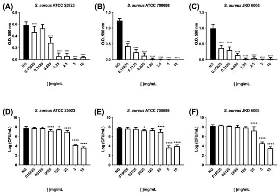
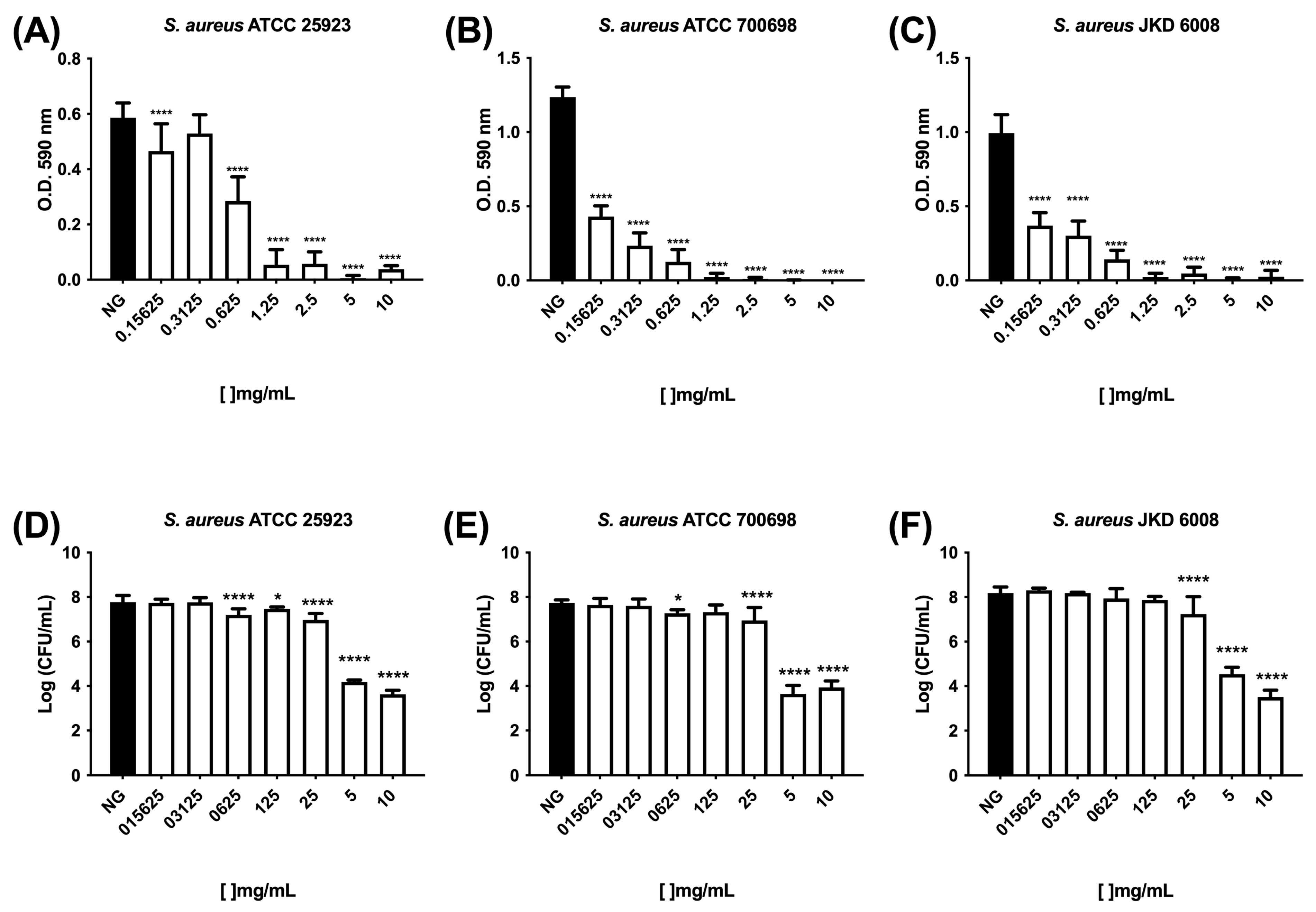
The effects of OCp on biofilm formation in three S. aureus strains were analyzed (Figure 1). The oil caused a significant reduction in biofilm biomass in all strains, with reduction percentages varying from 100% to 55%. Regarding the number of viable cells, all strains showed significant reductions, particularly at the three highest concentrations of OCp. The S. aureus ATCC 25923 strain treated with OCp showed a reduction of 4.0 and 3.6 logs at concentrations of 10 and 5 mg/mL, respectively, with reductions also observed at the lower concentrations. The S. aureus ATCC 700698 strain showed reductions of 3.8 and 4.0 logs at 10 and 5 mg/mL. For S. aureus JKD 6008, the reductions were 4.7 and 3.6 logs at 10 and 5 mg/mL.
Figure 1.
Effect of Croton pluriglandulosus essential oil on biofilm formation of Staphylococcus aureus strains. (A) S. aureus ATCC 25923 biomass; (B) S. aureus ATCC 700698 biomass; (C) S. aureus JKD 6008 biomass; (D) viable cells of S. aureus ATCC 25923 biofilm; (E) viable cells of S. aureus ATCC 700698 biofilm; (F) viable cells of S. aureus JKD 6008 biofilm. White bars indicate OCp-treated groups, and black bars indicate untreated controls. Data are presented as mean ± SD of three independent experiments performed in triplicate. Statistical significance was assessed by one-way ANOVA followed by Bonferroni post hoc test. Significance levels versus the respective control: * p < 0.05, **** p < 0.0001.
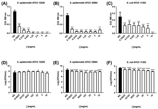
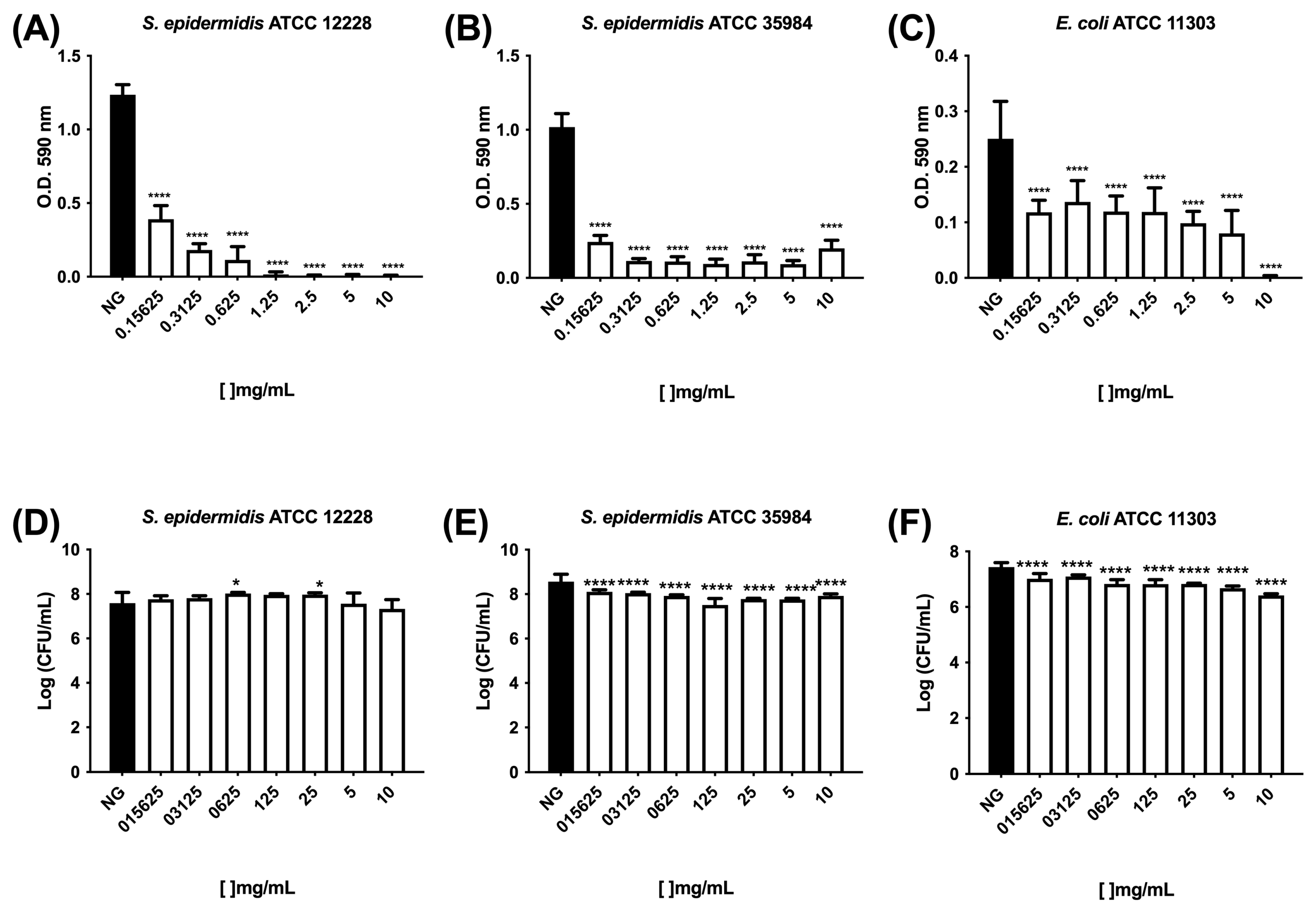
The biofilm analyses of the S. epidermidis and E. coli strains are shown in Figure 2. The results indicated a significant reduction in the total biomass of the biofilms at all OCp concentrations tested for all strains. For the S. epidermidis strains, biomass reductions ranged from 92% to 71%, while for E. coli ATCC 11303, the reductions ranged from 95% to 77%.
Figure 2.
Effect of Croton pluriglandulosus essential oil on biofilm formation of Staphylococcus epidermidis and Escherichia coli strains. (A) Biofilm biomass of S. epidermidis ATCC 12228; (B) biofilm biomass of S. epidermidis ATCC 35984; (C) biofilm biomass of E. coli ATCC 11303; (D) viable cells of S. epidermidis ATCC 12228 biofilm; (E) viable cells of S. epidermidis ATCC 35984 biofilm; (F) viable cells of E. coli ATCC 11303 biofilm. White bars indicate OCp-treated groups, and black bars indicate untreated controls. Data are presented as mean ± SD of three independent experiments performed in triplicate. Statistical significance was assessed by one-way ANOVA followed by Bonferroni post hoc test. Significance levels versus the respective control: * p < 0.05, **** p < 0.0001.
Regarding the number of viable cells in the biofilms, S. epidermidis ATCC 35984 and E. coli ATCC 11303 showed significant reductions at all tested concentrations, with decreases of up to 1 log (Figure 2E,F). For S. epidermidis ATCC 12228, significant reductions were observed at concentrations of 2.5 and 0.625 mg/mL (Figure 2D).
3.4. OCp Activity on Preformed Biofilms
No significant differences were detected between solvent-treated and untreated preformed biofilms.
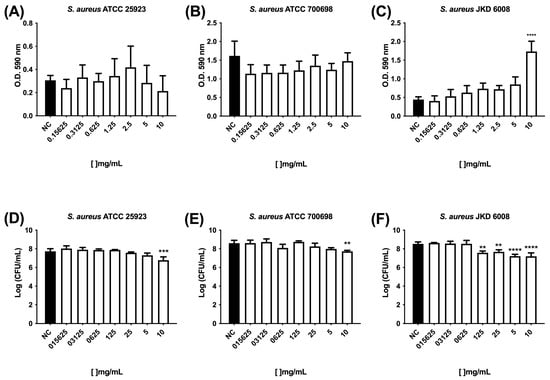
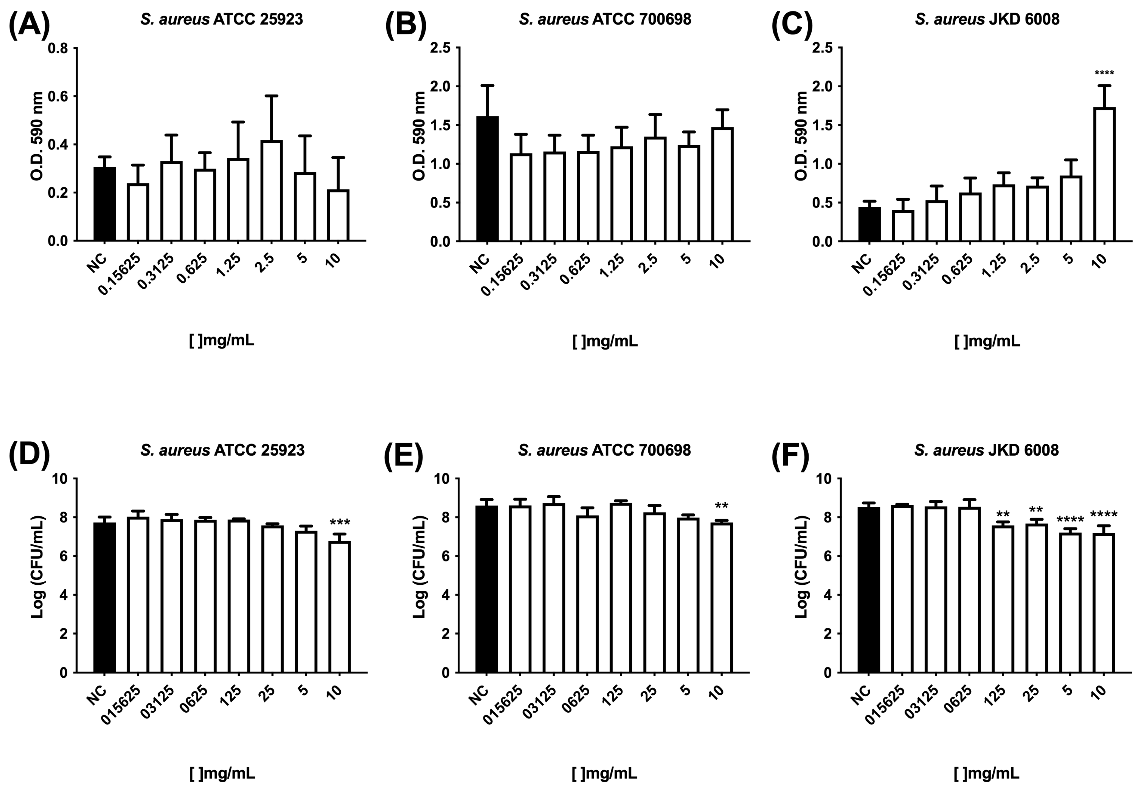
The results of OCp activity on preformed S. aureus biofilms can be seen in Figure 3. None of the tested concentrations resulted in a reduction in the biofilm biomass of S. aureus strains. On the other hand, a decrease in the number of viable cells was observed in all biofilm strains treated with OCp. S. aureus ATCC 25923 showed a reduction of 0.9 log at a concentration of 10 mg/mL (Figure 3D). For S. aureus ATCC 700698, the reduction was 0.8 log at 10 mg/mL (Figure 3E). In the case of S. aureus JKD 6008, the reductions were 1.3 log at 10 and 5 mg/mL, and 0.8 and 0.9 log at 2.5 mg/mL and 1.25 mg/mL, respectively (Figure 3F).
Figure 3.
Effect of Croton pluriglandulosus essential oil on preformed biofilms of different Staphylococcus aureus strains. (A) S. aureus ATCC 25923 biomass; (B) S. aureus ATCC 700698 biomass; (C) S. aureus JKD 6008 biomass; (D) viable cells of S. aureus ATCC 25923 biofilm; (E) viable cells of S. aureus ATCC 700698 biofilm; (F) viable cells of S. aureus JKD 6008 biofilm. White bars indicate OCp-treated groups, and black bars indicate untreated controls. Data are presented as mean ± SD of three independent experiments performed in triplicate. Statistical significance was assessed by one-way ANOVA followed by Bonferroni post hoc test. Significance levels versus the respective control: ** p < 0.01, *** p < 0.001, **** p < 0.0001.
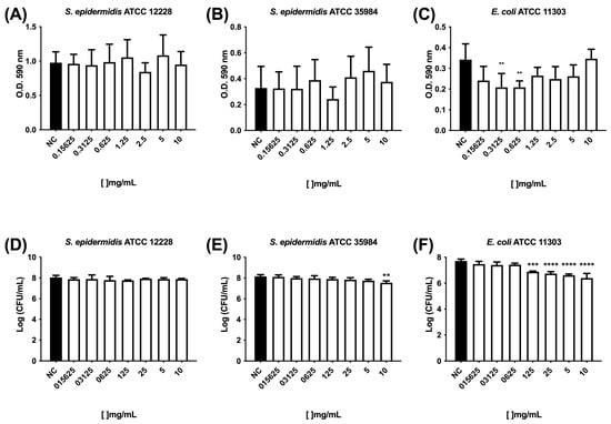
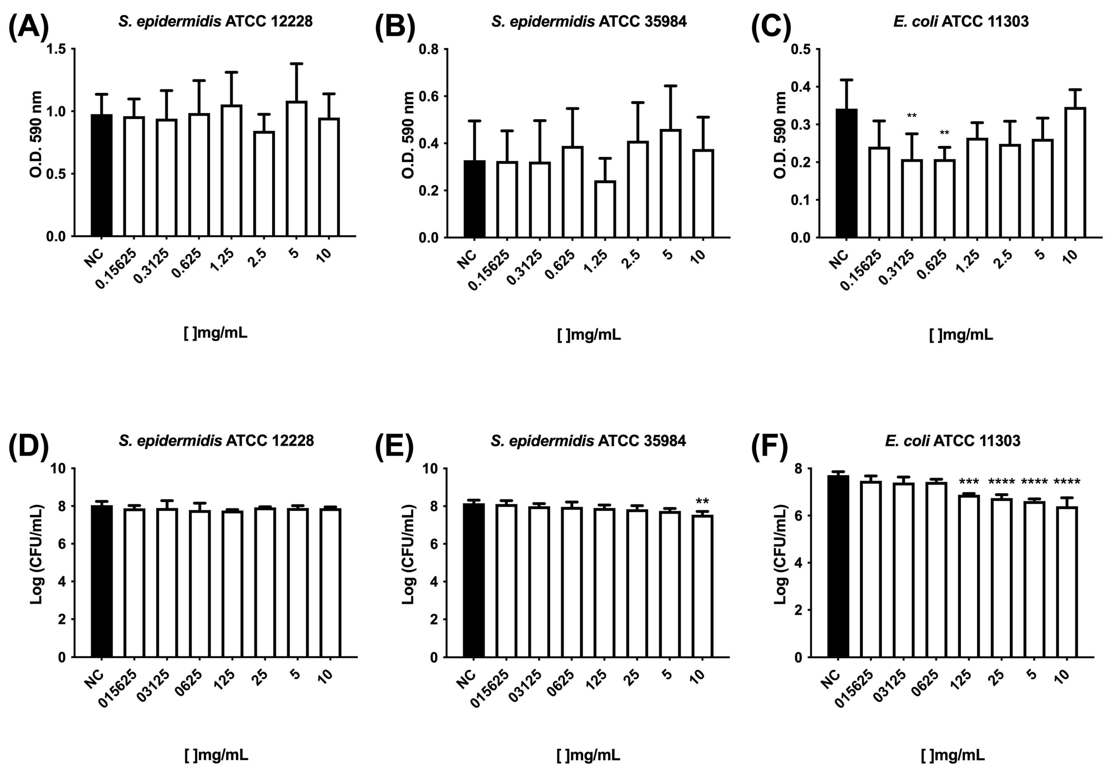
The activity of OCp on the preformed biofilms of the S. epidermidis and E. coli strains can be observed in Figure 4. Regarding biofilm biomass, no significant reductions were observed for the S. epidermidis biofilm strains treated with OCp. However, for the biofilms of E. coli ATCC 11303, there was a significant reduction at concentrations of 0.625 and 0.3125 mg/mL of approximately 39% in biomass for both concentrations (Figure 4C).
Figure 4.
Effect of Croton pluriglandulosus essential oil on preformed biofilms of Staphylococcus epidermidis and Escherichia coli strains. (A) Biofilm biomass of S. epidermidis ATCC 12228; (B) biofilm biomass of S. epidermidis ATCC 35984; (C) biofilm biomass of E. coli ATCC 11303; (D) viable cells of S. epidermidis ATCC 12228 biofilm; (E) viable cells of S. epidermidis ATCC 35984 biofilm; (F) viable cells of E. coli ATCC 11303 biofilm. White bars indicate OCp-treated groups, and black bars indicate untreated controls. Data are presented as mean ± SD of three independent experiments performed in triplicate. Statistical significance was assessed by one-way ANOVA followed by Bonferroni post hoc test. Significance levels versus the respective control: ** p < 0.01, *** p < 0.001, **** p < 0.0001.
Regarding the number of viable cells, it was observed that for the S. epidermidis ATCC 12228 biofilm, there was no significant reduction (Figure 4D). On the other hand, the S. epidermidis ATCC 35984 biofilm showed a reduction of 0.6 log at 10 mg/mL (Figure 4E). For E. coli ATCC 11303, reductions of 1.3 and 0.8 log were observed at concentrations of 10 and 1.25 mg/mL, respectively (Figure 4F).
3.5. Structural Analysis of Biofilms by Scanning Electron Microscopy (SEM)
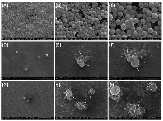
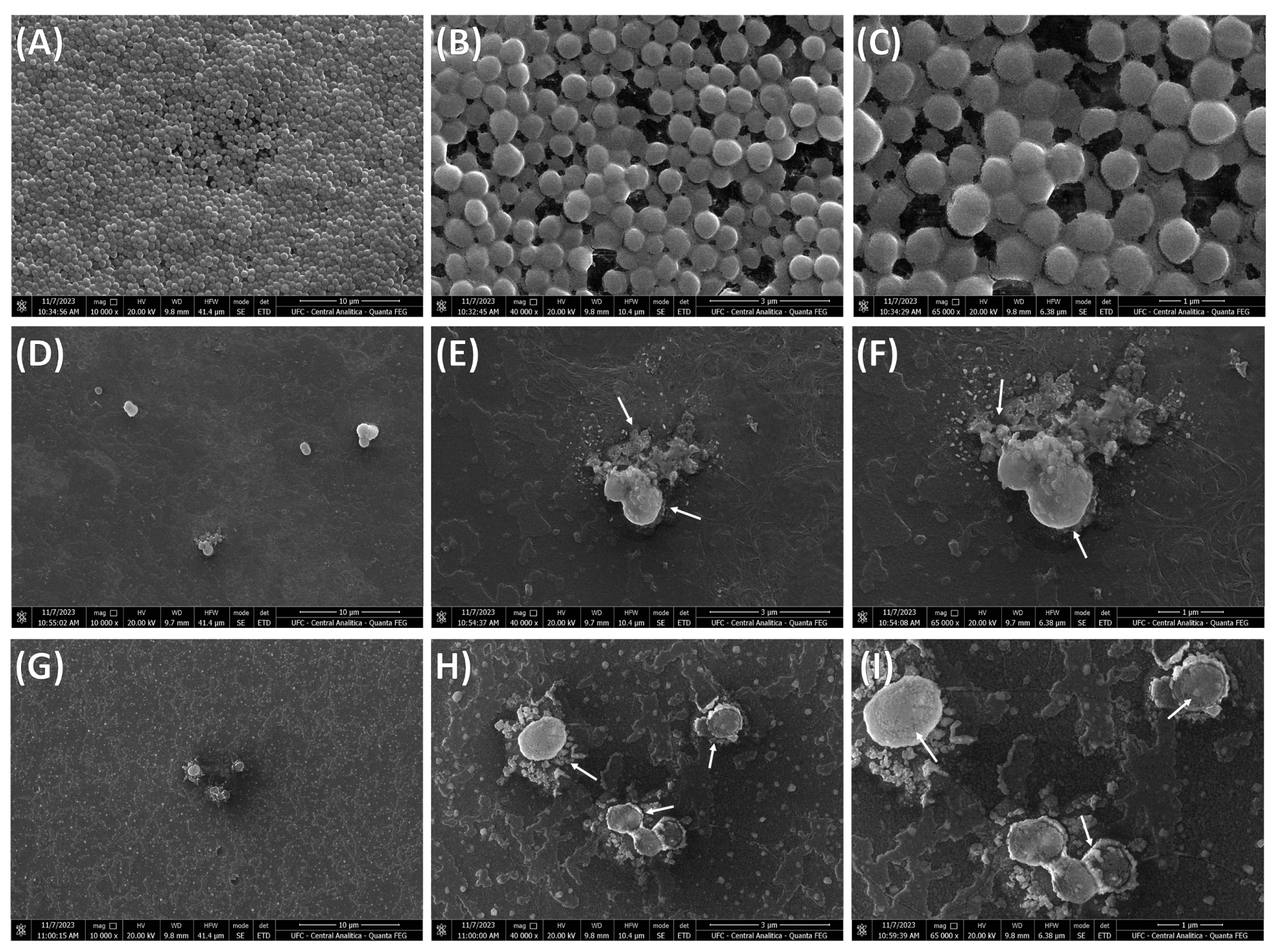
The electromicrographs obtained from S. aureus ATCC 700698 biofilms treated with OCp at 10 and 5 mg/mL and untreated (Control) can be observed in Figure 3. In the biofilms untreated (Figure 5A–C), the morphological regularity of the biofilm is evident, with the presence of grouped cocci, as is often observed in strains of the Staphylococcus genus, as well as a high cell density. However, in biofilms treated with OCp at a concentration of 5 mg/mL (Figure 5D–F), a significant reduction in cell clusters is observed, as structural damage to the bacterial surface and changes in morphology. A similar effect can be observed in biofilms treated with 10 mg/mL OCp (Figure 5G–I), with a significant reduction in cell clusters and changes in cell morphology and topology, as well as a significant increase in cell dimensions.
Figure 5.
Electron micrographs of biofilm formation by Staphylococcus aureus ATCC 700698. Untreated biofilms with Croton pluriglandulosus essential oil (A–C); treated with 5 mg/mL of Croton pluriglandulosus essential oil (D–F); and treated with 10 mg/mL of C. pluriglandulosus essential oil (G–I). Magnification: ×10,000, ×40,000, and ×65,000. White arrows indicate alterations in bacterial structures.
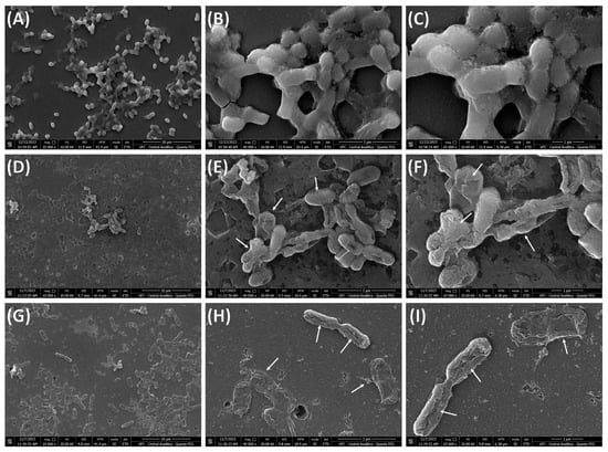
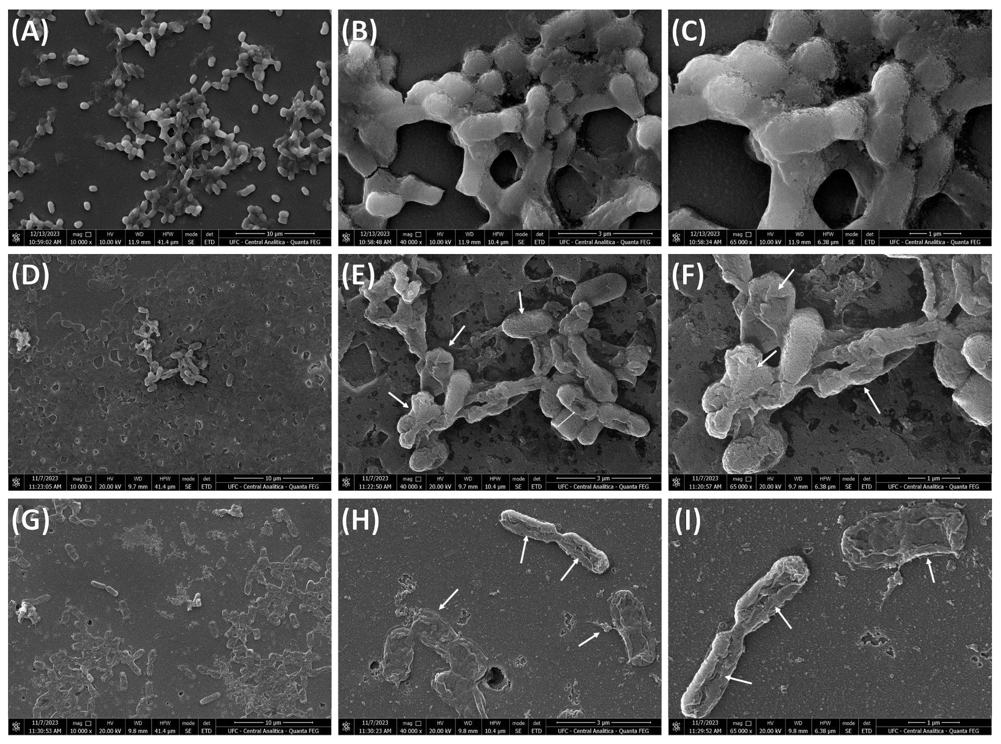
The electromicrographs E. coli ATCC 11303 biofilms treated with OCp at 10 and 5 mg/mL and untreated can be observed in Figure 6. In the untreated biofilm (Figure 6A–C), the morphological regularity of the bacterial cells is evident, with the cylindrical shape of bacilli, and high cell density can be observed. In contrast, biofilms treated with 5 mg/mL OCp exhibited a marked reduction in cell clusters and noticeable alterations on the cell surface, suggesting membrane damage (Figure 6D–F). Treatment with 10 mg/mL OCp further reduced cell density and induced morphological distortions similar to those observed at 5 mg/mL (Figure 6G–I).
Figure 6.
Electron micrographs of biofilm formation by Escherichia coli ATCC 11303. Untreated biofilms with Croton pluriglandulosus essential oil (A–C); treated with 5 mg/mL of Croton pluriglandulosus essential oil (D–F); and treated with 10 mg/mL of C. pluriglandulosus essential oil (G–I). Magnification: ×10,000, ×40,000, and ×65,000. White arrows indicate alterations in bacterial structures.
3.6. Structural and Viability Analysis of Biofilms by Confocal Laser Scanning Microscopy (CLSM)
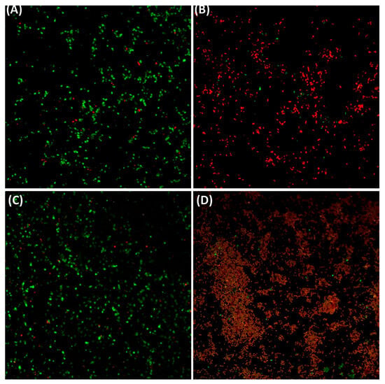
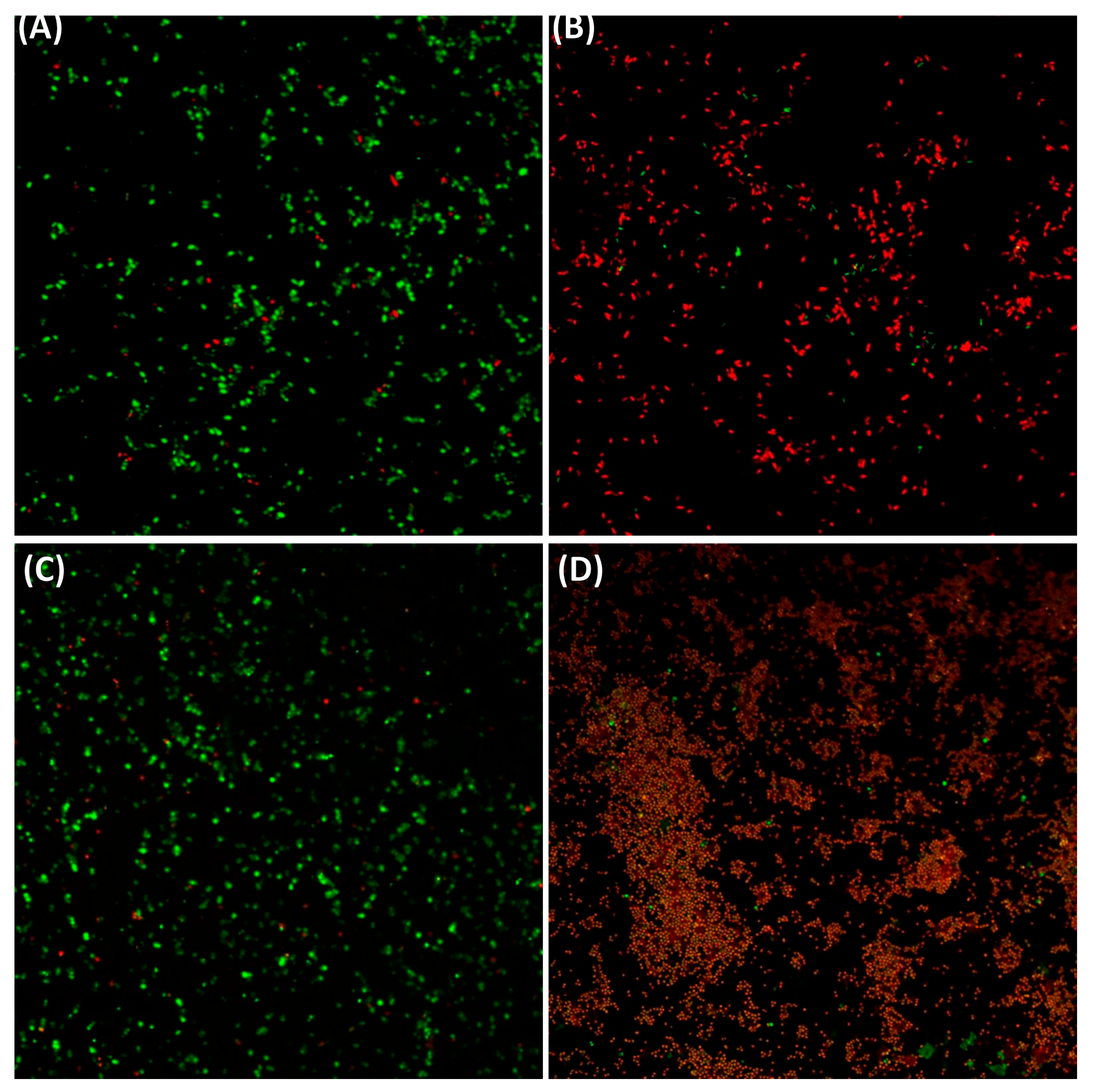
The images of S. aureus and E. coli biofilms treated and untreated with OCp (10 mg/mL) were analyzed by CLSM. In Figure 7, live cells can be observed in green fluorescence (SYTO 9) and dead cells in red (propidium iodide). Figure 7A shows the untreated E. coli ATCC 11303 biofilm, characterized by a high density of SYTO 9-stained cells, indicative of preserved membrane integrity. In contrast, Figure 7B (OCp treatment) shows a visible reduction in SYTO 9-stained cells accompanied by an increased presence of PI-stained cells, suggesting loss of membrane integrity and reduced cell viability. Figure 7C shows the untreated S. aureus ATCC 700698 biofilm, also displaying a dense layer of viable cells. Following treatment (Figure 7D), the biofilm structure remains relatively dense, but the fluorescence signal reveals a mixed pattern of SYTO 9- and PI-stained cells, consistent with a reduction in viable cell populations. These observations are based on qualitative CLSM fluorescence patterns rather than numerical quantification.
Figure 7.
Micrographs of preformed biofilms of Escherichia coli ATCC 11303 untreated with Croton pluriglandulosus essential oil (A) and treated with 10 mg/mL of Croton pluriglandulosus essential oil (B), and micrographs of preformed biofilms of Staphylococcus aureus ATCC 700698 untreated with Croton pluriglandulosus essential oil (C) and treated with 10 mg/mL of Croton pluriglandulosus essential oil (D).
4. Discussion
EOs are secondary metabolites produced by various plant species and consist of a wide variety of volatile chemical compounds, such as terpenes [28]. Currently, few studies evaluate the analyses of compounds presents in the C. pluriglandulosus EO. Carvalho et al. [29] analyzed the chemical composition of C. pluriglandulosus EO and observed that the main components were elmicin (25.77%), bicyclogermacrene (9.37%), and caryophyllene (8.99%). Additionally, according to Vasconcelos et al. [30], the main components present in C. pluriglandulosus EO were eucalyptol (17.41%), methyleugenol (16.06%), and elmicin (15.99%), demonstrating a variation compared to the predominant compounds in the present study.
This variation is not unexpected, as multiple studies have demonstrated that EO chemical compositions may vary even within a single Croton species. These changes in composition are influenced by several factors, such as temperature, solar incidence, time of year, type of soil, geographical location, climate, stage of plant development, method of collection and extraction, and intrinsic genetic characteristics of each population [31].
However, it is commonly reported in the literature that the compounds identified in this study (eucalyptol, α-pinene, and spathulenol) are also found in EOs from other species of the Croton genus. Medeiros et al. [32] reported that α-pinene was the major component of the EO of C. grewioides, accounting for 47.43% of the total compounds. García-Díaz et al. [33] also identified α-pinene in the EO of C. linearis, with 11.1%. Furthermore, Vidal et al. [34] identified spathulenol as the main constituent of the EO of C. rhamnifolioides (22.46%). Morais et al. [35] reported that spathulenol is predominant in C. argyrophylloides (42.54%) and C. jacobinensis (15.41%). Recently, Nunes et al. [36] identified the same major compounds in the EO of C. blanchetianus, although in different proportions: eucalyptol (20.97%), spathulenol (14.73%), and α-pinene (10.21%). Similarly, Malveira et al. [37] found that the EO of Croton urticifolius contained eucalyptol (19.03%) as one of its main constituents.
Among the compounds present in OCp, eucalyptol showed the highest concentration. The compound is characterized as a monoterpene widely found in the EOs of several plant species, especially in the Croton genus. Furthermore, eucalyptol is known for its antimicrobial activity against both Gram-positive and Gram-negative bacteria [35]. Spathulenol, in turn, is a sesquiterpene with antioxidant, antimicrobial, and anti-inflammatory properties [38]. α-pinene is a monoterpene commonly found in various plant species, and although Freitas et al. [39] did not observe antimicrobial activity for this compound, studies by Aćimović et al. [40] and Ramdani et al. [41] suggest that it does possess such activity.
OCp displayed relevant antibacterial activity against S. aureus, consistent with previous reports involving Croton-derived EOs, although differences in potency were observed [30]. Studies with EO obtained from different Croton species showed similar MIC and MBC values to those found for OCp. Siqueira et al. [42] showed that EOs of different genotypes from C. tetradenius showed MIC values between 2.8 and 5.6 mg/mL against E. coli ATCC 25922 and between 2.8 and 11.3 mg/mL against S. aureus ATCC 25923. Rocha et al. [43] reported MIC and MBC values of 4 mg/mL against S. aureus for C. tetradenius EO. In the same study, C. pulegiodorous EO showed an MIC value of 8 mg/mL against S. aureus, like the value found in the present study with OCp. Valarezo et al. [44] showed that the EO of C. ferrugineus exhibited no antimicrobial activity at the concentrations tested. Nunes et al. [36] reported that the C. blanchetianus EO showed MIC value of 0.78 mg/mL and MBC of 1.56 mg/mL against S. aureus and S. epidermidis strains, and a MIC of 1.56 mg/mL against E. coli ATCC 11303. These variations in the antibacterial activities of Croton EOs can be attributed to their chemical complexity, where synergistic and antagonistic interactions of the components influence the effects.
As observed for several Croton-derived EOs, OCp exhibited reduced activity against Gram-negative bacteria, including E. coli [23,45,46,47,48]. In fact, Gram-negative bacteria are less susceptible to the action of EOs from species of the Croton genus, unlike Gram-positive bacteria. These results have been observed in several studies [43,44,45,46]. The main hypothesis for the greater resistance of Gram-negative bacteria to EOs is the outer membrane, which acts as a barrier against microbial agents that target the cell membrane. This inhibits the action of most EOs, whose mechanism of action generally involves interaction with and damage to the membrane of microorganisms [23].
The analyses of biofilm formation treated with OCp showed a significant reduction in biomass across all tested bacterial strains, although, as previously mentioned, complete inhibition of planktonic cells of S. epidermidis and E. coli was not observed. For the S. aureus biofilm strains, the OCp showed reductions in biomass even at sub-inhibitory concentrations of OCp. These results are interesting since biomass reductions, represent a decrease in the extracellular polymeric matrix, an important resistance factor of biofilms [49].
Regarding C. pluriglandulosus EO against bacterial biofilms, Vasconcelos et al. [30] reported its combination with chlorhexidine against oral bacteria biofilms, including activity on S. mutans UA 159 strain, there was no reduction in biomass, but there was a decrease in the number of CFUs. In contrast, the S. salivarius ATCC 7073 and S. mutans ATCC 25175 strains showed biomass inhibition, but no significant reductions in CFUs. Therefore, our study is the first to analyze OCp in isolation against the formation of bacterial biofilms.
Nunes et al. [36] showed that C. blanchetianus EO reduced biofilm formation of S. aureus and E. coli strains at concentrations between 50 and 0.78 mg/mL, even though these concentrations were higher compared to the present study, its effects were similar to OCp. Vale et al. [23] showed that the C. piauhiensis EO inhibited the biofilm formation of S. aureus and E. coli.
The antimicrobial activity of EOs is attributed to their hydrophobic characteristics, which allow them to diffuse through the bacterial cell wall, causing membrane damage and altering its permeability. The components of EOs can cross the membrane, resulting in cell lysis, ion loss, and ATP depletion [50]. SEM electron micrographs show alterations in S. aureus and E. coli biofilms, evidencing surface damage, suggesting that the action of OCp directly affects the membrane, as observed with other EOs. Although OCp lacked bactericidal activity against E. coli, microscopy confirmed significant morphological damage.
Confocal microscopy analyses confirmed an increase in dead bacterial cells. The uptake of propidium iodide (PI), a fluorophore that penetrates cells only when membrane integrity is compromised, provides direct evidence that OCp induces structural damage to the bacterial membrane. This disruption can increase membrane permeability, leading to leakage of intracellular components, energetic collapse, and cell lysis—mechanisms consistent with the bactericidal effects of several membrane-active terpenes. In the context of biofilms, membrane destabilization is particularly meaningful because it circumvents matrix-associated tolerance and contributes to biomass reduction and impaired viability despite the physical barrier imposed by the extracellular polymeric substances.
It is important to note that the studies conducted by Vale et al. [23] with the EO of C. piauhiensis did not record bactericidal activity against S. epidermidis and E. coli strains. However, they did observe antibiofilm action, like the results found in our study. Other studies have also reported a lack of activity against planktonic cells but have highlighted the effectiveness of the action against biofilms. For example, the studies by Kavanaugh and Ribbeck [51] analyzed the effect of Thymus vulgaris EO on S. aureus and P. aeruginosa biofilms, demonstrating its effectiveness in inhibiting biofilm formation, although it showed no activity against planktonic cells. This ability to combat biofilms without affecting the development of planktonic cells is highly promising, as it may prevent the emergence of strains resistant to antimicrobial agents [23].
Few studies have explored the activity of EOs from the Croton genus against mature biofilms. Analysis of OCp against mature biofilms showed that, except for E. coli ATCC 11303, which had a significant reduction of around 39% in biomass, the other strains showed no reduction. Regarding the number of CFUs, all the strains showed decreases, except for S. epidermidis 12228. Oliveira et al. [45] observed that the EO of C. canduplicatus reduced the biomass of mature S. aureus biofilms by 32% and 27% in different strains. Firmino et al. [52] reported that the diterpene ICB4 from the EO of C. blanchetianus reduced the biomass of S. parasanguinis by 71.2% and S. mutans by 75%, with reductions in CFUs like those of OCp.
As observed, there was a reduction in the antibiofilm activity of OCp for mature biofilms. However, these reductions are expected, as microorganisms in this state can be up to 1000 times more resistant than in their planktonic form [53]. In these communities, they exhibit tolerance/resistance to multiple drugs and can survive in hostile environments, circumventing selective pressures [5]. It is now known that the metabolic activity of the cells within biofilms is variable: there is higher activity on the surface, while in the more internal regions, activity is lower, which hinders the action of antimicrobials [7].
The antibacterial and antibiofilm activity of OCp is likely related to its composition and the interaction between its compounds, such as eucalyptol (1,8-cineole), its major constituent. This widely studied compound is known for its ability to permeabilize the bacterial membrane and damage the cell structure [50]. Additionally, it can block receptors related to quorum sensing, which is essential for biofilm formation [54]. Several studies have investigated the antimicrobial mechanisms of eucalyptol. Moo et al. [55] observed that it significantly affects the bacterial membrane of Klebsiella pneumoniae, increasing its surface charge and causing the loss of lipopolysaccharides, resulting in increased membrane permeability and a decrease in ATP levels, as well as the release of intracellular content. Although it has weaker bacterial activity than conventional antibiotics, Hriouech et al. [56] showed that it has a synergistic effect with antibiotics such as gentamicin and amoxicillin, reducing the MIC of these drugs.
Studies by Moo et al. [55] indicate that eucalyptol affects the membrane of Klebsiella pneumoniae, increasing its permeability and decreasing ATP levels, although its bacterial activity is lower than that of antibiotics. In addition, the combination of eucalyptol with antibacterial drugs, such as gentamicin and amoxicillin, can enhance antimicrobial efficacy [56]. Colalto [57] shows that this volatile is safe in therapeutic doses, but excessive use can lead to acute toxicity.
Terpenes, including eucalyptol, are considered safe but can alter membrane fluidity and cause cell death [58]. Although reports of hepatotoxicity in animal models exist, the low toxicity of compounds such as α-pinene suggests possible safe applications for OCp [59,60].
Importantly, although the chemical composition of OCp has been previously characterized, its biological activities against clinically relevant biofilm-forming pathogens had not been investigated. To our knowledge, this is the first study to evaluate its antibacterial activity against resistant phenotypes such as MRSA and VISA, as well as methicillin-susceptible strains, including a direct comparison between Gram-positive and Gram-negative bacteria. Furthermore, we demonstrate both inhibition of biofilm formation and disruption of mature biofilms, which represents a clinically relevant scenario associated with persistent medical device-related infections. The microscopy analysis provides the first evidence of a bactericidal mechanism linked to cell membrane damage, expanding the current understanding of the biological potential of this species. However, the present study is limited by its in vitro design, which does not fully mimic clinical biofilm environments, and by the absence of cytotoxicity data. Further studies using clinical isolates and in vivo models are required to validate the therapeutic potential of the OCp.
5. Conclusions
The EO of C. pluriglandulosus (OCp) was characterized by a high abundance of eucalyptol and demonstrated relevant antibacterial and antibiofilm activities. OCp displayed antibacterial effects against planktonic S. aureus and significantly antibiofilm activity against all tested pathogens. Membrane damage confirmed by SEM and CLSM supports a bactericidal mechanism. Even in mature biofilms, where biomass reduction was limited, OCp impaired bacterial viability. These findings indicate that OCp is a promising natural candidate against biofilm-related infections, although translational studies remain necessary.
Author Contributions
G.L.C.M., E.A.M., C.R.d.S.S. and A.K.A.N.—Investigation; Writing—original draft; Formal analysis; W.M.B.d.S., S.M.d.M., H.S.d.S. and L.B.d.L.—Methodology; Formal analysis and Data curation; C.C.d.A. and D.N.d.N.S.—Visualization; Formal analysis; Writing—review and editing; E.H.T. and M.A.d.V.—Writing—review and editing; Supervision; Project administration; Funding acquisition and Conceptualization. All authors have read and agreed to the published version of the manuscript.
Funding
This work was supported by the Brazilian agencies CNPq (Conselho Nacional de Desenvolvimento Científico e Tecnológico). E. H. Teixeira (CNPq #303531/2022-3), M. A. Vasconcelos (CNPq #302261/2025-7).
Data Availability Statement
All data and materials are available with the corresponding authors.
Acknowledgments
The authors would like to thank the collaboration of the Central Analitica UFC/CT-INFRA/MCTI-SISNANO/Pro-Equipamentos CAPES.
Conflicts of Interest
The authors declare no conflicts of interest.
Abbreviations
The following abbreviations are used in this manuscript:
| AMR | Antimicrobial resistance |
| APG IV | Angiosperm Phylogeny Group IV |
| ATCC | American Type Culture Collection |
| ATP | Adenosine Triphosphate |
| CLSI | Clinical and Laboratory Standards Institute |
| CLSM | Confocal Laser Scanning Microscopy |
| CFU | Colony Forming Unit |
| CFU/mL | Colony Forming Units per milliliter |
| DMSO | Dimethyl Sulfoxide |
| EO(s) | Essential Oil (s) |
| EPS | Extracellular Polymeric Substances |
| GC–MS | Gas Chromatography–Mass Spectrometry |
| MBC | Minimum Bactericidal Concentration |
| MHA | Mueller–Hinton Agar |
| MHB | Mueller–Hinton Broth |
| MIC | Minimum Inhibitory Concentration |
| MRSA | Methicillin-resistant Staphylococcus aureus |
| MRSE | Methicillin-resistant Staphylococcus epidermidis |
| OCp | Croton pluriglandulosus essential oil |
| PBS | Phosphate-buffered saline |
| PI | Propidium Iodide |
| SEM | Scanning Electron Microscopy |
| TSA | Tryptone Soy Agar |
| TSB | Tryptone Soy Broth |
| USA | United States of America |
| VISA | Vancomycin-intermediate Staphylococcus aureus |
| WHO | World Health Organization |
References
- World Health Organization. Antimicrobial Resistance. Fact Sheet. 2020. Available online: https://www.who.int/news-room/fact-sheets/detail/antimicrobial-resistance (accessed on 15 December 2025).
- Subramaniam, G.; Girish, M. Antibiotic Resistance—A Cause for Reemergence of Infections. Indian J. Pediatr. 2020, 87, 937–944. [Google Scholar] [CrossRef] [Scilit]
- Aslam, B.; Wang, W.; Arshad, M.I.; Khurshid, M.; Muzammil, S.; Rasool, M.H.; Nisar, M.A.; Alvi, R.F.; Aslam, M.A.; Qamar, M.U.; et al. Antibiotic resistance: A rundown of a global crisis. Infect. Drug Resist. 2018, 11, 1645–1658. [Google Scholar] [CrossRef] [Scilit] [PubMed]
- Antimicrobial Resistance Collaborators. Global burden of bacterial antimicrobial resistance in 2019: A systematic analysis. Lancet 2022, 399, 629–655. [Google Scholar] [CrossRef] [Scilit] [PubMed]
- Flemming, H.-C.; Wuertz, S. Bacteria and archaea on Earth and their abundance in biofilms. Nat. Rev. Microbiol. 2019, 17, 247–260. [Google Scholar] [CrossRef] [Scilit] [PubMed]
- Olwal, C.O.; Ang’ienda, P.O.; Ochiel, D.O. Alternative sigma factor B (σB) and catalase enzyme contribute to Staphylococcus epidermidis biofilm’s tolerance against physico-chemical disinfection. Sci. Rep. 2019, 9, 5355. [Google Scholar] [CrossRef] [Scilit]
- Ciofu, O.; Tolker-Nielsen, T. Tolerance and Resistance of Pseudomonas aeruginosa Biofilms to Antimicrobial Agents—How P. aeruginosa Can Escape Antibiotics. Front. Microbiol. 2019, 10, 913. [Google Scholar] [CrossRef] [Scilit]
- Soares, A.; Roussel, V.; Pestel-Caron, M.; Barreau, M.; Caron, F.; Bouffartigues, E.; Chevalier, S.; Etienne, M. Understanding Ciprofloxacin Failure in Pseudomonas aeruginosa Biofilm: Persister Cells Survive Matrix Disruption. Front. Microbiol. 2019, 10, 2603. [Google Scholar] [CrossRef] [Scilit]
- Sauer, K.; Stoodley, P.; Goeres, D.M.; Hall-Stoodley, L.; Burmølle, M.; Stewart, P.S.; Bjarnsholt, T. The biofilm life cycle: Expanding the conceptual model of biofilm formation. Nat. Rev. Microbiol. 2022, 20, 608–620. [Google Scholar] [CrossRef] [Scilit]
- van Harten, R.M.; Willems, R.J.L.; Martin, N.I.; Hendrickx, A.P.A. Multidrug-Resistant Enterococcal Infections: New Compounds, Novel Antimicrobial Therapies? Trends Microbiol. 2017, 25, 467–479. [Google Scholar] [CrossRef] [Scilit]
- Botelho, J.; Grosso, F.; Peixe, L. Antibiotic resistance in Pseudomonas aeruginosa—Mechanisms, epidemiology and evolution. Drug Resist. Updat. 2019, 44, 100640. [Google Scholar] [CrossRef] [Scilit]
- Klein, E.Y.; Van Boeckel, T.P.; Martinez, E.M.; Pant, S.; Gandra, S.; Levin, S.A.; Goossens, H.; Laxminarayan, R. Global increase and geographic convergence in antibiotic consumption between 2000 and 2015. Proc. Natl. Acad. Sci. USA 2018, 115, E3463–E3470. [Google Scholar] [CrossRef] [Scilit] [PubMed]
- Tavares, W.R.; Seca, A.M.L.; Inula, L. Secondary Metabolites against Oxidative Stress-Related Human Diseases. Antioxidants 2019, 8, 122. [Google Scholar] [CrossRef] [Scilit] [PubMed]
- Melander, R.J.; Basak, A.K.; Melander, C. Natural products as inspiration for the development of bacterial antibiofilm agents. Nat. Prod. Rep. 2020, 37, 1454–1477. [Google Scholar] [CrossRef] [Scilit] [PubMed]
- Yang, L.; Wen, K.-S.; Ruan, X.; Zhao, Y.-X.; Wei, F.; Wang, Q. Response of Plant Secondary Metabolites to Environmental Factors. Molecules 2018, 23, 762. [Google Scholar] [CrossRef] [Scilit]
- Abbasi, M.S.; Tahir, M.A. Ecological and Phytochemical Significance of Croton bonplandianum (Bail). Asian J. Res. Bot. 2020, 4, 26–32. [Google Scholar]
- Ferreira, M.J.G.; Dias, F.G.B.; Santos, S.M.; Menezes, R.C.S.; Silva, L.M.R.; Figueiredo, E.A.T. Evaluation of medicinal plants as potential food antimicrobial additives. Res. Soc. Dev. 2020, 9, 153953295. [Google Scholar] [CrossRef] [Scilit]
- Riina, R.; Van Ee, B.; Caruzo, M.B.R.; Carneiro-Torres, D.S.; Dos Santos, R.F.; Berry, P.E. The Neotropical Croton sect. Geiseleria (Euphorbiaceae): Classification Update, Phylogenetic Framework, and Seven New Species from South America. Ann. Mo. Bot. Gard. 2021, 106, 111–166. [Google Scholar] [CrossRef] [Scilit]
- Apg, I.I. An update of the Angiosperm Phylogeny Group classification for the orders and families of flowering plants: APG IV. Bot. J. Linn. Soc. 2016, 181, 399–436. [Google Scholar] [CrossRef] [Scilit]
- Oliveira, A.R.d.; Jezler, C.N.; Oliveira, R.A.; Mielke, M.S.; Costa, L.C. Determinação do tempo de hidrodestilação e do horário de colheita no óleo essencial de menta. Hortic. Bras. 2012, 30, 155–159. [Google Scholar] [CrossRef] [Scilit]
- Approved Standard—M07-A10; Method for Dilution Antibacterial Susceptibility Tests for Bacteria That Grow Aerobically. 10th ed. Clinical and Laboratory Standards Institute (CLSI): Wayne, Pennsylvania, USA, 2015.
- Approved Standard—M100; Performance Standards for Antimicrobial Susceptibility Testing. 27th ed. Clinical and Laboratory Standards Institute (CLSI): Wayne, Pennsylvania, USA, 2017.
- do Vale, J.P.C.; Vasconcelos, M.A.; Arruda, F.V.S.; Firmino, N.C.S.; Pereira, A.L.; Andrade, A.L.; Saker-Sampaio, S.; Sampaio, A.H.; Marinho, E.S.; Teixeira, A.M.R.; et al. Evaluation of Antimicrobial and Antioxidant Potential of Essential Oil from Croton piauhiensis Müll. Arg. Curr. Microbiol. 2021, 78, 1926–1938. [Google Scholar] [CrossRef] [Scilit]
- Stepanović, S.; Vuković, D.; Dakić, I.; Savić, B.; Švabić-Vlahović, M. A modified microtiter-plate test for quantification of staphylococcal biofilm formation. J. Microbiol. Methods 2000, 40, 175–179. [Google Scholar] [CrossRef] [Scilit]
- Vasconcelos, M.A.; Arruda, F.V.S.; Santos, H.S.; Rodrigues, A.S.; Bandeira, P.N.; Albuquerque, M.R.J.R.; Cavada, B.S.; Teixeira, E.H.; Henriques, M.; Pereira, M.O. Effect of a casbane diterpene isolated from Croton nepetaefolius on the prevention and control of biofilms formed by bacteria and Candida species. Ind. Crops Prod. 2014, 61, 499–509. [Google Scholar] [CrossRef] [Scilit]
- Castelo-Branco, D.S.C.M.; Riello, G.B.; Vasconcelos, D.C.; Guedes, G.M.M.; Serpa, R.; Bandeira, T.J.P.G.; Monteiro, A.J.; Cordeiro, R.A.; Rocha, M.F.G.; Sidrim, J.J.C.; et al. Farnesol increases the susceptibility of Burkholderia pseudomallei biofilm to antimicrobials used to treat melioidosis. J. Appl. Microbiol. 2016, 120, 600–606. [Google Scholar] [CrossRef] [Scilit] [PubMed]
- Adams, R.P. Identification of Essential Oil Components by Gas Chromatography/Quadrupole Mass Spectroscopy, 4th ed.; Allured Publishing Corporation: Carol Stream, IL, USA, 2007. [Google Scholar]
- Stringaro, A.; Colone, M.; Angiolella, L. Antioxidant, Antifungal, Antibiofilm, and Cytotoxic Activities of Mentha spp. Essential Oils. Medicines 2018, 5, 112. [Google Scholar] [CrossRef] [Scilit] [PubMed]
- Carvalho, R.J.P.; Souza, P.F.N.; Malveira, E.A.; Neto, N.A.S.; Silva, R.R.S.; Melo, G.L.C.; Silva, A.F.B.; Lima, L.B.; de Albuquerque, C.C.; Bastos, R.W.; et al. Antimicrobial Activity the Essential Oil from Croton pluriglandulosus Carn. Leaves against Microorganisms of Clinical Interest. J. Fungi 2023, 9, 756. [Google Scholar] [CrossRef] [Scilit]
- Vasconcelos, B.M.; Pereira, A.M.G.; Coelho, P.A.T.; Cavalcante, R.M.B.; Carneiro-Torres, D.S.; Bandeira, P.N.; da Silva, F.F.; Rodrigues, T.H.S.; Gomes, G.A.; Carneiro, V.A. Enhancement of chlorhexidine activity against planktonic and biofilm forms of oral streptococci by two Croton spp. essential oils from the Caatinga biome. Biofouling 2023, 38, 984–993. [Google Scholar] [CrossRef] [Scilit]
- Moghaddam, M.; Mehdizadeh, L. Chemistry of Essential Oils and Factors Influencing Their Constituents. In Soft Chemistry and Food Fermentation; Elsevier: Amsterdam, The Netherlands, 2017; pp. 379–419. [Google Scholar]
- de Medeiros, V.M.; do Nascimento, Y.M.; Souto, A.L.; Madeiro, S.A.L.; Costa, V.C.d.O.; Silva, S.M.P.M.; Falcão Silva, V.d.S.; Agra, M.d.F.; de Siqueira-Júnior, J.P.; Tavares, J.F. Chemical composition and modulation of bacterial drug resistance of the essential oil from leaves of Croton grewioides. Microb. Pathog. 2017, 111, 468–471. [Google Scholar] [CrossRef] [Scilit]
- García-Díaz, J.; Escalona-Arranz, J.C.; Ochoa-Pacheco, A.; Santos, S.G.D.; González-Fernández, R.; Rojas-Vargas, J.A.; Monzote, L.; Setzer, W.N. Chemical Composition and In Vitro and In Silico Antileishmanial Evaluation of the Essential Oil from Croton linearis Jacq. Stems. Antibiotics 2022, 11, 1712. [Google Scholar] [CrossRef] [Scilit]
- Vidal, C.S.; Oliveira-Tintino, C.D.M.; Tintino, S.R.; Galvão, H.B.F.; Costa, J.G.M.d.; Coutinho, H.D.M.; Menezes, I.R.A.d. Chemical composition, antibacterial and modulatory action of the essential oil of Croton rhamnifolioides leaves Pax and Hoffman. Biosci. J. 2016, 32, 1632–1643. [Google Scholar] [CrossRef] [Scilit]
- Morais, S.; Cossolosso, D.; Silva, A.; de Moraes, M.; Teixeira, M.; Campello, C.; Bonilla, O.; de Paula, V.; Vila-Nova, N. Essential Oils from Croton Species: Chemical Composition, in vitro and in silico Antileishmanial Evaluation, Antioxidant and Cytotoxicity Activities. J. Braz. Chem. Soc. 2019, 30, 2155–2167. [Google Scholar] [CrossRef] [Scilit]
- Nunes, A.K.A.; Araújo Malveira, E.; Lopes Andrade, A.; Barbosa da Silva, W.M.; de Morais, S.M.; Silva dos Santos, H.; Cavalcanti de Albuquerque, C.; Holanda Teixeira, E.; do Nascimento Souza, D.N.; Alves de Vasconcelos, M. Chemical Composition Determination and Evaluation of the Antimicrobial Activity of Essential Oil from Croton blanchetianus (Euphorbiaceae) against Clinically Relevant Bacteria. Chem. Biodivers. 2022, 19, e202200777. [Google Scholar] [CrossRef] [Scilit] [PubMed]
- Malveira, E.A.; Nunes, A.K.A.; Andrade, A.L.; Melo, G.L.C.; da Silva, W.M.B.; de Morais, S.M.; dos Santos, H.S.; de Lima, L.B.; de Albuquerque, C.C.; do Nascimento Souza, D.N.; et al. Antibacterial and Antibiofilm Activity of Croton urticifolius Lam. Essential Oil Via Membrane Disruption. Curr. Microbiol. 2024, 81, 312. [Google Scholar] [CrossRef] [Scilit] [PubMed]
- do Nascimento, K.F.; Moreira, F.M.F.; Alencar Santos, J.; Kassuya, C.A.L.; Croda, J.H.R.; Cardoso, C.A.L.; Vieira, M.d.C.; Góis Ruiz, A.L.T.; Ann Foglio, M.; de Carvalho, J.E.; et al. Antioxidant, anti-inflammatory, antiproliferative and antimycobacterial activities of the essential oil of Psidium guineense Sw. and spathulenol. J. Ethnopharmacol. 2018, 210, 351–358. [Google Scholar] [CrossRef] [Scilit] [PubMed]
- Freitas, P.R.; de Araújo, A.C.J.; dos Santos Barbosa, C.R.; Muniz, D.F.; da Silva, A.C.A.; Rocha, J.E.; de Morais Oliveira-Tintino, C.D.; Ribeiro-Filho, J.; da Silva, L.E.; Confortin, C.; et al. GC-MS-FID and potentiation of the antibiotic activity of the essential oil of Baccharis reticulata (ruiz & pav.) pers. and α-pinene. Ind. Crops Prod. 2020, 145, 112106. [Google Scholar]
- Aćimović, M.G.; Pavlović, S.Đ.; Varga, A.O.; Filipović, V.M.; Cvetković, M.T.; Stanković, J.M.; Čabarkapa, I.S. Chemical Composition and Antibacterial Activity of Angelica archangelica Root Essential Oil. Nat. Prod. Commun. 2017, 12, 1934578X1701200. [Google Scholar] [CrossRef] [Scilit]
- Ramdani, M.; Lograda, T.; Silini, H.; Zeraib, A.; Chalard, P.; Figueredo, G.; Bouchaala, M.; Zerrar, S. Antibacterial activity of essential oils of Juniperus phoenicea from Eastern Algeria. J. Appl. Pharm. Sci. 2013, 3, 22–28. [Google Scholar]
- Siqueira, I.B.; Teixeira Barbosa, A.A.; Jain, S.; Miranda Fernandes, R.P.; Tavares Silva, A.R.S.; Ferreira Barbosa, F.H.; Schimieguel, D.M.; Blank, A.F.; Sacramento, A.G.; de Castro Nizio, D.A.; et al. In vitro Antibacterial Activity of Essential Oils of Croton tetradenius Baill. From the Brazilian Caatinga Biome and Its Synergistic Effect With Ciprofloxacin and Meropenem. J. Essent. Oil Bear. Plants 2021, 24, 12–21. [Google Scholar] [CrossRef] [Scilit]
- Rocha, R.R.; Matos, M.N.C.; Guerrero, J.A.P.; Cavalcante, R.M.B.; Melo, R.S.; Azevedo, Á.M.A.; Pereira, A.M.G.; Lopes, P.H.R.; Rodrigues, T.H.S.; Bandeira, P.N.; et al. Comparative study of the chemical composition, antibacterial activity and synergic effects of the essential oils of Croton tetradenius baill. And C. pulegiodorus baill. Against Staphylococcus aureus isolates. Microb. Pathog. 2021, 156, 104934. [Google Scholar] [CrossRef] [Scilit]
- Valarezo, E.; Gaona-Granda, G.; Morocho, V.; Cartuche, L.; Calva, J.; Meneses, M.A. Chemical Constituents of the Essential Oil from Ecuadorian Endemic Species Croton ferrugineus and Its Antimicrobial, Antioxidant and α-Glucosidase Inhibitory Activity. Molecules 2021, 26, 4608. [Google Scholar] [CrossRef] [Scilit]
- Oliveira, G.D.d.; Rocha, W.R.V.d.; Rodrigues, J.F.B.; Alves, H.d.S. Synergistic and Antibiofilm Effects of the Essential Oil from Croton conduplicatus (Euphorbiaceae) against Methicillin-Resistant Staphylococcus aureus. Pharmaceuticals 2022, 16, 55. [Google Scholar] [CrossRef] [Scilit]
- Araújo, F.M.; Dantas, M.C.S.M.; e Silva, L.S.; Aona, L.Y.S.; Tavares, I.F.; de Souza-Neta, L.C. Antibacterial activity and chemical composition of the essential oil of Croton heliotropiifolius Kunth from Amargosa, Bahia, Brazil. Ind. Crops Prod. 2017, 105, 203–206. [Google Scholar] [CrossRef] [Scilit]
- Daouda, T.; Prevost, K.; Gustave, B.; Joseph, D.A.; Nathalie, G.; Raphaël, O.; Rubens, D.; Claude, C.J.; Mireille, D.; Felix, T. Terpenes, Antibacterial and Modulatory Antibiotic Activity of Essential Oils from Croton hirtus L’ Hér. (Euphorbiaceae) from Ivory Coast. J. Essent. Oil Bear. Plants 2014, 17, 607–616. [Google Scholar] [CrossRef] [Scilit]
- Leite, T.R.; Silva, M.A.P.d.; Santos, A.C.B.d.; Coutinho, H.D.M.; Duarte, A.E.; Costa, J.G.M.d. Antimicrobial, modulatory and chemical analysis of the oil of Croton limae. Pharm. Biol. 2017, 55, 2015–2019. [Google Scholar] [CrossRef] [Scilit]
- Gupta, P.; Sarkar, S.; Das, B.; Bhattacharjee, S.; Tribedi, P. Biofilm, pathogenesis and prevention—A journey to break the wall: A review. Arch. Microbiol. 2015, 198, 1–15. [Google Scholar] [CrossRef] [Scilit] [PubMed]
- Angane, M.; Swift, S.; Huang, K.; Butts, C.A.; Quek, S.Y. Essential Oils and Their Major Components: An Updated Review on Antimicrobial Activities, Mechanism of Action and Their Potential Application in the Food Industry. Foods 2022, 11, 464. [Google Scholar] [CrossRef] [Scilit]
- Kavanaugh, N.L.; Ribbeck, K. Selected Antimicrobial Essential Oils Eradicate Pseudomonas spp. and Staphylococcus aureus Biofilms. Appl. Environ. Microbiol. 2012, 78, 4057–4061. [Google Scholar] [CrossRef] [Scilit] [PubMed]
- Sá Firmino, N.C.; Alexandre, F.S.O.; de Vasconcelos, M.A.; Pinheiro, A.A.; Arruda, F.V.S.; Guedes, M.L.S.; Silveira, E.R.; Teixeira, E.H. Diterpenes isolated from Croton blanchetianus Baill: Potential compounds in prevention and control of the oral Streptococci biofilms. Ind. Crops Prod. 2019, 131, 371–377. [Google Scholar] [CrossRef] [Scilit]
- Karygianni, L.; Ruf, S.; Hellwig, E.; Follo, M.; Vach, K.; Al-Ahmad, A. Antimicrobial Photoinactivation of In Situ Oral Biofilms by Visible Light Plus Water-Filtered Infrared A and Tetrahydroporphyrin-tetratosylate (THPTS). Microorganisms 2021, 9, 145. [Google Scholar] [CrossRef] [Scilit]
- Sybiya Vasantha Packiavathy, I.A.; Agilandeswari, P.; Musthafa, K.S.; Karutha Pandian, S.; Veera Ravi, A. Antibiofilm and quorum sensing inhibitory potential of Cuminum cyminum and its secondary metabolite methyl eugenol against Gram negative bacterial pathogens. Food Res. Int. 2012, 45, 85–92. [Google Scholar] [CrossRef] [Scilit]
- Moo, C.-L.; Osman, M.A.; Yang, S.-K.; Yap, W.-S.; Ismail, S.; Lim, S.-H.-E.; Chong, C.-M.; Lai, K.-S. Antimicrobial activity and mode of action of 1,8-cineol against carbapenemase-producing Klebsiella pneumoniae. Sci. Rep. 2021, 11, 21008. [Google Scholar] [CrossRef] [Scilit]
- Hriouech, S.; Akhmouch, A.A.; Mzabi, A.; Chefchaou, H.; Tanghort, M.; Oumokhtar, B.; Chami, N.; Remmal, A. The Antistaphylococcal Activity of Amoxicillin/Clavulanic Acid, Gentamicin, and 1,8-Cineole Alone or in Combination and Their Efficacy through a Rabbit Model of Methicillin-Resistant Staphylococcus aureus Osteomyelitis. Evid. Based Complement. Alternat. Med. 2020, 2020, 4271017. [Google Scholar] [CrossRef] [Scilit]
- Colalto, C. Volatile molecules for COVID-19: A possible pharmacological strategy? Drug Dev. Res. 2020, 81, 950–968. [Google Scholar] [CrossRef] [Scilit]
- Buchbauer, G.; Baser, K.H.C. Handbook of Essential Oils: Science, Technology, and Applications; Taylor & Francis Group: Boca Raton, FL, USA, 2020. [Google Scholar]
- Zárybnický, T.; Boušová, I.; Ambrož, M.; Skálová, L. Hepatotoxicity of monoterpenes and sesquiterpenes. Arch. Toxicol. 2017, 92, 1–13. [Google Scholar] [CrossRef] [Scilit]
- Rodrigues, K.A.d.F.; Amorim, L.V.; Dias, C.N.; Moraes, D.F.C.; Carneiro, S.M.P.; Carvalho, F.A.d.A. Syzygium cumini (L.) Skeels essential oil and its major constituent α-pinene exhibit anti-Leishmania activity through immunomodulation in vitro. J. Ethnopharmacol. 2015, 160, 32–40. [Google Scholar] [CrossRef] [Scilit]

Disclaimer/Publisher’s Note: The statements, opinions and data contained in all publications are solely those of the individual author(s) and contributor(s) and not of MDPI and/or the editor(s). MDPI and/or the editor(s) disclaim responsibility for any injury to people or property resulting from any ideas, methods, instructions or products referred to in the content. |
© 2026 by the authors. Licensee MDPI, Basel, Switzerland. This article is an open access article distributed under the terms and conditions of the Creative Commons Attribution (CC BY) license.