


Abstract
Introduction: Next-generation malaria vaccines are urgently needed to provide greater efficacy and longevity. Antibodies targeting blood-stage merozoites can confer protection against clinical malaria through multiple Fc-mediated functions. In particular, merozoite surface protein 2 (PfMSP2) is a known target of protective antibodies that can clear merozoites via multiple antibody Fc-mediated functions, making it a highly promising vaccine candidate. Methods: We developed PfMSP2 as a self-amplifying RNA (saRNA) vaccine, which was successfully validated for in vitro expression in mammalian cells. Subsequently, the PfMSP2-saRNA was formulated as lipid nanoparticles (LNPs) and evaluated for immunogenicity in mice following a three-dose regimen comparing 1 μg and 10 μg doses. We evaluated the induction of antibodies with functional activities relevant to protective immunity. Results: Our PfMSP2-saRNA vaccine induced antigen-specific IgG responses that recognised the surface of whole merozoites. Both 1 μg and 10 μg dosing regimens induced comparable antibodies to PfMSP2, and responses were predominantly murine cytophilic IgG subclasses. These vaccine-induced antibodies demonstrated potent Fc-mediated functions, including complement fixation and binding of human Fcγ-receptor I (FcγRI), after only two doses, which remained consistent after the third dose. Conclusions: PfMSP2 is highly immunogenic using the saRNA vaccine platform in a potential dose-sparing regimen, and induces antibodies with multiple Fc-mediated functions associated with protective immunity in humans. This saRNA platform is a promising strategy to develop highly efficacious vaccines requiring lower and fewer doses.
1. Introduction
Malaria has a high global health burden, having recorded more than 0.25 billion cases and 0.6 million deaths in 2024 [1], with the highest burden in Africa and among young children. Plasmodium falciparum causes most disease cases, and there are risks of resurgence in coming years. Therefore, effective vaccines are needed for malaria control and elimination [1].
P. falciparum has a complex lifecycle, whereby Anopheles mosquitoes transmit sporozoites through skin bites. Sporozoites infect hepatocytes and develop into merozoites, which are then released into the blood and undergo exponential asexual replication within red blood cells. This results in clinical malaria and can lead to severe illness and death [2]. There are only two approved vaccines for malaria, RTS,S/AS01 and R21/Matrix-M, both targeting sporozoites to prevent initial hepatic infection [3]. However, these vaccines provide only modest efficacy with breakthrough blood-stage parasitemia and clinical malaria still occurring, requiring booster doses annually [3,4,5,6,7,8]. Next-generation vaccines are needed for greater efficacy and longevity [9,10]. Blood-stage merozoite antigens are attractive vaccine targets as they have the potential to reduce blood-stage replication and clinical illness [11,12,13,14]. Antibodies targeting merozoites can inhibit blood-stage replication via Fc-mediated antibody functions including complement fixation and interaction with Fc receptors to promote opsonic phagocytosis and cellular cytotoxicity [15,16].
P. falciparum merozoite surface protein 2 (PfMSP2) is an abundant surface protein and potential vaccine candidate against blood-stage malaria [13,17,18]. PfMSP2 has a variable dimorphic region comprising two allelic families (3D7 and FC27), and conserved N- and C-terminal domains [19,20]. Naturally acquired antibodies to PfMSP2 have been associated with protection against malaria across different populations [17]. Antibodies function via the Fc region to mediate complement fixation and opsonic phagocytosis, leading to inhibition of blood-stage replication [17,21]. Therefore, inducing these protective antibody functions is important for future blood-stage vaccines.
Advances in vaccine technologies and the success of SARS-CoV-2 mRNA vaccines have introduced a new era of rapid vaccine development for infectious diseases [22]. mRNA technology offers efficient single- and multi-antigen formulations, and rapid adaptability to emerging pathogens with relatively small doses for sufficient immunogenicity to various infectious diseases [23,24,25,26]. The conventional mRNA-lipid nanoparticle (LNP) platform has been evaluated for liver and transmission stages in both P. falciparum and P. vivax, showing the feasibility of the platform for malaria vaccine development [27,28,29]. Although mRNA-LNP vaccines induce robust antibody responses, there are limitations with the conventional non-replicating linear mRNA [22], including the potential requirement for high dosage, multi-dose regimens, and inefficient endosomal escape of mRNA for translation [30]. To overcome these limitations, the next-generation self-amplifying RNA (saRNA) platform can be utilised [31,32,33,34,35]. saRNA vectors include a viral replicase complex consisting of four non-structural proteins (nsP1-4) derived from an alphavirus that enables self-limiting replication of RNA transcripts in situ. The complex replicates the positive-sense RNA (+ssRNA) into negative-sense RNA (-ssRNA), then back to +ssRNA for translation [31,36]. The Venezuelan Equine Encephalitis Virus (VEEV) replicon is one of the most widely used alphavirus-derived saRNA platform; however, replicons from other alphaviruses could also be utilised [37,38,39]. This replication feature offers potential advantages such as lower (and fewer) doses that prolong antigen expression for robust immune responses and antibody durability. Importantly, the recombination between saRNA vaccines and infectious viruses that could produce new functional chimeric viruses has not been reported [40].
A recent saRNA vaccine for SARS-CoV-2 was approved in Japan and demonstrated durable immune responses compared to conventional mRNA vaccines, despite a 6–20-fold lower dose [41,42,43,44]. Additionally, a rabies glycoprotein saRNA vaccine showed promise in preclinical models and a Phase I clinical trial, eliciting immune responses above the WHO-defined benchmark of protection with ultra-low and fewer doses of saRNA [45,46]. The findings suggest the saRNA platform could be an effective alternative to the currently approved whole-inactivated rabies virus vaccines. This promising vaccine platform has also been evaluated for other infectious diseases such as seasonal influenza, respiratory syncytial virus (RSV), and Neisseria meningitidis serogroup B (MenB), but has yet to be evaluated as a malaria vaccine [47,48,49,50,51].
Here, we designed and evaluated a PfMSP2 vaccine using two dosing regimens of the saRNA-LNP platform with the aim of generating functional antibodies against merozoites [45]. This work provides an important proof-of-concept for potential dose-sparing and shorter-regimen next-generation saRNA vaccines for malaria.
2. Materials and Methods
2.1. Production and Preparation of saRNA-LNP Vaccines
The codon-optimised P. falciparum MSP2 3D7 sequence (Δ19N, Δ23C; GenBank number JN248383, with C-terminal Flag-tag replacing the His-tag) was synthesised, as described previously [36], using the saRNA-2 vector with 5′ Cap1, 3′ Poly(A) sequence, and 5-methylcytidine substitution. The saRNA was used with the Moderna FDA-approved LNP formulation (SM-102) in 10% (w/v) sucrose in 1 × phosphate-buffered saline (PBS; pH 7.4) [30]. The saRNA-LNP formulation was produced at two concentrations (0.02 μg/μL and 0.2 μg/μL) and stored at −80 °C, and then thawed at 4 °C prior to use within 24–48 h. The saRNA synthesis, LNP formulation, and characterisation were performed by GenScript Biotech, Sydney, Australia (Table S1).
2.2. Protein Expression Analysis
PfMSP2 saRNA expression was assessed by transfecting Expi293F suspension cells. Transfected cells were collected at regular intervals, and cell lysates were separated by SDS-PAGE under reducing conditions as described previously [21]. Briefly, whole-cell suspensions were prepared with a final concentration of 100 mM dithiothreitol (Thermo Fisher Scientific, Vilnius, Lithuania), and 1 × NuPAGE LDS Sample Buffer (Invitrogen, Carlsbad, CA, USA), and then boiled at 95 °C for 5 min. The samples were vortexed for 30 s and then loaded onto a 4–12% Bis-Tris gel (Invitrogen, Carlsbad, CA, USA) with Precision Plus Protein Dual Color Standards (Bio-Rad, Hercules, CA, USA). The gel was run for 1.5 h at a voltage of 135 V and 200 mA in 1 × MES running buffer (Invitrogen, Carlsbad, CA, USA).
Western blot analysis was performed as described previously [21]. Briefly, the gel was transferred onto a nitrocellulose (NC) membrane at a voltage of 20 V for 7 min (default setting P3) using iBlot 2 Transfer Stacks (Thermo Fisher Scientific, Carlsbad, CA, USA). Membrane blocking was performed overnight at 4 °C with 1% (w/v) bovine serum albumin (BSA; Bovogen, Melbourne, Australia) in 1 × phosphate-buffered saline (PBS) containing 0.05% (v/v) Tween20 (Sigma-Aldrich, St. Louis, MO, USA) (PBS-T). The blocked membrane was washed with PBS-T and then probed with mouse anti-Flag DyLight 800 (1/1000; Invitrogen, Carlsbad, CA, USA) in 3% (w/v) BSA at RT to detect Flag-tagged PfMSP2 expression from saRNA. The membrane was analysed with the ChemiDoc MP (Bio-Rad, Hercules, CA, USA) using the default setting for DyLight 800 fluorescent dye visualisation.
2.3. Study Design
To evaluate the PfMSP2 saRNA-LNP vaccine, female C57BL/6 mice aged 8–12 weeks were used in this study (n = 5 per group). Mice were immunised with two regimens of saRNA-LNP: low-dose (3 × 1 μg) and high-dose (3 × 10 μg), as previously described [45]. The induction of antibodies was evaluated by comparing serum samples collected post-vaccination with serum from the same mice collected pre-vaccination. We did not include an empty LNP or irrelevant-saRNA control group in this study. We aimed to balance ethical considerations using animals in experimental research and followed the 3R concept (Replacement, Reduction, Refinement) [52]. In this descriptive analysis, we used sample sizes that were considered sufficient for proof-of-concept studies, and evaluated the saRNA-LNP vaccine for antibodies generated against P. falciparum merozoites. All vaccine formulations were injected using 50 μL/dose. The mice were randomly assigned to each group, and the immunisations were performed 28 days apart with prior blood collection before the injection (Figure 1B, Table S2). All injections of mice were administered intramuscularly to the calf muscle. The first two bleeds were retro-orbital collections, and the terminal collection was a cardiac bleed. The mice were anaesthetically euthanised using isoflurane mixed with oxygen at a concentration of 4% at a 1 L/min flow rate for 1–2 min as an inhaled dose to induce deep anaesthesia until the mice were unconscious. The mice remained anaesthetised during the full procedure, and the state of anaesthesia was confirmed by loss of the pedal/toe-pinch reflex. The mice remained anaesthetised and unresponsive, and to confirm death before performing cardiac puncture, a secondary physical method of cervical dislocation was utilised. All mice were housed, cared for, and handled (injection and bleeding) by the WEHI Antibody Facility in Bundoora, Australia.
2.4. ELISA and Plate-Based Assays
The recombinant PfMSP2 3D7 used in ELISA was manufactured by GroPep Pty Ltd. (Adelaide, Australia) as previously described [13,21]. Parasite culture (Plasmodium falciparum 3D7) and whole merozoite isolation were performed as previously described [14]. ELISA protocols for IgG, C1q fixation, and human Fc receptor binding were performed as described previously [17,21,53]. A background signal from blank wells containing no serum was subtracted from all measurements, and an assay-specific positive control was included on each plate to standardise results across plates. Plate washing between steps was performed using the automated 405 LS microplate washer (Agilent Biotek, Winooski, VT, USA) with PBS-T. Assays were normalised across plates within each experiment, and samples were tested in duplicates.
To measure total IgG, 0.5 µg/mL of recombinant PfMSP2 3D7 was used to coat flat-bottom 96-well Nunc Maxisorp microplates (Thermo Fisher Scientific, Roskilde, Denmark) at 4 °C with overnight incubation. The plates were then blocked with 0.1% (w/v) casein (Sigma-Aldrich, St. Louis, MO, USA) in 1 × PBS and further incubated for 2 h at 37 °C. Mouse serum samples, diluted in the same buffer, were added and incubated at RT for 2 h. Following incubation, HRP-conjugated antibodies (goat anti-mouse IgG HRP, Merck Millipore, Temecula, CA, USA; goat anti-mouse IgG subclasses HRP, Southern BioTech, Birmingham, AL, USA) were diluted in 0.1% (w/v) casein in 1 × PBS and then added and further incubated for 1 h at RT. Finally, TMB liquid substrate (Life Technologies, Carlsbad, CA, USA) was added to the plates and incubated in the dark for 10 min at RT to allow for the colour change. To stop the reaction, 1M H2SO4 was added to the plates and OD values were immediately read at 450 nm using a Multiskan GO plate reader (Thermo Fisher Scientific, Vantaa, Finland).
A modified ELISA protocol was followed for vaccine-induced IgG binding to merozoites. The modifications include coating wells with 5 × 10⁴ merozoites/mL diluted with 5% (w/v) skim milk in PBS-T for 2 h at 37 °C. The sera and antibody dilution steps used 5% (w/v) skim milk in PBS-T, and 10% (w/v) skim milk in PBS-T was used for plate blocking.
A modified ELISA protocol was followed to measure human FcγRI binding. The same standard IgG ELISA steps were followed except for using 1% (w/v) BSA in 1 × PBS as the diluent for all steps, and plate blocking reagent. After sera samples were incubated and washed, 0.125 µg/mL of biotinylated FcγRI 10256-H27H-B (Sino Biological, Beijing, China) was added to the plate and incubated for 1 h at 37 °C [54]. Streptavidin-HRP (ThermoFisher Scientific, Rockford, IL, USA), diluted 1/10,000, was then added and incubated for an additional hour at 37 °C. The substrate was then added, the reaction was stopped, and absorbance was measured.
For C1q fixation ELISA, a modified protocol was followed. Plates were coated with 1 µg/mL of recombinant antigen, and serum samples were incubated as described for the standard IgG ELISA. Human C1q (Merck Millipore, Darmstadt, Germany) was diluted to 10 µg/mL with 0.1% (w/v) casein in 1 × PBS and incubated at RT for 30 min. Following incubation and washing, rabbit anti-C1q IgG (Burnet Institute, Melbourne, Australia) [55] was diluted with 0.1% (w/v) casein in 1 × PBS and incubated at RT for 1 h. Goat anti-rabbit IgG HRP (Bio-Rad, Kidlington, UK) was diluted with 0.1% (w/v) casein in 1 × PBS and then incubated at RT for a further 1 h. The substrate was added, the reaction was stopped, and absorbance was immediately measured.
2.5. Statistical Analysis
Statistical analyses and data visualisation were conducted using GraphPad Prism 11 (MacOS) software. The unpaired nonparametric Mann–Whitney test was used to compare two independent groups, and the paired nonparametric Wilcoxon test was used to compare within groups. Spearman’s rho was calculated to assess correlations between parameters. p values were not adjusted for multiple comparisons.
3. Results
3.1. PfMSP2 saRNA Vaccine Shows High Immunogenicity in Mice
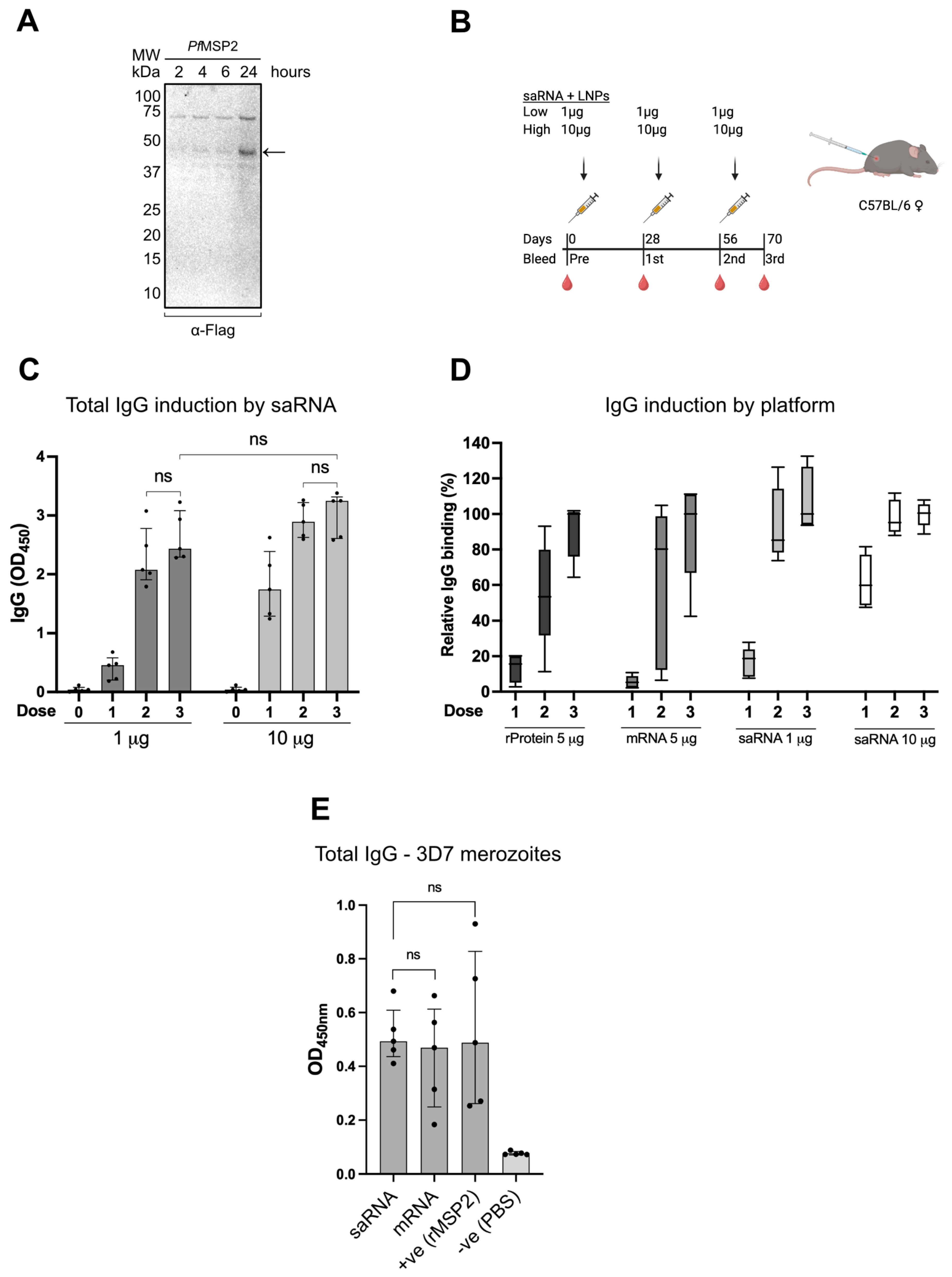
We designed a near-full-length PfMSP2 (3D7 allele) saRNA vaccine based on a previously described PfMSP2 construct with the addition of a C-terminal Flag-tag [13,21]. The saRNA was codon-optimised and synthesised using an alphavirus vector. We first transfected Expi293F (HEK293) suspension cells with naked saRNA to confirm antigen expression. PfMSP2 3D7 saRNA showed protein expression over time by detection of the C-terminal Flag-tag at the expected molecular weight size (Figure 1A and Figure S1). We then formulated the saRNA-LNP for in vivo testing in female C57BL/6 mice (n = 5 per group) which received either a fixed low dose (3 × 1 μg) or high dose (3 × 10 μg) on days 0, 28 and 56 (Figure 1B, Table S2). Mouse sera were collected at baseline (pre-bleed) and after each vaccine dose, with the peak response time-point collected on day 70 (third bleed). Vaccine-induced IgG to PfMSP2 increased after the second dose in both groups, but interestingly, there was no significant increase after the third dose (Figure 1C). When comparing the saRNA vaccine to recombinant protein and mRNA vaccines of PfMSP2, we observe broadly enhanced performance and lower biological variation in both saRNA regimens after the second and third doses (Figure 1D). The data suggest differences in the antibody-response trajectories relative to vaccine doses across platforms. This could indicate a dose-sparing advantage of the saRNA-LNP vaccine with fewer doses to achieve maximal IgG induction. Next, ELISA was performed using whole merozoites that were isolated from P. falciparum blood-stage culture, which express native PfMSP2 on their surface. We examined whether the saRNA vaccine platform can induce IgG that reacts with the merozoite surface. The saRNA vaccine-induced IgG from the high-dose 10 μg regimen at peak (day 70) was observed to recognise the merozoite surface (Figure 1E). There was no significant difference between the saRNA and the recombinant or mRNA PfMSP2 vaccine controls; however, the saRNA showed reduced variation between mice [21].
Figure 1.
PfMSP2 saRNA vaccine evaluation. (A) Expi293F cells were transfected with naked saRNA encoding Flag-tagged PfMSP2 (3D7 allele) and whole cells were collected. The samples were reduced for Western blot analysis, and protein expression was detected using α-Flag DyLight 800 monoclonal IgG (1/1000 dilution). The arrow indicates the PfMSP2 band. (B) Schematic of mouse immunisation regimens and timeline for blood collection (low dose: 3 × 1 μg; high dose: 3 × 10 μg saRNA PfMSP2). Mouse bleeds were collected one day prior to immunisation on days −1, 27, 55 (Table S2). (C) IgG reactivity in mice after immunisation with saRNA-LNP PfMSP2. Mouse sera measured at 1/3200 dilution by ELISA (n = 5 per group). Assays were performed in duplicates, and error bars represent median ± IQR. Statistical analysis was performed using an unpaired nonparametric Mann–Whitney test between groups, and paired nonparametric Wilcoxon test within groups; ns is not significant (p = 0.0625 for 1 μg group, dose 2 and 3 comparison; p = 0.3125 for 10 μg group, dose 2 and 3 comparison; p = 0.0952 for 1 μg and 10 μg groups, dose 3 comparison). (D) IgG reactivity in mice after immunisation with PfMSP2 using multiple platforms (previously published data of the recombinant protein and mRNA) [21]. Mouse sera measured at 1/3200 dilution by ELISA (n = 5 per group). Boxes represent interquartile range (IQR), and whiskers represent minimum and maximum values. Assays were performed in duplicates and standardised within each group as relative % IgG to the median OD value of the third bleed/dose. (E) ELISA analysis of IgG antibody reactivity to whole merozoite surface using mouse sera from the 10 μg high-dose saRNA vaccine group on day 70 (1/100 dilution; n = 5 per group), and control sera from recombinant and mRNA PfMSP2 vaccine groups on day 70 at 1/100 dilution (n = 5 per group) [21]. Error bars represent median ± 95% CI and assays were performed in duplicates. Statistical analysis was performed using an unpaired nonparametric Mann–Whitney test; ns is not significant (p = 0.6905 for saRNA and mRNA comparison; p > 0.9999 for saRNA and rMSP2 comparison).
3.2. PfMSP2 saRNA Vaccines Induce High Functional IgG with Fc-Mediated Activity
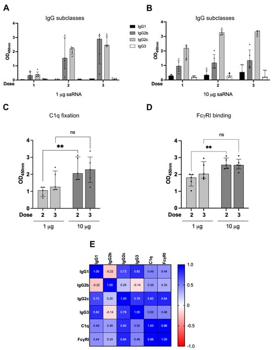
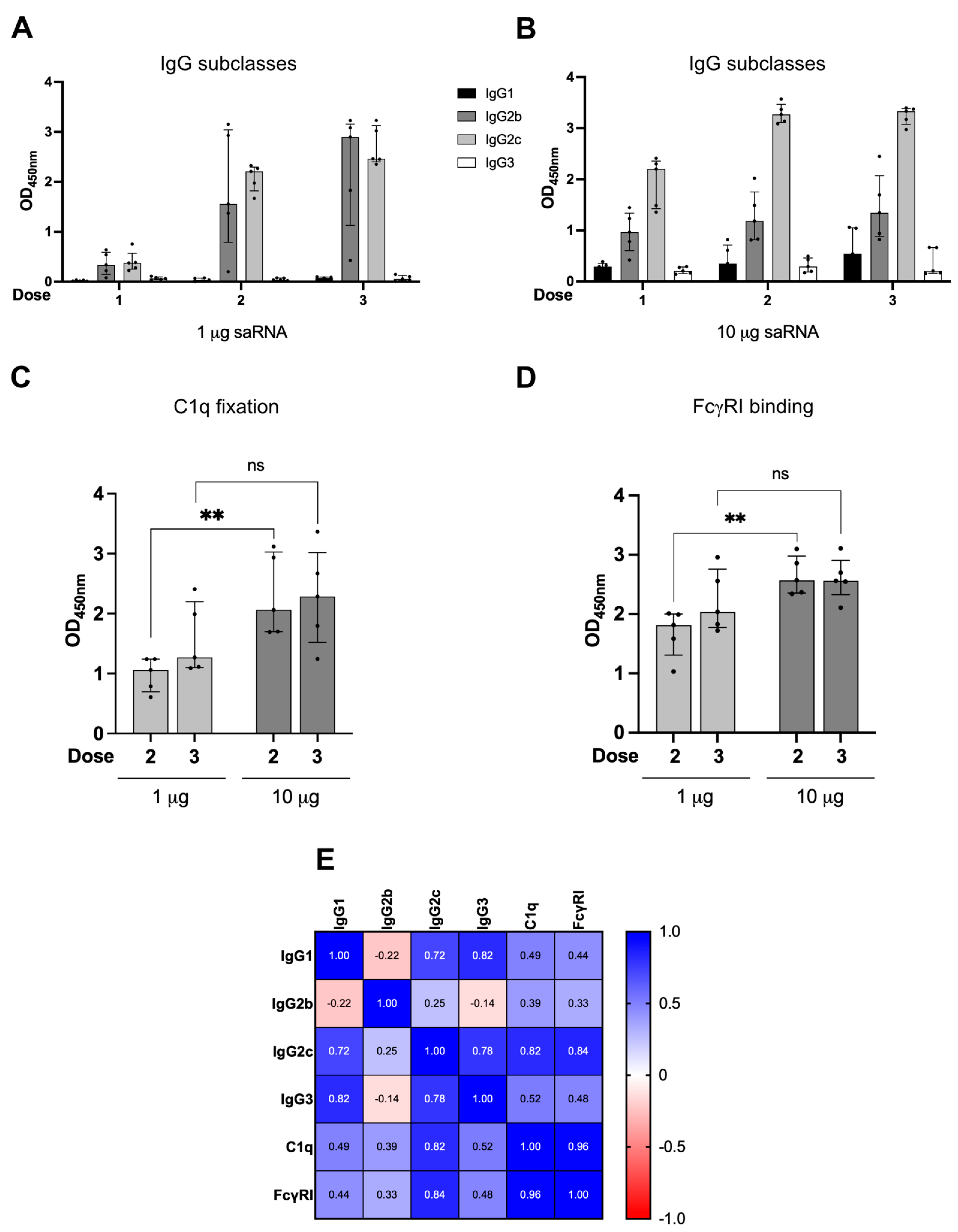
To understand the protective potential of PfMSP2 saRNA vaccine-induced immune responses, mouse IgG subclass profiles were examined. Remarkably, the cytophilic murine IgG2b and IgG2c that confer Fc-mediated protective functions were strongly induced and detected in serum following vaccination [21,56,57]. Testing IgG after each dose, the two dosing regimens showed comparable subclass profiles with IgG2b and IgG2c at higher magnitude, but low IgG1 and IgG3 levels (Figure 2A,B). The subclass levels only moderately increased after the third dose in both regimens, in line with the total IgG results (Figure S2). Previous studies have shown that antibodies to PfMSP2 have Fc-mediated functions that include complement fixation and activation, as well as antibody-dependent cellular inhibition (ADCI) and opsonic phagocytosis of merozoites that are mediated by IgG interactions with Fcγ receptors [13,14,15]. Therefore, we examined Fc-mediated functions of saRNA vaccine-induced mouse antibodies using established in vitro surrogate assays. Interestingly, the murine IgG demonstrated strong C1q fixation, which is the first step of classical complement activation (Figure 2C). Vaccine-induced antibodies also showed strong binding to Fcγ Receptor I, which can promote phagocytosis and other functions (Figure 2D). All functional activities were broadly comparable between groups, with the 10 μg saRNA regimen showing significantly higher activity after the second dose, albeit not 10-fold higher than the 1 μg saRNA regimen. No significant difference was observed after the third dose between the two regimens for functions, indicating a dose-sparing potential of the saRNA vaccine to induce functional IgG subclasses. In exploratory analysis, we found Fc-mediated functional activities (C1q fixation and FcγRI binding) most strongly correlated with IgG2c and moderately with the other IgG subclasses (Figure 2E, Table S3).
Figure 2.
Functional profile of PfMSP2 saRNA vaccine-induced IgG. (A) ELISA analysis of mouse IgG subclasses in sera from the 1 μg saRNA regimen (n = 5). Assays were performed in duplicates (1/3200 dilution) and error bars represent median ± IQR. (B) Mouse IgG subclasses in sera from the 10 μg saRNA vaccinated mice (n = 5). Assays were performed in duplicates (1/3200 dilution) and error bars represent median ± IQR. (C) Human C1q fixation by murine IgG from saRNA vaccine regimens (1/400 dilution). Assays were performed in duplicates and error bars represent median ± IQR. Statistical analysis was performed using an unpaired nonparametric Mann–Whitney test; ** is significant (p = 0.0079), ns is not significant (p = 0.2222). (D) Human FcγRI binding (1/12,800 dilution) by murine IgG from sera. Assays were performed in duplicates and error bars represent median ± IQR. Statistical analysis was performed using an unpaired nonparametric Mann–Whitney test; ** is significant (p = 0.0079), ns is not significant (p = 0.2222). (E) Correlation matrix (values are nonparametric Spearman’s rho) of mouse IgG subclasses and functions at peak response (day 70; n = 10 mice) with +1 indicating strong positive correlation, 0 indicating no correlation, and −1 indicating strong negative correlation. All coefficient/unadjusted p-values can be found in Table S3.
4. Discussion
Here, we demonstrate a new strategy to target the P. falciparum merozoite surface using saRNA vaccine technology to generate antibodies with Fc-mediated functional activity. PfMSP2 is an abundant merozoite surface antigen, making it a prime target for saRNA vaccines [58,59,60,61]. This antigen has been shown to be a target of functional antibodies that promote complement fixation, leading to merozoite replication inhibition and their opsonic phagocytosis [14,15,17]. Additionally, antibodies to PfMSP2 have been associated with protection in studies of naturally acquired and vaccine-induced immunity [17]. In this study, the vaccine results show effective antibodies with Fc-mediated functional activities generated using a dose-sparing 1 μg regimen with two doses. Using PfMSP2 served as a good proof-of-concept for saRNA-LNP vaccines for malaria and supports future studies evaluating additional antigens, including multi-antigen vaccine designs.
We validated the expression of PfMSP2 in mammalian cells before progressing the saRNA vaccine into animal studies. The saRNA-LNP formulation induced optimal levels of cytophilic IgG subclasses in mice following two doses for either 1 μg or 10 μg regimens, unlike conventional mRNA vaccines that require three doses [21]. The efficiency of conventional mRNA vaccine translation in vivo has been reported to be 1–2% due to limitation in endosomal escape, and therefore requires higher and repeated mRNA doses [62]. In contrast, saRNA offers self-replication in situ, allowing prolonged and increased antigen presentation with a lower dose, and thereby decreasing the impact of low endosomal escape [31]. Remarkably, the immunogenicity of the saRNA vaccine was comparable between both dosing regimens (1 μg and 10 μg), with reduced biological variation between mice compared to the recombinant protein and conventional mRNA vaccines [21]. Using higher or additional doses of saRNA did not show improvement in immunogenicity, which demonstrates potential dose-sparing advantages as reported in multiple infectious diseases [45,46,47,49]. However, further dose optimisation studies using lower amounts of saRNA, larger sample sizes, and control groups are needed to further demonstrate the advantages of the platform and formally evaluate different dosing regimens and increase statistical power.
saRNA vaccine-induced antibodies showed strong IgG Fc-mediated engagement with functional mediators, namely complement fixation and interaction with FcγRI [63]. Similarities were observed between functional profiles of the saRNA vaccine-induced antibodies and previously reported antibodies in clinical trials of a recombinant PfMSP2 protein vaccine and naturally acquired immunity, as well as in preclinical studies of the conventional PfMSP2 mRNA vaccine [15,17,21]. Fcγ receptor binding and complement fixation functions have been shown to be key to parasite inhibition and clearance in vitro and in vivo [14,15,17]. In C57BL/6 mice, IgG2b and IgG2c subclasses can mediate antibody functions effectively, and were nearly exclusively induced using the saRNA platform. This indicates an advantage in the functional IgG subclasses that are preferentially induced following saRNA vaccination, which may enhance vaccine-induced immune responses.
Naturally acquired antibodies reportedly target the variable allele-specific regions of PfMSP2, whereas vaccine-induced antibodies have shown greater targeting of conserved epitopes [17]. Further construct optimisation and inclusion of the FC27 allele are needed in future studies. Additionally, examining the cellular immune kinetics may provide insights into vaccine longevity. This includes the stimulation of CD4+ T-follicular helper cells that support antibody generation, and expansion of B cell memory population [64]. Further studies could also evaluate antibody avidity by comparing conventional mRNA and saRNA vaccines and different doses. Animal models could be used to evaluate protective efficacy of the vaccine; however, established mouse models have some limitations for performing these analyses. Therefore, exploring humanised mice and human organoid models to analyse vaccine responses would be valuable in future studies. Further, due to the absence of PfMSP2 or an orthologue in rodent malaria species, and differences in C1q and Fc-mediated functions between humans and mice, laboratory mouse models pose limitations for assessing the efficacy of PfMSP2-based vaccines. In future studies, this saRNA vaccine could be further evaluated in non-human primate models or controlled human malaria infection (CHMI) vaccine trials.
5. Conclusions
Our study provides a proof-of-concept for using saRNA-LNP as a platform for malaria vaccines, demonstrating strong immunogenicity and antibody functional activities. We showed that the PfMSP2 saRNA-LNP vaccine could be used in dose-sparing and fewer-dose regimens of 1 μg that induce functional IgG. A low dose and two doses induced high IgG broadly comparable to the 10 μg regimen. This platform can be extended to include other malaria vaccine antigens in development, such as PfRh5, to induce antibodies with both growth-inhibitory and Fc-mediated functional activities. This platform could offer protective vaccine efficacy with short- and low-dose vaccine regimens.
Supplementary Materials
The following supporting information can be downloaded at https://www.mdpi.com/article/10.3390/vaccines14110948/s1, Figure S1: PfMSP2 saRNA expression validation; Figure S2: Murine IgG subclass profiles induced by PfMSP2 (3D7) vaccine platforms; Table S1: saRNA-LNP characterisation (summarised values); Table S2: Mice immunisation and bleed schedules; Table S3: Correlation matrix coefficient and unadjusted p-values (non-parametric Spearman’s) of mouse subclasses at peak and function (day 70; n = 10 mice).
Author Contributions
Conceptualisation, A.T. and J.G.B.; methodology, A.T., T.K.C.H., S.C., L.K. and T.R.; validation, A.T., T.K.C.H. and T.R.; formal analysis, A.T. and T.K.C.H.; investigation, A.T., T.K.C.H. and J.G.B.; resources, A.T., L.K., C.S.D. and J.G.B.; data curation, A.T., C.S.D. and J.G.B.; writing—original draft preparation, A.T.; writing—review and editing, A.T., T.K.C.H., S.C., L.K., T.R., C.S.D. and J.G.B.; visualisation, A.T. and J.G.B.; supervision, A.T. and J.G.B.; project administration, A.T., C.S.D. and J.G.B.; funding acquisition, A.T. and J.G.B. All authors have read and agreed to the published version of the manuscript.
Funding
This work was supported by the State of Victoria mRNA Activation Program grant (Burnet Institute Vaccine Initiative) awarded to J.G.B., National Health and Medical Research Council of Australia grant awarded to J.G.B. (2033320), a CASS Foundation Medicine/Science grant awarded to A.T. (11959), a GenScript Life Science Research grant awarded to A.T., a Monash University postgraduate scholarship awarded to T.K.C.H., and a DAAD PROMOS scholarship from the University of Bonn awarded to T.R. Burnet Institute is supported by an Institute Operational Support grant from the Victorian State Government and the NHMRC Independent Institutes Infrastructure Support Scheme.
Institutional Review Board Statement
Burnet Institute malaria antigens saRNA handling approval (IBC 48659). WEHI Antibody Facility animal in Bundoora Scientific Procedure Premises Licence (SPPL201904), and ethics approval (2020.019 and 2023.012).
Informed Consent Statement
Not applicable.
Data Availability Statement
The raw data supporting conclusions of this article will be made available by the authors, without undue reservation. The recombinant and mRNA PfMSP2 preclinical data were previously published [21], and were re-analysed in this manuscript for comparison purposes with the saRNA vaccine platform following the same protocols, mouse strain, reagents, and immunisation/bleeding schedules.
Acknowledgments
We thank Paul Masendycz, Ridouan Bouhbouh, and Myha Huynh at WEHI Antibody Facility for their support with our animal studies. Schematics were created using BioRender. Burnet Institute is located on the traditional lands of the Boonwurrung people of the Kulin nation.
Conflicts of Interest
The authors declare no conflicts of interest.
References
- World Health Organization. World Malaria Report 2025; World Health Organization: Geneva, Switzerland, 2025; ISBN 978-92-4-011782-2. [Google Scholar]
- Beeson, J.G.; Drew, D.R.; Boyle, M.J.; Feng, G.; Fowkes, F.J.I.; Richards, J.S. Merozoite surface proteins in red blood cell invasion, immunity and vaccines against malaria. FEMS Microbiol. Rev. 2016, 40, 343–372. [Google Scholar] [CrossRef] [Scilit] [PubMed]
- Beeson, J.G.; Kurtovic, L.; Valim, C.; Asante, K.P.; Boyle, M.J.; Mathanga, D.; Dobano, C.; Moncunill, G. The RTS,S malaria vaccine: Current impact and foundation for the future. Sci. Transl. Med. 2022, 14, eabo6646. [Google Scholar] [CrossRef] [Scilit] [PubMed]
- Bundi, C.; Bellamy, D.; Kibwana, E.; Nyamako, L.; Ogwang, R.; Keter, K.; Kimani, D.; Salman, A.M.; Provstgaard-Morys, S.; Stockdale, L.; et al. Vaccine-induced responses to R21/Matrix-M—An analysis of samples from a phase 1b age de-escalation, dose-escalation trial. Front. Immunol. 2025, 16, 1620366. [Google Scholar] [CrossRef] [Scilit] [PubMed]
- Neafsey, D.E.; Juraska, M.; Bedford, T.; Benkeser, D.; Valim, C.; Griggs, A.; Lievens, M.; Abdulla, S.; Adjei, S.; Agbenyega, T.; et al. Genetic Diversity and Protective Efficacy of the RTS,S/AS01 Malaria Vaccine. N. Engl. J. Med. 2015, 373, 2025–2037. [Google Scholar] [CrossRef] [Scilit] [PubMed]
- Datoo, M.S.; Dicko, A.; Tinto, H.; Ouédraogo, J.B.; Hamaluba, M.; Olotu, A.; Beaumont, E.; Lopez, F.R.; Natama, H.M.; Weston, S.; et al. Safety and efficacy of malaria vaccine candidate R21/Matrix-M in African children: A multicentre, double-blind, randomised, phase 3 trial. Lancet 2024, 403, 533–544. [Google Scholar] [CrossRef] [Scilit] [PubMed]
- RTS,S Clinical Trials Partnership. Efficacy and safety of RTS,S/AS01 malaria vaccine with or without a booster dose in infants and children in Africa: Final results of a phase 3, individually randomised, controlled trial. Lancet 2015, 386, 31–45, Erratum in Lancet 2015, 386, 30. [Google Scholar] [CrossRef] [Scilit] [PubMed]
- Hysa, A.; Opi, D.H.; Waterhouse, J.; Chishimba, S.; Horton, J.L.; Kingston, N.; Netter, H.J.; Wetzel, D.; Piontek, M.; Feng, G.; et al. Antibody fine specificity correlates with protection from malaria for the RTS,S vaccine in young African children: A post hoc analysis of a phase IIb randomised controlled trial. PLoS Med. 2026, 23, e1004877. [Google Scholar] [CrossRef] [Scilit] [PubMed]
- Kurtovic, L.; Reiling, L.; Opi, D.H.; Beeson, J.G. Recent clinical trials inform the future for malaria vaccines. Commun. Med. 2021, 1, 26. [Google Scholar] [CrossRef] [Scilit] [PubMed]
- Beeson, J.G.; Kurtovic, L.; Dobaño, C.; Opi, D.H.; Chan, J.A.; Feng, G.; Good, M.F.; Reiling, L.; Boyle, M.J. Challenges and strategies for developing efficacious and long-lasting malaria vaccines. Sci. Transl. Med. 2019, 11, eaau1458. [Google Scholar] [CrossRef] [Scilit] [PubMed]
- Natama, H.M.; Salkeld, J.; Somé, A.; Soremekun, S.; Diallo, S.; Traoré, O.; Rouamba, T.; Ouédraogo, F.; Ouédraogo, E.; Daboné, K.C.S.; et al. Safety and efficacy of the blood-stage malaria vaccine RH5.1/Matrix-M in Burkina Faso: Interim results of a double-blind, randomised, controlled, phase 2b trial in children. Lancet Infect. Dis. 2025, 25, 495–506. [Google Scholar] [CrossRef] [Scilit] [PubMed]
- Silk, S.E.; Kalinga, W.F.; Salkeld, J.; Mtaka, I.M.; Ahmed, S.; Milando, F.; Diouf, A.; Bundi, C.K.; Balige, N.; Hassan, O.; et al. Blood-stage malaria vaccine candidate RH5.1/Matrix-M in healthy Tanzanian adults and children; an open-label, non-randomised, first-in-human, single-centre, phase 1b trial. Lancet Infect. Dis. 2024, 24, 1105–1117. [Google Scholar] [CrossRef] [Scilit] [PubMed]
- McCarthy, J.S.; Marjason, J.; Elliott, S.; Fahey, P.; Bang, G.; Malkin, E.; Tierney, E.; Aked-Hurditch, H.; Adda, C.; Cross, N.; et al. A phase 1 trial of MSP2-C1, a blood-stage malaria vaccine containing 2 isoforms of MSP2 formulated with montanide® ISA 720. PLoS ONE 2011, 6, e24413. [Google Scholar] [CrossRef] [Scilit] [PubMed]
- Osier, F.H.A.; Feng, G.; Boyle, M.J.; Langer, C.; Zhou, J.; Richards, J.S.; McCallum, F.J.; Reiling, L.; Jaworowski, A.; Anders, R.F.; et al. Opsonic phagocytosis of Plasmodium falciparum merozoites: Mechanism in human immunity and a correlate of protection against malaria. BMC Med. 2014, 12, 108. [Google Scholar] [CrossRef] [Scilit] [PubMed]
- Boyle, M.J.; Reiling, L.; Feng, G.; Langer, C.; Osier, F.H.; Aspeling-Jones, H.; Cheng, Y.S.; Stubbs, J.; Tetteh, K.K.A.; Conway, D.J.; et al. Human antibodies fix complement to inhibit Plasmodium falciparum invasion of erythrocytes and are associated with protection against malaria. Immunity 2015, 42, 580–590. [Google Scholar] [CrossRef] [Scilit] [PubMed]
- Nkumama, I.N.; Ogwang, R.; Odera, D.; Musasia, F.; Mwai, K.; Nyamako, L.; Murungi, L.; Tuju, J.; Fürle, K.; Rosenkranz, M.; et al. Breadth of Fc-mediated effector function correlates with clinical immunity following human malaria challenge. Immunity 2024, 57, 1215–1224.e6. [Google Scholar] [CrossRef] [Scilit] [PubMed]
- Feng, G.; Boyle, M.J.; Cross, N.; Chan, J.A.; Reiling, L.; Osier, F.; Stanisic, D.I.; Mueller, I.; Anders, R.F.; McCarthy, J.S.; et al. Human Immunization with a Polymorphic Malaria Vaccine Candidate Induced Antibodies to Conserved Epitopes That Promote Functional Antibodies to Multiple Parasite Strains. J. Infect. Dis. 2018, 218, 35–43. [Google Scholar] [CrossRef] [Scilit] [PubMed]
- Genton, B.; Betuela, I.; Felger, I.; Al-Yaman, F.; Anders, R.F.; Saul, A.; Rare, L.; Baisor, M.; Lorry, K.; Brown, G.V.; et al. A Recombinant Blood-Stage Malaria Vaccine Reduces Plasmodium falciparum Density and Exerts Selective Pressure on Parasite Populations in a Phase 1-2b Trial in Papua New Guinea. J. Infect. Dis. 2002, 185, 820–827. [Google Scholar] [CrossRef] [Scilit] [PubMed]
- Krishnarjuna, B.; Andrew, D.; MacRaild, C.A.; Morales, R.A.V.; Beeson, J.G.; Anders, R.F.; Richards, J.S.; Norton, R.S. Strain-transcending immune response generated by chimeras of the malaria vaccine candidate merozoite surface protein 2. Sci. Rep. 2016, 6, 20613. [Google Scholar] [CrossRef] [Scilit] [PubMed]
- Henshall, I.G.; Chmielewski, J.; Angage, D.; Romeo, O.; Lai, K.H.; Turland, K.R.; Badii, N.; Foley, M.; Anders, R.F.; Beeson, J.G.; et al. An abundant merozoite surface protein of Plasmodium falciparum modulates susceptibility to inhibitory antibodies. eLife 2025, 14, RP107603. [Google Scholar] [CrossRef] [PubMed]
- Thomas, A.; Rünz, T.; Ho, T.K.C.; Fabb, S.; Lee, C.L.; Chishimba, S.; Mugan, R.S.; Reiling, L.; Kurtovic, L.; D’Souza, C.S.; et al. Targeting a malaria merozoite surface protein with mRNA vaccine generates multifunctional antibodies. Front. Immunol. 2026, 17, 1840217. [Google Scholar] [CrossRef] [Scilit] [PubMed]
- Zhang, L.; More, K.R.; Ojha, A.; Jackson, C.B.; Quinlan, B.D.; Li, H.; He, W.; Farzan, M.; Pardi, N.; Choe, H. Effect of mRNA-LNP components of two globally-marketed COVID-19 vaccines on efficacy and stability. npj Vaccines 2023, 8, 156. [Google Scholar] [CrossRef] [Scilit] [PubMed]
- Heinz, F.X.; Stiasny, K. Distinguishing features of current COVID-19 vaccines: Knowns and unknowns of antigen presentation and modes of action. npj Vaccine 2021, 6, 104. [Google Scholar] [CrossRef] [Scilit] [PubMed]
- Jackson, N.A.C.; Kester, K.E.; Casimiro, D.; Gurunathan, S.; DeRosa, F. The promise of mRNA vaccines: A biotech and industrial perspective. npj Vaccines 2020, 5, 11. [Google Scholar] [CrossRef] [Scilit] [PubMed]
- Al-Wassiti, H.A.; Fabb, S.A.; Grimley, S.L.; Kochappan, R.; Ho, J.K.; Wong, C.Y.; Tan, C.W.; Payne, T.J.; Takanashi, A.; Lee, C.L.; et al. mRNA vaccines encoding membrane-anchored RBDs of SARS-CoV-2 mutants induce strong humoral responses and can overcome immune imprinting. Mol. Ther. Methods Clin. Dev. 2024, 32, 101380. [Google Scholar] [CrossRef] [Scilit] [PubMed]
- Skerritt, J.H.; Tucek-Szabo, C.; Sutton, B.; Nolan, T. The Platform Technology Approach to mRNA Product Development and Regulation. Vaccines 2024, 12, 528. [Google Scholar] [CrossRef] [Scilit] [PubMed]
- Hayashi, C.T.H.; Cao, Y.; Clark, L.C.; Tripathi, A.K.; Zavala, F.; Dwivedi, G.; Knox, J.; Alameh, M.G.; Lin, P.J.C.; Tam, Y.K.; et al. mRNA-LNP expressing PfCSP and Pfs25 vaccine candidates targeting infection and transmission of Plasmodium falciparum. npj Vaccine 2022, 7, 155, Correction in npj Vaccines 2023, 8, 115. [Google Scholar] [CrossRef] [Scilit] [PubMed]
- Limsalakpetch, A.; Kum-Arb, U.; Yongvanitchit, K.; Im-Erbsin, R.; Ubalee, R.; Waters, N.; Vesely, B.A.; Muramatsu, H.; Weissman, D.; Tam, Y.K.; et al. mRNA-LNP vaccine encoding the Plasmodium vivax circumsporozoite protein is highly immunogenic and confers protection in mice. Mol. Ther. Nucleic Acids 2025, 36. [Google Scholar] [CrossRef] [Scilit] [PubMed]
- Dietrich, M.H.; Chmielewski, J.; Chan, L.J.; Tan, L.L.; Adair, A.; Lyons, F.M.T.; Gabriela, M.; Lopaticki, S.; Dite, T.A.; Dagley, L.F.; et al. Cryo-EM structure of endogenous Plasmodium falciparum Pfs230 and Pfs48/45 fertilization complex. Science 2025, 389. [Google Scholar] [CrossRef] [Scilit] [PubMed]
- Schoenmaker, L.; Witzigmann, D.; Kulkarni, J.A.; Verbeke, R.; Kersten, G.; Jiskoot, W.; Crommelin, D.J.A. mRNA-lipid nanoparticle COVID-19 vaccines: Structure and stability. Int. J. Pharm. 2021, 601, 120586. [Google Scholar] [CrossRef] [Scilit] [PubMed]
- Vallet, T.; Vignuzzi, M. Self-Amplifying RNA: Advantages and Challenges of a Versatile Platform for Vaccine Development. Viruses 2025, 17, 566. [Google Scholar] [CrossRef] [Scilit] [PubMed]
- Azizi, H.; Renner, T.M.; Agbayani, G.; Simard, B.; Dudani, R.; Harrison, B.A.; Iqbal, U.; Jia, Y.; McCluskie, M.J.; Akache, B. Self-amplifying RNAs generated with the modified nucleotides 5-methylcytidine and 5-methyluridine mediate strong expression and immunogenicity in vivo. NAR Mol. Med. 2024, 1, ugae004. [Google Scholar] [CrossRef] [Scilit] [PubMed]
- Algaissi, A.; Moni, S.S. Next-generation mRNA vaccines: Strategies to overcome stability challenges. npj Vaccine 2026, 11, 192. [Google Scholar] [CrossRef] [Scilit] [PubMed]
- Singh, O.N.; Berry, U.; Joshi, G.; Asuru, T.R.; Chandrasekar, K.; Narayanan, S.; Srivastava, P.; Tiwari, M.; Chattopadhyay, S.; Mehdi, F.; et al. Comparison of immunogenicity and protection efficacy of self-amplifying and circular mRNA vaccines against SARS-CoV-2. iScience 2025, 28, 113498. [Google Scholar] [CrossRef] [Scilit] [PubMed]
- Comes, J.D.G.; Pijlman, G.P.; Hick, T.A.H. Rise of the RNA machines–Self-amplification in mRNA vaccine design. Trends Biotechnol. 2023, 41, 1417–1429. [Google Scholar] [CrossRef] [Scilit] [PubMed]
- Sun, Z.; Liu, Y.; Zhang, H.; Ge, T.; Pan, Y.; Liu, Y.; Wu, M.; Shan, T.; Zhu, G.; Wu, Q.; et al. A novel alphavirus-based saRNA platform for enhanced vaccination efficacy and organ-specific targeting. Vaccine 2026, 72, 128055. [Google Scholar] [CrossRef] [Scilit] [PubMed]
- Casmil, I.C.; Bathula, N.V.; Huang, C.; Wayne, C.J.; Cairns, E.S.; Friesen, J.J.; Soriano, S.K.; Liao, S.; Ho, C.H.; Kong, K.Y.S.; et al. Alphaviral backbone of self-amplifying RNA enhances protein expression and immunogenicity against SARS-CoV-2 antigen. Mol. Ther. 2025, 33, 514–528. [Google Scholar] [CrossRef] [Scilit] [PubMed]
- Boghici, D.T.; Yong, D.; Vidal, S.M.; Liu, G. Alphavirus replicase and regulatory RNA elements in host interactions and viral vector engineering. J. Virol. 2026, 100, e0177825. [Google Scholar] [CrossRef] [Scilit] [PubMed]
- Azizi, H.; Agbayani, G.; Renner, T.M.; Simard, B.; Iqbal, U.; Dudani, R.; Jia, Y.; McCluskie, M.J.; Akache, B. Sindbis Virus Self-Amplifying Replicon is Compatible with Modified Nucleotides Mediating Protein Expression and Vaccine Responses In Vivo. Mol. Ther. Adv. 2026, 34, 201789. [Google Scholar] [CrossRef] [Scilit] [PubMed]
- Hick, T.A.H.; Geertsema, C.; Nguyen, W.; Bishop, C.R.; van Oosten, L.; Abbo, S.R.; Dumenil, T.; van Kuppeveld, F.J.M.; Langereis, M.A.; Rawle, D.J.; et al. Safety concern of recombination between self-amplifying mRNA vaccines and viruses is mitigated in vivo. Mol. Ther. 2024, 32, 2519–2534. [Google Scholar] [CrossRef] [Scilit] [PubMed]
- Oda, Y.; Kumagai, Y.; Kanai, M.; Iwama, Y.; Okura, I.; Minamida, T.; Yagi, Y.; Kurosawa, T.; Greener, B.; Zhang, Y.; et al. Immunogenicity and safety of a booster dose of a self-amplifying RNA COVID-19 vaccine (ARCT-154) versus BNT162b2 mRNA COVID-19 vaccine: A double-blind, multicentre, randomised, controlled, phase 3, non-inferiority trial. Lancet Infect. Dis. 2024, 24, 351–360. [Google Scholar] [CrossRef] [Scilit] [PubMed]
- Levine, K.S.; Blanc, R.; Wang, Q.; Malca, H.; Sekulovich, R.; Jin, H.; Liu, S.; Verhoeven, C.; Sullivan, B.; Smolenov, I.; et al. Self-amplifying COVID-19 mRNA vaccination induces longitudinally enhanced antibody function in a Phase 3 trial. npj Vaccine 2026, 11, 106. [Google Scholar] [CrossRef] [Scilit] [PubMed]
- Akahata, W.; Sekida, T.; Nogimori, T.; Ode, H.; Tamura, T.; Kono, K.; Kazami, Y.; Washizaki, A.; Masuta, Y.; Suzuki, R.; et al. Safety and immunogenicity of SARS-CoV-2 self-amplifying RNA vaccine expressing an anchored RBD: A randomized, observer-blind phase 1 study. Cell Rep. Med. 2023, 4, 101134. [Google Scholar] [CrossRef] [Scilit] [PubMed]
- Kitonsa, J.; Serwanga, J.; Cheeseman, H.M.; Abaasa, A.; Lunkuse, J.F.; Ruzagira, E.; Kato, L.; Nambaziira, F.; Oluka, G.K.; Gombe, B.; et al. Safety and Immunogenicity of a Modified Self-Amplifying Ribonucleic Acid (saRNA) Vaccine Encoding SARS-CoV-2 Spike Glycoprotein in SARS-CoV-2 Seronegative and Seropositive Ugandan Individuals. Vaccine 2025, 13, 553. [Google Scholar] [CrossRef] [Scilit] [PubMed]
- Maine, C.J.; Miyake-Stoner, S.J.; Spasova, D.S.; Picarda, G.; Chou, A.C.; Brand, E.D.; Olesiuk, M.D.; Domingo, C.C.; Little, H.J.; Goodman, T.T.; et al. Safety and immunogenicity of an optimized self-replicating RNA platform for low dose or single dose vaccine applications: A randomized, open label Phase I study in healthy volunteers. Nat. Commun. 2025, 16, 456. [Google Scholar] [CrossRef] [Scilit] [PubMed]
- Zhao, D.; Chen, G.; Wang, S.; Suo, Y.; Ye, Q.; Xing, R.; Yang, Y.; Li, Z.; Wang, X.; Zhou, R.; et al. A single ultra-low dose self-amplifying RNA rabies vaccine confers rapid and complete protection in mice. Vaccine 2026, 73, 128150. [Google Scholar] [CrossRef] [Scilit] [PubMed]
- Huang, M.; Quan, Y.; Chen, R.; Gu, H.; Song, W.; Zhu, F.; Feng, S.; Lin, J.; Lu, J.; Li, X.; et al. Enhanced immunogenicity and dose-sparing efficacy of self-amplifying RNA vaccines against seasonal influenza across subtypes. Emerg. Microbes Infect. 2026, 15, 2668752. [Google Scholar] [CrossRef] [Scilit] [PubMed]
- Chang, C.; Music, N.; Cheung, M.; Rossignol, E.; Bedi, S.; Patel, H.; Safari, M.; Lee, C.; Otten, G.R.; Settembre, E.C.; et al. Self-amplifying mRNA bicistronic influenza vaccines raise cross-reactive immune responses in mice and prevent infection in ferrets. Mol. Ther. Methods Clin. Dev. 2022, 27, 195–205, Errata in Mol. Ther. Methods Clin. Dev. 2023, 28, 11. [Google Scholar] [CrossRef] [Scilit] [PubMed]
- Chen, G.; Chen, G.; Wang, S.; Xing, R.; Yang, Y.; Zhou, R.; Ma, L.; Wang, X.; Wang, Y.; Song, W.; et al. Modular design of a self-amplifying mRNA vaccine for multivalent immunization against Neisseria meningitidis B. Vaccine 2025, 68, 127964. [Google Scholar] [CrossRef] [Scilit] [PubMed]
- Chang, C.; Patel, H.; Ferrari, A.; Scalzo, T.; Petkov, D.; Xu, H.; Rossignol, E.; Palladino, G.; Wen, Y. Sa-mRNA influenza vaccine raises a higher and more durable immune response than mRNA vaccine in preclinical models. Vaccine 2025, 51, 126883. [Google Scholar] [CrossRef] [Scilit] [PubMed]
- Zuo, L.; Liu, Q.; Zhang, K.; Zhao, L.; Lin, S.; Dai, Y.; Sun, Y.; Li, Y.; Zhang, P.; Shen, H.; et al. Self-amplifying mRNA vaccines protect elderly BALB/c mice against a lethal respiratory syncytial virus infection. Mol. Ther. 2025, 33, 499–513. [Google Scholar] [CrossRef] [Scilit] [PubMed]
- Smith, A.J. Guidelines for planning and conducting high-quality research and testing on animals. Lab Anim. Res. 2020, 36, 21. [Google Scholar] [CrossRef] [Scilit] [PubMed]
- Chishimba, S.; Reiling, L.; Boyle, M.J.; Kurtovic, L.; Beeson, J.G. Complement-Fixation by Antibodies to Plasmodium falciparum; Springer Nature: Berlin/Heidelberg, Germany, 2024. [Google Scholar] [CrossRef] [Scilit] [PubMed]
- Wines, B.D.; Vanderven, H.A.; Esparon, S.E.; Kristensen, A.B.; Kent, S.J.; Hogarth, P.M. Dimeric FcγR Ectodomains as Probes of the Fc Receptor Function of Anti-Influenza Virus IgG. J. Immunol. 2016, 197, 1507–1516. [Google Scholar] [CrossRef] [Scilit] [PubMed]
- Kurtovic, L.; Agius, P.A.; Feng, G.; Drew, D.R.; Ubillos, I.; Sacarlal, J.; Aponte, J.J.; Fowkes, F.J.I.; Dobaño, C.; Beeson, J.G. Induction and decay of functional complement-fixing antibodies by the RTS,S malaria vaccine in children, and a negative impact of malaria exposure. BMC Med. 2019, 17, 45. [Google Scholar] [CrossRef] [Scilit] [PubMed]
- Dekkers, G.; Bentlage, A.E.H.; Stegmann, T.C.; Howie, H.L.; Lissenberg-Thunnissen, S.; Zimring, J.; Rispens, T.; Vidarsson, G. Affinity of human IgG subclasses to mouse Fc gamma receptors. MAbs 2017, 9, 767–773. [Google Scholar] [CrossRef] [Scilit] [PubMed]
- Han, J.; Liu, Q.; Xu, X.; Qin, W.; Pan, Y.; Qin, R.; Zhao, R.; Gu, Y.; Gu, J.; Ren, S. Relative Quantitation of Subclass-Specific Murine IgG Fc N-Glycoforms by Multiple Reaction Monitoring. ACS Omega 2020, 5, 8564–8571. [Google Scholar] [CrossRef] [Scilit] [PubMed]
- Balam, S.; Olugbile, S.; Servis, C.; Diakité, M.; D’Alessandro, A.; Frank, G.; Moret, R.; Nebie, I.; Tanner, M.; Felger, I.; et al. Plasmodium falciparum merozoite surface protein 2: Epitope mapping and fine specificity of human antibody response against non-polymorphic domains. Malar. J. 2014, 13, 510. [Google Scholar] [CrossRef] [Scilit] [PubMed]
- Adda, C.G.; MacRaild, C.A.; Reiling, L.; Wycherley, K.; Boyle, M.J.; Kienzle, V.; Masendycz, P.; Foley, M.; Beeson, J.G.; Norton, R.S.; et al. Antigenic characterization of an intrinsically unstructured protein, Plasmodium falciparum merozoite surface protein 2. Infect. Immun. 2012, 80, 4177–4185. [Google Scholar] [CrossRef] [Scilit] [PubMed]
- MacRaild, C.A.; Pedersen, M.E.; Anders, R.F.; Norton, R.S. Lipid interactions of the malaria antigen merozoite surface protein 2. Biochim Biophys. Acta 2012, 1818, 2572–2578. [Google Scholar] [CrossRef] [Scilit] [PubMed]
- Zhang, X.; Perugini, M.A.; Yao, S.; Adda, C.G.; Murphy, V.J.; Low, A.; Anders, R.F.; Norton, R.S. Solution Conformation, Backbone Dynamics and Lipid Interactions of the Intrinsically Unstructured Malaria Surface Protein MSP2. J. Mol. Biol. 2008, 379, 105–121. [Google Scholar] [CrossRef] [Scilit] [PubMed]
- Gilleron, J.; Querbes, W.; Zeigerer, A.; Borodovsky, A.; Marsico, G.; Schubert, U.; Manygoats, K.; Seifert, S.; Andree, C.; Stöter, M.; et al. Image-based analysis of lipid nanoparticle-mediated siRNA delivery, intracellular trafficking and endosomal escape. Nat. Biotechnol. 2013, 31, 638–646. [Google Scholar] [CrossRef] [Scilit] [PubMed]
- Temming, A.R.; Bentlage, A.E.H.; de Taeye, S.W.; Bosman, G.P.; Lissenberg-Thunnissen, S.N.; Derksen, N.I.L.; Brasser, G.; Mok, J.Y.; van Esch, W.J.E.; Howie, H.L.; et al. Cross-reactivity of mouse IgG subclasses to human Fc gamma receptors: Antibody deglycosylation only eliminates IgG2b binding. Mol. Immunol. 2020, 127, 79–86. [Google Scholar] [CrossRef] [Scilit] [PubMed]
- Dhenni, R.; Hoppé, A.C.; Reynaldi, A.; Kyaw, W.; Handoko, N.T.; Grootveld, A.K.; Keith, Y.H.; Bhattacharyya, N.D.; Ahel, H.I.; Telfser, A.J.; et al. Macrophages direct location-dependent recall of B cell memory to vaccination. Cell 2025, 188, 3477–3496.e22. [Google Scholar] [CrossRef] [Scilit] [PubMed]
Disclaimer/Publisher’s Note: The statements, opinions and data contained in all publications are solely those of the individual author(s) and contributor(s) and not of MDPI and/or the editor(s). MDPI and/or the editor(s) disclaim responsibility for any injury to people or property resulting from any ideas, methods, instructions or products referred to in the content. |
© 2026 by the authors. Licensee MDPI, Basel, Switzerland. This article is an open access article distributed under the terms and conditions of the Creative Commons Attribution (CC BY) license.