


Abstract
One approach to therapeutic cancer vaccine development uses whole tumor cells or their lysates as sources of a broad repertoire of tumor antigens. A key advantage of this strategy is that it does not require prior identification of tumor antigens, unlike personalized neoantigen peptide or mRNA vaccines, potentially reducing the time and cost of vaccine development. However, late-phase clinical trials have not demonstrated sufficient efficacy of tumor cell-based cancer vaccines. Currently, strategies to enhance the therapeutic potential of vaccines based on whole tumor cells and their derived lysates are being actively developed, and the vaccine potential of individual tumor cell components, such as cell membranes and extracellular vesicles, is under investigation. In this review, we systematize data on how whole tumor cells and their derivatives (lysates, cell membranes, and extracellular vesicles) are used to generate therapeutic cancer vaccines, analyze the results of experimental and clinical studies, and evaluate promising engineering and combination therapy strategies capable of overcoming the current limitations of this platform.
1. Introduction
Cancer treatment remains one of the major challenges in modern medicine. Conventional therapeutic approaches, including surgery, chemotherapy, and radiotherapy, are often associated with substantial adverse effects and may not always provide durable clinical benefit [1,2,3]. In recent years, cancer immunotherapy has become an increasingly important component of cancer treatment because it harnesses the immune system to generate targeted antitumor responses. A broad range of immunotherapeutic strategies has been developed, including monoclonal and bispecific antibodies, immune checkpoint inhibitors, cytokine-based therapies, cancer vaccines, oncolytic viruses, and adoptive cell therapies involving tumor-infiltrating lymphocytes (TILs), T-cell receptor-engineered T cells (TCR-T), chimeric antigen receptor T cells (CAR-T), chimeric antigen receptor NK cells (CAR-NK), and chimeric antigen receptor macrophages [4,5,6].
Cancer vaccines represent a promising area of contemporary cancer immunotherapy [7,8]. Depending on their intended application, they can be broadly classified into two main categories: prophylactic and therapeutic vaccines [7]. Prophylactic vaccines are often designed to induce protective immunity against pathogens associated with cancer development. For example, vaccination against human papillomavirus significantly reduces the risk of cervical cancer, whereas hepatitis B virus vaccination is associated with a reduced incidence of hepatocellular carcinoma [9,10]. Considerable attention is currently focused on the development of therapeutic cancer vaccines, which are designed to activate immune responses against established malignancies [11].
To prepare this review, we searched PubMed, Scopus, and ClinicalTrials.gov for publications available up to October 2026. The search covered clinical studies of whole-tumor-cell and tumor-lysate-based cancer vaccines, as well as preclinical and clinical studies of vaccine platforms based on tumor-cell derivatives, including tumor-cell membranes, tumor-derived extracellular vesicles, dendritic cells loaded with tumor lysates or tumor-derived extracellular vesicles, and hybrid vaccines. Priority was given to original experimental and clinical studies, systematic reviews, and meta-analyses. Publications devoted exclusively to peptide, neoantigen, mRNA, or DNA vaccines, oncolytic viruses, or dendritic-cell vaccines not loaded with tumor lysates or tumor-derived extracellular vesicles were considered only for the brief overview of cancer vaccine platforms and were not the primary focus of this review.
2. Therapeutic Cancer Vaccines: Mechanisms of Action and Platform Diversity
The primary goal of therapeutic cancer vaccines is to induce effective antitumor immune responses by promoting efficient tumor antigen presentation, overcoming immunological tolerance, and enhancing immune recognition of malignant cells, ultimately facilitating their elimination.
2.1. Activation of Antitumor Immunity
This section provides a brief overview of the key steps involved in the generation of antitumor immune responses, which is essential for understanding the mechanisms of action of cancer vaccines and their ability to harness the host immune system to recognize and eliminate tumor cells.
During neoplastic transformation, tumor cells acquire features that distinguish them from normal cells. These alterations may be accompanied by the expression of tumor antigens that can be recognized by the immune system. Following tumor cell death, released antigens can be captured by dendritic cells (DCs), processed into peptide fragments, and loaded onto major histocompatibility complex class I or class II molecules (MHC-I and MHC-II) for presentation to T cells [12]. Among the different DC subsets, conventional type 1 dendritic cells (cDC1s) are particularly important for the initiation of antitumor immunity because they efficiently cross-present exogenous tumor-derived antigens on MHC-I molecules and provide the costimulatory signals required for the activation of tumor-specific CD8+ T cells [13].
One of the key factors during the antigen-capture stage is the presence of immunostimulatory signals that promote DC maturation. Damage-associated molecular patterns (DAMPs) released by dying tumor cells, pathogen-associated molecular patterns (PAMPs), type I interferons (IFN-α and IFN-β), type II interferon (IFN-γ), tumor necrosis factor-α (TNF-α), and other mediators can induce DC maturation, which is characterized by the upregulation of MHC-I and MHC-II, costimulatory molecules (CD40, CD80, and CD86), migration-related molecules (C-C chemokine receptor type 7), and a broad range of cytokines [14,15].
Antigen-loaded dendritic cells migrate to tumor-draining lymph nodes, which represent the primary sites of T-cell priming. cDC1s present exogenous antigens on MHC-II to CD4+ T cells. Upon antigen recognition, CD4+ T cells provide activating signals to DCs in a process termed DC licensing, which is required for subsequent efficient priming of CD8+ T cells. DC licensing is primarily mediated by the interaction between CD40 expressed on DCs and CD40 ligand expressed on CD4+ T cells, resulting in the upregulation of costimulatory molecules, including CD80, CD86, and 4-1BB ligand, as well as MHC-I and MHC-II molecules. The interaction of cDC1s with CD4+ T cells preferentially promotes the differentiation of CD4+ T cells into type 1 helper T cells (Th1). Following maturation and licensing, cDC1s activate CD8+ T cells through T-cell receptor (TCR)-mediated recognition of exogenous tumor-derived antigens cross-presented on MHC-I molecules. However, full CD8+ T-cell activation additionally requires costimulatory signals mediated by molecules such as CD80, CD86, and 4-1BB ligand, as well as cytokine signals, including interleukin (IL)-12, IL-18, and IL-15 trans-presented in complex with IL-15 receptor alpha. Together, these activating signals promote the survival, proliferation, and differentiation of CD8+ T cells into cytotoxic T lymphocytes (CTLs) and memory T cells [13,15].
CTLs infiltrate tumor tissue and recognize malignant cells through the specific interaction of their TCRs with peptide-MHC-I complexes. Target-cell killing is mediated predominantly through the exocytosis of cytotoxic granules containing perforin and granzymes or through death-receptor signaling, for example, via the interaction of Fas ligand expressed on CTLs with Fas receptors on tumor cells [16]. In addition, CTLs secrete proinflammatory cytokines, including IFN-γ and TNF-α, which can contribute to tumor-cell apoptosis and modulate antitumor immune responses [17]. Tumor-cell killing releases additional tumor antigens that can be captured by dendritic cells, thereby restarting the cancer-immunity cycle and promoting the expansion of antitumor immune responses [12]. The process of antitumor immune response generation is schematically illustrated in Figure 1.
Figure 1.
Schematic representation of the cancer-immunity cycle and the generation of antitumor T-cell responses. Tumor-cell death leads to the release of tumor antigens, which are captured by dendritic cells (DCs), processed, and presented on major histocompatibility complex class I and class II molecules (MHC-I and MHC-II). Mature conventional type 1 dendritic cells (cDC1s) migrate to tumor-draining lymph nodes, where they activate CD4+ T cells and undergo CD40-dependent licensing, thereby acquiring an enhanced capacity to prime CD8+ T cells. In the presence of costimulatory signals and cytokines, activated CD8+ T cells differentiate into cytotoxic T lymphocytes (CTLs), which traffic to the tumor and eliminate malignant cells through perforin/granzyme release and Fas-Fas ligand (FasL)-mediated cytotoxicity. The release of additional tumor antigens following tumor-cell killing sustains the cancer-immunity cycle and promotes the expansion of antitumor immune responses. Created in BioRender (https://BioRender.com/9tjnco7, accessed on 5 September 2026).
2.2. Selection of Tumor Antigens for Vaccine Development
A critical step in the design of therapeutic cancer vaccines is the selection of antigens that can elicit effective antitumor immune responses. Tumor antigens used in vaccine formulations can be broadly classified into two main categories: tumor-associated antigens (TAAs) and tumor-specific antigens (TSAs) [18,19].
Tumor-associated antigens are non-mutated proteins or peptides that are either overexpressed in tumor tissues relative to normal tissues, represent differentiation antigens, or belong to the cancer-germline (cancer-testis) antigen family [19,20,21,22]. TAAs that are overexpressed in tumor tissues relative to normal tissues often play functional roles in tumor growth and survival. A well-characterized example is human epidermal growth factor receptor 2 (HER2), a receptor tyrosine kinase that regulates multiple cellular processes, including proliferation and resistance to apoptosis. The gene encoding HER2 is overexpressed in breast cancer, yet it is also expressed in normal tissues, albeit at substantially lower levels [23].
Differentiation antigens are gene products that are expressed in both normal tissue of a given type and in tumors arising from that tissue. Examples of differentiation antigens include proteins involved in melanin synthesis and melanosome biogenesis, such as glycoprotein 100 (gp100) and tyrosinase-related protein 1 (TRP1), which are expressed by normal melanocytes and by melanoma cells [20]. This group of TAAs also includes prostate-specific antigen (PSA) and prostatic acid phosphatase (PAP), which are targeted in prostate cancer immunotherapy [19]. Defining the threshold level of antigen expression at which an overexpressed or differentiation antigen becomes recognizable by the immune system (i.e., overcomes immunological tolerance) represents a major challenge. In addition, the induction of an immune response against such antigens may carry a risk of toxicity to normal tissues [19,20].
Another subgroup of tumor-associated antigens, namely cancer-germline (cancer-testis) antigens, is normally expressed only in germ cells and trophoblastic cells, yet can be aberrantly expressed in a wide range of tumor types [24,25]. Representative examples include the melanoma-associated antigen (MAGE) family and New York esophageal squamous cell carcinoma 1 (NY-ESO-1) [26,27]. Because these proteins are normally restricted to immunologically privileged tissues, they are considered safer targets for cancer vaccines and are associated with a lower risk of toxicity [19].
Tumor-specific antigens include neoantigens, which are mutant protein products whose expression is restricted to tumor cells, thereby enabling selective targeting of tumor tissue with minimal damage to healthy tissues. A key advantage of neoantigens over tumor-associated antigens is their higher immunogenicity, which arises from the absence of central immunological tolerance to the corresponding epitopes. One of the most common sources of neoantigens is single-nucleotide variants in protein-coding regions of the genome that result in alterations of the amino acid sequence of proteins (missense mutations). Amino acid substitutions can alter peptide antigenicity and generate neoepitopes recognizable by T-cell receptors. They can also create or enhance MHC-binding motifs, allowing peptides that previously bound MHC poorly or not at all to be presented and recognized by T cells [28].
Mutations are commonly classified as “driver” mutations, which promote tumor development, or “passenger” mutations, which do not exert a substantial effect on tumor-cell fitness [29]. Neoantigens derived from driver mutations represent promising targets for immunotherapy because they are more likely to be shared across patients and their expression is often critical for tumor progression. Examples of such neoantigens include mutant variants of p53, KRAS, BRAF, EGFR, HER2, and other proteins [30]. In contrast, neoantigens arising from passenger mutations are abundant in tumors but are typically patient-specific and frequently subclonal. Immunotherapy directed against a single neoantigen resulting from a passenger mutation may lead either to the elimination of only the tumor-cell clone expressing that neoantigen or to downregulation of the MHC molecule that presents it. Therefore, to elicit a potent and durable antitumor response, vaccines incorporating multiple personalized neoantigens are employed [29].
Insertions and deletions can also generate neoantigens, particularly when they induce a frameshift that results in the production of an entirely novel polypeptide chain. Another source of neoantigens is gene fusions arising from the high chromosomal instability of tumor cells [29,31,32]. A representative example is the BCR-ABL fusion protein, which is generated by a translocation between chromosomes 9 and 22 and contributes to tumor progression in patients with chronic myeloid leukemia [33]. Alternative splicing of mRNA, driven by mutations in splice sites or splicing factors, can also serve as a source of neoantigens. In addition, alterations in post-translational protein modifications and proteasomal processing can contribute to the generation of novel immunogenic antigens [29,31,32].
Despite the considerable potential of neoantigens for the development of therapeutic cancer vaccines, their identification as sources of immunogenic neoepitopes remains a challenging task. It is necessary not only to identify mutations that give rise to neoantigens, but also to comprehensively predict neoantigen processing, binding to MHC molecules, and recognition by the T-cell receptor [11]. Bioinformatic algorithms that enable patient-specific neoantigen prediction for the subsequent design of personalized vaccines are currently under active development [19]. However, the development of such vaccines is a complex and time-consuming process that does not always lead to successful outcomes. Only about 6% of neoantigens predicted using computational methods prove to be truly immunogenic [34].
Infection with oncogenic viruses is one of the causes of cancer development [35]. Virus-specific antigens expressed in tumor cells are also considered TSAs, as they are absent from normal tissues and can be recognized by the immune system as foreign [25]. Given the substantial contribution of oncogenic viral infections to the global cancer burden, the use of these antigens to stimulate antitumor immunity represents a promising direction [36].
2.3. The Immunosuppressive Tumor Microenvironment as a Barrier to Therapeutic Cancer Vaccines
Selecting immunogenic tumor antigens is essential but insufficient to induce effective antitumor immunity. One of the key factors limiting the efficacy of therapeutic cancer vaccines is the immunosuppressive tumor microenvironment (TME).
The tumor microenvironment is a complex, heterogeneous system composed of tumor cells, cells surrounding the tumor, extracellular matrix components, extracellular vesicles, cytokines, various metabolites, and blood and lymphatic vessels [37]. During tumor progression, the TME undergoes dynamic changes that facilitate tumor immune evasion [38]. It suppresses the function and proliferation of immune cells involved in antitumor responses and may induce their pro-tumorigenic reprogramming [39]. The TME also supports tumor cell survival, proliferation, and metastasis while reducing the efficacy of anticancer therapies.
Important components of the TME include immunosuppressive immune cell populations, such as myeloid-derived suppressor cells (MDSCs), tumor-associated macrophages (TAMs), tumor-associated neutrophils (TANs), tumor-associated dendritic cells (tDCs), and regulatory T cells (Tregs), as well as cancer-associated fibroblasts (CAFs) [40,41]. Tumor cells and cells with immunosuppressive functions secrete a variety of soluble factors that suppress antitumor immune responses and promote tumor progression, including transforming growth factor-β (TGF-β), IL-10, vascular endothelial growth factor (VEGF), epidermal growth factor (EGF), matrix metalloproteinases (MMPs), indoleamine 2,3-dioxygenase (IDO), and other mediators [40,42,43,44].
Activation of immune checkpoint signaling pathways promotes the suppression of antitumor immune responses and the maintenance of an immunosuppressive tumor microenvironment. Programmed death-ligand 1 (PD-L1), expressed by tumor cells and immunosuppressive cells, interacts with programmed cell death protein 1 (PD-1) on T cells, thereby inhibiting their activation, proliferation, and effector functions [39,45,46]. In addition, tumor-infiltrating T cells exhibiting features of functional exhaustion may show increased expression of other immune checkpoint receptors, including cytotoxic T-lymphocyte-associated protein 4 (CTLA-4), lymphocyte-activation gene 3 (LAG-3), T-cell immunoglobulin and mucin-domain-containing protein 3 (TIM-3), and T-cell immunoreceptor with Ig and ITIM domains (TIGIT) [39].
Cancer-associated fibroblasts contribute significantly to the formation of an immunosuppressive TME. CAFs participate in extracellular matrix deposition and remodeling, thereby forming a dense physical barrier that restricts immune cell infiltration and the penetration of therapeutic agents into tumor tissue [47].
Metabolic features of the TME, including hypoxia and acidosis, also promote tumor progression and suppress the activation of effector immune cells [48]. Another mechanism through which tumors evade immune responses is the transfer of dysfunctional mitochondria, together with mitophagy-inhibitory molecules, from tumor cells to tumor-infiltrating lymphocytes, resulting in immune cell exhaustion and impaired effector function [49].
Currently, one of the most promising strategies to enhance the efficacy of cancer vaccines is their combination with immune checkpoint inhibitors (ICIs), such as antibodies targeting PD-1, PD-L1, or CTLA-4 [50]. Blockade of these signaling pathways helps restore the proliferative capacity and effector functions of tumor-specific T cells, thereby reducing the severity of their functional exhaustion within the immunosuppressive TME [51].
The efficacy of cancer vaccines depends substantially on the choice of adjuvant, which determines the magnitude and duration of the induced immune response [52,53]. Toll-like receptor (TLR) agonists, such as certain PAMPs and DAMPs, promote dendritic cell maturation and activation, thereby enhancing the initiation of antitumor immune responses [54]. Agonists of the cGAS-STING signaling pathway (for example, cyclic GMP-AMP (cGAMP)) are considered promising adjuvants for cancer vaccines, as they trigger a potent antitumor immune response by inducing robust production of type I interferons and subsequent activation of effector immune cells, particularly CD8+ T cells [55]. In addition, cytokines such as IL-2, IL-12, and granulocyte-macrophage colony-stimulating factor (GM-CSF) can be used as vaccine adjuvants, as they promote the activation and proliferation of T and NK cells, drive Th1 polarization of the immune response, and support the recruitment and maturation of dendritic cells [53].
2.4. Types of Therapeutic Cancer Vaccines
Currently, a wide range of platforms has been developed for therapeutic cancer vaccines, including cellular vaccines (based on dendritic cells, tumor cells, hybrid cells), viral and bacterial vaccines, peptide vaccines, and nucleic acid vaccines [50,56].
Peptide vaccines and nucleic acid vaccines are designed to elicit antitumor immunity against identified TAAs and TSAs [57]. Peptide vaccines are constructed from synthetic peptides that contain specific epitopes of target antigens [58]. Nucleic acid vaccines are based on mRNA or DNA sequences that encode TAAs or TSAs. Following expression of these antigens, dendritic cells are activated and adaptive antitumor immunity is induced [59]. Nucleic acid vaccines can encode full-length tumor antigens, enabling presentation of multiple epitopes and eliciting a broader T-cell response [60]. In addition to nucleic acids encoding individual TAAs and TSAs, total tumor mRNA can be used to develop cancer vaccines and serves as starting material for the generation of tumor mRNA libraries [61].
Viral and bacterial vaccines include oncolytic viruses and bacterial cells. Oncolytic viruses selectively replicate within tumor cells and destroy them while minimally damaging healthy tissues [50]. In addition, the viral genome can be engineered to encode a broad range of therapeutic molecules, including cytokines, immunomodulators, and tumor antigens, thereby enhancing the local antitumor immune response and improving therapeutic efficacy [62]. A representative of this cancer vaccine class is talimogene laherparepvec, an FDA-approved drug for the treatment of patients with unresectable stage IIIB/IV melanoma [63]. Talimogene laherparepvec is an attenuated herpes simplex virus type 1 (HSV-1) that has been genetically modified to replicate within tumor cells and to produce GM-CSF. Viral replication in tumor cells leads to their lysis and release of new viral particles that infect neighboring cells. Moreover, the site of lysis contains high levels of PAMPs, DAMPs, and cytokines, including GM-CSF, which attract immune cells. Disintegrating tumor cells release TAAs and TSAs, which are captured by dendritic cells and presented to T cells in the context of MHC molecules [64].
The use of bacteria in cancer therapy represents a promising direction in modern antitumor treatment. Bacteria can selectively accumulate in tumors, directly kill tumor cells, and activate both innate and adaptive immunity. Using genetic engineering approaches, bacteria can be modified to locally produce a wide range of therapeutic molecules, including cytotoxic agents, immunomodulators, TAAs, TSAs, and nanobodies targeting immune checkpoint blockade [65]. Currently, an intravesical immunomodulatory drug based on live attenuated Mycobacterium bovis cells (Bacillus Calmette-Guérin, BCG) is used for the treatment of bladder cancer [66].
Cellular cancer vaccines encompass a broad spectrum of platforms, including dendritic cell-based vaccines, tumor cell-based vaccines, and hybrid cell-based vaccines [67].
The immunosuppressive tumor microenvironment often impairs dendritic cell maturation, migration, and the efficient presentation of tumor antigens on MHC molecules. Dendritic cell-based cancer vaccines are designed to overcome these limitations through ex vivo differentiation and activation of DCs using cytokines and other stimulants, followed by loading with tumor antigens (e.g., tumor lysates, peptides, proteins, DNA/RNA transfection, or transduction with viral vectors carrying tumor antigens). After administration, such cells can migrate to lymphoid organs and initiate the activation of tumor-specific T cells [68,69]. Currently, sipuleucel-T, an FDA-approved autologous dendritic cell-based vaccine, is used for the treatment of metastatic prostate cancer. The manufacturing of dendritic cell-based cancer vaccines requires a complex leukapheresis process and is associated with high treatment costs [70,71].
One approach to the development of cancer vaccines is based on the use of whole tumor cells or lysates derived from these cells [72]. Tumor cells express a broad repertoire of tumor antigens, which promotes the generation of a polyclonal pool of CTLs capable of recognizing diverse tumor antigens. An important advantage of tumor cell-based vaccines is the absence of a need for prior identification of individual tumor antigens, which can shorten development timelines and reduce manufacturing costs.
In the following sections, we discuss tumor cell-based cancer vaccines and promising strategies employing tumor cell components, including tumor lysates, cell membranes, and extracellular vesicles.
3. Therapeutic Cancer Vaccines Based on Whole Tumor Cells and Tumor Cell Lysates
Tumor cell-based vaccines represent platforms carrying a broad repertoire of tumor antigens. These antigens are taken up and presented by dendritic cells, leading to the subsequent activation of tumor-specific T cells.
Tumor cell-based vaccines can be manufactured using either autologous tumor cells from the patient or allogeneic tumor cell lines. To enhance the antitumor effect, the cells are often modified, for example, by introducing genetic constructs for the production of cytokines or co-stimulatory molecules. In addition, adjuvants that enhance innate and adaptive immune activation can be incorporated into vaccine platforms.
Cancer vaccines based on autologous tumor cells represent personalized therapeutic products containing an individual repertoire of tumor antigens, including patient-specific neoantigens. This provides a basis for inducing a more specific and targeted antitumor immune response. However, the use of autologous tumor cells is associated with several challenges. First, sufficient tumor material and viable tumor cells cannot be obtained from all patients. Second, when genetic modification is required, additional challenges include efficient delivery of genetic constructs, stable transgene expression, and in vitro cell expansion. Moreover, the efficiency of genetic material introduction may vary among patients, leading to heterogeneous levels of transgene expression. Finally, individualized manufacturing is associated with high costs and prolonged vaccine production times, which can substantially limit the applicability of this approach in patients with rapidly progressive disease.
Cancer vaccines based on allogeneic tumor cells are of considerable interest because this approach offers broad opportunities for standardization and scale-up of vaccine manufacturing in the clinical setting. Allogeneic tumor cells derived from the same histological tumor type as the patient’s tumor may share a similar repertoire of tumor antigens (common TAAs and TSAs). However, this approach has several limitations. First, the antigenic profile of the allogeneic cell line may not adequately match that of the patient’s tumor. Second, MHC mismatch can elicit a pronounced alloimmune response directed predominantly against alloantigens expressed by the vaccine cells. This response may accelerate vaccine-cell clearance, limit the efficacy of repeated administration, and hinder the development of immune responses against antigens shared with the patient’s tumor.
Thus, autologous vaccines enable presentation of a more diverse antigenic repertoire reflecting intratumoral heterogeneity in the patient’s tumor and carry a minimal risk of alloimmune reactions. Nevertheless, their use is constrained by the requirement for sufficient tumor material. Allogeneic vaccines, by contrast, are more readily standardized and scalable, enabling the development of off-the-shelf products; however, their efficacy may be limited by incomplete antigenic overlap with the patient’s tumor and by a strong alloimmune response.
A concept has been proposed to use a universal MHC-negative cell line stably expressing immunostimulatory molecules as a component of cancer vaccines [73,74]. Such a cell line can be used in combination with autologous tumor cells from the patient or with allogeneic tumor cell lines serving as antigen sources.
Cancer vaccines based on tumor cells and lysates derived from these cells have been investigated in experimental and clinical studies for several decades. Table 1 summarizes phase II/III and phase III clinical trials registered in the ClinicalTrials.gov database.
Table 1.
Phase III and phase II/III clinical trials of therapeutic cancer vaccines based on tumor cells and tumor lysates.
Vaccines based on unmodified allogeneic melanoma tumor cell lines, Melacine® and Canvaxin™, did not demonstrate a convincing clinically meaningful benefit in the overall patient population in controlled studies [88,89]. VACCIMEL (CSF-470), which consists of four irradiated allogeneic melanoma cell lines and is administered in combination with BCG and GM-CSF, showed promising results in a phase II clinical trial [90]. The median distant metastasis-free survival was 96 months in the vaccination group (n = 19), compared with 13 months in the IFNα2b group (n = 11; p = 0.035). However, results from the phase III clinical trial of VACCIMEL have not yet been reported.
Genetic modification of tumor cells represents a promising approach for enhancing the immunogenicity and potentially improving the clinical efficacy of therapeutic cancer vaccines. Cancer vaccines genetically engineered to produce GM-CSF have been extensively investigated because of the ability of this cytokine to recruit dendritic cells to the vaccination site and promote their activation. Using murine B16 melanoma cells, Glenn Dranoff et al. [91] evaluated ten genetic constructs encoding molecules with the potential to enhance antitumor immunity, including murine IL-2, IL-4, IL-5, IL-6, GM-CSF, IFN-γ, IL-1RA, ICAM, and CD2, as well as human TNF-α. B16 cells secreting GM-CSF induced the most potent, durable, and tumor-specific antitumor immunity in syngeneic mouse models. The authors also demonstrated the efficacy of vaccination with GM-CSF-secreting tumor cells in models of colon carcinoma, renal cell carcinoma, and fibrosarcoma.
In animal models, vaccination with GM-CSF-secreting tumor cells was more effective than administration of a combination of tumor cells and soluble GM-CSF [92,93]. It also outperformed an approach involving the concomitant administration of tumor cells and cutaneous transfection of GM-CSF cDNA at the vaccination site [94].
GVAX® vaccines consist of whole autologous and/or allogeneic irradiated tumor cells genetically engineered to produce GM-CSF. Early-phase clinical trials of GVAX® cancer vaccines yielded promising results, demonstrating an acceptable safety profile and the induction of antigen-specific immune responses [95]. To date, two phase III clinical trials (NCT00089856 (VITAL-1) and NCT00133224 (VITAL-2)) have been registered to evaluate an allogeneic GVAX® cancer vaccine for metastatic hormone-refractory prostate cancer. The VITAL-1 trial evaluated the efficacy of the GVAX® vaccine compared with docetaxel plus prednisone in patients who had not previously received chemotherapy. The trial was terminated early based on a futility analysis [96]. In the VITAL-2 trial, GVAX® in combination with docetaxel was compared with docetaxel plus prednisone in patients with pain who had not previously received taxane-based chemotherapy. The trial was terminated early owing to higher mortality in the GVAX® plus docetaxel arm than in the control arm [96]. Clinical trials of GVAX® vaccines in combination with other therapeutic modalities are currently ongoing. For example, the phase II clinical trial NCT02451982 evaluates an allogeneic GVAX vaccine in combination with low-dose cyclophosphamide, anti-PD-1 therapy (nivolumab), and a CD137 (4-1BB) agonist (urelumab) in patients with resectable pancreatic adenocarcinoma. This therapeutic regimen demonstrated an acceptable safety profile and increased the proportion of activated intratumoral cytotoxic T cells [97]. Although the median disease-free survival and overall survival in the GVAX® plus nivolumab plus urelumab group were higher than those in the GVAX® and GVAX® plus nivolumab groups, the differences were numerical only and did not reach statistical significance owing to the small sample size. Thus, these findings suggest a potentially promising but preliminary efficacy signal that requires confirmation in further studies.
The Lucanix™ vaccine (belagenpumatucel-L) is a therapeutic cancer vaccine composed of four irradiated allogeneic non-small cell lung cancer (NSCLC) cell lines in which TGFB2 gene expression has been reduced by antisense transfection [98]. Because TGF-β isoforms impair the antigen-presenting function of dendritic cells through downregulation of MHC expression and inhibit CD8+ T-cell activity, their knockdown in tumor cells may enhance tumor-specific T-cell priming and thereby improve the antitumor efficacy of the vaccine [99]. In the randomized phase III trial, the Lucanix™ vaccine did not demonstrate a statistically significant improvement in overall survival compared with placebo (median overall survival was 20.3 months in the Lucanix™ group versus 17.8 months in the placebo group; p = 0.594) [78]. However, a potential clinical signal was observed in prespecified subgroups: among patients randomized within 12 weeks after completion of induction chemotherapy, median overall survival was 20.7 months in the Lucanix™ group versus 13.3 months in the placebo group (p = 0.092), and among patients who had received prior chemoradiotherapy, median overall survival was 28.4 versus 16.0 months, respectively (p = 0.032).
The Vigil® vaccine (gemogenovatucel-T) consists of autologous tumor cells from the patient transfected with a plasmid encoding GM-CSF and a bi-shRNA construct targeting furin [84]. Furin is a protease involved in the processing of TGF-β precursors [99]. Reduction in furin levels limits the maturation and secretion of TGF-β1 and TGF-β2. Vigil® combines and extends the approaches implemented in the GVAX and Lucanix™ vaccines. Vigil® has demonstrated promising results in phase I/II clinical trials in Ewing sarcoma and in phase IIA/IIB trials in ovarian cancer [84]. However, the registered phase III trial in Ewing sarcoma (NCT03495921) was terminated early due to insufficient patient accrual, which precluded a robust evaluation of vaccine efficacy in a larger cohort.
The immunogenicity of cellular vaccines can be enhanced by engineering tumor cells to express murine α1,3-galactosyltransferase (αGT), which is responsible for the synthesis of the α-galactosyl (αGal) epitope. Because αGal is absent on human cells but present in antigens of the gut flora, humans naturally circulate anti-αGal antibodies. Binding of these antibodies to αGal-positive vaccine cells mediates hyperacute rejection and promotes the development of an antitumor immune response [80]. This approach has been implemented in the HyperAcute® platform of allogeneic whole tumor cell-based vaccines. Vaccine variants have been developed for pancreatic cancer (HyperAcute®-Pancreas; algenpantucel-L), prostate cancer (HyperAcute®-Prostate), non-small cell lung cancer (HyperAcute®-Lung; tergenpumatucel-L), melanoma (HyperAcute®-Melanoma; dorgenmeltucel-L), and renal cell carcinoma (HyperAcute®-Renal) [100]. Phase III trials of algenpantucel-L did not demonstrate an improvement in overall survival when the vaccine was added to standard therapy in patients with resectable, borderline resectable, or locally advanced unresectable pancreatic cancer [101,102].
One approach to enhancing the antitumor T-cell response is to combine vaccination with the adoptive transfer of T cells that have been preactivated ex vivo [103]. This strategy not only induces an immune response against a broad repertoire of tumor antigens, but also expands the number of potentially tumor-reactive effector cells prior to their reinfusion into the patient. This principle underlies the TVI-Brain-1 therapeutic platform, which has been developed for the treatment of glioblastoma [85]. Vaccination is performed using autologous irradiated tumor cells obtained from the patient after surgical tumor resection, followed by leukapheresis and ex vivo activation and expansion of T cells, their subsequent reinfusion into the patient, and a short course of IL-2. A randomized phase IIb/III clinical trial of TVI-Brain-1 (NCT05685004) is currently ongoing in patients with newly diagnosed MGMT-unmethylated glioblastoma.
Overall, published phase III and phase II/III clinical trials of whole tumor cell-based and tumor lysate-based vaccines have not demonstrated significant clinical efficacy, and many of these studies were terminated early. However, in these trials, vaccine therapy was not combined with immune checkpoint inhibitors. It is possible that new clinical trials incorporating ICIs may enhance the therapeutic potential of this type of vaccine. One example of this approach is a randomized, open-label, multicenter Phase III study (NCT06072612; status: Recruiting) evaluating the efficacy of the Bria-IMT™ vaccine, an irradiated allogeneic SV-BR-1 breast cancer cell line genetically modified for stable GM-CSF expression, in combination with the checkpoint inhibitor retifanlimab versus treatment of patients’/physicians’ choice in patients with advanced metastatic or locally recurrent breast cancer [86].
A published meta-analysis of 36 phase II and III clinical trials of cellular cancer vaccines, including whole tumor cell-based and dendritic cell-based vaccines (16 single-arm and 20 controlled), did not reveal a statistically significant clinical benefit [104]. Nevertheless, individual successful studies suggest that cellular vaccine therapy may still hold promise.
Currently, strategies aimed at enhancing the therapeutic potential of whole tumor cell-based vaccines are under active development. Meng et al. [105] demonstrated that vaccination with tumor cells loaded with photothermal nanoparticles, followed by near-infrared (NIR) irradiation, induces moderate local hyperthermia and upregulates heat shock proteins, which act as endogenous adjuvants. This, in turn, promotes dendritic cell recruitment and the development of a robust antitumor immune response. The efficacy of this vaccine was demonstrated in mouse models of breast cancer (4T1), colon carcinoma (CT26), lung carcinoma (LLC), and pancreatic carcinoma (Luc-Pan02), as well as in a humanized patient-derived xenograft tumor model obtained from a patient with pancreatic cancer. An important advantage of this platform is the possibility of single vaccination followed by repeated irradiation, which contributes to the induction of durable and long-lasting immunity.
Tingting Wang et al. [106] developed a whole tumor cell-based vaccine in which tumor cells were co-loaded with the photoabsorber indocyanine green (ICG) and JQ1, a small-molecule inhibitor of the bromodomain and extraterminal (BET) protein BRD4. By suppressing PD-L1 expression, JQ1 attenuated PD-1/PD-L1-mediated immunosuppression within the tumor. The loaded tumor cells were encapsulated in a hydrogel matrix designed to provide local, controlled release of the vaccine components. Upon NIR irradiation, ICG generated a photothermal effect, triggering the release of tumor antigens and JQ1. In a murine 4T1 breast cancer model, vaccination promoted dendritic cell maturation, increased intratumoral infiltration by CD8+ cytotoxic T lymphocytes, and induced antitumor immunological memory. In addition, the vaccine suppressed postoperative tumor recurrence and metastasis.
Tumor cell lysates represent an accessible and technologically relatively simple source of a broad repertoire of tumor antigens. The use of biomaterial-based carriers containing tumor lysates and adjuvants ensures localized and sustained release of tumor antigens and immunostimulants, as well as protection of antigens from degradation. Carriers can include poly(lactic-co-glycolic acid) (PLGA) particles, chitosan, calcium carbonate, hydrogels, and liposomes [107,108,109,110,111,112]. An important factor enhancing vaccine immunogenicity is optimization of the tumor lysate preparation method. The pretreatment of tumor cells prior to lysis and the lysis conditions determine the profile of released DAMPs and, consequently, the efficiency of dendritic cell stimulation. Approaches such as multiple freeze–thaw cycles, UV irradiation, heat shock, and other methods can be employed [113]. Oxidation of tumor cells prior to lysis using hypochlorous acid represents an alternative approach that also modulates lysate immunogenicity [113].
Spatiotemporally controlled release of tumor antigens can substantially improve the efficacy of cancer vaccines. Ji Eun Won et al. [114] developed CO2-bubble-generating thermosensitive liposomes encapsulating tumor cell lysate. Lysate release was triggered by NIR irradiation. The released lysate activated dendritic cells and induced a tumor-specific CD8+ T-cell response. Combined administration of liposomes loaded with tumor lysate and liposomes containing doxorubicin enhanced the antitumor effect in the mouse B16-F10 melanoma model.
A phase I clinical trial evaluated the WDVAX vaccine, which is based on a tumor lysate and a biomaterial carrier (NCT01753089). WDVAX consists of a macroporous PLGA polymer scaffold containing autologous tumor cell lysate prepared by freeze–thaw cycles, GM-CSF, and CpG oligodeoxynucleotide (CpG ODN) (a TLR9 agonist) [115]. Subcutaneous implantation of this scaffold is designed to recruit dendritic cells to the implantation site, activate them, and enable subsequent presentation of tumor lysate antigens. Results from the WDVAX clinical trial demonstrated the feasibility of manufacturing a personalized vaccine product and an acceptable safety profile [115]. The authors also noted the potential of this platform for use in combination with immune checkpoint inhibitors.
In addition to being directly incorporated into cancer vaccines, tumor cells and their lysates can be used to load dendritic cells ex vivo for the generation of DC-based vaccines. Details of DC vaccine technology are described in other articles [68,69,116,117].
Dendritic cell vaccines loaded with tumor lysates have been investigated in late-phase trials: DCVax®-L (NCT00045968)—a phase III trial in glioblastoma, and MesoPher (NCT03610360)—a phase II/III trial in pleural mesothelioma. DCVax®-L, which consists of autologous dendritic cells loaded with autologous tumor lysate, was administered in combination with standard therapy in patients with newly diagnosed and recurrent glioblastoma. This combined treatment was associated with a clinically meaningful and statistically significant extension of survival in patients with glioblastoma: median overall survival was 19.3 months from randomization (22.4 months from surgery) in newly diagnosed glioblastoma versus 16.5 months in the external control population (p = 0.002), and 13.2 months from relapse in recurrent glioblastoma versus 7.8 months in the external control population (p < 0.001) [118]. However, interpretation of the results is limited by the study design: owing to the crossover of patients from the placebo group to DCVax-L after disease progression, comparisons were made with external control groups rather than with an internal control group. In addition, the primary endpoint was changed from progression-free survival to overall survival during the study. Although the results are encouraging, they require confirmation in further randomized studies.
MesoPher is a vaccine composed of autologous dendritic cells loaded with a lysate derived from five allogeneic human pleural mesothelioma cell lines [119]. It was evaluated as maintenance therapy after first-line chemotherapy in patients with unresectable pleural mesothelioma without evidence of disease progression. The addition of MesoPher to best supportive care did not result in a statistically significant improvement in overall survival: median overall survival was 16.8 months in the MesoPher group versus 18.3 months with best supportive care alone (p = 0.62) [119]. The authors consider the most promising directions for further investigation to be the combination of MesoPher with immune checkpoint inhibitors and the use of the vaccine at earlier stages of treatment.
Hybrid cells generated by the fusion of dendritic cells with whole tumor cells combine the advantages of both approaches. They express MHC class I and II molecules, co-stimulatory molecules, cell adhesion molecules, and a full repertoire of tumor antigens. In such hybrids, multiple tumor peptides are processed and subsequently presented in the context of MHC-I and MHC-II, enabling simultaneous activation of CD8+ and CD4+ T cells [120]. Hybrid cells can be produced by several methods, including electrofusion, virus-induced fusion, and chemical fusion using polyethylene glycol [121].
Hybrid cell-based cancer vaccines have been investigated in early-phase clinical trials (phase I/II and II) in solid and hematologic malignancies. In a randomized phase II trial (NCT02728102), vaccination with a hybrid cell vaccine composed of autologous DCs and autologous multiple myeloma cells, administered with GM-CSF and lenalidomide after upfront autologous hematopoietic cell transplantation, did not result in a statistically significant increase in the rate of complete response at 1 year in patients with multiple myeloma [122]. However, vaccination was associated with a statistically significant expansion of multiple myeloma-reactive T cells, confirming the immunogenicity of the platform. Promising results were observed in a phase II trial (NCT03059485) evaluating a hybrid vaccine consisting of autologous DCs and autologous acute myeloid leukemia cells: 2-year progression-free survival was 36% among vaccinated patients compared with 23% in the control arm, and 2-year overall survival was 73% versus 61%, respectively [123]. However, premature trial closure and the small sample size precluded a statistically robust assessment of differences in clinical endpoints. A phase I/II trial evaluated a hybrid vaccine composed of allogeneic DCs and autologous tumor cells in patients with renal cell carcinoma [124]. Vaccination induced clinical responses in a subset of patients. However, the small sample size and single-arm study design limit conclusions regarding its clinical efficacy.
In addition to whole tumor cells and their lysates, total mRNA isolated from tumor tissue, which contains transcripts encoding a broad spectrum of tumor antigens, can be used for the development of cancer vaccines. Total tumor mRNA can be used to load dendritic cells for the generation of cell-based vaccines and can also serve as starting material for the development of nucleic acid-based cancer vaccines. These technologies are described in greater detail elsewhere [61,125,126,127,128,129].
4. Therapeutic Cancer Nanovaccines Based on Tumor Cell Membranes
Tumor cell membranes present a wide repertoire of antigens to the immune system. The most common approach for developing cancer vaccines based on tumor cell membranes is the engineering of core–shell nanoplatforms, in which nanoparticles serve as the structural core and the tumor cell membrane forms the outer shell [130]. This architecture substantially enhances the stability of the membrane component in vivo. Biodegradable polymeric or inorganic nanoparticles are typically used as the structural core. The physicochemical properties of the core, including photothermal and magnetic characteristics, provide additional opportunities to improve the therapeutic efficacy of cancer vaccines [131].
As with most vaccines, adjuvant selection is an important factor that can substantially enhance the therapeutic efficacy of tumor membrane-based nanovaccines. The most widely used adjuvants are Toll-like receptor agonists (for example, CpG ODN for TLR9, R837 and imiquimod for TLR7, R848 for TLR7/8, and monophosphoryl lipid A for TLR4), along with cGAS-STING pathway agonists [132,133,134,135,136,137,138,139,140,141].
Targeted binding of nanovaccines to dendritic cells ensures their selective uptake by DCs, thereby improving the efficiency of antigen processing and presentation and enhancing the induction of a specific T-cell response. Shanshan Gou et al. [142] developed a nanovaccine in which PLGA nanoparticles loaded with cGAMP (a cGAS-STING agonist) served as the structural core, while the outer shell was formed by a tumor cell membrane engineered for selective binding to C-type lectin domain containing 9a (Clec9a) on dendritic cells. Clec9a is a specific marker of cDC1, a dendritic cell subset that performs cross-presentation of exogenous antigens in the context of MHC class I molecules, leading to the activation of CD8+ T cells [143]. Targeted binding of the nanovaccine to dendritic cells was achieved by pre-transfecting tumor cells with a vector encoding the 12-mer Clec9a-binding peptide (CBP-12) [142]. This vaccine enabled efficient delivery of tumor antigens to DCs, stimulation of type I IFN secretion via the cGAS-STING agonist, and enhancement of the antigen-specific CD8+ T-cell response. The therapeutic potential of the nanovaccine was demonstrated in mouse models of B16-OVA melanoma and highly metastatic 4T1 breast cancer, and its combined use with local radiotherapy further augmented the antitumor effect, resulting in more pronounced tumor growth suppression and increased animal survival in the B16-OVA and TC-1 (lung cancer) models.
Another promising target for directing nanovaccine binding to DCs is the dendritic cell-specific intercellular adhesion molecule-3-grabbing non-integrin (DC-SIGN). Bo Liu et al. [144] developed a nanovaccine based on cell membranes of B16-OVA mouse melanoma tumor cells or CT26 mouse colon carcinoma cells, in which CpG ODN was used as the adjuvant and a cholesterol-modified aptamer against the DC-SIGN receptor was incorporated into the nanovaccine formulations to enable targeted binding to dendritic cells. Vaccination elicited a pronounced antitumor effect, whereas combining this nanovaccine with checkpoint blockade (anti-PD-1) produced an even more robust therapeutic effect, including the elimination of most tumors in animals and the establishment of long-term immune memory.
The presence of co-stimulatory signals on the surface of tumor cell membranes that ensure activation of DCs can substantially enhance the efficacy of nanovaccines. Yuanke Li et al. [145] genetically modified tumor cells to express a single-chain variable fragment of an anti-CD40 antibody (anti-CD40 scFv) on their membrane to stimulate dendritic cells via interaction with CD40, and used the resulting membrane fractions to construct PLGA nanoparticle-based nanovaccines. Vaccination of mice led to accelerated dendritic cell maturation and was accompanied by enhanced antitumor activity in both a “hot” tumor model (MC38 colorectal cancer) and a “cold” tumor model (Panc02 pancreatic adenocarcinoma). Further inclusion of cell lysates into the vaccine formulation further augmented the nanovaccine’s capacity to induce an immune response against tumors.
Pretreatment of tumor cells, leading to alterations in their protein expression profile, can substantially enhance the immunogenicity of nanovaccines. Promising results were obtained with a nanovaccine based on nanoparticles of the hyperbranched polymer PEI25k, loaded with the CpG ODN adjuvant and coated with the membrane of CD47-knockout B16-F10 mouse melanoma cells exhibiting elevated calreticulin levels [146]. CD47 is a transmembrane protein expressed on many tumor cells that serves as a “don’t eat me” signal, helping them avoid uptake by antigen-presenting cells (APCs), including dendritic cells. CRISPR-Cas9-mediated knockout of the CD47 gene abolishes this inhibitory signal between tumor cells and APCs. Additional treatment of B16-F10 tumor cells with the chemotherapeutic agent mitoxantrone (an inducer of immunogenic cell death) promotes translocation of calreticulin to the cell membrane, providing an “eat me” signal for APCs and facilitating immune response activation. The engineered vaccine was shown to be efficiently taken up by dendritic cells and to stimulate their maturation. Combined treatment with the vaccine and anti-PD-L1 antibodies resulted in significant tumor growth retardation and increased numbers and activity of central memory CD8+ T cells.
Yuwei Li et al. [147] proposed a promising and rapid approach for vaccine development based on nanoparticles coated with membranes of IFN-γ-pretreated tumor cells. IFN-γ acts as a potent inducer of MHC-I expression and enhances antigen presentation across a broad spectrum of tumor cells, enabling the preparation of antigen-enriched tumor cell membranes (AECM). The AECMs were used to coat nanoparticles composed of the synthetic polymer PC7A, a multivalent cGAS-STING agonist that combines adjuvant and carrier functions and ensures robust activation of innate immunity followed by T-cell response activation. The resulting nanovaccine demonstrated therapeutic efficacy in mouse models of B16-OVA melanoma, MC38 and CT26 colorectal cancer, as well as in a humanized cell line-derived xenograft model of triple-negative breast cancer (MDA-MB-231).
The structural “core” of a nanovaccine can substantially enhance its therapeutic potential. Feng Li et al. [148] developed a vaccine based on Fe3O4 nanoclusters exhibiting high magnetization and superparamagnetic properties, which enables retention of the particles in lymph nodes under an external magnetic field and their noninvasive tracking by magnetic resonance imaging. CpG ODN is adsorbed onto the surface of the nanoclusters, serving as an adjuvant and enhancing the activation of APCs. The nanovaccine is coated with the membrane of 4T1 tumor cells, modified with anti-CD205 antibodies via click conjugation, thereby targeting dendritic cells capable of MHC-I-dependent cross-presentation and activation of CD8+ T cells. Magnetic retention of the nanovaccine in lymph nodes under an external magnetic field led to increased vaccine uptake by dendritic cells, their maturation, and, consequently, stimulation of CD8+ T-cell proliferation. Combined treatment with the nanovaccine and anti-PD-1 therapy significantly prolonged mouse survival in a 4T1 breast cancer model that is poorly responsive to immune checkpoint inhibitor therapy.
The use of photothermally active materials as the core of nanovaccines is a promising strategy, as thermal damage to the tumor can enhance cell death and the release of tumor antigens and DAMPs. Tan Li et al. [139] created a nanovaccine based on mesoporous polydopamine (MPDA) particles loaded with the TLR7/8 agonist resiquimod (R848) and coated with 4T1 tumor cell membrane. Coating the nanovaccine with tumor cell membrane promoted its targeting to tumor cells via homotypic cellular adhesion, while irradiation of the tumor with a NIR laser induced photothermal heating and triggered tumor cell death. In in vivo experiments in mice bearing 4T1 breast cancer tumors, application of the vaccine in combination with anti-PD-L1 therapy significantly prolonged animal survival compared with the control groups. In addition, an increase in the number of mature DCs, enhanced tumor infiltration by CD8+ T cells, elevated secretion of TNF-α and IFN-γ, and induction of memory CD8+ T cells were observed.
An original approach to nanovaccine design was proposed by Phung et al. [140]. The authors isolated membrane proteins from B16-F10 melanoma tumor cells and, together with the adjuvant imiquimod (a TLR7 agonist), incorporated them into a synthetic phospholipid bilayer. To enhance targeting to dendritic cells, the nanovaccine surface was modified with an Fc-binding peptide, followed by noncovalent conjugation with an anti-CD205 antibody. The vaccine formulation additionally included siRNA against IL-10 to suppress secretion of the immunosuppressive cytokine IL-10 and to stimulate DC maturation. Immunization with this vaccine effectively slowed the progression of B16-F10 tumors in a mouse model and promoted the formation of a memory T-cell population.
Shi-Hao Zhou et al. [149] developed a liposomal nanovaccine that, in addition to tumor cell membrane proteins, incorporated synthetic antigens. The synthetic antigens enhanced the vaccine’s therapeutic effect and increased the likelihood of controlling tumor growth. The vaccine was further augmented by including an adjuvant module based on the TLR4 agonist GAP-112 and QS-21, which improves the efficiency of antigen presentation, stimulates T-cell proliferation, and enhances cytokine secretion. The nanovaccine was evaluated in three models using membrane proteins from B16-MUC1 (melanoma), B16-OVA (melanoma), and EAC (Ehrlich ascites carcinoma) mouse lines in combination with the corresponding synthetic peptide antigens MUC1, OVA, and MG3. Vaccination induced a comprehensive antitumor immune response, which prevented tumor recurrence in the postoperative period.
The tumor cell membrane can be used as a biomimetic shell for an oncolytic virus, combining the advantages of viral oncolytic therapy and membrane-based cancer vaccines. A hybrid nanovaccine was created consisting of an oncolytic adenovirus coated with tumor cell membrane [150]. The virus induced tumor cell lysis and acted as an immunological adjuvant, while the membrane shell provided presentation of a broad spectrum of tumor antigens, protected the virus from neutralizing antibodies, and enhanced entry into target cells. The antitumor efficacy of this platform was demonstrated in vivo in syngeneic models of B16-OVA and B16-F10 melanoma, LL/2 Lewis lung carcinoma, as well as in a xenograft model of human lung adenocarcinoma A549.
Nanovaccines based on hybrid membranes combine tumor cell antigens and immunostimulatory molecules from immune or bacterial membranes within a single platform, thereby enhancing the antitumor response.
4.1. Fusion of the Tumor Cell Membrane with a Bacterial Cell Membrane
PAMPs present on bacterial membranes promote the induction of antigen-presenting cell maturation. Wei Fu et al. created a nanovaccine that combines membranes of senescent tumor cells, the cytoplasmic membrane of E. coli, and hypercross-linked polymer nanoparticles encapsulating GM-CSF to stimulate DCs [151]. Tumor cell senescence was induced by cisplatin treatment, resulting in cells acquiring a senescence-associated secretory phenotype characterized by high immunogenicity. The developed vaccine, containing the bacterial component, demonstrated a favorable safety profile upon administration to mice and did not cause signs of systemic toxicity. Application of the vaccine in mouse models of B16-F10 melanoma and Lewis lung carcinoma, in combination with anti-PD-1 therapy, led to pronounced suppression of tumor growth and reduced the frequency of postoperative recurrences.
The efficacy of incorporating the E. coli membrane component into tumor cell membrane-based nanovaccines was also demonstrated in a mouse model of 4T1 breast cancer [152,153].
4.2. Fusion of the Tumor Cell Membrane with the Dendritic Cell Membrane
Fusion of tumor cell membranes with dendritic cell membranes ensures high expression of the full repertoire of tumor antigens and co-stimulatory molecules on a single hybrid shell. Nanovaccines coated with such a membrane can, on one hand, directly activate T-cell immunity via preformed peptide-MHC complexes and co-stimulation, and on the other hand, deliver tumor antigens for additional uptake and processing by dendritic cells, thereby initiating T-cell activation [154]. The simultaneous engagement of these two pathways generates a potent antitumor immune response, while the hybrid shell provides homotypic targeting to the original tumor and homing to lymph nodes [155].
There are two main approaches to generating hybrid membranes for such nanovaccines: either tumor cells and dendritic cells are first fused, and membranes are then isolated from the resulting hybrid cells, or membranes are first obtained separately from dendritic cells and tumor cells and subsequently combined [154,155,156,157,158,159].
Peiqi Zhao et al. [156] developed a nanovaccine consisting of mesoporous silica nanoparticles loaded with the adjuvant R837 and coated with a hybrid membrane derived from 4T1 mouse breast cancer tumor cells and dendritic cells. Combination of the nanovaccine with anti-PD-1 blockers suppressed tumor growth and prolonged mouse survival by activating CD8+ T cells and modulating the immunosuppressive tumor microenvironment.
To enhance the therapeutic effect of hybrid nanovaccines, beyond combination therapy with checkpoint inhibitors, photothermal therapy approaches have been proposed based on the incorporation of a photothermal “core” into the hybrid nanovaccine formulation [155,157].
Tan Li et al. [158] combined the advantages of a hybrid nanovaccine with chemodynamic therapy. The nanovaccine was based on poly(2-diisopropylaminoethyl methacrylate) nanoparticles loaded with manganese oxide (MnOX). In the tumor microenvironment, these nanoparticles catalyze the formation of hydroxyl radicals (·OH), which induce tumor cell death. The nanovaccine shell consisted of two components: membranes of 4T1 tumor cells pretreated with MnOX nanoparticles, leading to enhanced DAMP expression and, consequently, increased immunogenicity; and membranes of dendritic cells that had been pre-incubated with the treated 4T1 cells to promote their maturation. On one hand, this vaccine acted on the tumor, inducing its death via chemodynamic properties; on the other hand, in lymph nodes it directly activated T cells through co-stimulatory molecules and peptide-MHC complexes, as well as indirectly via dendritic cell activation.
He Ren et al. [160] proposed a personalized triple-membrane nanovaccine assembled from dendritic cell membranes, E. coli bacterial membranes, and tumor cell membranes. This design simultaneously combines antigen presentation/immune activation, an adjuvant signal, and a source of tumor antigens, rendering the vaccine more immunogenic than binary or single-membrane systems.
Despite the promise of hybrid tumor cell-dendritic cell membranes, this technology remains relatively labor-intensive, as it requires the isolation, culture, and ex vivo maturation of dendritic cells. Pei Yu et al. [161] proposed an approach in which tumor cells are reprogrammed into dendritic-like cells. The authors showed that the product of the syntaxin 11 gene (STX11) enhances surface expression of MHC I and the co-stimulatory molecules CD80 and CD86, thereby endowing tumor cells with dendritic cell-like properties. Based on these findings, they developed a PLGA-based vaccine loaded with R837 and coated with a membrane derived from 4T1 tumor cells transfected with a plasmid driving STX11 expression. To accelerate the generation of patient-derived nanovaccines, a strategy was proposed involving pretreatment of tumor cells with the natural compound deoxypodophyllotoxin (DPT), which upregulates STX11 expression. This strategy holds high potential for the development of personalized cancer vaccines, as it enables the induction of dendritic-like properties in tumor cells while preserving their antigenic profile.
Another way to endow tumor cells with antigen-presenting cell properties is their genetic modification to express co-stimulatory molecules. For example, a biomimetic nanovaccine was developed consisting of a PLGA-based polymer core coated with the membrane of B16-F10 mouse melanoma cells genetically modified to express the co-stimulatory molecule CD80 [162]. Such nanoparticles directly activated antigen-specific CD8+ T cells and exhibited antitumor activity.
Thorough characterization is an important prerequisite for the application of nanovaccines based on tumor cell membranes. It is necessary to determine particle size, zeta potential, morphology, and preservation of the antigenic composition, as well as to evaluate the stability of nanovaccines under physiological conditions. Comprehensive assessment of these parameters is essential to ensure the reproducibility and predictable immunogenicity of nanovaccines.
Despite rapid progress, nanovaccines based on tumor cell membranes are currently predominantly at the experimental stage, and their clinical efficacy has not yet been confirmed.
5. Therapeutic Cancer Vaccines Based on Tumor Cell-Derived Extracellular Vesicles
Extracellular vesicles (EVs) are heterogeneous membrane structures of nano- or microscale size released by cells [163]. EVs include exosomes, which form within the endosomal compartment as part of multivesicular bodies; ectosomes (microvesicles), which bud directly from the cell surface; and other membrane structures [163]. The composition of EVs is highly diverse and varies substantially both within a single cell type and between different cell types. The major components of EVs are proteins, nucleic acids, lipids, and cellular metabolites [164]. EVs released by tumor cells play a role in mediating intercellular communication and regulating the tumor microenvironment. On one hand, EVs promote tumor progression by stimulating proliferation, invasion, drug resistance, neovascularization, metastasis, and immunosuppression [165,166]. On the other hand, they can induce an antitumor immune response by delivering tumor antigens, co-stimulatory molecules, and adjuvants to immune cells [166,167,168].
EVs from tumors, in particular tumor-derived exosomes (TEXs), are considered promising platforms for the development of therapeutic cancer vaccines [168]. Various strategies have been proposed to enhance the antitumor efficacy of TEX-based vaccines. Jung-ah Cho et al. [169] showed that exosomes derived from heat-treated CT26-MUC1 colon carcinoma cells were enriched in heat shock protein 70 (Hsp70), which acts as a DAMP. These exosomes exhibited increased immunogenicity compared with exosomes from non-heat-treated tumor cells, effectively stimulated dendritic cell maturation, induced a Th1 response, and suppressed tumor growth in both autologous and allogeneic mouse models. In another study, exosomes from cells expressing membrane-bound Hsp70 were shown to be more effective than exosomes from heat-treated cells [170].
An alternative strategy to enhance the immunogenicity of TEXs is to load them with adjuvants. Muzaffer Yildirim et al. [171] evaluated the therapeutic potential of exosomes isolated from 4T1/Her2 breast cancer cells and loaded with CpG ODN (a TLR9 agonist) and poly(I:C) (a TLR3 agonist). Vaccination with these exosomes induced a robust cellular and humoral antitumor immune response and significantly slowed 4T1 tumor growth in mice.
Another approach to improving the efficacy of tumor exosome-based cancer vaccines involves genetic modification of the source cells to express immunostimulatory molecules that can then be incorporated into the exosomes. For example, exosomes isolated from E.G7-OVA lymphoma cells genetically modified to express IL-2 were shown to contain this cytokine and to exhibit enhanced antitumor activity [172]. Exosomes from RC-2 renal cell carcinoma cells expressing glycolipid-anchored IL-12 carry a membrane-bound form of IL-12 and are characterized by increased immunogenicity and antitumor activity [173].
Genetic modification of tumor cells to suppress the expression of immunosuppressive molecules can enhance the therapeutic efficacy of vaccines based on exosomes derived from these cells. Fang Huang et al. demonstrated that downregulating exosomal TGF-β1 via lentiviral shRNA silencing of TGF-β1 in L1210 leukemia cells increases the immunogenicity of the exosomal vaccine [174]. Administration of the engineered TEXs enhanced DC maturation, CD4+ T-cell proliferation, Th1 responses, and CTL and NK cytotoxicity, resulting in both prophylactic and therapeutic antitumor effects in a leukemia model.
Enrichment of tumor exosomes with microRNAs can enhance their immunostimulatory properties. Exosomes isolated from 4T1 breast cancer cells and loaded with miR-155, miR-142, or let-7i have been shown to induce dendritic cell maturation by increasing the expression of MHC-II, CD80, and CD40 [175]. The most pronounced effect was observed with exosomes containing let-7i. Promising results in stimulating antitumor immune responses have been obtained using TEX loaded with miR-34a, miR-124-3p, miR-211a, miR-375-3p, and miR-155 in various tumor models [176,177,178,179,180].
Qi Chen et al. [181] combined tumor-derived extracellular vesicles, serving as a source of tumor antigens, with immunostimulatory molecules (the adjuvant CDA, a cGAS-STING pathway agonist, and GM-CSF) and a hydrogel scaffold based on FK12 peptide nanofibers (FK12F). The hydrogel scaffold formed a local depot that enabled sustained release of the components and recruitment of dendritic cells, leading to a potent tumor-specific CD8+ T-cell response and suppression of tumor growth in mouse models of RM-1 prostate cancer and 4T1 breast cancer.
In addition to the direct use of tumor-derived extracellular vesicles for vaccination, EVs can also be employed to load dendritic cells and generate DC-based cancer vaccines [182,183]. Xiaoyu Gu et al. [184] showed that this approach outperforms loading DCs with tumor lysates.
Cancer vaccines based on tumor cell-derived extracellular vesicles are predominantly at the experimental development stage. However, the ability of these vesicles to recapitulate the antigenic composition of the parent tumor cells makes this platform promising for clinical translation. Nevertheless, there are limitations related to obtaining sufficient quantities of tumor-derived vesicles, as well as to the potentially longer and more labor-intensive process of generating personalized vaccines. An additional limitation is the poor reproducibility of extracellular vesicle-based experiments. According to the MISEV2023 guidelines, the interpretation of EV studies is complicated by differences in terminology, producer-cell culture conditions, isolation methods, storage conditions, and the depth of vesicle characterization [163]. The choice of isolation and purification method affects the yield, size, composition, and purity of the preparations, as well as the content of contaminating proteins, lipoproteins, cell fragments, and free nucleic acids. Consequently, differences in the immunogenicity and antitumor activity of EVs may reflect not only the biological properties of the vesicles but also variability in the preparative procedures. Therefore, future studies should follow the principles of MISEV2023 and include standardized characterization and quality control of EV preparations, which is essential for the accurate evaluation of the therapeutic potential of this platform.
Most positive results obtained for cancer vaccines based on tumor-cell derivatives have been demonstrated in syngeneic mice bearing subcutaneous or orthotopic transplanted tumors. Although such models are convenient and allow the induction of antitumor immune responses to be evaluated, they only partially recapitulate tumor architecture and the characteristics of the tumor microenvironment. Transplanted tumors often grow more rapidly and develop a less pronounced desmoplastic, hypoxic, and immunosuppressive stroma than tumors in patients, which may lead to an overestimation of vaccine efficacy [185]. In addition, transplanted cell lines are characterized by a relatively homogeneous antigenic profile, whereas patient tumors exhibit pronounced intratumoral heterogeneity. Therefore, results obtained in transplanted murine tumor models should be interpreted as evidence of the principal immunogenicity and feasibility of a vaccine platform rather than as a direct prediction of its clinical efficacy.
Tumor cells and their derivatives represent a diverse and adaptable platform for therapeutic cancer vaccine development, encompassing whole-cell vaccines; formulations based on tumor lysates, membranes, nucleic acids, and extracellular vesicles; and dendritic cell-based vaccines generated by loading DCs with tumor-derived material (Figure 2). This review focuses primarily on vaccines based on whole tumor cells, their lysates, cell membranes, and extracellular vesicles, as well as related dendritic cell-based technologies; approaches based on total tumor mRNA are discussed only briefly.
Figure 2.
Application of tumor cells and their derivatives for the development of cancer vaccines. Created in BioRender (https://BioRender.com/f1is16z, accessed on 5 September 2026).
6. Conclusions
Whole tumor cells serve as a source of a broad spectrum of tumor-associated and tumor-specific antigens and are used for the development of therapeutic cancer vaccines. The main advantage of this approach is that it does not require prior identification of tumor antigens, which fundamentally distinguishes it from personalized neoantigen peptide and mRNA vaccines designed against predefined targets. This approach may shorten vaccine development timelines, which is particularly important for rapidly progressing malignancies.
Both autologous tumor material, containing the patient’s individual antigenic repertoire, and allogeneic cell lines, which serve as standardized and readily scalable carriers of shared TAAs and TSAs, can be used as sources of tumor cells. Vaccines can be developed based on either whole tumor cells or their derivatives, including lysates, cell membranes, and extracellular vesicles.
Vaccines based on whole tumor cells and tumor cell-derived lysates have been tested in late-stage clinical trials; however, they have not demonstrated significant therapeutic effects. This is likely due to insufficient vaccine immunogenicity, the lack of strong co-stimulation, and suppression of the induced immune response by the immunosuppressive tumor microenvironment. A promising strategy is to enhance the immunogenicity of vaccines based on whole tumor cells and their derivatives through the use of potent adjuvants, such as TLR agonists or cGAS-STING activators; genetic modification of tumor cells, including the expression of cytokines and co-stimulatory molecules or the suppression of immunosuppressive molecules; and the use of biomaterials, such as scaffolds and nanoparticles. These biomaterials can provide controlled release of antigens and adjuvants, improve vaccine delivery and stability, and enhance efficacy through carrier-specific properties, such as photothermal effects.
Tumor cells and their components are used to load dendritic cells and generate DC-based vaccines. Another strategy involves generating hybrid vaccines by fusing tumor cells with dendritic cells or by fusing tumor cell membranes with DC membranes. Such approaches combine the broad antigenic profile of tumor cells with the professional antigen-presenting function and co-stimulatory potential of dendritic cells.
Combination therapy, particularly with immune checkpoint inhibitors (anti-PD-1/PD-L1 and anti-CTLA-4), is currently being actively used to improve vaccination efficacy [186]. These agents can prevent and/or reverse immune-cell dysfunction within the immunosuppressive tumor microenvironment. A notable example of the potential synergy between therapeutic cancer vaccines and immune checkpoint inhibitors is the personalized mRNA vaccine mRNA-4157/V940 in combination with pembrolizumab (anti-PD-1). In a randomized phase 2 clinical trial (NCT03897881), this combination improved recurrence-free survival and distant metastasis-free survival in patients with completely resected high-risk melanoma [187].
It is also important to note the strategy of administering therapeutic cancer vaccines at earlier stages of disease, when tumor burden is low, the immune system is less exhausted, and the tumor microenvironment is less immunosuppressive.
Further improvement of vaccine platforms based on whole tumor cells and their derivatives, along with their rational combination with immune checkpoint inhibitors, provides a basis for their integration into clinical practice and may contribute to improved treatment outcomes in patients with cancer.
Author Contributions
Conceptualization, D.M., A.B. and D.G.; writing—original draft preparation, D.M. and M.M.; writing—review and editing, A.E. and T.B.; visualization, D.G. and D.M.; supervision, A.E.; project administration, D.G. All authors have read and agreed to the published version of the manuscript.
Funding
This review was funded by the state assignment of the Federal State Budgetary Scientific Institution “Research Centre for Medical Genetics” of the Ministry of Science and Higher Education of the Russian Federation.
Institutional Review Board Statement
Not applicable.
Informed Consent Statement
Not applicable.
Data Availability Statement
No new data were created or analyzed in this study. Data sharing is not applicable to this article.
Conflicts of Interest
The authors declare no conflicts of interest.
Abbreviations
The following abbreviations are used in this manuscript:
| DCs | Dendritic cells |
| MHC-I | Major histocompatibility complex class I |
| MHC-II | Major histocompatibility complex class II |
| cDC1s | Conventional type 1 dendritic cells |
| DAMPs | Damage-associated molecular patterns |
| PAMPs | Pathogen-associated molecular patterns |
| IFN | Interferon |
| TNF-α | Tumor necrosis factor-α |
| Th1 | Type 1 helper T cells |
| TCR | T-cell receptor |
| CTLs | Cytotoxic T lymphocytes |
| IL | Interleukin |
| TAAs | Tumor-associated antigens |
| TSAs | Tumor-specific antigens |
| HER2 | Human epidermal growth factor receptor 2 |
| TME | Tumor microenvironment |
| TGF-β | Transforming growth factor-β |
| PD-L1 | Programmed death-ligand 1 |
| PD-1 | Programmed cell death protein 1 |
| CTLA-4 | Cytotoxic T-lymphocyte-associated protein 4 |
| ICIs | Immune checkpoint inhibitors |
| TLR | Toll-like receptor |
| GM-CSF | Granulocyte-macrophage colony-stimulating factor |
| BCG | Bacillus Calmette-Guérin |
| NSCLC | Non-small cell lung cancer |
| αGal | α-galactosyl |
| NIR | Near-infrared |
| PLGA | Poly(lactic-co-glycolic acid) |
| CpG ODN | CpG oligodeoxynucleotide |
| Clec9a | C-type lectin domain containing 9a |
| APCs | Antigen-presenting cells |
| STX11 | Syntaxin 11 |
| EVs | Extracellular vesicles |
| TEXs | Tumor-derived exosomes |
References
- Zafar, A.; Khatoon, S.; Khan, M.J.; Abu, J.; Naeem, A. Advancements and Limitations in Traditional Anti-Cancer Therapies: A Comprehensive Review of Surgery, Chemotherapy, Radiation Therapy, and Hormonal Therapy. Discov. Oncol. 2025, 16, 607. [Google Scholar] [CrossRef] [Scilit] [PubMed]
- Anand, U.; Dey, A.; Chandel, A.K.S.; Sanyal, R.; Mishra, A.; Pandey, D.K.; De Falco, V.; Upadhyay, A.; Kandimalla, R.; Chaudhary, A.; et al. Cancer Chemotherapy and beyond: Current Status, Drug Candidates, Associated Risks and Progress in Targeted Therapeutics. Genes Dis. 2022, 10, 1367–1401. [Google Scholar] [CrossRef] [Scilit] [PubMed]
- Guo, S.; Yao, Y.; Tang, Y.; Xin, Z.; Wu, D.; Ni, C.; Huang, J.; Wei, Q.; Zhang, T. Radiation-Induced Tumor Immune Microenvironments and Potential Targets for Combination Therapy. Signal Transduct. Target. Ther. 2023, 8, 205. [Google Scholar] [CrossRef] [Scilit] [PubMed]
- Bandara, S.; Raveendran, S. Current Landscape and Future Directions in Cancer Immunotherapy: Therapies, Trials, and Challenges. Cancers 2025, 17, 821. [Google Scholar] [CrossRef] [Scilit] [PubMed]
- Zhang, Y.; Zhang, Z. The History and Advances in Cancer Immunotherapy: Understanding the Characteristics of Tumor-Infiltrating Immune Cells and Their Therapeutic Implications. Cell. Mol. Immunol. 2020, 17, 807–821. [Google Scholar] [CrossRef] [Scilit] [PubMed]
- Du, S.; Yan, J.; Xue, Y.; Zhong, Y.; Dong, Y. Adoptive Cell Therapy for Cancer Treatment. Exploration 2023, 3, 20210058. [Google Scholar] [CrossRef] [Scilit] [PubMed]
- Becker, W.; Rathmell, W.K. Vaccines in Cancer Treatment and Prevention: The Time Is Now. J. Clin. Investig. 2025, 135, e195673. [Google Scholar] [CrossRef] [Scilit] [PubMed]
- Lei, W.; Zhou, K.; Lei, Y.; Li, Q.; Zhu, H. Cancer Vaccines: Platforms and Current Progress. Mol. Biomed. 2025, 6, 3. [Google Scholar] [CrossRef] [Scilit] [PubMed]
- Wu, S.; Deng, Y.; Lepp, T.; Ask, L.S.; Sparen, P.; Clements, M.; Dillner, J.; Lei, J. Extended Follow-up of Invasive Cervical Cancer Risk after Quadrivalent HPV Vaccination: Nationwide, Register Based Study. BMJ 2026, 392, e087326. [Google Scholar] [CrossRef] [Scilit] [PubMed]
- Flores, J.E.; Thompson, A.J.; Ryan, M.; Howell, J. The Global Impact of Hepatitis B Vaccination on Hepatocellular Carcinoma. Vaccines 2022, 10, 793. [Google Scholar] [CrossRef] [Scilit] [PubMed]
- Fan, T.; Zhang, M.; Yang, J.; Zhu, Z.; Cao, W.; Dong, C. Therapeutic Cancer Vaccines: Advancements, Challenges and Prospects. Signal Transduct. Target. Ther. 2023, 8, 450. [Google Scholar] [CrossRef] [Scilit] [PubMed]
- Chen, D.S.; Mellman, I. Oncology Meets Immunology: The Cancer-Immunity Cycle. Immunity 2013, 39, 1–10. [Google Scholar] [CrossRef] [Scilit] [PubMed]
- Luri-Rey, C.; Teijeira, Á.; Wculek, S.K.; de Andrea, C.; Herrero, C.; Lopez-Janeiro, A.; Rodríguez-Ruiz, M.E.; Heras, I.; Aggelakopoulou, M.; Berraondo, P.; et al. Cross-Priming in Cancer Immunology and Immunotherapy. Nat. Rev. Cancer 2025, 25, 249–273. [Google Scholar] [CrossRef] [Scilit] [PubMed]
- Moon, C.Y.; Belabed, M.; Park, M.D.; Mattiuz, R.; Puleston, D.; Merad, M. Dendritic Cell Maturation in Cancer. Nat. Rev. Cancer 2025, 25, 225–248. [Google Scholar] [CrossRef] [Scilit] [PubMed]
- Xie, Z.; Fang, Y.; Zhang, X.; Fang, Y.; Li, R.; Guo, Y.; Yang, Y.; Yang, S.; Song, L. The Emerging Role of Dendritic Cells in the Tumor Microenvironment: From Antigen Presentation to Targeted Immunotherapy. Cell Death Dis. 2025, 16, 900. [Google Scholar] [CrossRef] [Scilit] [PubMed]
- Raskov, H.; Orhan, A.; Christensen, J.P.; Gögenur, I. Cytotoxic CD8+ T Cells in Cancer and Cancer Immunotherapy. Br. J. Cancer 2021, 124, 359–367. [Google Scholar] [CrossRef] [Scilit] [PubMed]
- Huse, M. Mechanoregulation of Lymphocyte Cytotoxicity. Nat. Rev. Immunol. 2025, 25, 680–695. [Google Scholar] [CrossRef] [Scilit] [PubMed]
- Feng, W.; Zhao, Y.; Yu, D.; Jia, W.; Cao, H.; Zhang, Y.; Cao, J.; Li, Z. Advances in Tumor Antigen Vaccines: A New Frontier in Cancer Immunotherapy. Int. J. Med. Sci. 2025, 22, 4406–4417. [Google Scholar] [CrossRef] [Scilit] [PubMed]
- Feola, S.; Chiaro, J.; Martins, B.; Cerullo, V. Uncovering the Tumor Antigen Landscape: What to Know about the Discovery Process. Cancers 2020, 12, 1660. [Google Scholar] [CrossRef] [Scilit] [PubMed]
- Vigneron, N.; Habib, J.A.; Eynde, B.J.V. den Learning from the Proteasome How To Fine-Tune Cancer Immunotherapy. Trends Cancer 2017, 3, 726–741. [Google Scholar] [CrossRef] [Scilit] [PubMed]
- Munhoz, R.R.; Postow, M.A. Recent Advances in Understanding Antitumor Immunity. F1000Research 2016, 5, 2545. [Google Scholar] [CrossRef] [Scilit] [PubMed]
- Wickland, D.P.; Jessen, E.; Nair, A.; Necela, B.; Lauer, K.; Mangalaparthi, K.K.; Arokia Balaya, R.D.; Pandey, A.; Mansfield, A.S.; Knutson, K.L.; et al. Identification of Tumor-Associated Antigens with Multi-Cancer Therapeutic Potential. Front. Immunol. 2026, 17, 1839980. [Google Scholar] [CrossRef] [Scilit] [PubMed]
- Alrhmoun, S.; Sennikov, S. The Role of Tumor-Associated Antigen HER2/Neu in Tumor Development and the Different Approaches for Using It in Treatment: Many Choices and Future Directions. Cancers 2022, 14, 6173. [Google Scholar] [CrossRef] [Scilit] [PubMed]
- Yi, X.; Zhao, H.; Hu, S.; Dong, L.; Dou, Y.; Li, J.; Gao, Q.; Zhang, B. Tumor-Associated Antigen Prediction Using a Single-Sample Gene Expression State Inference Algorithm. Cell Rep. Methods 2024, 4, 100906. [Google Scholar] [CrossRef] [Scilit] [PubMed]
- Zhao, Y.; Baldin, A.V.; Isayev, O.; Werner, J.; Zamyatnin, A.A.; Bazhin, A.V. Cancer Vaccines: Antigen Selection Strategy. Vaccines 2021, 9, 85. [Google Scholar] [CrossRef] [Scilit] [PubMed]
- Schooten, E.; Di Maggio, A.; van Bergen en Henegouwen, P.M.P.; Kijanka, M.M. MAGE-A Antigens as Targets for Cancer Immunotherapy. Cancer Treat. Rev. 2018, 67, 54–62. [Google Scholar] [CrossRef] [Scilit] [PubMed]
- Alsalloum, A.; Shevchenko, J.A.; Sennikov, S. NY-ESO-1 Antigen: A Promising Frontier in Cancer Immunotherapy. Clin. Transl. Med. 2024, 14, e70020. [Google Scholar] [CrossRef] [Scilit] [PubMed]
- Aparicio, B.; Theunissen, P.; Hervas-Stubbs, S.; Fortes, P.; Sarobe, P. Relevance of Mutation-Derived Neoantigens and Non-Classical Antigens for Anticancer Therapies. Hum. Vaccines Immunother. 2024, 20, 2303799. [Google Scholar] [CrossRef] [Scilit] [PubMed]
- Nagel, R.; Pataskar, A.; Champagne, J.; Agami, R. Boosting Antitumor Immunity with an Expanded Neoepitope Landscape. Cancer Res. 2022, 82, 3637–3649. [Google Scholar] [CrossRef] [Scilit] [PubMed]
- Goloudina, A.; Le Chevalier, F.; Authié, P.; Charneau, P.; Majlessi, L. Shared Neoantigens for Cancer Immunotherapy. Mol. Ther. Oncol. 2025, 33, 200978. [Google Scholar] [CrossRef] [Scilit] [PubMed]
- Emilius, L.; Bremm, F.; Binder, A.K.; Schaft, N.; Dörrie, J. Tumor Antigens beyond the Human Exome. Int. J. Mol. Sci. 2024, 25, 4673. [Google Scholar] [CrossRef] [Scilit] [PubMed]
- Xie, N.; Shen, G.; Gao, W.; Huang, Z.; Huang, C.; Fu, L. Neoantigens: Promising Targets for Cancer Therapy. Signal Transduct. Target. Ther. 2023, 8, 9. [Google Scholar] [CrossRef] [Scilit] [PubMed]
- Kamizela, A.E.; Leongamornlert, D.; Williams, N.; Wang, X.; Nyamondo, K.; Dawson, K.; Spencer Chapman, M.; Guo, J.; Lee, J.; Mane, K.; et al. Timing and Trajectory of BCR::ABL1-Driven Chronic Myeloid Leukaemia. Nature 2025, 640, 982–990. [Google Scholar] [CrossRef] [Scilit] [PubMed]
- Cho, S.; Lee, J.; Lee, Y.-M.; Nam, J.-H. Take Five: Harmonization in Personalized Cancer Vaccines for Cancer Immunotherapy. Exp. Mol. Med. 2026, 58, 2550–2561. [Google Scholar] [CrossRef] [Scilit] [PubMed]
- Zhang, W.; Zeng, M.; Li, Y.; Yu, L. Leveraging Oncovirus-Derived Antigen against the Viral Malignancies in Adoptive Cell Therapies. Biomark. Res. 2024, 12, 71. [Google Scholar] [CrossRef] [Scilit] [PubMed]
- de Martel, C.; Georges, D.; Bray, F.; Ferlay, J.; Clifford, G.M. Global Burden of Cancer Attributable to Infections in 2018: A Worldwide Incidence Analysis. Lancet Glob. Health 2020, 8, e180–e190. [Google Scholar] [CrossRef] [Scilit] [PubMed]
- Baghban, R.; Roshangar, L.; Jahanban-Esfahlan, R.; Seidi, K.; Ebrahimi-Kalan, A.; Jaymand, M.; Kolahian, S.; Javaheri, T.; Zare, P. Tumor Microenvironment Complexity and Therapeutic Implications at a Glance. Cell Commun. Signal. 2020, 18, 59. [Google Scholar] [CrossRef] [Scilit] [PubMed]
- de Visser, K.E.; Joyce, J.A. The Evolving Tumor Microenvironment: From Cancer Initiation to Metastatic Outgrowth. Cancer Cell 2023, 41, 374–403. [Google Scholar] [CrossRef] [Scilit] [PubMed]
- Tufail, M.; Jiang, C.-H.; Li, N. Immune Evasion in Cancer: Mechanisms and Cutting-Edge Therapeutic Approaches. Signal Transduct. Target. Ther. 2025, 10, 227. [Google Scholar] [CrossRef] [Scilit] [PubMed]
- Tie, Y.; Tang, F.; Wei, Y.; Wei, X. Immunosuppressive Cells in Cancer: Mechanisms and Potential Therapeutic Targets. J. Hematol. Oncol. 2022, 15, 61. [Google Scholar] [CrossRef] [Scilit] [PubMed]
- Mao, X.; Xu, J.; Wang, W.; Liang, C.; Hua, J.; Liu, J.; Zhang, B.; Meng, Q.; Yu, X.; Shi, S. Crosstalk between Cancer-Associated Fibroblasts and Immune Cells in the Tumor Microenvironment: New Findings and Future Perspectives. Mol. Cancer 2021, 20, 131. [Google Scholar] [CrossRef] [Scilit] [PubMed]
- Xu, S.; Wang, Q.; Ma, W. Cytokines and Soluble Mediators as Architects of Tumor Microenvironment Reprogramming in Cancer Therapy. Cytokine Growth Factor Rev. 2024, 76, 12–21. [Google Scholar] [CrossRef] [Scilit] [PubMed]
- Yi, M.; Li, T.; Niu, M.; Zhang, H.; Wu, Y.; Wu, K.; Dai, Z. Targeting Cytokine and Chemokine Signaling Pathways for Cancer Therapy. Signal Transduct. Target. Ther. 2024, 9, 176. [Google Scholar] [CrossRef] [Scilit] [PubMed]
- Guo, Y.; Liu, Y.; Wu, W.; Ling, D.; Zhang, Q.; Zhao, P.; Hu, X. Indoleamine 2,3-Dioxygenase (Ido) Inhibitors and Their Nanomedicines for Cancer Immunotherapy. Biomaterials 2021, 276, 121018. [Google Scholar] [CrossRef] [Scilit] [PubMed]
- Aliazis, K.; Christofides, A.; Shah, R.; Yeo, Y.Y.; Jiang, S.; Charest, A.; Boussiotis, V.A. The Tumor Microenvironment’s Role in the Response to Immune Checkpoint Blockade. Nat. Cancer 2025, 6, 924–937. [Google Scholar] [CrossRef] [Scilit] [PubMed]
- He, X.; Xu, C. Immune Checkpoint Signaling and Cancer Immunotherapy. Cell Res. 2020, 30, 660–669. [Google Scholar] [CrossRef] [Scilit] [PubMed]
- Jia, H.; Chen, X.; Zhang, L.; Chen, M. Cancer Associated Fibroblasts in Cancer Development and Therapy. J. Hematol. Oncol. 2025, 18, 36. [Google Scholar] [CrossRef] [Scilit] [PubMed]
- Racacho, K.J.; Shiau, Y.-P.; Villa, R.; Mahri, S.; Tang, M.; Lin, T.-Y.; Li, Y. The Tumor Immune Microenvironment: Implications for Cancer Immunotherapy, Treatment Strategies, and Monitoring Approaches. Front. Immunol. 2025, 16, 1621812. [Google Scholar] [CrossRef] [Scilit] [PubMed]
- Ikeda, H.; Kawase, K.; Nishi, T.; Watanabe, T.; Takenaga, K.; Inozume, T.; Ishino, T.; Aki, S.; Lin, J.; Kawashima, S.; et al. Immune Evasion through Mitochondrial Transfer in the Tumour Microenvironment. Nature 2025, 638, 225–236. [Google Scholar] [CrossRef] [Scilit] [PubMed]
- Roy, S.; Sethi, T.K.; Taylor, D.; Kim, Y.J.; Johnson, D.B. Breakthrough Concepts in Immune-Oncology: Cancer Vaccines at the Bedside. J. Leukoc. Biol. 2020, 108, 1455–1489. [Google Scholar] [CrossRef] [Scilit] [PubMed]
- Chen, H.; Yang, H.; Guo, L.; Sun, Q. The Role of Immune Checkpoint Inhibitors in Cancer Therapy: Mechanism and Therapeutic Advances. MedComm 2025, 6, e70412. [Google Scholar] [CrossRef] [Scilit] [PubMed]
- Cuzzubbo, S.; Mangsbo, S.; Nagarajan, D.; Habra, K.; Pockley, A.G.; McArdle, S.E.B. Cancer Vaccines: Adjuvant Potency, Importance of Age, Lifestyle, and Treatments. Front. Immunol. 2021, 11, 615240. [Google Scholar] [CrossRef] [Scilit] [PubMed]
- Ren, D.; Xiong, S.; Ren, Y.; Yang, X.; Zhao, X.; Jin, J.; Xu, M.; Liang, T.; Guo, L.; Weng, L. Advances in Therapeutic Cancer Vaccines: Harnessing Immune Adjuvants for Enhanced Efficacy and Future Perspectives. Comput. Struct. Biotechnol. J. 2024, 23, 1833–1843. [Google Scholar] [CrossRef] [Scilit] [PubMed]
- Jeon, D.; Hill, E.; McNeel, D.G. Toll-like Receptor Agonists as Cancer Vaccine Adjuvants. Hum. Vaccines Immunother. 2024, 20, 2297453. [Google Scholar] [CrossRef] [Scilit] [PubMed]
- Yu, X.; Cai, L.; Yao, J.; Li, C.; Wang, X. Agonists and Inhibitors of the cGAS-STING Pathway. Molecules 2024, 29, 3121. [Google Scholar] [CrossRef] [Scilit] [PubMed]
- Kaczmarek, M.; Poznańska, J.; Fechner, F.; Michalska, N.; Paszkowska, S.; Napierała, A.; Mackiewicz, A. Cancer Vaccine Therapeutics: Limitations and Effectiveness—A Literature Review. Cells 2023, 12, 2159. [Google Scholar] [CrossRef] [Scilit] [PubMed]
- Pail, O.; Lin, M.J.; Anagnostou, T.; Brown, B.D.; Brody, J.D. Cancer Vaccines and the Future of Immunotherapy. Lancet 2025, 406, 189–202. [Google Scholar] [CrossRef] [Scilit] [PubMed]
- Abd-Aziz, N.; Poh, C.L. Development of Peptide-Based Vaccines for Cancer. J. Oncol. 2022, 2022, 9749363. [Google Scholar] [CrossRef] [Scilit] [PubMed]
- Melo, A.R.d.S.; de Macêdo, L.S.; Invenção, M.d.C.V.; de Moura, I.A.; da Gama, M.A.T.M.; de Melo, C.M.L.; Silva, A.J.D.; Batista, M.V.d.A.; Freitas, A.C.d. Third-Generation Vaccines: Features of Nucleic Acid Vaccines and Strategies to Improve Their Efficiency. Genes 2022, 13, 2287. [Google Scholar] [CrossRef] [Scilit] [PubMed]
- Liao, H.-C.; Liu, S.-J. Advances in Nucleic Acid-Based Cancer Vaccines. J. Biomed. Sci. 2025, 32, 10. [Google Scholar] [CrossRef] [Scilit] [PubMed]
- Carralot, J.-P.; Weide, B.; Schoor, O.; Probst, J.; Scheel, B.; Teufel, R.; Hoerr, I.; Garbe, C.; Rammensee, H.-G.; Pascolo, S. Production and Characterization of Amplified Tumor-Derived cRNA Libraries to Be Used as Vaccines against Metastatic Melanomas. Genet. Vaccines Ther. 2005, 3, 6. [Google Scholar] [CrossRef] [Scilit] [PubMed]
- Lin, D.; Shen, Y.; Liang, T. Oncolytic Virotherapy: Basic Principles, Recent Advances and Future Directions. Signal Transduct. Target. Ther. 2023, 8, 156. [Google Scholar] [CrossRef] [Scilit] [PubMed]
- Andtbacka, R.H.I.; Collichio, F.; Harrington, K.J.; Middleton, M.R.; Downey, G.; Öhrling, K.; Kaufman, H.L. Final Analyses of OPTiM: A Randomized Phase III Trial of Talimogene Laherparepvec versus Granulocyte-Macrophage Colony-Stimulating Factor in Unresectable Stage III-IV Melanoma. J. Immunother. Cancer 2019, 7, 145. [Google Scholar] [CrossRef] [Scilit] [PubMed]
- Kohlhapp, F.J.; Kaufman, H.L. Molecular Pathways: Mechanism of Action for Talimogene Laherparepvec, a New Oncolytic Virus Immunotherapy. Clin. Cancer Res. 2016, 22, 1048–1054. [Google Scholar] [CrossRef] [Scilit] [PubMed]
- Chen, N.; Yang, Z.; Arpaia, N. Bacterial Engineering for Cancer Therapy. Nat. Cancer 2026, 7, 1340–1349. [Google Scholar] [CrossRef] [Scilit] [PubMed]
- Han, J.; Gu, X.; Li, Y.; Wu, Q. Mechanisms of BCG in the Treatment of Bladder Cancer-Current Understanding and the Prospect. Biomed. Pharmacother. 2020, 129, 110393. [Google Scholar] [CrossRef] [Scilit] [PubMed]
- Li, X.; Song, R.; Zhu, J.; He, J.; Liu, X. A Multidimensional Analysis and Future Perspectives of Cellular Cancer Vaccines. Biochem. Biophys. Res. Commun. 2025, 780, 152380. [Google Scholar] [CrossRef] [Scilit] [PubMed]
- Nava, S.; Lisini, D.; Frigerio, S.; Bersano, A. Dendritic Cells and Cancer Immunotherapy: The Adjuvant Effect. Int. J. Mol. Sci. 2021, 22, 12339. [Google Scholar] [CrossRef] [Scilit] [PubMed]
- Santos, P.M.; Butterfield, L.H. Dendritic Cell-Based Cancer Vaccines. J. Immunol. 2018, 200, 443–449. [Google Scholar] [CrossRef] [Scilit] [PubMed]
- Sims, R.B. Development of Sipuleucel-T: Autologous Cellular Immunotherapy for the Treatment of Metastatic Castrate Resistant Prostate Cancer. Vaccine 2012, 30, 4394–4397. [Google Scholar] [CrossRef] [Scilit] [PubMed]
- Fong, L.; Carroll, P.; Weinberg, V.; Chan, S.; Lewis, J.; Corman, J.; Amling, C.L.; Stephenson, R.A.; Simko, J.; Sheikh, N.A.; et al. Activated Lymphocyte Recruitment into the Tumor Microenvironment Following Preoperative Sipuleucel-T for Localized Prostate Cancer. J. Natl. Cancer Inst. 2014, 106, dju268. [Google Scholar] [CrossRef] [Scilit] [PubMed]
- Diao, L.; Liu, M. Rethinking Antigen Source: Cancer Vaccines Based on Whole Tumor Cell/Tissue Lysate or Whole Tumor Cell. Adv. Sci. 2023, 10, 2300121. [Google Scholar] [CrossRef] [Scilit] [PubMed]
- Borrello, I.; Sotomayor, E.M.; Cooke, S.; Levitsky, H.I. A Universal Granulocyte-Macrophage Colony-Stimulating Factor-Producing Bystander Cell Line for Use in the Formulation of Autologous Tumor Cell-Based Vaccines. Hum. Gene Ther. 1999, 10, 1983–1991. [Google Scholar] [CrossRef] [Scilit] [PubMed]
- Creelan, B.C.; Antonia, S.; Noyes, D.; Hunter, T.B.; Simon, G.R.; Bepler, G.; Williams, C.C.; Tanvetyanon, T.; Haura, E.B.; Schell, M.J.; et al. Phase II Trial of a GM-CSF-Producing and CD40L-Expressing Bystander Cell Line Combined with an Allogeneic Tumor Cell-Based Vaccine for Refractory Lung Adenocarcinoma. J. Immunother. 2013, 36, 442–450. [Google Scholar] [CrossRef] [Scilit] [PubMed]
- Sosman, J.A.; Sondak, V.K. Melacine®: An Allogeneic Melanoma Tumor Cell Lysate Vaccine. Expert Rev. Vaccines 2003, 2, 353–368. [Google Scholar] [CrossRef] [Scilit] [PubMed]
- Faries, M.B.; Mozzillo, N.; Kashani-Sabet, M.; Thompson, J.F.; Kelley, M.C.; DeConti, R.C.; Lee, J.E.; Huth, J.F.; Wagner, J.; Dalgleish, A.; et al. Long-Term Survival after Complete Surgical Resection and Adjuvant Immunotherapy for Distant Melanoma Metastases. Ann. Surg. Oncol. 2017, 24, 3991–4000. [Google Scholar] [CrossRef] [Scilit] [PubMed]
- Higano, C.S.; Corman, J.M.; Smith, D.C.; Centeno, A.S.; Steidle, C.P.; Gittleman, M.; Simons, J.W.; Sacks, N.; Aimi, J.; Small, E.J. Phase 1/2 Dose-Escalation Study of a GM-CSF-Secreting, Allogeneic, Cellular Immunotherapy for Metastatic Hormone-Refractory Prostate Cancer. Cancer 2008, 113, 975–984. [Google Scholar] [CrossRef] [Scilit] [PubMed]
- Giaccone, G.; Bazhenova, L.A.; Nemunaitis, J.; Tan, M.; Juhász, E.; Ramlau, R.; van den Heuvel, M.M.; Lal, R.; Kloecker, G.H.; Eaton, K.D.; et al. A Phase III Study of Belagenpumatucel-L, an Allogeneic Tumour Cell Vaccine, as Maintenance Therapy for Non-Small Cell Lung Cancer. Eur. J. Cancer 2015, 51, 2321–2329. [Google Scholar] [CrossRef] [Scilit] [PubMed]
- Mordoh, J.; Pampena, M.B.; Aris, M.; Blanco, P.A.; Lombardo, M.; von Euw, E.M.; Mac Keon, S.; Yépez Crow, M.; Bravo, A.I.; O’Connor, J.M.; et al. Phase II Study of Adjuvant Immunotherapy with the CSF-470 Vaccine Plus Bacillus Calmette–Guerin Plus Recombinant Human Granulocyte Macrophage-Colony Stimulating Factor vs Medium-Dose Interferon Alpha 2B in Stages IIB, IIC, and III Cutaneous Melanoma Patients: A Single Institution, Randomized Study. Front. Immunol. 2017, 8, 625. [Google Scholar] [CrossRef] [Scilit] [PubMed]
- McCormick, K.A.; Coveler, A.L.; Rossi, G.R.; Vahanian, N.N.; Link, C.; Chiorean, E.G. Pancreatic Cancer: Update on Immunotherapies and Algenpantucel-L. Hum. Vaccines Immunother. 2015, 12, 563–575. [Google Scholar] [CrossRef] [Scilit] [PubMed]
- Malhotra, J.; Jabbour, S.K.; Aisner, J. Current State of Immunotherapy for Non-Small Cell Lung Cancer. Transl. Lung Cancer Res. 2017, 6, 196–211. [Google Scholar] [CrossRef] [Scilit] [PubMed]
- Lokhov, P.G.; Lichtenberg, S.; Balashova, E.E. Changing Landscape of Cancer Vaccines—Novel Proteomics Platform for New Antigen Compositions. Int. J. Mol. Sci. 2022, 23, 4401. [Google Scholar] [CrossRef] [Scilit] [PubMed]
- Berd, D. M-Vax: An Autologous, Hapten-Modified Vaccine for Human Cancer. Expert Opin. Biol. Ther. 2002, 2, 335–342. [Google Scholar] [CrossRef] [Scilit] [PubMed]
- Nemunaitis, J.; Stanbery, L.; Walter, A.; Wallraven, G.; Nemunaitis, A.; Horvath, S.; Bognar, E.; Rao, D.; Engle, S.; Brun, S.; et al. Gemogenovatucel-T (Vigil): Bi-shRNA Plasmid-Based Targeted Immunotherapy. Future Oncol. 2024, 20, 2149–2164. [Google Scholar] [CrossRef] [Scilit] [PubMed]
- ClinicalTrials.gov. Available online: https://clinicaltrials.gov/study/NCT05685004 (accessed on 5 September 2026).
- Lacher, M.D.; Bauer, G.; Fury, B.; Graeve, S.; Fledderman, E.L.; Petrie, T.D.; Coleal-Bergum, D.P.; Hackett, T.; Perotti, N.H.; Kong, Y.Y.; et al. SV-BR-1-GM, a Clinically Effective GM-CSF-Secreting Breast Cancer Cell Line, Expresses an Immune Signature and Directly Activates CD4+ T Lymphocytes. Front. Immunol. 2018, 9, 776. [Google Scholar] [CrossRef] [Scilit] [PubMed]
- Hanna, M.G., Jr. Immunotherapy with Autologous Tumor Cell Vaccines for Treatment of Occult Disease in Early Stage Colon Cancer. Hum. Vaccines Immunother. 2012, 8, 1156–1160. [Google Scholar] [CrossRef] [Scilit] [PubMed]
- Zhang, J.; Cao, Q.; Cen, Y.; Zhang, L.; Chen, J. Vaccines as Treatment for Melanoma: An Update Review. Front. Immunol. 2026, 17, 1724490. [Google Scholar] [CrossRef] [Scilit] [PubMed]
- Sondak, V.K.; Sosman, J.A. Results of Clinical Trials with an Allogenic Melanoma Tumor Cell Lysate Vaccine: Melacine. Semin. Cancer Biol. 2003, 13, 409–415. [Google Scholar] [CrossRef] [Scilit] [PubMed]
- Mordoh, A.; Aris, M.; Carri, I.; Bravo, A.I.; Podaza, E.; Pardo, J.C.T.; Cueto, G.R.; Barrio, M.M.; Mordoh, J. An Update of Cutaneous Melanoma Patients Treated in Adjuvancy With the Allogeneic Melanoma Vaccine VACCIMEL and Presentation of a Selected Case Report With In-Transit Metastases. Front. Immunol. 2022, 13, 842555. [Google Scholar] [CrossRef] [Scilit] [PubMed]
- Dranoff, G.; Jaffee, E.; Lazenby, A.; Golumbek, P.; Levitsky, H.; Brose, K.; Jackson, V.; Hamada, H.; Pardoll, D.; Mulligan, R.C. Vaccination with Irradiated Tumor Cells Engineered to Secrete Murine Granulocyte-Macrophage Colony-Stimulating Factor Stimulates Potent, Specific, and Long-Lasting Anti-Tumor Immunity. Proc. Natl. Acad. Sci. USA 1993, 90, 3539–3543. [Google Scholar] [CrossRef] [Scilit] [PubMed]
- Sanda, M.G.; Ayyagari, S.R.; Jaffee, E.M.; Epstein, J.I.; Clift, S.L.; Cohen, L.K.; Dranoff, G.; Pardoll, D.M.; Mulligan, R.C.; Simons, J.W. Demonstration of a Rational Strategy for Human Prostate Cancer Gene Therapy. J. Urol. 1994, 151, 622–628. [Google Scholar] [CrossRef] [Scilit] [PubMed]
- Simmons, A.D.; Li, B.; Gonzalez-Edick, M.; Lin, C.; Moskalenko, M.; Du, T.; Creson, J.; VanRoey, M.J.; Jooss, K. GM-CSF-Secreting Cancer Immunotherapies: Preclinical Analysis of the Mechanism of Action. Cancer Immunol. Immunother. CII 2007, 56, 1653–1665. [Google Scholar] [CrossRef] [Scilit] [PubMed]
- Shi, F.S.; Weber, S.; Gan, J.; Rakhmilevich, A.L.; Mahvi, D.M. Granulocyte-Macrophage Colony-Stimulating Factor (GM-CSF) Secreted by cDNA-Transfected Tumor Cells Induces a More Potent Antitumor Response than Exogenous GM-CSF. Cancer Gene Ther. 1999, 6, 81–88. [Google Scholar] [CrossRef] [Scilit] [PubMed]
- Nemunaitis, J. Vaccines in Cancer: GVAX, a GM-CSF Gene Vaccine. Expert Rev. Vaccines 2005, 4, 259–274. [Google Scholar] [CrossRef] [Scilit] [PubMed]
- Vishnu, P.; Tan, W.W. Update on Options for Treatment of Metastatic Castration-Resistant Prostate Cancer. OncoTargets Ther. 2010, 3, 39–51. [Google Scholar] [CrossRef] [Scilit] [PubMed][Green Version]
- Heumann, T.; Judkins, C.; Li, K.; Lim, S.J.; Hoare, J.; Parkinson, R.; Cao, H.; Zhang, T.; Gai, J.; Celiker, B.; et al. A Platform Trial of Neoadjuvant and Adjuvant Antitumor Vaccination Alone or in Combination with PD-1 Antagonist and CD137 Agonist Antibodies in Patients with Resectable Pancreatic Adenocarcinoma. Nat. Commun. 2023, 14, 3650. [Google Scholar] [CrossRef] [Scilit] [PubMed]
- Nemunaitis, J.; Dillman, R.O.; Schwarzenberger, P.O.; Senzer, N.; Cunningham, C.; Cutler, J.; Tong, A.; Kumar, P.; Pappen, B.; Hamilton, C.; et al. Phase II Study of Belagenpumatucel-L, a Transforming Growth Factor Beta-2 Antisense Gene-Modified Allogeneic Tumor Cell Vaccine in Non-Small-Cell Lung Cancer. J. Clin. Oncol. 2006, 24, 4721–4730. [Google Scholar] [CrossRef] [Scilit] [PubMed]
- Deng, Z.; Fan, T.; Xiao, C.; Tian, H.; Zheng, Y.; Li, C.; He, J. TGF-β Signaling in Health, Disease, and Therapeutics. Signal Transduct. Target. Ther. 2024, 9, 61. [Google Scholar] [CrossRef] [Scilit] [PubMed]
- Hahn, A.W.; Drake, C.; Denmeade, S.R.; Zakharia, Y.; Maughan, B.L.; Kennedy, E.; Link, C.; Vahanian, N.; Hammers, H.; Agarwal, N. A Phase I Study of Alpha-1,3-Galactosyltransferase-Expressing Allogeneic Renal Cell Carcinoma Immunotherapy in Patients with Refractory Metastatic Renal Cell Carcinoma. Oncologist 2020, 25, 121-e213. [Google Scholar] [CrossRef] [Scilit] [PubMed]
- Hewitt, D.B.; Nissen, N.; Hatoum, H.; Musher, B.; Seng, J.; Coveler, A.L.; Al-Rajabi, R.; Yeo, C.J.; Leiby, B.; Banks, J.; et al. A Phase 3 Randomized Clinical Trial of Chemotherapy With or Without Algenpantucel-L (HyperAcute-Pancreas) Immunotherapy in Subjects With Borderline Resectable or Locally Advanced Unresectable Pancreatic Cancer. Ann. Surg. 2022, 275, 45–53. [Google Scholar] [CrossRef] [Scilit] [PubMed]
- Elaileh, A.; Saharia, A.; Potter, L.; Baio, F.; Ghafel, A.; Abdelrahim, M.; Heyne, K. Promising New Treatments for Pancreatic Cancer in the Era of Targeted and Immune Therapies. Am. J. Cancer Res. 2019, 9, 1871–1888. [Google Scholar] [PubMed]
- Tuncer, T.; Salacz, M.; Milligan, B.; Sloan, A.; Mammoser, A.; Yu, J.; Aguiar, J.; Skikne, B.; Carter, W.; Wood, G. CTIM-25. PHASE2B/3 STUDY OF NEOANTIGEN-SPECIFIC T CELL IMMUNOTHERAPY IN NEWLY DIAGNOSED O6—METHYLGUANINE METHYLTRANSFERASE UNMETHYLATED (MGMT-) GLIOBLASTOMA. Neuro-Oncol. 2024, 26, viii91. [Google Scholar] [CrossRef] [Scilit]
- Tiwari, A.; Alcover, K.; Carpenter, E.; Thomas, K.; Krum, J.; Nissen, A.; Van Decar, S.; Smolinsky, T.; Valdera, F.; Vreeland, T.; et al. Utility of Cell-Based Vaccines as Cancer Therapy: Systematic Review and Meta-Analysis. Hum. Vaccines Immunother. 2024, 20, 2323256. [Google Scholar] [CrossRef] [Scilit] [PubMed]
- Meng, J.; Lv, Y.; Bao, W.; Meng, Z.; Wang, S.; Wu, Y.; Li, S.; Jiao, Z.; Tian, Z.; Ma, G.; et al. Generation of Whole Tumor Cell Vaccine for On-Demand Manipulation of Immune Responses against Cancer under near-Infrared Laser Irradiation. Nat. Commun. 2023, 14, 4505. [Google Scholar] [CrossRef] [Scilit] [PubMed]
- Wang, T.; Wang, D.; Yu, H.; Feng, B.; Zhou, F.; Zhang, H.; Zhou, L.; Jiao, S.; Li, Y. A Cancer Vaccine-Mediated Postoperative Immunotherapy for Recurrent and Metastatic Tumors. Nat. Commun. 2018, 9, 1532. [Google Scholar] [CrossRef] [Scilit] [PubMed]
- Gross, B.P.; Wongrakpanich, A.; Francis, M.B.; Salem, A.K.; Norian, L.A. A Therapeutic Microparticle-Based Tumor Lysate Vaccine Reduces Spontaneous Metastases in Murine Breast Cancer. AAPS J. 2014, 16, 1194–1203. [Google Scholar] [CrossRef] [Scilit] [PubMed]
- Shi, G.-N.; Zhang, C.-N.; Xu, R.; Niu, J.-F.; Song, H.-J.; Zhang, X.-Y.; Wang, W.-W.; Wang, Y.-M.; Li, C.; Wei, X.-Q.; et al. Enhanced Antitumor Immunity by Targeting Dendritic Cells with Tumor Cell Lysate-Loaded Chitosan Nanoparticles Vaccine. Biomaterials 2017, 113, 191–202. [Google Scholar] [CrossRef] [Scilit] [PubMed]
- Lybaert, L.; Ryu, K.A.; Nuhn, L.; De Rycke, R.; De Wever, O.; Chon, A.C.; Esser-Kahn, A.P.; De Geest, B.G. Cancer Cell Lysate Entrapment in CaCO3 Engineered with Polymeric TLR-Agonists: Immune-Modulating Microparticles in View of Personalized Antitumor Vaccination. Chem. Mater. 2017, 29, 4209–4217. [Google Scholar] [CrossRef] [Scilit]
- Yang, K.; Zhou, Y.; Huang, B.; Zhao, G.; Geng, Y.; Wan, C.; Jiang, F.; Jin, H.; Ye, C.; Chen, J. Sustained Release of Tumor Cell Lysate and CpG from an Injectable, Cytotoxic Hydrogel for Melanoma Immunotherapy. Nanoscale Adv. 2023, 5, 2071–2084. [Google Scholar] [CrossRef] [Scilit] [PubMed]
- Song, H.; Huang, P.; Niu, J.; Shi, G.; Zhang, C.; Kong, D.; Wang, W. Injectable Polypeptide Hydrogel for Dual-Delivery of Antigen and TLR3 Agonist to Modulate Dendritic Cells in Vivo and Enhance Potent Cytotoxic T-Lymphocyte Response against Melanoma. Biomaterials 2018, 159, 119–129. [Google Scholar] [CrossRef] [Scilit] [PubMed]
- Callmann, C.E.; Cole, L.E.; Kusmierz, C.D.; Huang, Z.; Horiuchi, D.; Mirkin, C.A. Tumor Cell Lysate-Loaded Immunostimulatory Spherical Nucleic Acids as Therapeutics for Triple-Negative Breast Cancer. Proc. Natl. Acad. Sci. USA 2020, 117, 17543–17550. [Google Scholar] [CrossRef] [Scilit] [PubMed]
- Seong, J.; Kim, K. Activation of Cellular Players in Adaptive Immunity via Exogenous Delivery of Tumor Cell Lysates. Pharmaceutics 2022, 14, 1358. [Google Scholar] [CrossRef] [Scilit] [PubMed]
- Won, J.E.; Byeon, Y.; Wi, T.I.; Lee, J.M.; Kang, T.H.; Lee, J.W.; Shin, B.C.; Han, H.D.; Park, Y.-M. Enhanced Antitumor Immunity Using a Tumor Cell Lysate-Encapsulated CO2-Generating Liposomal Carrier System and Photothermal Irradiation. ACS Appl. Bio Mater. 2019, 2, 2481–2489. [Google Scholar] [CrossRef] [Scilit] [PubMed]
- Hodi, F.S.; Giobbie-Hurder, A.; Adu-Berchie, K.; Ranasinghe, S.; Lako, A.; Severgnini, M.; Thrash, E.M.; Weirather, J.L.; Baginska, J.; Manos, M.P.; et al. First-in-Human Clinical Trial of Vaccination with WDVAX, a Dendritic Cell-Activating Scaffold Incorporating Autologous Tumor Cell Lysate, in Patients with Metastatic Melanoma. Cancer Immunol. Res. 2025, 13, 978–989. [Google Scholar] [CrossRef] [Scilit] [PubMed]
- Lee, K.-W.; Yam, J.W.P.; Mao, X. Dendritic Cell Vaccines: A Shift from Conventional Approach to New Generations. Cells 2023, 12, 2147. [Google Scholar] [CrossRef] [Scilit] [PubMed]
- Geskin, L.J.; Damiano, J.J.; Patrone, C.C.; Butterfield, L.H.; Kirkwood, J.M.; Falo, L.D. Three Antigen Loading Methods in Dendritic Cell Vaccines for Metastatic Melanoma. Melanoma Res. 2018, 28, 211–221. [Google Scholar] [CrossRef] [Scilit] [PubMed]
- Liau, L.M.; Ashkan, K.; Brem, S.; Campian, J.L.; Trusheim, J.E.; Iwamoto, F.M.; Tran, D.D.; Ansstas, G.; Cobbs, C.S.; Heth, J.A.; et al. Association of Autologous Tumor Lysate-Loaded Dendritic Cell Vaccination With Extension of Survival Among Patients With Newly Diagnosed and Recurrent Glioblastoma: A Phase 3 Prospective Externally Controlled Cohort Trial. JAMA Oncol. 2023, 9, 112–121. [Google Scholar] [CrossRef] [Scilit] [PubMed]
- Aerts, J.G.; Belderbos, R.; Baas, P.; Scherpereel, A.; Bezemer, K.; Enninga, I.; Meijer, R.; Willemsen, M.; Berardi, R.; Fennell, D.; et al. Dendritic Cells Loaded with Allogeneic Tumour Cell Lysate plus Best Supportive Care versus Best Supportive Care Alone in Patients with Pleural Mesothelioma as Maintenance Therapy after Chemotherapy (DENIM): A Multicentre, Open-Label, Randomised, Phase 2/3 Study. Lancet Oncol. 2024, 25, 865–878. [Google Scholar] [CrossRef] [Scilit] [PubMed]
- Koido, S. Dendritic-Tumor Fusion Cell-Based Cancer Vaccines. Int. J. Mol. Sci. 2016, 17, 828. [Google Scholar] [CrossRef] [Scilit] [PubMed]
- Zhang, S.; Mo, S.; Huang, W.; Zhong, D.; Yang, X.; Xie, S.; Liu, A.; Mo, F.; Huang, X.; Liu, H.; et al. Dendritic Cell Vaccines: Current Research Progress, Challenges, and Opportunities. Genes Dis. 2026, 13, 101913. [Google Scholar] [CrossRef] [Scilit] [PubMed]
- Chung, D.J.; Shah, N.; Wu, J.; Logan, B.; Bisharat, L.; Callander, N.; Cheloni, G.; Anderson, K.; Chodon, T.; Dhakal, B.; et al. Randomized Phase II Trial of Dendritic Cell/Myeloma Fusion Vaccine with Lenalidomide Maintenance after Upfront Autologous Hematopoietic Cell Transplantation for Multiple Myeloma: BMT CTN 1401. Clin. Cancer Res. 2023, 29, 4784–4796. [Google Scholar] [CrossRef] [Scilit] [PubMed]
- Pophali, P.; Cheloni, G.; Stone, R.M.; Wucherpfennig, K.W.; Hall, A.C.; Fathi, A.T.; Bisharat, L.; Logan, E.K.; Liu, Y.; Neuberg, D.S. Randomized Phase II Trial of Dendritic Cell/AML Fusion Cell Vaccination Compared to Standard of Care Therapy in AML CR1. Blood 2024, 144, 4256. [Google Scholar] [CrossRef] [Scilit]
- Avigan, D.E.; Vasir, B.; George, D.J.; Oh, W.K.; Atkins, M.B.; McDermott, D.F.; Kantoff, P.W.; Figlin, R.A.; Vasconcelles, M.J.; Xu, Y.; et al. Phase I/II Study of Vaccination with Electrofused Allogeneic Dendritic Cells/Autologous Tumor-Derived Cells in Patients with Stage IV Renal Cell Carcinoma. J. Immunother. 2007, 30, 749–761. [Google Scholar] [CrossRef] [Scilit] [PubMed]
- Figlin, R.A.; Tannir, N.M.; Uzzo, R.G.; Tykodi, S.S.; Chen, D.Y.T.; Master, V.; Kapoor, A.; Vaena, D.; Lowrance, W.; Bratslavsky, G.; et al. Results of the ADAPT Phase 3 Study of Rocapuldencel-T in Combination with Sunitinib as First-Line Therapy in Patients with Metastatic Renal Cell Carcinoma. Clin. Cancer Res. 2020, 26, 2327–2336. [Google Scholar] [CrossRef] [Scilit] [PubMed]
- Beck, J.D.; Reidenbach, D.; Salomon, N.; Sahin, U.; Türeci, Ö.; Vormehr, M.; Kranz, L.M. mRNA Therapeutics in Cancer Immunotherapy. Mol. Cancer 2021, 20, 69. [Google Scholar] [CrossRef] [Scilit] [PubMed]
- McNamara, M.A.; Nair, S.K.; Holl, E.K. RNA-Based Vaccines in Cancer Immunotherapy. J. Immunol. Res. 2015, 2015, 1–9. [Google Scholar] [CrossRef] [Scilit] [PubMed]
- Chiang, C.L.-L.; Coukos, G.; Kandalaft, L.E. Whole Tumor Antigen Vaccines: Where Are We? Vaccines 2015, 3, 344–372. [Google Scholar] [CrossRef] [Scilit] [PubMed]
- Dörrie, J.; Schaft, N.; Schuler, G.; Schuler-Thurner, B. Therapeutic Cancer Vaccination with Ex Vivo RNA-Transfected Dendritic Cells—An Update. Pharmaceutics 2020, 12, 92. [Google Scholar] [CrossRef] [Scilit] [PubMed]
- Zhang, X.; Cui, H.; Zhang, W.; Li, Z.; Gao, J. Engineered Tumor Cell-Derived Vaccines against Cancer: The Art of Combating Poison with Poison. Bioact. Mater. 2022, 22, 491–517. [Google Scholar] [CrossRef] [Scilit] [PubMed]
- Yang, M.; Zhou, J.; Lu, L.; Deng, D.; Huang, J.; Tang, Z.; Shi, X.; Lo, P.; Lovell, J.F.; Zheng, Y.; et al. Tumor Cell Membrane-based Vaccines: A Potential Boost for Cancer Immunotherapy. Exploration 2024, 4, 20230171. [Google Scholar] [CrossRef] [Scilit] [PubMed]
- Zhao, G.; Jiang, Y.; Ma, P.; Wang, S.; Nie, G.; Li, N. Membrane-Based Cancer Nanovaccines: The Time Is Now. QJM Int. J. Med. 2023, 116, 621–624. [Google Scholar] [CrossRef] [Scilit] [PubMed]
- Kroll, A.V.; Fang, R.H.; Jiang, Y.; Zhou, J.; Wei, X.; Yu, C.L.; Gao, J.; Luk, B.T.; Dehaini, D.; Gao, W.; et al. Nanoparticulate Delivery of Cancer Cell Membrane Elicits Multi-Antigenic Antitumor Immunity. Adv. Mater. 2017, 29, 1703969. [Google Scholar] [CrossRef] [Scilit] [PubMed]
- Johnson, D.T.; Zhou, J.; Kroll, A.V.; Fang, R.H.; Yan, M.; Xiao, C.; Chen, X.; Kline, J.; Zhang, L.; Zhang, D.-E. Acute Myeloid Leukemia Cell Membrane-Coated Nanoparticles for Cancer Vaccination Immunotherapy. Leukemia 2022, 36, 994–1005. [Google Scholar] [CrossRef] [Scilit] [PubMed]
- Gan, J.; Du, G.; He, C.; Jiang, M.; Mou, X.; Xue, J.; Sun, X. Tumor Cell Membrane Enveloped Aluminum Phosphate Nanoparticles for Enhanced Cancer Vaccination. J. Control. Release 2020, 326, 297–309. [Google Scholar] [CrossRef] [Scilit] [PubMed]
- Huang, D.; Wu, T.; Lan, S.; Liu, C.; Guo, Z.; Zhang, W. In Situ Photothermal Nano-Vaccine Based on Tumor Cell Membrane-Coated Black Phosphorus-Au for Photo-Immunotherapy of Metastatic Breast Tumors. Biomaterials 2022, 289, 121808. [Google Scholar] [CrossRef] [Scilit] [PubMed]
- Li, S.; Feng, X.; Wang, J.; Xu, W.; Islam, M.A.; Sun, T.; Xie, Z.; Wang, C.; Ding, J.; Chen, X. Multiantigenic Nanoformulations Activate Anticancer Immunity Depending on Size. Adv. Funct. Mater. 2019, 29, 1903391. [Google Scholar] [CrossRef] [Scilit]
- Xiong, X.; Zhao, J.; Pan, J.; Liu, C.; Guo, X.; Zhou, S. Personalized Nanovaccine Coated with Calcinetin-Expressed Cancer Cell Membrane Antigen for Cancer Immunotherapy. Nano Lett. 2021, 21, 8418–8425. [Google Scholar] [CrossRef] [Scilit] [PubMed]
- Li, T.; Chen, G.; Xiao, Z.; Li, B.; Zhong, H.; Lin, M.; Cai, Y.; Huang, J.; Xie, X.; Shuai, X. Surgical Tumor-Derived Photothermal Nanovaccine for Personalized Cancer Therapy and Prevention. Nano Lett. 2022, 22, 3095–3103. [Google Scholar] [CrossRef] [Scilit] [PubMed]
- Phung, C.D.; Tran, T.H.; Nguyen, H.T.; Nguyen, T.T.; Jeong, J.-H.; Ku, S.K.; Yong, C.S.; Choi, H.-G.; Kim, J.O. Nanovaccines Silencing IL-10 Production at Priming Phase for Boosting Immune Responses to Melanoma. J. Control. Release 2021, 338, 211–223. [Google Scholar] [CrossRef] [Scilit] [PubMed]
- Fang, R.H.; Hu, C.-M.J.; Luk, B.T.; Gao, W.; Copp, J.A.; Tai, Y.; O’Connor, D.E.; Zhang, L. Cancer Cell Membrane-Coated Nanoparticles for Anticancer Vaccination and Drug Delivery. Nano Lett. 2014, 14, 2181–2188. [Google Scholar] [CrossRef] [Scilit] [PubMed]
- Gou, S.; Liu, W.; Wang, S.; Chen, G.; Chen, Z.; Qiu, L.; Zhou, X.; Wu, Y.; Qi, Y.; Gao, Y. Engineered Nanovaccine Targeting Clec9a+ Dendritic Cells Remarkably Enhances the Cancer Immunotherapy Effects of STING Agonist. Nano Lett. 2021, 21, 9939–9950. [Google Scholar] [CrossRef] [Scilit] [PubMed]
- Jin, F.; Xie, L.; Zhang, H.; Fan, X.; Tian, J.; Liu, W.; Xiao, Y.; Fan, X. Dendritic Cells: Origin, Classification, Development, Biological Functions, and Therapeutic Potential. MedComm 2025, 6, e70455. [Google Scholar] [CrossRef] [Scilit] [PubMed]
- Liu, B.; Yang, Y.; Chao, Y.; Xiao, Z.; Xu, J.; Wang, C.; Dong, Z.; Hou, L.; Li, Q.; Liu, Z. Equipping Cancer Cell Membrane Vesicles with Functional DNA as a Targeted Vaccine for Cancer Immunotherapy. Nano Lett. 2021, 21, 9410–9418. [Google Scholar] [CrossRef] [Scilit] [PubMed]
- Li, Y.; Zhang, H.; Wang, R.; Wang, Y.; Li, R.; Zhu, M.; Zhang, X.; Zhao, Z.; Wan, Y.; Zhuang, J.; et al. Tumor Cell Nanovaccines Based on Genetically Engineered Antibody-Anchored Membrane. Adv. Mater. 2023, 35, 2208923. [Google Scholar] [CrossRef] [PubMed]
- Liu, S.; Wu, J.; Feng, Y.; Guo, X.; Li, T.; Meng, M.; Chen, J.; Chen, D.; Tian, H. CD47KO/CRT Dual-Bioengineered Cell Membrane-Coated Nanovaccine Combined with Anti-PD-L1 Antibody for Boosting Tumor Immunotherapy. Bioact. Mater. 2023, 22, 211–224. [Google Scholar] [CrossRef] [Scilit] [PubMed]
- Li, Y.; Fang, M.; Yu, H.; Wang, X.; Xue, S.; Jiang, Z.; Huang, Z.; Rong, S.; Wei, X.; Lu, Z. Neoantigen Enriched Biomimetic Nanovaccine for Personalized Cancer Immunotherapy. Nat. Commun. 2025, 16, 4783. [Google Scholar] [CrossRef] [Scilit] [PubMed]
- Li, F.; Nie, W.; Zhang, F.; Lu, G.; Lv, C.; Lv, Y.; Bao, W.; Zhang, L.; Wang, S.; Gao, X.; et al. Engineering Magnetosomes for High-Performance Cancer Vaccination. ACS Cent. Sci. 2019, 5, 796–807. [Google Scholar] [CrossRef] [Scilit] [PubMed]
- Zhou, S.-H.; Lei, Y.-J.; Wen, Y.; Ding, D.; Luo, M.-Q.; Cui, H.-Y.; Guo, J. Engineered Hybrid Membrane Vesicles Combined with Autologous and Synthetic Antigens as Therapeutic Vaccines to Efficiently Suppress Tumor Recurrence. J. Control. Release 2025, 385, 113979. [Google Scholar] [CrossRef] [Scilit] [PubMed]
- Fusciello, M.; Fontana, F.; Tähtinen, S.; Capasso, C.; Feola, S.; Martins, B.; Chiaro, J.; Peltonen, K.; Ylösmäki, L.; Ylösmäki, E.; et al. Artificially Cloaked Viral Nanovaccine for Cancer Immunotherapy. Nat. Commun. 2019, 10, 5747. [Google Scholar] [CrossRef] [Scilit] [PubMed]
- Fu, W.; Cai, X.; Yang, J.; Yang, L.; Pan, Y.; Tuo, Z. A Bifunctional Fusion Membrane-Based Biocompatible Nanovaccine to Potentiate Cancer Immunotherapy. Theranostics 2025, 15, 5719–5737. [Google Scholar] [CrossRef] [Scilit] [PubMed]
- Zou, M.-Z.; Li, Z.-H.; Bai, X.-F.; Liu, C.-J.; Zhang, X.-Z. Hybrid Vesicles Based on Autologous Tumor Cell Membrane and Bacterial Outer Membrane To Enhance Innate Immune Response and Personalized Tumor Immunotherapy. Nano Lett. 2021, 21, 8609–8618. [Google Scholar] [CrossRef] [Scilit] [PubMed]
- Pan, P.; Dong, X.; Chen, Y.; Ye, J.-J.; Sun, Y.-X.; Zhang, X.-Z. A Heterogenic Membrane-Based Biomimetic Hybrid Nanoplatform for Combining Radiotherapy and Immunotherapy against Breast Cancer. Biomaterials 2022, 289, 121810. [Google Scholar] [CrossRef] [Scilit] [PubMed]
- Liu, W.-L.; Zou, M.-Z.; Liu, T.; Zeng, J.-Y.; Li, X.; Yu, W.-Y.; Li, C.-X.; Ye, J.-J.; Song, W.; Feng, J.; et al. Cytomembrane Nanovaccines Show Therapeutic Effects by Mimicking Tumor Cells and Antigen Presenting Cells. Nat. Commun. 2019, 10, 3199. [Google Scholar] [CrossRef] [Scilit] [PubMed]
- Liu, W.; Zou, M.; Liu, T.; Zeng, J.; Li, X.; Yu, W.; Li, C.; Ye, J.; Song, W.; Feng, J.; et al. Expandable Immunotherapeutic Nanoplatforms Engineered from Cytomembranes of Hybrid Cells Derived from Cancer and Dendritic Cells. Adv. Mater. 2019, 31, 1900499. [Google Scholar] [CrossRef] [Scilit] [PubMed]
- Zhao, P.; Xu, Y.; Ji, W.; Li, L.; Qiu, L.; Zhou, S.; Qian, Z.; Zhang, H. Hybrid Membrane Nanovaccines Combined with Immune Checkpoint Blockade to Enhance Cancer Immunotherapy. Int. J. Nanomed. 2022, 17, 73–89. [Google Scholar] [CrossRef] [Scilit] [PubMed]
- Xu, C.; Jiang, Y.; Han, Y.; Pu, K.; Zhang, R. A Polymer Multicellular Nanoengager for Synergistic NIR-II Photothermal Immunotherapy. Adv. Mater. 2021, 33, 2008061. [Google Scholar] [CrossRef] [Scilit] [PubMed]
- Li, T.; Chen, G.; Lin, L.; Li, B.; Wang, X.; Chen, Y.; Huang, W.; Cai, M.; Xiao, Z.; Shuai, X. Manganese Oxide-Constructed Multifunctional Biomimetic Nanovaccine for Robust Tumor-Specific T Cell Priming and Chemodynamic Therapy. Biomaterials 2024, 309, 122626. [Google Scholar] [CrossRef] [Scilit] [PubMed]
- Zhang, L.; Zhao, W.; Huang, J.; Li, F.; Sheng, J.; Song, H.; Chen, Y. Development of a Dendritic Cell/Tumor Cell Fusion Cell Membrane Nano-Vaccine for the Treatment of Ovarian Cancer. Front. Immunol. 2022, 13, 828263. [Google Scholar] [CrossRef] [Scilit] [PubMed]
- Ren, H.; Li, J.; Zhang, J.; Liu, J.; Yang, X.; Zhang, N.; Qiu, Q.; Li, D.; Yu, Y.; Liu, X.; et al. Anti-Tumor Immunity Induced by a Ternary Membrane System Derived From Cancer Cells, Dendritic Cells, and Bacteria. Small 2023, 19, e2302756. [Google Scholar] [CrossRef] [Scilit] [PubMed]
- Yu, P.; Jin, Z.; Meng, L.; Shi, Z.; Li, M.; Luo, J.; Zhu, X.; Yang, L.; Yin, Y.; Zhang, C.; et al. Biomimetic Vesicles Engineered from Modified Tumour Cells Act as Personalized Vaccines for Post-Surgical Cancer Immunotherapy. Nat. Nanotechnol. 2026, 21, 443–454. [Google Scholar] [CrossRef] [Scilit] [PubMed]
- Jiang, Y.; Krishnan, N.; Zhou, J.; Chekuri, S.; Wei, X.; Kroll, A.V.; Yu, C.L.; Duan, Y.; Gao, W.; Fang, R.H.; et al. Engineered Cell-Membrane-Coated Nanoparticles Directly Present Tumor Antigens to Promote Anticancer Immunity. Adv. Mater. 2020, 32, 2001808. [Google Scholar] [CrossRef] [Scilit] [PubMed]
- Welsh, J.A.; Goberdhan, D.C.I.; O’Driscoll, L.; Buzas, E.I.; Blenkiron, C.; Bussolati, B.; Cai, H.; Di Vizio, D.; Driedonks, T.A.P.; Erdbrügger, U.; et al. Minimal Information for Studies of Extracellular Vesicles (MISEV2023): From Basic to Advanced Approaches. J. Extracell. Vesicles 2024, 13, e12404. [Google Scholar] [CrossRef] [Scilit] [PubMed]
- Wu, P.; Zhang, B.; Ocansey, D.K.W.; Xu, W.; Qian, H. Extracellular Vesicles: A Bright Star of Nanomedicine. Biomaterials 2021, 269, 120467. [Google Scholar] [CrossRef] [Scilit] [PubMed]
- Clancy, J.W.; D’Souza-Schorey, C. Tumor-Derived Extracellular Vesicles: Multifunctional Entities in the Tumor Microenvironment. Annu. Rev. Pathol. 2023, 18, 205–229. [Google Scholar] [CrossRef] [Scilit] [PubMed]
- Kuang, L.; Wu, L.; Li, Y. Extracellular Vesicles in Tumor Immunity: Mechanisms and Novel Insights. Mol. Cancer 2025, 24, 45. [Google Scholar] [CrossRef] [Scilit] [PubMed]
- Wang, X.; Liu, Y.; Jiang, Y.; Li, Q. Tumor-Derived Exosomes as Promising Tools for Cancer Diagnosis and Therapy. Front. Pharmacol. 2025, 16, 1596217. [Google Scholar] [CrossRef] [Scilit] [PubMed]
- Naseri, M.; Bozorgmehr, M.; Zöller, M.; Ranaei Pirmardan, E.; Madjd, Z. Tumor-Derived Exosomes: The next Generation of Promising Cell-Free Vaccines in Cancer Immunotherapy. OncoImmunology 2020, 9, 1779991. [Google Scholar] [CrossRef] [Scilit] [PubMed]
- Cho, J.; Lee, Y.-S.; Kim, S.-H.; Ko, J.-K.; Kim, C.-W. MHC Independent Anti-Tumor Immune Responses Induced by Hsp70-Enriched Exosomes Generate Tumor Regression in Murine Models. Cancer Lett. 2009, 275, 256–265. [Google Scholar] [CrossRef] [Scilit] [PubMed]
- Xie, Y.; Bai, O.; Zhang, H.; Yuan, J.; Zong, S.; Chibbar, R.; Slattery, K.; Qureshi, M.; Wei, Y.; Deng, Y.; et al. Membrane-Bound HSP70-Engineered Myeloma Cell-Derived Exosomes Stimulate More Efficient CD8+ CTL- and NK-Mediated Antitumour Immunity than Exosomes Released from Heat-Shocked Tumour Cells Expressing Cytoplasmic HSP70. J. Cell. Mol. Med. 2010, 14, 2655–2666. [Google Scholar] [CrossRef] [Scilit] [PubMed]
- Yildirim, M.; Yildirim, T.C.; Turay, N.; Bildik, T.; Ibibik, B.; Evcili, I.; Ersan, P.G.; Tokat, U.M.; Sahin, O.; Gursel, I. TLR Ligand Loaded Exosome Mediated Immunotherapy of Established Mammary Tumor in Mice. Immunol. Lett. 2021, 239, 32–41. [Google Scholar] [CrossRef] [Scilit] [PubMed]
- Yang, Y.; Xiu, F.; Cai, Z.; Wang, J.; Wang, Q.; Fu, Y.; Cao, X. Increased Induction of Antitumor Response by Exosomes Derived from Interleukin-2 Gene-Modified Tumor Cells. J. Cancer Res. Clin. Oncol. 2007, 133, 389–399. [Google Scholar] [CrossRef] [Scilit] [PubMed]
- Zhang, Y.; Luo, C.-L.; He, B.-C.; Zhang, J.-M.; Cheng, G.; Wu, X.-H. Exosomes Derived from IL-12-Anchored Renal Cancer Cells Increase Induction of Specific Antitumor Response in Vitro: A Novel Vaccine for Renal Cell Carcinoma. Int. J. Oncol. 2010, 36, 133–140. [Google Scholar] [CrossRef] [Scilit]
- Huang, F.; Wan, J.; Hu, W.; Hao, S. Enhancement of Anti-Leukemia Immunity by Leukemia–Derived Exosomes via Downregulation of TGF-B1 Expression. Cell. Physiol. Biochem. 2018, 44, 240–254. [Google Scholar] [CrossRef] [Scilit] [PubMed]
- Taghikhani, A.; Hassan, Z.M.; Ebrahimi, M.; Moazzeni, S. microRNA Modified Tumor-derived Exosomes as Novel Tools for Maturation of Dendritic Cells. J. Cell. Physiol. 2019, 234, 9417–9427. [Google Scholar] [CrossRef] [Scilit] [PubMed]
- Hosseini, M.; Baghaei, K.; Hajivalili, M.; Zali, M.R.; Ebtekar, M.; Amani, D. The Anti-Tumor Effects of CT-26 Derived Exosomes Enriched by MicroRNA-34a on Murine Model of Colorectal Cancer. Life Sci. 2022, 290, 120234. [Google Scholar] [CrossRef] [Scilit] [PubMed]
- Rezaei, R.; Baghaei, K.; Hashemi, S.M.; Zali, M.R.; Ghanbarian, H.; Amani, D. Tumor-Derived Exosomes Enriched by miRNA-124 Promote Anti-Tumor Immune Response in CT-26 Tumor-Bearing Mice. Front. Med. 2021, 8, 619939. [Google Scholar] [CrossRef] [Scilit] [PubMed]
- Atashzar, M.R.; Ataollahi, M.R.; Asad, A.G.; Doroudgar, P.; Amani, D. The Effects of Tumor-Derived Exosomes Enriched with miRNA-211a on B16F10 Cells. Contemp. Oncol. 2024, 28, 121–129. [Google Scholar] [CrossRef] [Scilit] [PubMed]
- Rezaei, R.; Baghaei, K.; Amani, D.; Piccin, A.; Hashemi, S.M.; Asadzadeh Aghdaei, H.; Zali, M.R. Exosome-Mediated Delivery of Functionally Active miRNA-375-3p Mimic Regulate Epithelial Mesenchymal Transition (EMT) of Colon Cancer Cells. Life Sci. 2021, 269, 119035. [Google Scholar] [CrossRef] [Scilit] [PubMed]
- Asadirad, A.; Hashemi, S.M.; Baghaei, K.; Ghanbarian, H.; Mortaz, E.; Zali, M.R.; Amani, D. Phenotypical and Functional Evaluation of Dendritic Cells after Exosomal Delivery of miRNA-155. Life Sci. 2019, 219, 152–162. [Google Scholar] [CrossRef] [Scilit] [PubMed]
- Chen, Q.; Jiang, C.; Du, X.; Tang, M.; Shang, Q.; Hu, C.; Peng, Z.; Xue, W.; Dong, L.; Wang, F. Spatiotemporally Engineered Tumor-Derived Extracellular Vesicle-Based Scaffold Vaccine for Personalized Cancer Immunotherapy. Nat. Commun. 2026, 17, 4310. [Google Scholar] [CrossRef] [Scilit] [PubMed]
- Xiao, L.; Erb, U.; Zhao, K.; Hackert, T.; Zöller, M. Efficacy of Vaccination with Tumor-Exosome Loaded Dendritic Cells Combined with Cytotoxic Drug Treatment in Pancreatic Cancer. Oncoimmunology 2017, 6, e1319044. [Google Scholar] [CrossRef] [Scilit] [PubMed]
- Li, J.; Lin, W.; Huang, T.; Chen, M.; Lin, Q. IL-12 Improves the Anti-HCC Efficacy of Dendritic Cells Loaded with Exosomes from Overexpressing Rab27a Tumor Cells. Exp. Cell Res. 2024, 439, 114073. [Google Scholar] [CrossRef] [Scilit] [PubMed]
- Gu, X.; Erb, U.; Büchler, M.W.; Zöller, M. Improved Vaccine Efficacy of Tumor Exosome Compared to Tumor Lysate Loaded Dendritic Cells in Mice. Int. J. Cancer 2015, 136, E74–E84. [Google Scholar] [CrossRef] [Scilit] [PubMed]
- Guerin, M.V.; Finisguerra, V.; Van den Eynde, B.J.; Bercovici, N.; Trautmann, A. Preclinical Murine Tumor Models: A Structural and Functional Perspective. eLife 2020, 9, e50740. [Google Scholar] [CrossRef] [Scilit] [PubMed]
- Oladejo, M.; Paulishak, W.; Wood, L. Synergistic Potential of Immune Checkpoint Inhibitors and Therapeutic Cancer Vaccines. Semin. Cancer Biol. 2023, 88, 81–95. [Google Scholar] [CrossRef] [Scilit] [PubMed]
- Khattak, A.; Carlino, M.S.; Meniawy, T.; Ansstas, G.; Taylor, M.H.; Kim, K.B.; McKean, M.; Long, G.V.; Sullivan, R.J.; Faries, M.; et al. Intismeran Autogene Plus Pembrolizumab Versus Pembrolizumab Alone in High-Risk Resected Melanoma: 5-Year Update of the Randomized Phase IIb KEYNOTE-942 Study. J. Clin. Oncol. 2026, 44, 2482–2489. [Google Scholar] [CrossRef] [Scilit] [PubMed]
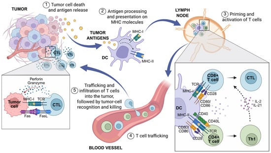
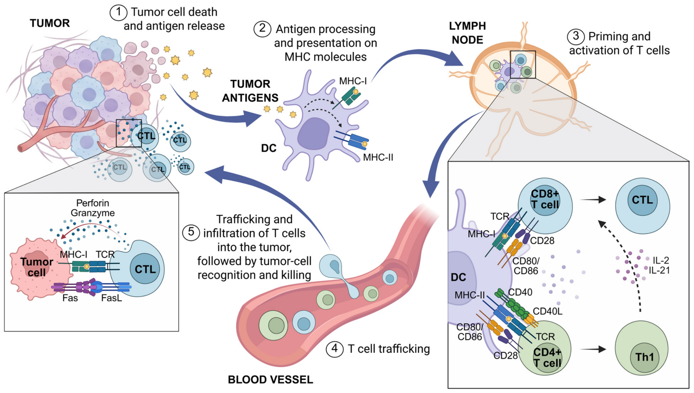
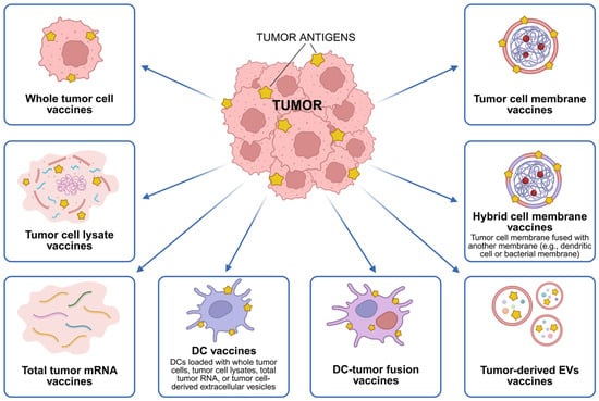
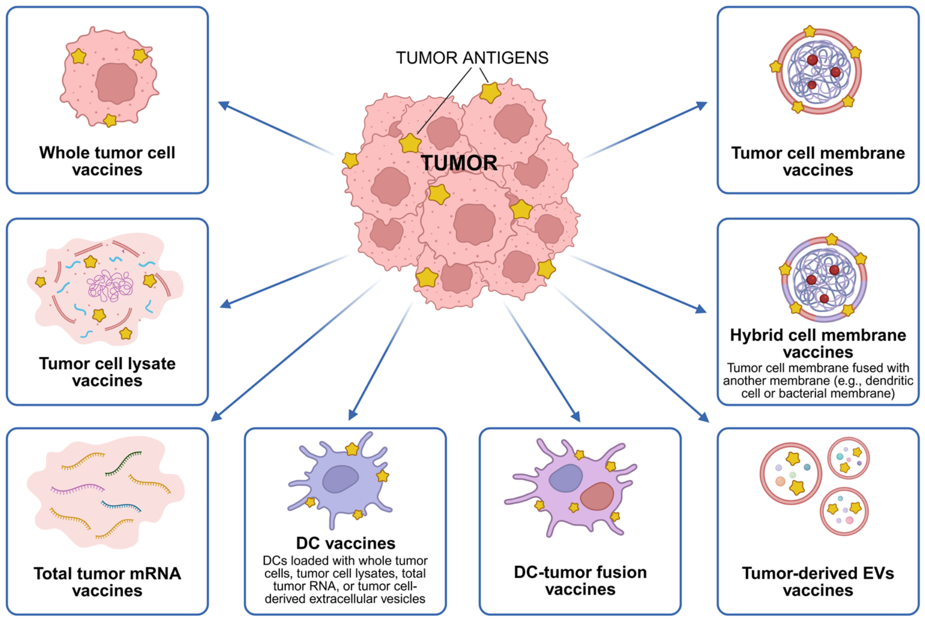

Disclaimer/Publisher’s Note: The statements, opinions and data contained in all publications are solely those of the individual author(s) and contributor(s) and not of MDPI and/or the editor(s). MDPI and/or the editor(s) disclaim responsibility for any injury to people or property resulting from any ideas, methods, instructions or products referred to in the content. |
© 2026 by the authors. Licensee MDPI, Basel, Switzerland. This article is an open access article distributed under the terms and conditions of the Creative Commons Attribution (CC BY) license.