Next Issue
Volume 9, August
Previous Issue
Volume 9, June


Viruses, Volume 9, Issue 7 (July 2017) – 36 articles

Cover Story (view full-size image): 
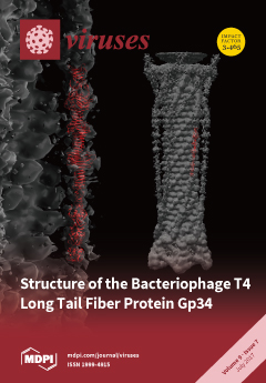
Long tail fibers of phage T4 are formed by proteins gp34, gp35, gp36, and gp37, with gp34 located at the phage-proximal end. A partial structure of gp34 revealed an extended triple β-helix domain punctuated by three β-prism domains. Between the N-terminal and the β-helix, three mixed α-β domains are located. More copies of this mixed α-β domain are present in the unsolved part of gp34 and in other T4 fiber proteins. View this paper
  • Issues are regarded as officially published after their release is announced to the table of contents alert mailing list.
  • You may sign up for e-mail alerts to receive table of contents of newly released issues.
  • PDF is the official format for papers published in both, html and pdf forms. To view the papers in pdf format, click on the "PDF Full-text" link, and use the free Adobe Reader to open them.
Order results
Result details
Section
Select all
Export citation of selected articles as:
10 pages, 1221 KiB  
Article
Garlic Organosulfur Compounds Reduce Inflammation and Oxidative Stress during Dengue Virus Infection
by Alex Hall, Andrea Troupin, Berlin Londono-Renteria and Tonya M. Colpitts *
1 Department of Pathology, Microbiology and Immunology, University of South Carolina School of Medicine, Columbia, SC 29209, USA
Current address: Department of Entomology, Kansas State University, Manhattan, KS 66506, USA
Viruses 2017, 9(7), 159; https://doi.org/10.3390/v9070159 - 23 Jun 2017
Cited by 61 | Viewed by 7619
Abstract
Dengue virus (DENV) is a mosquito-borne flavivirus that causes significant global human disease and mortality. One approach to develop treatments for DENV infection and the prevention of severe disease is through investigation of natural medicines. Inflammation plays both beneficial and harmful roles during [...] Read more.
Dengue virus (DENV) is a mosquito-borne flavivirus that causes significant global human disease and mortality. One approach to develop treatments for DENV infection and the prevention of severe disease is through investigation of natural medicines. Inflammation plays both beneficial and harmful roles during DENV infection. Studies have proposed that the oxidative stress response may be one mechanism responsible for triggering inflammation during DENV infection. Thus, blocking the oxidative stress response could reduce inflammation and the development of severe disease. Garlic has been shown to both reduce inflammation and affect the oxidative stress response. Here, we show that the garlic active compounds diallyl disulfide (DADS), diallyl sulfide (DAS) and alliin reduced inflammation during DENV infection and show that this reduction is due to the effects on the oxidative stress response. These results suggest that garlic could be used as an alternative treatment for DENV infection and for the prevention of severe disease development. Full article
(This article belongs to the Section Viral Immunology, Vaccines, and Antivirals)
Show Figures

Figure 1

14 pages, 2293 KiB  
Review
HHV-6A/B Integration and the Pathogenesis Associated with the Reactivation of Chromosomally Integrated HHV-6A/B
by Vanessa Collin 1 and Louis Flamand 1,2,*
1 Division of Infectious and Immune Diseases, CHU de Quebec Research Center-Laval University, Québec, QC G1V 4G2, Canada
2 Department of Microbiology, Infectious Disease and Immunology, Faculty of Medicine, Laval University, Québec, QC G1V 0A6, Canada
Viruses 2017, 9(7), 160; https://doi.org/10.3390/v9070160 - 26 Jun 2017
Cited by 25 | Viewed by 7070
Abstract
Unlike other human herpesviruses, human herpesvirus 6A and 6B (HHV-6A/B) infection can lead to integration of the viral genome in human chromosomes. When integration occurs in germinal cells, the integrated HHV-6A/B genome can be transmitted to 50% of descendants. Such individuals, carrying one [...] Read more.
Unlike other human herpesviruses, human herpesvirus 6A and 6B (HHV-6A/B) infection can lead to integration of the viral genome in human chromosomes. When integration occurs in germinal cells, the integrated HHV-6A/B genome can be transmitted to 50% of descendants. Such individuals, carrying one copy of the HHV-6A/B genome in every cell, are referred to as having inherited chromosomally-integrated HHV-6A/B (iciHHV-6) and represent approximately 1% of the world’s population. Interestingly, HHV-6A/B integrate their genomes in a specific region of the chromosomes known as telomeres. Telomeres are located at chromosomes’ ends and play essential roles in chromosomal stability and the long-term proliferative potential of cells. Considering that the integrated HHV-6A/B genome is mostly intact without any gross rearrangements or deletions, integration is likely used for viral maintenance into host cells. Knowing the roles played by telomeres in cellular homeostasis, viral integration in such structure is not likely to be without consequences. At present, the mechanisms and factors involved in HHV-6A/B integration remain poorly defined. In this review, we detail the potential biological and medical impacts of HHV-6A/B integration as well as the possible chromosomal integration and viral excision processes. Full article
(This article belongs to the Special Issue Viruses and Telomeres)
Show Figures

Figure 1

19 pages, 6910 KiB  
Article
Characterization of Sinorhizobium sp. LM21 Prophages and Virus-Encoded DNA Methyltransferases in the Light of Comparative Genomic Analyses of the Sinorhizobial Virome
by Przemyslaw Decewicz 1, Monika Radlinska 2 and Lukasz Dziewit 1,*
1 Department of Bacterial Genetics, Institute of Microbiology, Faculty of Biology, University of Warsaw, Miecznikowa 1, 02-096 Warsaw, Poland
2 Department of Virology, Institute of Microbiology, Faculty of Biology, University of Warsaw, Miecznikowa 1, 02-096 Warsaw, Poland
Viruses 2017, 9(7), 161; https://doi.org/10.3390/v9070161 - 26 Jun 2017
Cited by 11 | Viewed by 5955
Abstract
The genus Sinorhizobium/Ensifer mostly groups nitrogen-fixing bacteria that create root or stem nodules on leguminous plants and transform atmospheric nitrogen into ammonia, which improves the productivity of the plants. Although these biotechnologically-important bacteria are commonly found in various soil environments, little is known [...] Read more.
The genus Sinorhizobium/Ensifer mostly groups nitrogen-fixing bacteria that create root or stem nodules on leguminous plants and transform atmospheric nitrogen into ammonia, which improves the productivity of the plants. Although these biotechnologically-important bacteria are commonly found in various soil environments, little is known about their phages. In this study, the genome of Sinorhizobium sp. LM21 isolated from a heavy-metal-contaminated copper mine in Poland was investigated for the presence of prophages and DNA methyltransferase-encoding genes. In addition to the previously identified temperate phage, ΦLM21, and the phage-plasmid, pLM21S1, the analysis revealed the presence of three prophage regions. Moreover, four novel phage-encoded DNA methyltransferase (MTase) genes were identified and the enzymes were characterized. It was shown that two of the identified viral MTases methylated the same target sequence (GANTC) as cell cycle-regulated methyltransferase (CcrM) of the bacterial host strain, LM21. This discovery was recognized as an example of the evolutionary convergence between enzymes of sinorhizobial viruses and their host, which may play an important role in virus cycle. In the last part of the study, thorough comparative analyses of 31 sinorhizobial (pro)phages (including active sinorhizobial phages and novel putative prophages retrieved and manually re-annotated from Sinorhizobium spp. genomes) were performed. The networking analysis revealed the presence of highly conserved proteins (e.g., holins and endolysins) and a high diversity of viral integrases. The analysis also revealed a large number of viral DNA MTases, whose genes were frequently located within the predicted replication modules of analyzed prophages, which may suggest their important regulatory role. Summarizing, complex analysis of the phage protein similarity network enabled a new insight into overall sinorhizobial virome diversity. Full article
(This article belongs to the Special Issue Viruses of Microbes)
Show Figures

Figure 1

37 pages, 923 KiB  
Review
Type W Human Endogenous Retrovirus (HERV-W) Integrations and Their Mobilization by L1 Machinery: Contribution to the Human Transcriptome and Impact on the Host Physiopathology
by Nicole Grandi 1 and Enzo Tramontano 1,2,*
1 Department of Life and Environmental Sciences, University of Cagliari, Cittadella Universitaria di Monserrato SS554, 09042 Monserrato, Cagliari, Italy
2 Istituto di Ricerca Genetica e Biomedica, Consiglio Nazionale delle Ricerche (CNR), 09042 Monserrato, Cagliari, Italy
Viruses 2017, 9(7), 162; https://doi.org/10.3390/v9070162 - 27 Jun 2017
Cited by 61 | Viewed by 16290
Abstract
Human Endogenous Retroviruses (HERVs) are ancient infection relics constituting ~8% of our DNA. While HERVs’ genomic characterization is still ongoing, impressive amounts of data have been obtained regarding their general expression across tissues. Among HERVs, one of the most studied is the W [...] Read more.
Human Endogenous Retroviruses (HERVs) are ancient infection relics constituting ~8% of our DNA. While HERVs’ genomic characterization is still ongoing, impressive amounts of data have been obtained regarding their general expression across tissues. Among HERVs, one of the most studied is the W group, which is the sole HERV group specifically mobilized by the long interspersed element-1 (LINE-1) machinery, providing a source of novel insertions by retrotransposition of HERV-W processed pseudogenes, and comprising a member encoding a functional envelope protein coopted for human placentation. The HERV-W group has been intensively investigated for its putative role in several diseases, such as cancer, inflammation, and autoimmunity. Despite major interest in the link between HERV-W expression and human pathogenesis, no conclusive correlation has been demonstrated so far. In general, (i) the absence of a proper identification of the specific HERV-W sequences expressed in a given condition, and (ii) the lack of studies attempting to connect the various observations in the same experimental conditions are the major problems preventing the definitive assessment of the HERV-W impact on human physiopathology. In this review, we summarize the current knowledge on the HERV-W group presence within the human genome and its expression in physiological tissues as well as in the main pathological contexts. Full article
Show Figures

Figure 1

16 pages, 1738 KiB  
Article
Susceptibility and Lethality of Western Equine Encephalitis Virus in Balb/c Mice When Infected by the Aerosol Route
by Amanda L. Phelps 1,*, Lyn M. O’Brien 1, Lin S. Eastaugh 1, Carwyn Davies 1, Mark S. Lever 1, Jane Ennis 2, Larry Zeitlin 2, Alejandro Nunez 3 and David O. Ulaeto 1
1 CBR Division, Defence Science and Technology Laboratory (Dstl), Room 201, Building 7a, Porton Down, Salisbury, Wiltshire SP4 0JQ, UK
2 Mapp Biopharmaceutical Inc., 6160 Lusk Blvd. #C105, San Diego, CA 92121, USA
3 Pathology Department, Animal and Plant Health Agency, Weybrige, Woodham Lane, New Haw, Addlestone, Surrey, KT15 3NB, UK
Viruses 2017, 9(7), 163; https://doi.org/10.3390/v9070163 - 27 Jun 2017
Cited by 13 | Viewed by 4588
Abstract
Western equine encephalitis virus (WEEV) naturally cycles between mosquitos and birds or rodents, with a case fatality rate of up to 15% in humans during epizootic outbreaks. There are no medical countermeasures to treat WEEV infection, and accidental aerosol exposure increases the case [...] Read more.
Western equine encephalitis virus (WEEV) naturally cycles between mosquitos and birds or rodents, with a case fatality rate of up to 15% in humans during epizootic outbreaks. There are no medical countermeasures to treat WEEV infection, and accidental aerosol exposure increases the case fatality rate up to 40%. Understanding the pathogenesis of infection is required to develop and assess medical countermeasures. This study describes the clinical and pathological findings of mice infected with WEEV by the aerosol route, and use as a model for WEEV infection in humans. Balb/c mice were infected by the aerosol route with a dose range of high-virulence WEEV strain Fleming to establish the median lethal dose (MLD). The disease course was acute, culminating in severe clinical signs, neuroinvasion, and dose-dependent mortality. Further groups of mice were exposed by the aerosol route, periodically sacrificed, and tissues excised for histopathological examination and virology. Viral titres peaked four days post-challenge in the brain and lungs, corresponding with severe bilateral lesions in rostroventral regions of the encephalon, especially in the olfactory bulb and piriform cortex. Recapitulation of the most serious clinical presentations of human WEEV disease in mice may prove a useful tool in the evaluation of medical countermeasures. Full article
(This article belongs to the Section Viral Immunology, Vaccines, and Antivirals)
Show Figures

Graphical abstract

12 pages, 4889 KiB  
Review
LMP1 and Dynamic Progressive Telomere Dysfunction: A Major Culprit in EBV-Associated Hodgkin’s Lymphoma
by Hans Knecht 1,2,* and Sabine Mai 2
1 Division of Haematology, Department of Medicine, Jewish General Hospital, McGill University, Montréal, QC H3T 1E2, Canada
2 Manitoba Institute of Cell Biology, The Genomic Centre for Cancer Research and Diagnosis, University of Manitoba, Winnipeg, MB R3E 0V9, Canada
Viruses 2017, 9(7), 164; https://doi.org/10.3390/v9070164 - 27 Jun 2017
Cited by 18 | Viewed by 5548
Abstract
Epstein–Barr virus (EBV)-encoded latent membrane protein 1 (LMP1) is expressed in germinal-center-derived, mononuclear Hodgkin (H) and multinuclear, diagnostic Reed–Sternberg (RS) cells in classical EBV-positive Hodgkin’s lymphoma (cHL). LMP1 expression in EBV-negative H-cell lines results in a significantly increased number of RS cells. In [...] Read more.
Epstein–Barr virus (EBV)-encoded latent membrane protein 1 (LMP1) is expressed in germinal-center-derived, mononuclear Hodgkin (H) and multinuclear, diagnostic Reed–Sternberg (RS) cells in classical EBV-positive Hodgkin’s lymphoma (cHL). LMP1 expression in EBV-negative H-cell lines results in a significantly increased number of RS cells. In a conditional, germinal-center-derived B-cell in vitro system, LMP1 reversibly down-regulates the shelterin proteins, telomeric repeat binding factor (TRF)1, TRF2, and protection of telomeres (POT)1. This down-regulation is associated with progressive 3D shelterin disruption, resulting in telomere dysfunction, progression of complex chromosomal rearrangements, and multinuclearity. TRF2 appears to be the key player. Thus, we hypothesize that the 3D interaction of telomeres and TRF2 is disrupted in H cells, and directly associated with the formation of H and RS cells. Using quantitative 3D co-immuno-TRF2-telomere fluorescent in situ hybridization (3D TRF2/Telo-Q-FISH) applied to monolayers of primary H and RS cells, we demonstrate TRF2-telomere dysfunction in EBV-positive cHL. However, in EBV-negative cHL a second molecular mechanism characterized by massive up-regulation of TRF2, but attrition of telomere signals, is also identified. These facts point towards a shelterin-related pathogenesis of cHL, where two molecularly disparate mechanisms converge at the level of 3D Telomere–TRF2 interactions, leading to the formation of RS cells. Full article
(This article belongs to the Special Issue Viruses and Telomeres)
Show Figures

Figure 1

18 pages, 12709 KiB  
Article
Effective Suckling C57BL/6, Kunming, and BALB/c Mouse Models with Remarkable Neurological Manifestation for Zika Virus Infection
by Jianhai Yu 1,†, Xuling Liu 1,†, Changwen Ke 2,†, Qinghua Wu 1,†, Weizhi Lu 1, Zhiran Qin 1, Xiaoen He 1, Yujing Liu 1, Jieli Deng 1, Suiqi Xu 1, Ying Li 1, Li Zhu 1, Chengsong Wan 1, Qiwei Zhang 1, Weiwei Xiao 1, Qian Xie 1, Bao Zhang 1,* and Wei Zhao 1,3,*
1 Guangdong Provincial Key Laboratory of Tropical Disease Research, School of Public Health, Southern Medical University, Guangzhou 510515, China
2 Institute of Microbiology, Center for Diseases Control and Prevention of Guangdong Province, 176 Xin Gang West Road, Guangzhou, Guangdong 510300, China
3 Guangzhou Key Laboratory of Drug Research for Emerging Virus Prevention and Treatment, School of Pharmacy, Southern Medical University, Guangzhou 510515, China
These authors contributed equally to this work.
Viruses 2017, 9(7), 165; https://doi.org/10.3390/v9070165 - 29 Jun 2017
Cited by 31 | Viewed by 8657
Abstract
Since 2015, 84 countries and territories reported evidence of vector-borne Zika Virus (ZIKV) transmission. The World Health Organization (WHO) declared that ZIKV and associated consequences especially the neurological autoimmune disorder Guillain–Barré syndrome (GBS) and microcephaly will remain a significant enduring public health challenge [...] Read more.
Since 2015, 84 countries and territories reported evidence of vector-borne Zika Virus (ZIKV) transmission. The World Health Organization (WHO) declared that ZIKV and associated consequences especially the neurological autoimmune disorder Guillain–Barré syndrome (GBS) and microcephaly will remain a significant enduring public health challenge requiring intense action. We apply a standardization of the multi-subcutaneous dorsal inoculation method to systematically summarize clinical neurological manifestation, viral distribution, and tissue damage during the progress of viremia and systemic spread in suckling mouse models. We found that C57BL/6 and Kunming mice (KM) both showed remarkable and uniform neurologic manifestations. C57BL/6 owned the highest susceptibility and pathogenicity to the nervous system, referred to as movement disorders, with 100% incidence, while KM was an economic model for a Chinese study characterized by lower limb weakness with 62% morbidity. Slight yellow extraocular exudates were observed in BALB/c, suggesting the association with similar ocular findings to those of clinical cases. The virus distribution and pathological changes in the sera, brains, livers, kidneys, spleens, and testes during disease progression had strong regularity and uniformity, demonstrating the effectiveness and plasticity of the animal models. The successful establishment of these animal models will be conducive to expound the pathogenic mechanism of GBS. Full article
Show Figures

Figure 1

19 pages, 2781 KiB  
Article
Systemic Propagation of a Fluorescent Infectious Clone of a Polerovirus Following Inoculation by Agrobacteria and Aphids
by Sylvaine Boissinot 1, Elodie Pichon 1,2, Céline Sorin 3,4, Céline Piccini 3, Danièle Scheidecker 3, Véronique Ziegler-Graff 3 and Véronique Brault 1,*
1 Université de Strasbourg, Institut National de la Recherche Agronomique, SVQV UMR-A 1131, 68000 Colmar, France
2 UMR 385 BGPI, Institut National de la Recherche Agronomique—Centre de Coopération Internationale en Recherche Agronomique pour le Développement, SupAgro, CIRAD TA-A54/K, Campus International de Baillarguet, 34398 Montpellier, France
3 Institut de Biologie Moléculaire des Plantes, Centre National de la Recherche Scientifique, UPR 2357, Université de Strasbourg, 12 rue du Général Zimmer, 67084 Strasbourg, France
4 Institute of Plant Science Paris Saclay (IPS2), CNRS, INRA, University Paris Diderot, University of Paris-Saclay, 91405 Orsay, France
Viruses 2017, 9(7), 166; https://doi.org/10.3390/v9070166 - 29 Jun 2017
Cited by 24 | Viewed by 6971
Abstract
A fluorescent viral clone of the polerovirus Turnip yellows virus (TuYV) was engineered by introducing the Enhanced Green Fluorescent Protein (EGFP) sequence into the non-structural domain sequence of the readthrough protein, a minor capsid protein. The resulting recombinant virus, referred to as TuYV-RT [...] Read more.
A fluorescent viral clone of the polerovirus Turnip yellows virus (TuYV) was engineered by introducing the Enhanced Green Fluorescent Protein (EGFP) sequence into the non-structural domain sequence of the readthrough protein, a minor capsid protein. The resulting recombinant virus, referred to as TuYV-RTGFP, was infectious in several plant species when delivered by agroinoculation and invaded efficiently non-inoculated leaves. As expected for poleroviruses, which infect only phloem cells, the fluorescence emitted by TuYV-RTGFP was restricted to the vasculature of infected plants. In addition, TuYV-RTGFP was aphid transmissible and enabled the observation of the initial sites of infection in the phloem after aphid probing in epidermal cells. The aphid-transmitted virus moved efficiently to leaves distant from the inoculation sites and importantly retained the EGFP sequence in the viral genome. This work reports on the first engineered member in the Luteoviridae family that can be visualized by fluorescence emission in systemic leaves of different plant species after agroinoculation or aphid transmission. Full article
(This article belongs to the Section Viruses of Plants, Fungi and Protozoa)
Show Figures

Figure 1

15 pages, 3343 KiB  
Article
Rotavirus Genomic RNA Complex Forms via Specific RNA–RNA Interactions: Disruption of RNA Complex Inhibits Virus Infectivity
by Teodoro Fajardo Jr., Po-Yu Sung, Cristina C. Celma and Polly Roy *
1 Department of Pathogen Molecular Biology, Faculty of Infectious and Tropical Diseases, London School of Hygiene and Tropical Medicine, London WC1E 7HT, UK
These authors contributed equally.
Viruses 2017, 9(7), 167; https://doi.org/10.3390/v9070167 - 29 Jun 2017
Cited by 24 | Viewed by 6103
Abstract
Rotavirus (RV), a member of the Reoviridae family, causes infection in children and infants, with high morbidity and mortality. To be viable, the virus particle must package a set of eleven RNA segments. In order to understand the packaging mechanism, here, we co-synthesized [...] Read more.
Rotavirus (RV), a member of the Reoviridae family, causes infection in children and infants, with high morbidity and mortality. To be viable, the virus particle must package a set of eleven RNA segments. In order to understand the packaging mechanism, here, we co-synthesized sets of RNA segments in vitro in different combinations and detected by two alternate methods: the electrophoretic mobility shift assay (EMSA) and the RNA-bead pull-down assay. We showed that viral positive-sense RNA segments interact with each other in a specific manner, forming RNA complexes, and that the RNA–RNA interactions followed a sequential order initiated by small RV segments. Further, we demonstrated that RNA complexes were perturbed by targeted specific antisense oligoribonucleotides (ORNs) complementary to short RNA sequences, indicating that the RNA–RNA interactions between different segments were sequence-specific. The same inhibitory ORNs also had the capability to inhibit virus replication. The combined in vitro and in vivo data inferred that RNA–RNA interactions and specific complex formation are essential for sorting different segments, possibly prior to, or during, genome packaging. As genome assembly is a universal requirement in the Reoviridae family, this work offers an approach towards a further understanding of the sorting and packaging mechanisms of RV and related dsRNA (double-stranded RNA) viruses. Full article
(This article belongs to the Section Animal Viruses)
Show Figures

Figure 1

12 pages, 4836 KiB  
Article
Crystal Structure of the Carboxy-Terminal Region of the Bacteriophage T4 Proximal Long Tail Fiber Protein Gp34
by Meritxell Granell 1,†, Mikiyoshi Namura 2, Sara Alvira 1,3,‡, Shuji Kanamaru 2,* and Mark J. Van Raaij 1,3,*
1 Departmento de Estructura de Macromoleculas, Centro Nacional de Biotecnologia (CNB-CSIC), Calle Darwin 3, E-28049 Madrid, Spain
2 Department of Life Science and Technology, Tokyo Institute of Technology, M6-11 2-12-1 Ookayama, Meguro-ku Tokyo 152-8550, Japan
3 Departmento Bioquimica y Biologia Molecular, Facultad de Farmacia, Universidad de Santiago de Compostela, E-15782 Santiago de Compostela, Spain
Current address: Centre de Biochimie Structurale, CNRS UMR 5048-UM-INSERM U 1054, 29 rue de Navacelles, 34090 Montpellier, France
Current address: School of Biochemistry, University of Bristol, Biomedical Sciences Building, University Walk, Clifton, Bristol BS8 1TD, UK
Viruses 2017, 9(7), 168; https://doi.org/10.3390/v9070168 - 30 Jun 2017
Cited by 25 | Viewed by 9083
Abstract
Long tail fibers of bacteriophage T4 are formed by proteins gp34, gp35, gp36, and gp37, with gp34 located at the phage-proximal end and gp37 at the phage-distal, receptor-binding end. We have solved the structure of the carboxy-terminal region of gp34, consisting of amino [...] Read more.
Long tail fibers of bacteriophage T4 are formed by proteins gp34, gp35, gp36, and gp37, with gp34 located at the phage-proximal end and gp37 at the phage-distal, receptor-binding end. We have solved the structure of the carboxy-terminal region of gp34, consisting of amino acids 894–1289, by single-wavelength anomalous diffraction and extended the structure to amino acids 744–1289 using data collected from crystals containing longer gp34-fragments. The structure reveals three repeats of a mixed α-β fibrous domain in residues 744 to 877. A triple-helical neck connects to an extended triple β-helix domain (amino acids 900–1127) punctuated by two β-prism domains. Next, a β-prism domain decorated with short helices and extended β-helices is present (residues 1146–1238), while the C-terminal end is capped with another short β-helical region and three β-hairpins. The structure provides insight into the stability of the fibrous gp34 protein. Full article
(This article belongs to the Special Issue Viruses of Microbes)
Show Figures

Graphical abstract

15 pages, 3803 KiB  
Article
2BC Non-Structural Protein of Enterovirus A71 Interacts with SNARE Proteins to Trigger Autolysosome Formation
by Jeffrey K. F. Lai 1, I-Ching Sam 1, Pauline Verlhac 2,3,4,5,6, Joël Baguet 2,3,4,5,6, Eeva-Liisa Eskelinen 7, Mathias Faure 2,3,4,5,6,8,9 and Yoke Fun Chan 1,*
1 Department of Medical Microbiology, Faculty of Medicine, University Malaya, 50603 Kuala Lumpur, Malaysia
2 CIRI, International Center for Infectiology Research, Université de Lyon, 69007 Lyon, France
3 INSERM, U1111, 69007 Lyon, France
4 CNRS, UMR5308, 69007 Lyon, France
5 Ecole Normale Supérieure de Lyon, 69007 Lyon, France
6 Université Lyon 1, Centre International de Recherche en Infectiologie, 69365 Lyon, France
7 Department of Biosciences, Division of Biochemistry and Biotechnology, University of Helsinki, 00014 Helsinki, Finland
8 Institut Universitaire de France, 75231 Paris, France
9 Equipe labellisée Fondation pour la Recherche Médicale FRM, 75007 Paris, France
Viruses 2017, 9(7), 169; https://doi.org/10.3390/v9070169 - 4 Jul 2017
Cited by 31 | Viewed by 8525
Abstract
Viruses have evolved unique strategies to evade or subvert autophagy machinery. Enterovirus A71 (EV-A71) induces autophagy during infection in vitro and in vivo. In this study, we report that EV-A71 triggers autolysosome formation during infection in human rhabdomyosarcoma (RD) cells to facilitate its [...] Read more.
Viruses have evolved unique strategies to evade or subvert autophagy machinery. Enterovirus A71 (EV-A71) induces autophagy during infection in vitro and in vivo. In this study, we report that EV-A71 triggers autolysosome formation during infection in human rhabdomyosarcoma (RD) cells to facilitate its replication. Blocking autophagosome-lysosome fusion with chloroquine inhibited virus RNA replication, resulting in lower viral titres, viral RNA copies and viral proteins. Overexpression of the non-structural protein 2BC of EV-A71 induced autolysosome formation. Yeast 2-hybrid and co-affinity purification assays showed that 2BC physically and specifically interacted with a N-ethylmaleimide-sensitive factor attachment receptor (SNARE) protein, syntaxin-17 (STX17). Co-immunoprecipitation assay further showed that 2BC binds to SNARE proteins, STX17 and synaptosome associated protein 29 (SNAP29). Transient knockdown of STX17, SNAP29, and microtubule-associated protein 1 light chain 3B (LC3B), crucial proteins in the fusion between autophagosomes and lysosomes) as well as the lysosomal-associated membrane protein 1 (LAMP1) impaired production of infectious EV-A71 in RD cells. Collectively, these results demonstrate that the generation of autolysosomes triggered by the 2BC non-structural protein is important for EV-A71 replication, revealing a potential molecular pathway targeted by the virus to exploit autophagy. This study opens the possibility for the development of novel antivirals that specifically target 2BC to inhibit formation of autolysosomes during EV-A71 infection. Full article
(This article belongs to the Special Issue Viruses and Autophagy)
Show Figures

Graphical abstract

15 pages, 3281 KiB  
Review
Update on Senecavirus Infection in Pigs
by Raquel A. Leme 1,2, Alice F. Alfieri 1,2 and Amauri A. Alfieri 1,2,*
1 Laboratory of Animal Virology, Department of Veterinary Preventive Medicine, Universidade Estadual de Londrina, P.O. Box 10011, Paraná 86057-970, Brazil
2 Multi-User Animal Health Laboratory, Molecular Biology Unit, Department of Veterinary Preventive Medicine, Universidade Estadual de Londrina, P.O. Box 10011, Paraná 86057-970, Brazil
Viruses 2017, 9(7), 170; https://doi.org/10.3390/v9070170 - 3 Jul 2017
Cited by 97 | Viewed by 12560
Abstract
Senecavirus A (SVA) is a positive-sense single-stranded RNA virus that belongs to the Senecavirus genus within the Picornaviridae family. The virus has been silently circulating in pig herds of the USA since 1988. However, cases of senecavirus-associated vesicular disease were reported in Canada [...] Read more.
Senecavirus A (SVA) is a positive-sense single-stranded RNA virus that belongs to the Senecavirus genus within the Picornaviridae family. The virus has been silently circulating in pig herds of the USA since 1988. However, cases of senecavirus-associated vesicular disease were reported in Canada in 2007 and in the USA in 2012. Since late 2014 and early 2015, an increasing number of senecavirus outbreaks have been reported in pigs in different producing categories, with this virus being detected in Brazil, China, and Thailand. Considering the novel available data on senecavirus infection and disease, 2015 may be a divisor in the epidemiology of the virus. Among the aspects that reinforce this hypothesis are the geographical distribution of the virus, the affected pig-producing categories, clinical signs associated with the infection, and disease severity. This review presents the current knowledge regarding the senecavirus infection and disease, especially in the last two years. Senecavirus epidemiology, pathogenic potential, host immunological response, diagnosis, and prophylaxis and control measures are addressed. Perspectives are focused on the need for complete evolutionary, epidemiological and pathogenic data and the capability for an immediate diagnosis of senecavirus infection. The health risks inherent in the swine industry cannot be neglected. Full article
(This article belongs to the Special Issue Porcine Viruses)
Show Figures

Figure 1

15 pages, 4128 KiB  
Review
Complex Virus–Host Interactions Involved in the Regulation of Classical Swine Fever Virus Replication: A Minireview
by Su Li, Jinghan Wang, Qian Yang, Muhammad Naveed Anwar, Shaoxiong Yu and Hua-Ji Qiu *
1 State Key Laboratory of Veterinary Biotechnology, Harbin Veterinary Research Institute, Chinese Academy of Agricultural Sciences, 678 Haping Road, Harbin 150069, China
S.L. and J.W. contributed equally to this article.
Viruses 2017, 9(7), 171; https://doi.org/10.3390/v9070171 - 5 Jul 2017
Cited by 33 | Viewed by 11747
Abstract
Classical swine fever (CSF), caused by classical swine fever virus (CSFV), is one of the most devastating epizootic diseases of pigs in many countries. Viruses are small intracellular parasites and thus rely on the cellular factors for replication. Fundamental aspects of CSFV–host interactions [...] Read more.
Classical swine fever (CSF), caused by classical swine fever virus (CSFV), is one of the most devastating epizootic diseases of pigs in many countries. Viruses are small intracellular parasites and thus rely on the cellular factors for replication. Fundamental aspects of CSFV–host interactions have been well described, such as factors contributing to viral attachment, modulation of genomic replication and translation, antagonism of innate immunity, and inhibition of cell apoptosis. However, those host factors that participate in the viral entry, assembly, and release largely remain to be elucidated. In this review, we summarize recent progress in the virus–host interactions involved in the life cycle of CSFV and analyze the potential mechanisms of viral entry, assembly, and release. We conclude with future perspectives and highlight areas that require further understanding. Full article
(This article belongs to the Special Issue Porcine Viruses)
Show Figures

Figure 1

19 pages, 1460 KiB  
Review
Hepatitis Delta Virus: Replication Strategy and Upcoming Therapeutic Options for a Neglected Human Pathogen
by Florian A. Lempp 1,2 and Stephan Urban 1,2,*
1 Department of Infectious Diseases, Molecular Virology, Heidelberg University, Im Neuenheimer Feld 345, 69120 Heidelberg, Germany
2 German Centre for Infection Research (DZIF), Partner Site Heidelberg, 69120 Heidelberg, Germany
Viruses 2017, 9(7), 172; https://doi.org/10.3390/v9070172 - 4 Jul 2017
Cited by 32 | Viewed by 20549
Abstract
The human Hepatitis Delta Virus (HDV) is unique among all viral pathogens. Encoding only one protein (Hepatitis Delta Antigen; HDAg) within its viroid-like self-complementary RNA, HDV constitutes the smallest known virus in the animal kingdom. To disseminate in its host, HDV depends on [...] Read more.
The human Hepatitis Delta Virus (HDV) is unique among all viral pathogens. Encoding only one protein (Hepatitis Delta Antigen; HDAg) within its viroid-like self-complementary RNA, HDV constitutes the smallest known virus in the animal kingdom. To disseminate in its host, HDV depends on a helper virus, the human Hepatitis B virus (HBV), which provides the envelope proteins required for HDV assembly. HDV affects an estimated 15–20 million out of the 240 million chronic HBV-carriers and disperses unequally in disparate geographical regions of the world. The disease it causes (chronic Hepatitis D) presents as the most severe form of viral hepatitis, leading to accelerated progression of liver dysfunction including cirrhosis and hepatocellular carcinoma and a high mortality rate. The lack of approved drugs interfering with specific steps of HDV replication poses a high burden for gaining insights into the molecular biology of the virus and, consequently, the development of specific novel medications that resiliently control HDV replication or, in the best case, functionally cure HDV infection or HBV/HDV co-infection. This review summarizes our current knowledge of HBV molecular biology, presents an update on novel cell culture and animal models to study the virus and provides updates on the clinical development of the three developmental drugs Lonafarnib, REP2139-Ca and Myrcludex B. Full article
(This article belongs to the Special Issue Recent Advances in Hepatitis B Virus Research)
Show Figures

Figure 1

13 pages, 1111 KiB  
Review
Telomeres and Telomerase: Role in Marek’s Disease Virus Pathogenesis, Integration and Tumorigenesis
by Ahmed Kheimar 1,2, Renato L. Previdelli 1, Darren J. Wight 1 and Benedikt B. Kaufer 1,*
1 Institut für Virologie, Freie Universität Berlin, Robert von Ostertag-Straße 7–13, 14163 Berlin, Germany
2 Department of Poultry Diseases, Faculty of Veterinary Medicine, Sohag University, Sohag 82424, Egypt
Viruses 2017, 9(7), 173; https://doi.org/10.3390/v9070173 - 4 Jul 2017
Cited by 26 | Viewed by 7513
Abstract
Telomeres protect the ends of vertebrate chromosomes from deterioration and consist of tandem nucleotide repeats (TTAGGG)n that are associated with a number of proteins. Shortening of the telomeres occurs during genome replication, thereby limiting the replication potential of somatic cells. To counteract [...] Read more.
Telomeres protect the ends of vertebrate chromosomes from deterioration and consist of tandem nucleotide repeats (TTAGGG)n that are associated with a number of proteins. Shortening of the telomeres occurs during genome replication, thereby limiting the replication potential of somatic cells. To counteract this shortening, vertebrates encode the telomerase complex that maintains telomere length in certain cell types via de novo addition of telomeric repeats. Several herpesviruses, including the highly oncogenic alphaherpesvirus Marek’s disease virus (MDV), harbor telomeric repeats (TMR) identical to the host telomere sequences at the ends of their linear genomes. These TMR facilitate the integration of the MDV genome into host telomeres during latency, allowing the virus to persist in the host for life. Integration into host telomeres is critical for disease and tumor induction by MDV, but also enables efficient reactivation of the integrated virus genome. In addition to the TMR, MDV also encodes a telomerase RNA subunit (vTR) that shares 88% sequence identity with the telomerase RNA in chicken (chTR). vTR is highly expressed during all stages of the virus lifecycle, enhances telomerase activity and plays an important role in MDV-induced tumor formation. This review will focus on the recent advances in understanding the role of viral TMR and vTR in MDV pathogenesis, integration and tumorigenesis. Full article
(This article belongs to the Special Issue Viruses and Telomeres)
Show Figures

Figure 1

11 pages, 885 KiB  
Review
Virus/Host Cell Crosstalk in Hypoxic HPV-Positive Cancer Cells
by Karin Hoppe-Seyler 1, Julia Mändl 1,2, Svenja Adrian 1, Bianca J. Kuhn 1 and Felix Hoppe-Seyler 1,*
1 Molecular Therapy of Virus-Associated Cancers (F065), German Cancer Research Center (DKFZ), D-69120 Heidelberg, Germany
2 Viral Transformation Mechanisms (F030), German Cancer Research Center (DKFZ), D-69120 Heidelberg, Germany
Viruses 2017, 9(7), 174; https://doi.org/10.3390/v9070174 - 5 Jul 2017
Cited by 15 | Viewed by 8506
Abstract
Oncogenic types of human papillomaviruses (HPVs) are major human carcinogens. The expression of the viral E6/E7 oncogenes plays a key role for HPV-linked oncogenesis. It recently has been found that low oxygen concentrations (“hypoxia”), as present in sub-regions of HPV-positive cancers, [...] Read more.
Oncogenic types of human papillomaviruses (HPVs) are major human carcinogens. The expression of the viral E6/E7 oncogenes plays a key role for HPV-linked oncogenesis. It recently has been found that low oxygen concentrations (“hypoxia”), as present in sub-regions of HPV-positive cancers, strongly affect the interplay between the HPV oncogenes and their transformed host cell. As a result, a state of dormancy is induced in hypoxic HPV-positive cancer cells, which is characterized by a shutdown of viral oncogene expression and a proliferative arrest that can be reversed by reoxygenation. In this review, these findings are put into the context of the current concepts of both HPV-linked carcinogenesis and of the effects of hypoxia on tumor biology. Moreover, we discuss the consequences for the phenotype of HPV-positive cancer cells as well as for their clinical behavior and response towards established and prospective therapeutic strategies. Full article
(This article belongs to the Special Issue Expert Views on HPV Infection)
Show Figures

Figure 1

17 pages, 2343 KiB  
Article
SOX2 as a New Regulator of HPV16 Transcription
by Imelda Martínez-Ramírez 1, Víctor Del-Castillo-Falconi 1, Irma B. Mitre-Aguilar 2, Alfredo Amador-Molina 1, Adela Carrillo-García 1, Elizabeth Langley 1, Alejandro Zentella-Dehesa 2,3, Ernesto Soto-Reyes 1, Alejandro García-Carrancá 1, Luis A. Herrera 1,3 and Marcela Lizano 1,3,*
1 Unidad de Investigación Biomédica en Cáncer, Instituto Nacional de Cancerología (INCan)/Instituto de Investigaciones Biomédicas, Universidad Nacional Autónoma de México (UNAM), Ciudad de México 14080, Mexico
2 Unidad de Bioquímica, Instituto Nacional de Ciencias Médicas y Nutrición Salvador Zubirán (INCMNSZ)/Unidad Periférica del Instituto de Investigaciones Biomédicas, Universidad Nacional Autónoma de México (UNAM), Ciudad de México 14080, Mexico
3 Departamento de Medicina Genómica y Toxicología Ambiental, Instituto de Investigaciones Biomédicas, Universidad Nacional Autónoma de México (UNAM), Ciudad de México 04510, Mexico
Viruses 2017, 9(7), 175; https://doi.org/10.3390/v9070175 - 5 Jul 2017
Cited by 21 | Viewed by 5970
Abstract
Persistent infections with high-risk human papillomavirus (HPV) constitute the main risk factor for cervical cancer development. HPV16 is the most frequent type associated to squamous cell carcinomas (SCC), followed by HPV18. The long control region (LCR) in the HPV genome contains the replication [...] Read more.
Persistent infections with high-risk human papillomavirus (HPV) constitute the main risk factor for cervical cancer development. HPV16 is the most frequent type associated to squamous cell carcinomas (SCC), followed by HPV18. The long control region (LCR) in the HPV genome contains the replication origin and sequences recognized by cellular transcription factors (TFs) controlling viral transcription. Altered expression of E6 and E7 viral oncogenes, modulated by the LCR, causes modifications in cellular pathways such as proliferation, leading to malignant transformation. The aim of this study was to identify specific TFs that could contribute to the modulation of high-risk HPV transcriptional activity, related to the cellular histological origin. We identified sex determining region Y (SRY)-box 2 (SOX2) response elements present in HPV16-LCR. SOX2 binding to the LCR was demonstrated by in vivo and in vitro assays. The overexpression of this TF repressed HPV16-LCR transcriptional activity, as shown through reporter plasmid assays and by the down-regulation of endogenous HPV oncogenes. Site-directed mutagenesis revealed that three putative SOX2 binding sites are involved in the repression of the LCR activity. We propose that SOX2 acts as a transcriptional repressor of HPV16-LCR, decreasing the expression of E6 and E7 oncogenes in a SCC context. Full article
(This article belongs to the Section Animal Viruses)
Show Figures

Figure 1

21 pages, 583 KiB  
Review
Interplay between Autophagy, Exosomes and HIV-1 Associated Neurological Disorders: New Insights for Diagnosis and Therapeutic Applications
by Chet Raj Ojha 1, Jessica Lapierre 1, Myosotys Rodriguez 1, Seth M. Dever 1, Mohammad Asad Zadeh 2, Catherine DeMarino 2, Michelle L. Pleet 2, Fatah Kashanchi 2 and Nazira El-Hage 1,*
1 Department of Immunology, Herbert Wertheim College of Medicine, Florida International University, Miami, FL 33199, USA
2 Laboratory of Molecular Virology, School of Systems Biology, George Mason University, Manassas, VA 20110, USA
Viruses 2017, 9(7), 176; https://doi.org/10.3390/v9070176 - 6 Jul 2017
Cited by 50 | Viewed by 9893
Abstract
The autophagy–lysosomal pathway mediates a degradative process critical in the maintenance of cellular homeostasis as well as the preservation of proper organelle function by selective removal of damaged proteins and organelles. In some situations, cells remove unwanted or damaged proteins and RNAs through [...] Read more.
The autophagy–lysosomal pathway mediates a degradative process critical in the maintenance of cellular homeostasis as well as the preservation of proper organelle function by selective removal of damaged proteins and organelles. In some situations, cells remove unwanted or damaged proteins and RNAs through the release to the extracellular environment of exosomes. Since exosomes can be transferred from one cell to another, secretion of unwanted material to the extracellular environment in exosomes may have an impact, which can be beneficial or detrimental, in neighboring cells. Exosome secretion is under the influence of the autophagic system, and stimulation of autophagy can inhibit exosomal release and vice versa. Neurons are particularly vulnerable to degeneration, especially as the brain ages, and studies indicate that imbalances in genes regulating autophagy are a common feature of many neurodegenerative diseases. Cognitive and motor disease associated with severe dementia and neuronal damage is well-documented in the brains of HIV-infected individuals. Neurodegeneration seen in the brain in HIV-1 infection is associated with dysregulation of neuronal autophagy. In this paradigm, we herein provide an overview on the role of autophagy in HIV-associated neurodegenerative disease, focusing particularly on the effect of autophagy modulation on exosomal release of HIV particles and how this interplay impacts HIV infection in the brain. Specific autophagy–regulating agents are being considered for therapeutic treatment and prevention of a broad range of human diseases. Various therapeutic strategies for modulating specific stages of autophagy and the current state of drug development for this purpose are also evaluated. Full article
(This article belongs to the Special Issue Viruses and Autophagy)
Show Figures

Figure 1

13 pages, 1390 KiB  
Article
Porcine Epidemic Diarrhea in Europe: In-Detail Analyses of Disease Dynamics and Molecular Epidemiology
by Dennis Hanke 1, Anne Pohlmann 1, Carola Sauter-Louis 2, Dirk Höper 1, Julia Stadler 3, Mathias Ritzmann 3, Adi Steinrigl 4, Bernd-Andreas Schwarz 5, Valerij Akimkin 6, Robert Fux 7, Sandra Blome 1,* and Martin Beer 1
1 Friedrich-Loeffler-Institut, Institute of Diagnostic Virology, D-17493 Greifswald—Insel Riems, Germany
2 Friedrich-Loeffler-Institut, Institute of Epidemiology, D-17493 Greifswald—Insel Riems, Germany
3 Clinic for Swine, Ludwig-Maximilians-University Munich, D-85764 Oberschleissheim, Germany
4 Österreichische Agentur für Gesundheit und Ernährungssicherheit GmbH, A-2340 Mödling, Austria
5 Vaxxinova GmbH, Standort Leipzig, D-04103 Leipzig, Germany
6 Chemisches und Veterinäruntersuchungsamt Stuttgart, Fellbach, D-70736 Fellbach, Germany, Valerij.Akimkin@cvuas.bwl.de
7 Institute for Infectious Diseases and Zoonoses, Ludwig-Maximilians-University Munich, D-80539 Munich, Germany
Viruses 2017, 9(7), 177; https://doi.org/10.3390/v9070177 - 6 Jul 2017
Cited by 70 | Viewed by 8338
Abstract
Porcine epidemic diarrhea (PED) is an acute and highly contagious enteric disease of swine caused by the eponymous virus (PEDV) which belongs to the genus Alphacoronavirus within the Coronaviridae virus family. Following the disastrous outbreaks in Asia and the United States, PEDV has [...] Read more.
Porcine epidemic diarrhea (PED) is an acute and highly contagious enteric disease of swine caused by the eponymous virus (PEDV) which belongs to the genus Alphacoronavirus within the Coronaviridae virus family. Following the disastrous outbreaks in Asia and the United States, PEDV has been detected also in Europe. In order to better understand the overall situation, the molecular epidemiology, and factors that might influence the most variable disease impact; 40 samples from swine feces were collected from different PED outbreaks in Germany and other European countries and sequenced by shot-gun next-generation sequencing. A total of 38 new PEDV complete coding sequences were generated. When compared on a global scale, all investigated sequences from Central and South-Eastern Europe formed a rather homogeneous PEDV S INDEL cluster, suggesting a recent re-introduction. However, in-detail analyses revealed two new clusters and putative ancestor strains. Based on the available background data, correlations between clusters and location, farm type or clinical presentation could not be established. Additionally, the impact of secondary infections was explored using the metagenomic data sets. While several coinfections were observed, no correlation was found with disease courses. However, in addition to the PEDV genomes, ten complete viral coding sequences from nine different data sets were reconstructed each representing new virus strains. In detail, three pasivirus A strains, two astroviruses, a porcine sapelovirus, a kobuvirus, a porcine torovirus, a posavirus, and an enterobacteria phage were almost fully sequenced. Full article
(This article belongs to the Section Animal Viruses)
Show Figures

Figure 1

15 pages, 3164 KiB  
Article
Almond Skin Extracts Abrogate HSV-1 Replication by Blocking Virus Binding to the Cell
by Carlo Bisignano, Giuseppina Mandalari, Antonella Smeriglio, Domenico Trombetta, Maria Musarra Pizzo, Rosamaria Pennisi and Maria Teresa Sciortino *
Department of Chemical Biological Pharmaceutical and Environmental Sciences, University of Messina, Messina 98166, Italy
Viruses 2017, 9(7), 178; https://doi.org/10.3390/v9070178 - 10 Jul 2017
Cited by 62 | Viewed by 7898
Abstract
The aim of the present research was to determine the effect of almond skin extracts on herpes simplex virus 1 (HSV-1) replication. Drug-resistant strains of HSV frequently develop following therapeutic treatment. Therefore, the discovery of novel anti-HSV drugs deserves great effort. Here, we [...] Read more.
The aim of the present research was to determine the effect of almond skin extracts on herpes simplex virus 1 (HSV-1) replication. Drug-resistant strains of HSV frequently develop following therapeutic treatment. Therefore, the discovery of novel anti-HSV drugs deserves great effort. Here, we tested both natural (NS) and blanched (BS) polyphenols-rich almond skin extracts against HSV-1. HPLC analysis showed that the prevalent compounds in NS and BS extracts contributing to their antioxidant activity were quercetin, epicatechin and catechin. Results of cell viability indicated that NS and BS extracts were not toxic to cultured Vero cells. Furthermore, NS extracts were more potent inhibitors of HSV-1 than BS extracts, and this trend was in agreement with different concentrations of flavonoids. The plaque forming assay, Western blot and real-time PCR were used to demonstrate that NS extracts were able to block the production of infectious HSV-1 particles. In addition, the viral binding assay demonstrated that NS extracts inhibited HSV-1 adsorption to Vero cells. Our conclusion is that natural products from almond skin extracts are an extraordinary source of antiviral agents and provide a novel treatment against HSV-1 infections. Full article
(This article belongs to the Section Viral Immunology, Vaccines, and Antivirals)
Show Figures

Figure 1

12 pages, 4316 KiB  
Article
Development of Polioencephalomyelitis in Cesarean-Derived Colostrum-Deprived Pigs Following Experimental Inoculation with Either Teschovirus A Serotype 2 or Serotype 11
by Franco Matias Ferreyra, Bailey Arruda, Gregory Stevenson, Kent Schwartz, Darin Madson, Kyoung-Jin Yoon, Jianqiang Zhang, Pablo Piñeyro, Qi Chen and Paulo Arruda *
Veterinary Diagnostic Laboratory, Department of Veterinary Diagnostic and Production Animal Medicine, Iowa State University, 1850 Christensen Drive, Ames, IA 50011-1134, USA
Viruses 2017, 9(7), 179; https://doi.org/10.3390/v9070179 - 8 Jul 2017
Cited by 17 | Viewed by 5365
Abstract
Teschovirus encephalomyelitis is a sporadic disease associated with Teschovirus A (PTV) serotype 1 and, less frequently, other serotypes. In recent years, the number of cases submitted to the Iowa State University Veterinary Diagnostic Laboratory with a history of posterior paresis has increased. Submission [...] Read more.
Teschovirus encephalomyelitis is a sporadic disease associated with Teschovirus A (PTV) serotype 1 and, less frequently, other serotypes. In recent years, the number of cases submitted to the Iowa State University Veterinary Diagnostic Laboratory with a history of posterior paresis has increased. Submission histories from various regions of the United States suggest a trend for clinical disease to persist in herds and affect a wider age-range of pigs than historically reported. Polioencephalitis and/or myelitis was consistently present and PTV was detected in affected neural tissue by PCR in a portion of cases. Sequencing from two clinical cases identified PTV-2 and PTV-11. To assess neuropathogenicity of these isolates, 5-week-old cesarean derived and colostrum-deprived pigs were assigned to three groups: negative control (n = 4), PTV-2-inoculated (n = 7), and PTV-11-inoculated (n = 7). Three PTV-2-inoculated pigs developed mild incoordination of the hind limbs, one of which progressed to posterior ataxia. While all PTV-11-inoculated pigs showed severe neurological signs consistent with Teschovirus encephalomyelitis, no evidences of neurological signs were observed in sham-inoculated animals. All PTV-2- and PTV-11-inoculated pigs had microscopic lesions consistent with Teschovirus encephalomyelitis. To our knowledge, this is the first description of PTV-11 and experimental study demonstrating the neuropathogenicity of PTV-11 in the United States. Full article
(This article belongs to the Section Animal Viruses)
Show Figures

Figure 1

12 pages, 481 KiB  
Review
Telomerase Induction in HPV Infection and Oncogenesis
by Rachel Katzenellenbogen 1,2
1 Center for Global Infectious Disease Research, Seattle Children’s Research Institute, Seattle, WA 98101, USA
2 Department of Pediatrics, Division of Adolescent Medicine, University of Washington, Seattle, WA 98195, USA
Viruses 2017, 9(7), 180; https://doi.org/10.3390/v9070180 - 10 Jul 2017
Cited by 44 | Viewed by 7005
Abstract
Telomerase extends the repetitive DNA at the ends of linear chromosomes, and it is normally active in stem cells. When expressed in somatic diploid cells, it can lead to cellular immortalization. Human papillomaviruses (HPVs) are associated with and high-risk for cancer activate telomerase [...] Read more.
Telomerase extends the repetitive DNA at the ends of linear chromosomes, and it is normally active in stem cells. When expressed in somatic diploid cells, it can lead to cellular immortalization. Human papillomaviruses (HPVs) are associated with and high-risk for cancer activate telomerase through the catalytic subunit of telomerase, human telomerase reverse transcriptase (hTERT). The expression of hTERT is affected by both high-risk HPVs, E6 and E7. Seminal studies over the last two decades have identified the transcriptional, epigenetic, and post-transcriptional roles high-risk E6 and E7 have in telomerase induction. This review will summarize these findings during infection and highlight the importance of telomerase activation as an oncogenic pathway in HPV-associated cancer development and progression. Full article
(This article belongs to the Special Issue Expert Views on HPV Infection)
Show Figures

Figure 1

24 pages, 3762 KiB  
Article
PCR-DGGE Analysis: Unravelling Complex Mixtures of Badnavirus Sequences Present in Yam Germplasm
by Aliyu A. Turaki 1,2,†, Moritz Bömer 1,*,†, Gonçalo Silva 1, P. Lava Kumar 3 and Susan E. Seal 1
1 Natural Resources Institute, University of Greenwich, Central Avenue, Chatham Maritime, Kent ME4 4TB, UK
2 Kebbi State University of Science and Technology Aliero, Sokoto Road, PMB 1144 Birnin Kebbi, Nigeria
3 International Institute of Tropical Agriculture (IITA), Oyo Road, PMB 5320 Ibadan, Nigeria
These authors contributed equally to the work.
Viruses 2017, 9(7), 181; https://doi.org/10.3390/v9070181 - 11 Jul 2017
Cited by 12 | Viewed by 10486
Abstract
Badnaviruses (family Caulimoviridae, genus Badnavirus) have emerged as serious pathogens especially affecting the cultivation of tropical crops. Badnavirus sequences can be integrated in host genomes, complicating the detection of episomal infections and the assessment of viral genetic diversity in samples containing [...] Read more.
Badnaviruses (family Caulimoviridae, genus Badnavirus) have emerged as serious pathogens especially affecting the cultivation of tropical crops. Badnavirus sequences can be integrated in host genomes, complicating the detection of episomal infections and the assessment of viral genetic diversity in samples containing a complex mixture of sequences. Yam (Dioscorea spp.) plants are hosts to a diverse range of badnavirus species, and recent findings have suggested that mixed infections occur frequently in West African yam germplasm. Historically, the determination of the diversity of badnaviruses present in yam breeding lines has been achieved by cloning and sequencing of polymerase chain reaction (PCR) products. In this study, the molecular diversity of partial reverse transcriptase (RT)-ribonuclease H (RNaseH) sequences from yam badnaviruses was analysed using PCR-dependent denaturing gradient gel electrophoresis (PCR-DGGE). This resulted in the identification of complex ‘fingerprints’ composed of multiple sequences of Dioscorea bacilliform viruses (DBVs). Many of these sequences show high nucleotide identities to endogenous DBV (eDBV) sequences deposited in GenBank, and fall into six monophyletic species groups. Our findings highlight PCR-DGGE as a powerful tool in badnavirus diversity studies enabling a rapid indication of sequence diversity as well as potential candidate integrated sequences revealed by their conserved nature across germplasm. Full article
(This article belongs to the Section Viruses of Plants, Fungi and Protozoa)
Show Figures

Graphical abstract

14 pages, 1178 KiB  
Review
The Interaction between Nidovirales and Autophagy Components
by Yingying Cong, Pauline Verlhac and Fulvio Reggiori *
1 Department of Cell Biology, University of Groningen, University Medical Center Groningen, A. Deusinglaan 1, 9713 AV Groningen, The Netherlands
Equal contribution.
Viruses 2017, 9(7), 182; https://doi.org/10.3390/v9070182 - 11 Jul 2017
Cited by 33 | Viewed by 19893
Abstract
Autophagy is a conserved intracellular catabolic pathway that allows cells to maintain homeostasis through the degradation of deleterious components via specialized double-membrane vesicles called autophagosomes. During the past decades, it has been revealed that numerous pathogens, including viruses, usurp autophagy in order to [...] Read more.
Autophagy is a conserved intracellular catabolic pathway that allows cells to maintain homeostasis through the degradation of deleterious components via specialized double-membrane vesicles called autophagosomes. During the past decades, it has been revealed that numerous pathogens, including viruses, usurp autophagy in order to promote their propagation. Nidovirales are an order of enveloped viruses with large single-stranded positive RNA genomes. Four virus families (Arterividae, Coronaviridae, Mesoniviridae, and Roniviridae) are part of this order, which comprises several human and animal pathogens of medical and veterinary importance. In host cells, Nidovirales induce membrane rearrangements including autophagosome formation. The relevance and putative mechanism of autophagy usurpation, however, remain largely elusive. Here, we review the current knowledge about the possible interplay between Nidovirales and autophagy. Full article
(This article belongs to the Special Issue Viruses and Autophagy)
Show Figures

Figure 1

11 pages, 1333 KiB  
Article
Variability of Emaravirus Species Associated with Sterility Mosaic Disease of Pigeonpea in India Provides Evidence of Segment Reassortment
by Basavaprabhu L. Patil *, Meenakshi Dangwal and Ritesh Mishra
ICAR-National Research Centre on Plant Biotechnology, IARI, Pusa Campus, New Delhi 110012, India
Viruses 2017, 9(7), 183; https://doi.org/10.3390/v9070183 - 11 Jul 2017
Cited by 32 | Viewed by 6018
Abstract
Sterility mosaic disease (SMD) of pigeonpea is a serious constraint for cultivation of pigeonpea in India and other South Asian countries. SMD of pigeonpea is associated with two distinct emaraviruses, Pigeonpea sterility mosaic virus 1 (PPSMV-1) and Pigeonpea sterility mosaic virus 2 (PPSMV-2), [...] Read more.
Sterility mosaic disease (SMD) of pigeonpea is a serious constraint for cultivation of pigeonpea in India and other South Asian countries. SMD of pigeonpea is associated with two distinct emaraviruses, Pigeonpea sterility mosaic virus 1 (PPSMV-1) and Pigeonpea sterility mosaic virus 2 (PPSMV-2), with genomes consisting of five and six negative-sense RNA segments, respectively. The recently published genome sequences of both PPSMV-1 and PPSMV-2 are from a single location, Patancheru from the state of Telangana in India. However, here we present the first report of sequence variability among 23 isolates of PPSMV-1 and PPSMV-2, collected from ten locations representing six states of India. Both PPSMV-1 and PPSMV-2 are shown to be present across India and to exhibit considerable sequence variability. Variability of RNA3 sequences was higher than the RNA4 sequences for both PPSMV-1 and PPSMV-2. Additionally, the sixth RNA segment (RNA6), previously reported to be associated with only PPSMV-2, is also associated with isolates of PPSMV-1. Multiplex reverse transcription PCR (RT-PCR) analyses show that PPSMV-1 and PPSMV-2 frequently occur as mixed infections. Further sequence analyses indicated the presence of reassortment of RNA4 between isolates of PPSMV-1 and PPSMV-2. Full article
(This article belongs to the Section Viruses of Plants, Fungi and Protozoa)
Show Figures

Figure 1

14 pages, 1297 KiB  
Review
Chromosomally Integrated Human Herpesvirus 6: Models of Viral Genome Release from the Telomere and Impacts on Human Health
by Michael L. Wood and Nicola J. Royle *
Department of Genetics, University of Leicester, University Road, Leicester LE1 7RH, UK
Viruses 2017, 9(7), 184; https://doi.org/10.3390/v9070184 - 12 Jul 2017
Cited by 20 | Viewed by 6449
Abstract
Human herpesvirus 6A and 6B, alongside some other herpesviruses, have the striking capacity to integrate into telomeres, the terminal repeated regions of chromosomes. The chromosomally integrated forms, ciHHV-6A and ciHHV-6B, are proposed to be a state of latency and it has been shown [...] Read more.
Human herpesvirus 6A and 6B, alongside some other herpesviruses, have the striking capacity to integrate into telomeres, the terminal repeated regions of chromosomes. The chromosomally integrated forms, ciHHV-6A and ciHHV-6B, are proposed to be a state of latency and it has been shown that they can both be inherited if integration occurs in the germ line. The first step in full viral reactivation must be the release of the integrated viral genome from the telomere and here we propose various models of this release involving transcription of the viral genome, replication fork collapse, and t-circle mediated release. In this review, we also discuss the relationship between ciHHV-6 and the telomere carrying the insertion, particularly how the presence and subsequent partial or complete release of the ciHHV-6 genome may affect telomere dynamics and the risk of disease. Full article
(This article belongs to the Special Issue Viruses and Telomeres)
Show Figures

Figure 1

28 pages, 738 KiB  
Review
Vertebrate Reservoirs of Arboviruses: Myth, Synonym of Amplifier, or Reality?
by Goro Kuno 1,*, John S. Mackenzie 2,3, Sandra Junglen 4, Zdeněk Hubálek 5, Alexander Plyusnin 6 and Duane J. Gubler 7
1 Formerly at the Division of Vector-Borne Infectious Diseases, Centers for Control and Prevention, Fort Collins, CO, USA
2 Faculty of Medical Sciences, Curtin University, GPO Box U1987, Perth, WA 6845, Australia
3 Division of Microbiology & Infectious Diseases, PathWest, Nedlands, Western Australia 6009
4 Institute of Virology, Charité-Universitätsmedizin Berlin, Helmut-Ruska-Haus, Chariteplatz 1, 10117 Berlin, Germany
5 Institute of Vertebrate Biology, Academy of Sciences of Czech Republic, 60365 Brno, Czech Republic
6 Department of Virology, University of Helsinki, Haartmaninkatu 3, University of Helsinki, 00014 Helsinki, Finland
7 Program in Emerging Infectious Diseases, Duke-NUS Medical School, 8 College Rd., Singapore 169857 Singapore
Viruses 2017, 9(7), 185; https://doi.org/10.3390/v9070185 - 13 Jul 2017
Cited by 65 | Viewed by 12181
Abstract
The rapid succession of the pandemic of arbovirus diseases, such as dengue, West Nile fever, chikungunya, and Zika fever, has intensified research on these and other arbovirus diseases worldwide. Investigating the unique mode of vector-borne transmission requires a clear understanding of the roles [...] Read more.
The rapid succession of the pandemic of arbovirus diseases, such as dengue, West Nile fever, chikungunya, and Zika fever, has intensified research on these and other arbovirus diseases worldwide. Investigating the unique mode of vector-borne transmission requires a clear understanding of the roles of vertebrates. One major obstacle to this understanding is the ambiguity of the arbovirus definition originally established by the World Health Organization. The paucity of pertinent information on arbovirus transmission at the time contributed to the notion that vertebrates played the role of reservoir in the arbovirus transmission cycle. Because this notion is a salient feature of the arbovirus definition, it is important to reexamine its validity. This review addresses controversial issues concerning vertebrate reservoirs and their role in arbovirus persistence in nature, examines the genesis of the problem from a historical perspective, discusses various unresolved issues from multiple points of view, assesses the present status of the notion in light of current knowledge, and provides options for a solution to resolve the issue. Full article
(This article belongs to the Section Invertebrate Viruses)
Show Figures

Figure 1

17 pages, 3020 KiB  
Review
Targeting Pattern Recognition Receptors (PRR) for Vaccine Adjuvantation: From Synthetic PRR Agonists to the Potential of Defective Interfering Particles of Viruses
by Andri Vasou 1,*, Nazife Sultanoglu 1, Stephen Goodbourn 2, Richard E. Randall 3 and Leondios G. Kostrikis 1,*
1 Department of Biological Sciences, University of Cyprus, 1 University Avenue, Aglatzia, Nicosia 2109, Cyprus
2 Institute for Infection and Immunity, St George’s, University of London, London SW17 0RE, UK
3 School of Biology, University of St Andrews, The North Haugh, St Andrews KY16 9ST, UK
Viruses 2017, 9(7), 186; https://doi.org/10.3390/v9070186 - 13 Jul 2017
Cited by 69 | Viewed by 10790
Abstract
Modern vaccinology has increasingly focused on non-living vaccines, which are more stable than live-attenuated vaccines but often show limited immunogenicity. Immunostimulatory substances, known as adjuvants, are traditionally used to increase the magnitude of protective adaptive immunity in response to a pathogen-associated antigen. Recently [...] Read more.
Modern vaccinology has increasingly focused on non-living vaccines, which are more stable than live-attenuated vaccines but often show limited immunogenicity. Immunostimulatory substances, known as adjuvants, are traditionally used to increase the magnitude of protective adaptive immunity in response to a pathogen-associated antigen. Recently developed adjuvants often include substances that stimulate pattern recognition receptors (PRRs), essential components of innate immunity required for the activation of antigen-presenting cells (APCs), which serve as a bridge between innate and adaptive immunity. Nearly all PRRs are potential targets for adjuvants. Given the recent success of toll-like receptor (TLR) agonists in vaccine development, molecules with similar, but additional, immunostimulatory activity, such as defective interfering particles (DIPs) of viruses, represent attractive candidates for vaccine adjuvants. This review outlines some of the recent advances in vaccine development related to the use of TLR agonists, summarizes the current knowledge regarding DIP immunogenicity, and discusses the potential applications of DIPs in vaccine adjuvantation. Full article
(This article belongs to the Section Viral Immunology, Vaccines, and Antivirals)
Show Figures

Figure 1

11 pages, 3109 KiB  
Communication
Molecular Mechanisms of Human Papillomavirus Induced Skin Carcinogenesis
by Martin Hufbauer and Baki Akgül *
Institute of Virology, University of Cologne, Fürst-Pückler-Str. 56, 50935 Cologne, Germany
Viruses 2017, 9(7), 187; https://doi.org/10.3390/v9070187 - 14 Jul 2017
Cited by 59 | Viewed by 13898
Abstract
Infection of the cutaneous skin with human papillomaviruses (HPV) of genus betapapillomavirus (βHPV) is associated with the development of premalignant actinic keratoses and squamous cell carcinoma. Due to the higher viral loads of βHPVs in actinic keratoses than in cancerous lesions, it is [...] Read more.
Infection of the cutaneous skin with human papillomaviruses (HPV) of genus betapapillomavirus (βHPV) is associated with the development of premalignant actinic keratoses and squamous cell carcinoma. Due to the higher viral loads of βHPVs in actinic keratoses than in cancerous lesions, it is currently discussed that these viruses play a carcinogenic role in cancer initiation. In vitro assays performed to characterize the cell transforming activities of high-risk HPV types of genus alphapapillomavirus have markedly contributed to the present knowledge on their oncogenic functions. However, these assays failed to detect oncogenic functions of βHPV early proteins. They were not suitable for investigations aiming to study the interactive role of βHPV positive epidermis with mesenchymal cells and the extracellular matrix. This review focuses on βHPV gene functions with special focus on oncogenic mechanisms that may be relevant for skin cancer development. Full article
(This article belongs to the Special Issue Expert Views on HPV Infection)
Show Figures

Figure 1

15 pages, 6956 KiB  
Article
Novel Fri1-like Viruses Infecting Acinetobacter baumannii—vB_AbaP_AS11 and vB_AbaP_AS12—Characterization, Comparative Genomic Analysis, and Host-Recognition Strategy.
by Anastasia V. Popova 1,2,3,*, Daria G. Lavysh 1,4, Evgeniy I. Klimuk 1,4,5, Mikhail V. Edelstein 1, Alexander G. Bogun 3, Mikhail M. Shneider 6, Artemiy E. Goncharov 7,8,9, Sergey V. Leonov 2 and Konstantin V. Severinov 4,5,10,11
1 Institute of Antimicrobial Chemotherapy, Smolensk State Medical University, Smolensk, Russian Federation
2 Moscow Institute of Physics and Technology (State University), Moscow region, Dolgoprudny, Russian Federation
3 State Researсh Center for Applied Microbiology and Biotechnology, Moscow region, Obolensk, Russian Federation
4 Institute of Molecular Genetics and Gene Biology, Russian Academy of Sciences, Moscow, Russian Federation
5 Skolkovo Institute of Science and Technology, Skolkovo, Russian Federation
6 Shemyakin-Ovchinnikov Institute of Bioorganic Chemistry, Moscow, Russian Federation
7 North-Western State Medical University named after I.I. Mechnikov, Saint-Petersburg, Russian Federation
8 Institute of Experimental Medicine, Saint-Petersburg, Russian Federation
9 Saint Petersburg State University, Saint-Petersburg, Russian Federation
10 Peter the Great St. Petersburg Polytechnic University, St. Petersburg, Russian Federation
11 Waksman Institute, Rutgers, the State University of New Jersey, Piscataway, NJ, USA
add Show full affiliation list remove Hide full affiliation list
Viruses 2017, 9(7), 188; https://doi.org/10.3390/v9070188 - 17 Jul 2017
Cited by 39 | Viewed by 11335
Abstract
Acinetobacter baumannii is a gram-negative, non-fermenting aerobic bacterium which is often associated with hospital-acquired infections and known for its ability to develop resistance to antibiotics, form biofilms, and survive for long periods in hospital environments. In this study, we present two novel viruses, [...] Read more.
Acinetobacter baumannii is a gram-negative, non-fermenting aerobic bacterium which is often associated with hospital-acquired infections and known for its ability to develop resistance to antibiotics, form biofilms, and survive for long periods in hospital environments. In this study, we present two novel viruses, vB_AbaP_AS11 and vB_AbaP_AS12, specifically infecting and lysing distinct multidrug-resistant clinical A. baumannii strains with K19 and K27 capsular polysaccharide structures, respectively. Both phages demonstrate rapid adsorption, short latent periods, and high burst sizes in one-step growth experiments. The AS11 and AS12 linear double-stranded DNA genomes of 41,642 base pairs (bp) and 41,402 bp share 86.3% nucleotide sequence identity with the most variable regions falling in host receptor–recognition genes. These genes encode tail spikes possessing depolymerizing activities towards corresponding capsular polysaccharides which are the primary bacterial receptors. We described AS11 and AS12 genome organization and discuss the possible regulation of transcription. The overall genomic architecture and gene homology analyses showed that the phages are new representatives of the recently designated Fri1virus genus of the Autographivirinae subfamily within the Podoviridae family. Full article
(This article belongs to the Section Bacterial Viruses)
Show Figures

Figure 1

9 pages, 801 KiB  
Article
Synchronous Langat Virus Infection of Haemaphysalis longicornis Using Anal Pore Microinjection
by Melbourne Rio Talactac 1,2,5, Kentaro Yoshii 3, Emmanuel Pacia Hernandez 1,2, Kodai Kusakisako 1,2, Remil Linggatong Galay 4, Kozo Fujisaki 6, Masami Mochizuki 1,2 and Tetsuya Tanaka 1,2,*
1 Laboratory of Infectious Diseases, Joint Faculty of Veterinary Medicine, Kagoshima University, 1-21-24 Korimoto, Kagoshima 890-0065, Japan
2 Department of Pathological and Preventive Veterinary Science, The United Graduate School of Veterinary Science, Yamaguchi University, Yoshida, Yamaguchi 753-8515, Japan
3 Laboratory of Public Health, Faculty of Veterinary Medicine, Hokkaido University, Kita-ku Kita-18 Nishi-9, Sapporo, Hokkaido 060-0818, Japan
4 Department of Veterinary Paraclinical Sciences, College of Veterinary Medicine, University of the Philippines Los Baños, Los Baños, Laguna 4031, Philippines
5 Department of Clinical and Population Health, College of Veterinary Medicine and Biomedical Sciences, Cavite State University, Cavite 4122, Philippines
6 National Agriculture and Food Research Organization, 3-1-5 Kannondai, Tsukuba, Ibaraki 305-0856, Japan
Viruses 2017, 9(7), 189; https://doi.org/10.3390/v9070189 - 17 Jul 2017
Cited by 12 | Viewed by 7172
Abstract
The tick-borne encephalitis virus (TBEV) serocomplex of flaviviruses consists of arboviruses that cause important diseases in animals and humans. The transmission of this group of viruses is commonly associated with tick species such as Ixodes spp., Dermacentor spp., and Hyalomma spp. In the [...] Read more.
The tick-borne encephalitis virus (TBEV) serocomplex of flaviviruses consists of arboviruses that cause important diseases in animals and humans. The transmission of this group of viruses is commonly associated with tick species such as Ixodes spp., Dermacentor spp., and Hyalomma spp. In the case of Haemaphysalis longicornis, the detection and isolation of flaviviruses have been previously reported. However, studies showing survival dynamics of any tick-borne flavivirus in H. longicornis are still lacking. In this study, an anal pore microinjection method was used to infect adult H. longicornis with Langat virus (LGTV), a naturally attenuated member of the TBEV serocomplex. LGTV detection in ticks was done by real-time PCR, virus isolation, and indirect immunofluorescent antibody test. The maximum viral titer was recorded at 28 days post-inoculation, and midgut cells were shown to be the primary replication site. The tick can also harbor the virus for at least 120 days and can successfully transmit LGTV to susceptible mice as confirmed by detection of LGTV antibodies. However, no transovarial transmission was observed from the egg and larval samples. Taken together, our results highly suggest that anal pore microinjection can be an effective method in infecting adult H. longicornis, which can greatly assist in our efforts to study tick and virus interactions. Full article
(This article belongs to the Section Animal Viruses)
Show Figures

Figure 1

13 pages, 3925 KiB  
Article
DNA-Interacting Characteristics of the Archaeal Rudiviral Protein SIRV2_Gp1
by Eveline Peeters 1, Maarten Boon 2, Clare Rollie 3, Ronnie G. Willaert 4, Marleen Voet 2, Malcolm F. White 3, David Prangishvili 5, Rob Lavigne 2 and Tessa E.F. Quax 2,*
1 Research Group of Microbiology, Department of Bio-Engineering Sciences, Vrije Universiteit Brussel, Pleinlaan 2, B-1050 Brussels, Belgium
2 Laboratory of Gene Technology, Department of Biosystems, KU Leuven, Kasteelpark Arenberg 21 box 2462, Heverlee, 3001 Leuven, Belgium
3 Biomedical Sciences Research Complex, University of St Andrews, Fife, North Haugh, St. Andrews KY16 9AJ, UK
4 Alliance Research Group VUB-UGhent NanoMicrobiology, IJRG VUB-EPFL, BioNanotechnology & NanoMedicine, Research Group Structural Biology Brussels, Department of Bio-Engineering Sciences, Vrije Universiteit Brussel, Pleinlaan 2, B-1050 Brussels, Belgium
5 Department of Microbiology, Institut Pasteur, 75015 Paris, France
Viruses 2017, 9(7), 190; https://doi.org/10.3390/v9070190 - 18 Jul 2017
Cited by 12 | Viewed by 7595
Abstract
Whereas the infection cycles of many bacterial and eukaryotic viruses have been characterized in detail, those of archaeal viruses remain largely unexplored. Recently, studies on a few model archaeal viruses such as SIRV2 (Sulfolobus islandicus rod-shaped virus) have revealed an unusual lysis mechanism [...] Read more.
Whereas the infection cycles of many bacterial and eukaryotic viruses have been characterized in detail, those of archaeal viruses remain largely unexplored. Recently, studies on a few model archaeal viruses such as SIRV2 (Sulfolobus islandicus rod-shaped virus) have revealed an unusual lysis mechanism that involves the formation of pyramidal egress structures on the host cell surface. To expand understanding of the infection cycle of SIRV2, we aimed to functionally characterize gp1, which is a SIRV2 gene with unknown function. The SIRV2_Gp1 protein is highly expressed during early stages of infection and it is the only protein that is encoded twice on the viral genome. It harbours a helix-turn-helix motif and was therefore hypothesized to bind DNA. The DNA-binding behavior of SIRV2_Gp1 was characterized with electrophoretic mobility shift assays and atomic force microscopy. We provide evidence that the protein interacts with DNA and that it forms large aggregates, thereby causing extreme condensation of the DNA. Furthermore, the N-terminal domain of the protein mediates toxicity to the viral host Sulfolobus. Our findings may lead to biotechnological applications, such as the development of a toxic peptide for the containment of pathogenic bacteria, and add to our understanding of the Rudiviral infection cycle. Full article
(This article belongs to the Special Issue Viruses of Microbes)
Show Figures

Figure 1

16 pages, 5223 KiB  
Article
Perturbation of Human T-Cell Leukemia Virus Type 1 Particle Morphology by Differential Gag Co-Packaging
by José O. Maldonado 1,†, Isaac Angert 2,†, Sheng Cao 3, Serkan Berk 4, Wei Zhang 5, Joachim D. Mueller 6,* and Louis M. Mansky 7,*
1 Institute for Molecular Virology, D.D.S.-Ph.D. Dual Degree Program, University of Minnesota, Minneapolis, MN 55455, USA
2 Institute for Molecular Virology, Institute for Molecular Virology Training Program, School of Physics & Astronomy, University of Minnesota, Minneapolis, MN 55455, USA
3 Institute for Molecular Virology, Department of Diagnostic and Biological Sciences, School of Dentistry, University of Minnesota, Minneapolis, MN 55455, USA
4 Institute for Molecular Virology, Department of Biomedical Engineering, University of Minnesota, Minneapolis, MN 55455, USA
5 Institute for Molecular Virology, Department of Diagnostic and Biological Sciences, School of Dentistry, Characterization Facility, College of Science and Engineering, Masonic Cancer Center, University of Minnesota, Minneapolis, MN 55455, USA
6 Institute for Molecular Virology, School of Physics & Astronomy, Masonic Cancer Center, University of Minnesota, Minneapolis, MN 55455, USA
7 Institute for Molecular Virology, Division of Basic Sciences, School of Dentistry, Masonic Cancer Center, University of Minnesota, Minneapolis, MN 55455, USA
These authors contributed equally to this work.
Viruses 2017, 9(7), 191; https://doi.org/10.3390/v9070191 - 19 Jul 2017
Cited by 6 | Viewed by 5425
Abstract
Human T-cell leukemia virus type 1 (HTLV-1) is an important cancer-causing human retrovirus that has infected approximately 15 million individuals worldwide. Many aspects of HTLV-1 replication, including virus particle structure and assembly, are poorly understood. Group-specific antigen (Gag) proteins labeled at the carboxy [...] Read more.
Human T-cell leukemia virus type 1 (HTLV-1) is an important cancer-causing human retrovirus that has infected approximately 15 million individuals worldwide. Many aspects of HTLV-1 replication, including virus particle structure and assembly, are poorly understood. Group-specific antigen (Gag) proteins labeled at the carboxy terminus with a fluorophore protein have been used extensively as a surrogate for fluorescence studies of retroviral assembly. How these tags affect Gag stoichiometry and particle morphology has not been reported in detail. In this study, we used an HTLV-1 Gag expression construct with the yellow fluorescence protein (YFP) fused to the carboxy-terminus as a surrogate for the HTLV-1 Gag-Pol to assess the effects of co-packaging of Gag and a Gag-YFP on virus-like particle (VLP) morphology and analyzed particles by cryogenic transmission electron microscopy (cryo-TEM). Scanning transmission electron microscopy (STEM) and fluorescence fluctuation spectroscopy (FFS) were also used to determine the Gag stoichiometry. We found that ratios of 3:1 (Gag:Gag-YFP) or greater resulted in a particle morphology indistinguishable from that of VLPs produced with the untagged HTLV-1 Gag, i.e., a mean diameter of ~113 nm and a mass of 220 MDa as determined by cryo-TEM and STEM, respectively. Furthermore, FFS analysis indicated that HTLV-1 Gag-YFP was incorporated into VLPs in a predictable manner at the 3:1 Gag:Gag-YFP ratio. Both STEM and FFS analyses found that the Gag copy number in VLPs produced with a 3:1 ratio of Gag:Gag-YFP was is in the range of 1500–2000 molecules per VLP. The observations made in this study indicate that biologically relevant Gag–Gag interactions occur between Gag and Gag-YFP at ratios of 3:1 or higher and create a Gag lattice structure in VLPs that is morphologically indistinguishable from that of VLPs produced with just untagged Gag. This information is useful for the quantitative analysis of Gag–Gag interactions that occur during virus particle assembly and in released immature particles. Full article
(This article belongs to the Section Animal Viruses)
Show Figures

Graphical abstract

16 pages, 1353 KiB  
Review
Drosophila: Retrotransposons Making up Telomeres
by Elena Casacuberta
Institute of Evolutionary Biology, IBE, CSIC—Pompeu Fabra University, Barcelona Spain, Passeig de la Barceloneta 37-49, 08003 Barcelona, Spain
Viruses 2017, 9(7), 192; https://doi.org/10.3390/v9070192 - 19 Jul 2017
Cited by 68 | Viewed by 8959
Abstract
Drosophila and extant species are the best-studied telomerase exception. In this organism, telomere elongation is coupled with targeted retrotransposition of Healing Transposon (HeT-A) and Telomere Associated Retrotransposon (TART) with sporadic additions of Telomere Associated and HeT-A Related (TAHRE), all three specialized non-Long Terminal [...] Read more.
Drosophila and extant species are the best-studied telomerase exception. In this organism, telomere elongation is coupled with targeted retrotransposition of Healing Transposon (HeT-A) and Telomere Associated Retrotransposon (TART) with sporadic additions of Telomere Associated and HeT-A Related (TAHRE), all three specialized non-Long Terminal Repeat (non-LTR) retrotransposons. These three very special retroelements transpose in head to tail arrays, always in the same orientation at the end of the chromosomes but never in interior locations. Apparently, retrotransposon and telomerase telomeres might seem very different, but a detailed view of their mechanisms reveals similarities explaining how the loss of telomerase in a Drosophila ancestor could successfully have been replaced by the telomere retrotransposons. In this review, we will discover that although HeT-A, TART, and TAHRE are still the only examples to date where their targeted transposition is perfectly tamed into the telomere biology of Drosophila, there are other examples of retrotransposons that manage to successfully integrate inside and at the end of telomeres. Because the aim of this special issue is viral integration at telomeres, understanding the base of the telomerase exceptions will help to obtain clues on similar strategies that mobile elements and viruses could have acquired in order to ensure their survival in the host genome. Full article
(This article belongs to the Special Issue Viruses and Telomeres)
Show Figures

Figure 1

11 pages, 870 KiB  
Review
Applicability of Metal Nanoparticles in the Detection and Monitoring of Hepatitis B Virus Infection
by Maxim Shevtsov 1,2,*, Lili Zhao 3, Ulrike Protzer 3 and Maarten A. A. van de Klundert 3
1 Klinikum rechts der Isar, Technischen Universität München (TUM), Ismaniger Str. 22, 81675 Munich, Germany
2 Institute of Cytology of the Russian Academy of Sciences (RAS), Tikhoretsky Ave., 4, 194064 St. Petersburg, Russia
3 Institute of Virology, Technische Universität München/Helmholtz Zentrum München—German Center for Environmental Health, Trogerstr. 30, 81675 Munich, Germany
Viruses 2017, 9(7), 193; https://doi.org/10.3390/v9070193 - 21 Jul 2017
Cited by 29 | Viewed by 8048
Abstract
Chronic infection with the hepatitis B virus (HBV) can lead to liver failure and can cause liver cirrhosis and hepatocellular carcinoma (HCC). Reliable means for detecting and monitoring HBV infection are essential to identify patients in need of therapy and to prevent HBV [...] Read more.
Chronic infection with the hepatitis B virus (HBV) can lead to liver failure and can cause liver cirrhosis and hepatocellular carcinoma (HCC). Reliable means for detecting and monitoring HBV infection are essential to identify patients in need of therapy and to prevent HBV transmission. Nanomaterials with defined electrical, optical, and mechanical properties have been developed to detect and quantify viral antigens. In this review, we discuss the challenges in applying nanoparticles to HBV antigen detection and in realizing the bio-analytical potential of such nanoparticles. We discuss recent developments in generating detection platforms based on gold and iron oxide nanoparticles. Such platforms increase biological material detection efficiency by the targeted capture and concentration of HBV antigens, but the unique properties of nanoparticles can also be exploited for direct, sensitive, and specific antigen detection. We discuss several studies that show that nanomaterial-based platforms enable ultrasensitive HBV antigen detection. Full article
(This article belongs to the Special Issue Recent Advances in Hepatitis B Virus Research)
Show Figures

Figure 1

Previous Issue
Next Issue
Back to TopTop