Next Issue
Volume 15, November
Previous Issue
Volume 15, September


Viruses, Volume 15, Issue 10 (October 2023) – 163 articles

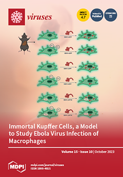
Cover Story (view full-size image): Among Orthoebolaviruses, Orthoebolavirus zairense (EBOV) represents the greatest public threat of medical significance. Severe EBOV disease occurs as a result of broad virus tropism, robust viral replication and a potent and dysregulated production of inflammatory mediators. Liver resident macrophages, Kupffer cells (KC), are thought to be one of the earliest targets for EBOV infection in vivo. Here, we examined EBOV infection in primary KCs and an immortalized Kupffer line, ImKC. KCs and ImKCs were readily infected with authentic EBOV-eGFP and interferon gamma (IFN-γ) polarization suppressed their permissiveness to infection. The effects of IFN-γ were further validated using a biologically contained model of EBOV, EBOV ΔVP30. Our studies define new cellular tools to study EBOV infection that can aid in the understanding of EBOV pathogenesis and development of antiviral therapies. View this paper
  • Issues are regarded as officially published after their release is announced to the table of contents alert mailing list.
  • You may sign up for e-mail alerts to receive table of contents of newly released issues.
  • PDF is the official format for papers published in both, html and pdf forms. To view the papers in pdf format, click on the "PDF Full-text" link, and use the free Adobe Reader to open them.
Order results
Result details
Section
Select all
Export citation of selected articles as:
11 pages, 1287 KiB  
Article
Assessment of Equine Influenza Virus Status in the Republic of Korea from 2020 to 2022
by Seong-In Lim, Min Ji Kim, Min-Ji Kim, Sang-Kyu Lee, Hyoung-Seok Yang, MiJung Kwon, Eui Hyeon Lim, In-Ohk Ouh, Eun-Jung Kim, Bang-Hun Hyun and Yoon-Hee Lee
Viruses 2023, 15(10), 2135; https://doi.org/10.3390/v15102135 - 23 Oct 2023
Cited by 1 | Viewed by 1373
Abstract
Equine influenza virus (EIV) causes acute respiratory disease in horses and belongs to the influenza A virus family Orthomyxoviridae, genus Orthomyxovirus. This virus may have severe financial implications for the horse industry owing to its highly contagious nature and rapid transmission. [...] Read more.
Equine influenza virus (EIV) causes acute respiratory disease in horses and belongs to the influenza A virus family Orthomyxoviridae, genus Orthomyxovirus. This virus may have severe financial implications for the horse industry owing to its highly contagious nature and rapid transmission. In the Republic of Korea, vaccination against EIV has been practiced with the active involvement of the Korea Racing Authority since 1974. In this study, we monitored the viral RNA for EIV using PCR, as well as the antibody levels against ‘A/equine/South Africa/4/03 (H3N8, clade 1)’, from 2020 to 2022. EIV was not detected using RT-PCR. The seropositivity rates detected using a hemagglutination inhibition assay were 90.3% in 2020, 96.7% in 2021, and 91.8% in 2022. The geometric mean of antibody titer (GMT) was 83.4 in 2020, 135.7 in 2021, and 95.6 in 2022. Yearlings and two-year-olds in training exhibited lower positive rates (59.1% in 2020, 38.9% in 2021, and 44.1% in 2022) than the average. These younger horses may require more attention for vaccination and vaccine responses against EIV. Continuous surveillance of EIV should be performed to monitor the prevalence and spread of this disease. Full article
(This article belongs to the Special Issue Advances in Animal Influenza Virus Research: Volume II)
Show Figures

Figure 1

14 pages, 3853 KiB  
Article
Evaluation of the Deletion of the African Swine Fever Virus Gene O174L from the Genome of the Georgia Isolate
by Elizabeth Ramirez-Medina, Lauro Velazquez-Salinas, Ayushi Rai, Nallely Espinoza, Alyssa Valladares, Ediane Silva, Leeanna Burton, Edward Spinard, Amanda Meyers, Guillermo Risatti, Sten Calvelage, Sandra Blome, Douglas P. Gladue and Manuel V. Borca
Viruses 2023, 15(10), 2134; https://doi.org/10.3390/v15102134 - 23 Oct 2023
Cited by 2 | Viewed by 1591
Abstract
African swine fever virus (ASFV) is a structurally complex, double-stranded DNA virus, which causes African swine fever (ASF), a contagious disease affecting swine. ASF is currently affecting pork production in a large geographical region, including Eurasia and the Caribbean. ASFV has a large [...] Read more.
African swine fever virus (ASFV) is a structurally complex, double-stranded DNA virus, which causes African swine fever (ASF), a contagious disease affecting swine. ASF is currently affecting pork production in a large geographical region, including Eurasia and the Caribbean. ASFV has a large genome, which harbors more than 160 genes, but most of these genes’ functions have not been experimentally characterized. One of these genes is the O174L gene which has been experimentally shown to function as a small DNA polymerase. Here, we demonstrate that the deletion of the O174L gene from the genome of the virulent strain ASFV Georgia2010 (ASFV-G) does not significantly affect virus replication in vitro or in vivo. A recombinant virus, having deleted the O174L gene, ASFV-G-∆O174L, was developed to study the effect of the O174L protein in replication in swine macrophages cultures in vitro and disease production when inoculated in pigs. The results demonstrated that ASFV-G-∆O174L has similar replication kinetics to parental ASFV-G in swine macrophage cultures. In addition, animals intramuscularly inoculated with 102 HAD50 of ASFV-G-∆O174L presented a clinical form of the disease that is indistinguishable from that induced by the parental virulent strain ASFV-G. All animals developed a lethal disease, being euthanized around day 7 post-infection. Therefore, although O174L is a well-characterized DNA polymerase, its function is apparently not critical for the process of virus replication, both in vitro and in vivo, or for disease production in domestic pigs. Full article
(This article belongs to the Special Issue African Swine Fever Virus 4.0)
Show Figures

Figure 1

15 pages, 2505 KiB  
Article
Increased Presence of Circulating Cell-Free, Fragmented, Host DNA in Pigs Infected with Virulent African Swine Fever Virus
by Ann Sofie Olesen, Louise Lohse, Camille Melissa Johnston, Thomas Bruun Rasmussen, Anette Bøtner and Graham J. Belsham
Viruses 2023, 15(10), 2133; https://doi.org/10.3390/v15102133 - 21 Oct 2023
Cited by 1 | Viewed by 1388
Abstract
African swine fever virus (ASFV) causes severe hemorrhagic disease in domestic pigs and wild boar, often with high case fatality rates. The virus replicates in the circulating cells of the monocyte–macrophage lineage and within lymphoid tissues. The infection leads to high fever and [...] Read more.
African swine fever virus (ASFV) causes severe hemorrhagic disease in domestic pigs and wild boar, often with high case fatality rates. The virus replicates in the circulating cells of the monocyte–macrophage lineage and within lymphoid tissues. The infection leads to high fever and a variety of clinical signs. In this study, it was observed that ASFV infection in pigs resulted in a >1000-fold increase in the level of circulating cell-free DNA (cfDNA), derived from the nuclei of host cells in the serum. This change occurred in parallel with the increase in circulating ASFV DNA. In addition, elevated levels (about 30-fold higher) of host mitochondrial DNA (mtDNA) were detected in the serum from ASFV-infected pigs. For comparison, the release of the cellular enzyme, lactate dehydrogenase (LDH), a commonly used marker of cellular damage, was also found to be elevated during ASFV infection, but later and less consistently. The sera from pigs infected with classical swine fever virus (CSFV), which causes a clinically similar disease to ASFV, were also tested but, surprisingly, this infection did not result in the release of cfDNA, mtDNA, or LDH. It was concluded that the level of cfDNA in the serum is a sensitive host marker of virulent ASFV infection. Full article
(This article belongs to the Section Animal Viruses)
Show Figures

Figure 1

13 pages, 2084 KiB  
Article
Emergence of Genomic Diversity in the Spike Protein of the “Omicron” Variant
by Surajit Basak, Pratanu Kayet, Manisha Ghosh, Joyeeta Chatterjee and Shanta Dutta
Viruses 2023, 15(10), 2132; https://doi.org/10.3390/v15102132 - 21 Oct 2023
Cited by 2 | Viewed by 1666
Abstract
SARS-CoV-2 (Severe Acute Respiratory Syndrome Coronavirus) has constantly been evolving into different forms throughout its spread in the population. Emerging SARS-CoV-2 variants, predominantly the variants of concern (VOCs), could have an impact on the virus spread, pathogenicity, and diagnosis. The recently emerged “Omicron” [...] Read more.
SARS-CoV-2 (Severe Acute Respiratory Syndrome Coronavirus) has constantly been evolving into different forms throughout its spread in the population. Emerging SARS-CoV-2 variants, predominantly the variants of concern (VOCs), could have an impact on the virus spread, pathogenicity, and diagnosis. The recently emerged “Omicron” variant has exhibited rapid transmission and divergence. The spike protein of SARS-CoV-2 has consistently been appearing as the mutational hotspot of all these VOCs. In order to determine a deeper understanding of the recently emerged and extremely divergent “Omicron”, a study of amino acid usage patterns and their substitution patterns was performed and compared with those of the other four successful variants of concern (“Alpha”, “Beta”, “Gamma”, and “Delta”). We observed that the amino acid usage of “Omicron” has a distinct pattern that distinguishes it from other VOCs and is significantly correlated with the increased hydrophobicity in spike proteins. We observed an increase in the non-synonymous substitution rate compared with the other four VOCs. Considering the phylogenetic relationship, we hypothesized about the functional interdependence between recombination and the mutation rate that might have resulted in a shift in the optimum of the mutation rate for the evolution of the “Omicron” variant. The results suggest that for improved disease prevention and control, more attention should be given to the significant genetic differentiation and diversity of newly emerging variants. Full article
(This article belongs to the Special Issue Coronavirus Genome Evolution, Recombination and Phylogeny)
Show Figures

Figure 1

7 pages, 2044 KiB  
Brief Report
Immunogenicity and Tolerability of a SARS-CoV-2 TNX-1800, a Live Recombinant Poxvirus Vaccine Candidate, in Syrian Hamsters and New Zealand White Rabbits
by Mayanka Awasthi, Anthony Macaluso, Scott J. Goebel, Erin Luea, Ryan S. Noyce, Farooq Nasar, Bruce Daugherty, Sina Bavari and Seth Lederman
Viruses 2023, 15(10), 2131; https://doi.org/10.3390/v15102131 - 21 Oct 2023
Cited by 1 | Viewed by 1754
Abstract
TNX-1800 is a preclinical stage synthetic-derived live attenuated chimeric horsepox virus vaccine engineered to express the SARS-CoV-2 spike (S) gene. The objectives of this study were to assess the safety, tolerability, and immunogenicity of TNX-1800 administration in Syrian golden hamsters and New Zealand [...] Read more.
TNX-1800 is a preclinical stage synthetic-derived live attenuated chimeric horsepox virus vaccine engineered to express the SARS-CoV-2 spike (S) gene. The objectives of this study were to assess the safety, tolerability, and immunogenicity of TNX-1800 administration in Syrian golden hamsters and New Zealand white rabbits. Animals were vaccinated at three doses via percutaneous inoculation. The data showed that the single percutaneous administration of three TNX-1800 vaccine dose levels was well tolerated in both hamsters and rabbits. At all dose levels, rabbits were more decerning regarding vaccine site reaction than hamsters. Lastly, no TNX-1800 genomes could be detected at the site of vaccination. Post-vaccination, all animals had anti-SARS-CoV-2 spike protein IgG specific antibody responses. These data demonstrate that TNX-1800 infection was limited, asymptomatic, and cleared by the end of this study, and a single dose was able to generate immune responses. Full article
(This article belongs to the Section SARS-CoV-2 and COVID-19)
Show Figures

Figure 1

18 pages, 1867 KiB  
Review
Advances of Reverse Vaccinology for mRNA Vaccine Design against SARS-CoV-2: A Review of Methods and Tools
by Maria Karolaynne da Silva, Daniel Melo de Oliveira Campos, Shopnil Akash, Shahina Akter, Leow Chiuan Yee, Umberto Laino Fulco and Jonas Ivan Nobre Oliveira
Viruses 2023, 15(10), 2130; https://doi.org/10.3390/v15102130 - 21 Oct 2023
Cited by 7 | Viewed by 2456
Abstract
mRNA vaccines are a new class of vaccine that can induce potent and specific immune responses against various pathogens. However, the design of mRNA vaccines requires the identification and optimization of suitable antigens, which can be challenging and time consuming. Reverse vaccinology is [...] Read more.
mRNA vaccines are a new class of vaccine that can induce potent and specific immune responses against various pathogens. However, the design of mRNA vaccines requires the identification and optimization of suitable antigens, which can be challenging and time consuming. Reverse vaccinology is a computational approach that can accelerate the discovery and development of mRNA vaccines by using genomic and proteomic data of the target pathogen. In this article, we review the advances of reverse vaccinology for mRNA vaccine design against SARS-CoV-2, the causative agent of COVID-19. We describe the steps of reverse vaccinology and compare the in silico tools used by different studies to design mRNA vaccines against SARS-CoV-2. We also discuss the challenges and limitations of reverse vaccinology and suggest future directions for its improvement. We conclude that reverse vaccinology is a promising and powerful approach to designing mRNA vaccines against SARS-CoV-2 and other emerging pathogens. Full article
(This article belongs to the Special Issue SARS-CoV-2 and mRNA Vaccines)
Show Figures

Figure 1

26 pages, 504 KiB  
Conference Report
The XVIII International Parvovirus Workshop Rimini, Italy, 14–17 June 2022
by Giorgio Gallinella and Antonio Marchini
Viruses 2023, 15(10), 2129; https://doi.org/10.3390/v15102129 - 20 Oct 2023
Viewed by 1185
Abstract
The XVIII International Parvovirus Workshop took place in Rimini, Italy, from 14 to 17 June 2022 as an on-site event, continuing the series of meetings started in 1985 and continuously held every two years. The communications dealt with all aspects of research in [...] Read more.
The XVIII International Parvovirus Workshop took place in Rimini, Italy, from 14 to 17 June 2022 as an on-site event, continuing the series of meetings started in 1985 and continuously held every two years. The communications dealt with all aspects of research in the field, from evolution and structure to receptors, from replication to trafficking, from virus–host interactions to clinical and veterinarian virology, including translational issues related to viral vectors, gene therapy and oncolytic parvoviruses. The oral communications were complemented by a poster exhibition available for view and discussion during the whole meeting. The XVIII International Parvovirus Workshop was dedicated to the memory of our dearest colleague Mavis Agbandje-McKenna (1963–2021). Full article
(This article belongs to the Special Issue Advances in Parvovirus Research 2022)
Show Figures

Figure 1

11 pages, 1833 KiB  
Article
Genetic Diversity of Domestic Cat Hepadnavirus in Southern Taiwan
by Benji Brayan Ilagan Silva, Jin-Yang Chen, Brian Harvey Avanceña Villanueva, Zi-Ying Lu, Hua-Zhen Hsing, Andrew D. Montecillo, Maya Shofa, Hoang Minh, Jen-Pin Chuang, Huai-Ying Huang, Akatsuki Saito and Kuo-Pin Chuang
Viruses 2023, 15(10), 2128; https://doi.org/10.3390/v15102128 - 20 Oct 2023
Cited by 1 | Viewed by 2090
Abstract
Domestic cat hepadnavirus (DCH) is an infectious disease associated with chronic hepatitis in cats, which suggests a similarity with hepatitis B virus infections in humans. Since its first identification in Australia in 2018, DCH has been reported in several countries with varying prevalence [...] Read more.
Domestic cat hepadnavirus (DCH) is an infectious disease associated with chronic hepatitis in cats, which suggests a similarity with hepatitis B virus infections in humans. Since its first identification in Australia in 2018, DCH has been reported in several countries with varying prevalence rates, but its presence in Taiwan has yet to be investigated. In this study, we aimed to identify the presence and genetic diversity of DCH infections in Taiwan. Among the 71 samples tested, eight (11.27%) were positive for DCH. Of these positive cases, three cats had elevated levels of alanine transaminase (ALT) and aspartate transaminase (AST), suggesting an association between DCH infection and chronic hepatitis. Four DCH-positive samples were also tested for feline immunodeficiency virus (FIV) and feline leukemia virus (FeLV) coinfection. One sample (25%) was positive for FIV, whereas there was no positive sample for FeLV (0%). In addition, we performed whole genome sequencing on six samples to determine the viral genome sequences. Phylogenetic analyses identified a distinct lineage compared with previously reported sequences. This study highlights the importance of continuous surveillance of DCH and further research to elucidate the pathophysiology and transmission route of DCH. Full article
(This article belongs to the Special Issue Viral Genetic Variation)
Show Figures

Figure 1

14 pages, 2915 KiB  
Article
Development of Polyclonal Antibodies and a Serological-Based Reverse-Transcription Loop-Mediated Isothermal Amplification (S-RT-LAMP) Assay for Rice Black-Streaked Dwarf Virus Detection in Both Rice and Small Brown Planthopper
by Yanhong Hua, Chenwei Feng, Tianxiao Gu, Haoyu Chen, Duxuan Liu, Kai Xu and Kun Zhang
Viruses 2023, 15(10), 2127; https://doi.org/10.3390/v15102127 - 20 Oct 2023
Cited by 3 | Viewed by 1303
Abstract
Rice black-streaked dwarf virus (RBSDV) infects rice and maize, and seriously affects rice yields in main rice-producing areas. It can be transmitted via small brown planthopper (SBPH: Laodelphax striatellus Fallén). To more rapidly, sensitively, and highly throughput diagnose RBSDV in the wild condition, [...] Read more.
Rice black-streaked dwarf virus (RBSDV) infects rice and maize, and seriously affects rice yields in main rice-producing areas. It can be transmitted via small brown planthopper (SBPH: Laodelphax striatellus Fallén). To more rapidly, sensitively, and highly throughput diagnose RBSDV in the wild condition, we first purified the recombinant His-CPRBSDV protein, and prepared the polyclonal antibodies against the His-CPRBSDV protein (PAb-CPRBSDV). Based on the PAb-CPRBSDV, we developed a series of serological detections, such as Western blot, an enzyme-linked immunosorbent assay (ELISA), and a dot immunoblotting assay (DIBA). Furthermore, we developed a serological-based reverse-transcription loop-mediated isothermal amplification assay (S-RT-LAMP) that could accurately detect RBSDV in the wild. Briefly, the viral genomic dsRNA together with viral CP were precipitated by co-immunoprecipitation using the PAb-CPRBSDV, then the binding RNAs were crudely isolated and used for RT-LAMP diagnosis. Using the prepared PAb-CPRBSDV, four serology-based detection methods were established to specifically detect RBSDV-infected rice plants or SBPHs in the wild. The method of S-RT-LAMP has also been developed to specifically, high-throughput, and likely detect RBSDV in rice seedlings and SBPHs simultaneously. The antiserum prepared here laid the foundation for the rapid and efficient detection of RBSDV-infected field samples, which will benefit for determination of the virulence rate of the transmission vector SBPH and outbreak and epidemic prediction of RBSDV in a rice production area. Full article
(This article belongs to the Section Viruses of Plants, Fungi and Protozoa)
Show Figures

Figure 1

20 pages, 6112 KiB  
Article
Identification and Analysis of a Four-Gene Set for Diagnosing SFTS Virus Infection Based on Machine Learning Methods and Its Association with Immune Cell Infiltration
by Tao Huang, Xueqi Wang, Yuqian Mi, Tiezhu Liu, Yang Li, Ruixue Zhang, Zhen Qian, Yanhan Wen, Boyang Li, Lina Sun, Wei Wu, Jiandong Li, Shiwen Wang and Mifang Liang
Viruses 2023, 15(10), 2126; https://doi.org/10.3390/v15102126 - 20 Oct 2023
Cited by 1 | Viewed by 1618
Abstract
Severe Fever with thrombocytopenia syndrome (SFTS) is a highly fatal viral infectious disease that poses a significant threat to public health. Currently, the phase and pathogenesis of SFTS are not well understood, and there are no specific vaccines or effective treatment available. Therefore, [...] Read more.
Severe Fever with thrombocytopenia syndrome (SFTS) is a highly fatal viral infectious disease that poses a significant threat to public health. Currently, the phase and pathogenesis of SFTS are not well understood, and there are no specific vaccines or effective treatment available. Therefore, it is crucial to identify biomarkers for diagnosing acute SFTS, which has a high mortality rate. In this study, we conducted differentially expressed genes (DEGs) analysis and WGCNA module analysis on the GSE144358 dataset, comparing the acute phase of SFTSV-infected patients with healthy individuals. Through the LASSO–Cox and random forest algorithms, a total of 2128 genes were analyzed, leading to the identification of four genes: ADIPOR1, CENPO, E2F2, and H2AC17. The GSEA analysis of these four genes demonstrated a significant correlation with immune cell function and cell cycle, aligning with the functional enrichment findings of DEGs. Furthermore, we also utilized CIBERSORT to analyze the immune cell infiltration and its correlation with characteristic genes. The results indicate that the combination of ADIPOR1, CENPO, E2F2, and H2AC17 genes has the potential as characteristic genes for diagnosing and studying the acute phase of SFTS virus (SFTSV) infection. Full article
(This article belongs to the Special Issue Severe Fever with Thrombocytopenia Syndrome Virus 3.0)
Show Figures

Figure 1

12 pages, 397 KiB  
Article
Uptake of Oral HIV Pre-Exposure Prophylaxis (PrEP) and Associated Factors among Female Sex Workers in Tanga, Tanzania
by Veronica O. Martin, Novatus A. Tesha and Bruno F. Sunguya
Viruses 2023, 15(10), 2125; https://doi.org/10.3390/v15102125 - 20 Oct 2023
Cited by 1 | Viewed by 1439
Abstract
Pre-exposure prophylaxis (PrEP) prevents HIV infection among female sex workers (FSW). WHO recommends the use of Tenofovir disoproxil fumarate for use in oral PrEP regimens (TDF). Emtricitabine (FTC) 200 mg/Tenofovir Disoproxil Fumarate (TDF) 300 mg (Truvada) daily is the approved PrEP regimen in [...] Read more.
Pre-exposure prophylaxis (PrEP) prevents HIV infection among female sex workers (FSW). WHO recommends the use of Tenofovir disoproxil fumarate for use in oral PrEP regimens (TDF). Emtricitabine (FTC) 200 mg/Tenofovir Disoproxil Fumarate (TDF) 300 mg (Truvada) daily is the approved PrEP regimen in Tanzania. Evidence is limited on oral PrEP uptake and its associated factors in countries with a high burden of HIV, such as Tanzania. This study aimed to examine the uptake of oral PrEP and its associated factors among FSW in the Tanga region of Tanzania. This community-based cross-sectional study was conducted among 428 FSW. Data were collected through face-to-face interviews and analysed using STATA version 17 and RDSAT. Logistic regression was used to examine the associations of independent factors and PrEP uptake among study participants. About 55% of the recruited FSW used oral PrEP. FSW with three or more children were 2.41 times more likely to take oral PrEP (AOR 2.41, 95% CI: 1.08–4.25, p < 0.05). Moreover, those with a positive attitude were more likely to use oral PrEP (AOR 2.8, 95% CI: 1.88–4.17, p < 0.05). Poor belief was a barrier to PrEP use, and side effects of the drugs were a reason for the discontinuation of PrEP services. Most of the participants preferred PrEP services to be provided in the community. Oral PrEP uptake was 55%. Efforts to scale up PrEP for FSW should address misconceptions regarding PrEP, PrEP sensitization, and improving access through community-based intervention. Full article
Show Figures

Figure 1

16 pages, 6480 KiB  
Article
The Transmembrane Protease Serine 2 (TMPRSS2) Non-Protease Domains Regulating Severe Acute Respiratory Syndrome Coronavirus 2 (SARS-CoV-2) Spike-Mediated Virus Entry
by Romano Strobelt, Julia Adler and Yosef Shaul
Viruses 2023, 15(10), 2124; https://doi.org/10.3390/v15102124 - 19 Oct 2023
Cited by 5 | Viewed by 1718
Abstract
The severe acute respiratory syndrome coronavirus 2 (SARS-CoV-2) enters cells by binding to the angiotensin-converting enzyme 2 (hACE2) receptor. This process is aided by the transmembrane protease serine 2 (TMPRSS2), which enhances entry efficiency and infectiousness by cleaving the SARS-CoV-2 surface glycoprotein (Spike). [...] Read more.
The severe acute respiratory syndrome coronavirus 2 (SARS-CoV-2) enters cells by binding to the angiotensin-converting enzyme 2 (hACE2) receptor. This process is aided by the transmembrane protease serine 2 (TMPRSS2), which enhances entry efficiency and infectiousness by cleaving the SARS-CoV-2 surface glycoprotein (Spike). The cleavage primes the Spike protein, promoting membrane fusion instead of receptor-mediated endocytosis. Despite the pivotal role played by TMPRSS2, our understanding of its non-protease distinct domains remains limited. In this report, we present evidence indicating the potential phosphorylation of a minimum of six tyrosine residues within the cytosolic tail (CT) of TMPRSS2. Via the use of TMPRSS2 CT phospho-mimetic mutants, we observed a reduction in TMPRSS2 protease activity, accompanied by a decrease in SARS-CoV-2 pseudovirus transduction, which was found to occur mainly via the endosomal pathway. We expanded our investigation beyond TMPRSS2 CT and discovered the involvement of other non-protease domains in regulating infection. Our co-immunoprecipitation experiments demonstrated a strong interaction between TMPRSS2 and Spike. We revealed a 21 amino acid long TMPRSS2-Spike-binding region (TSBR) within the TMPRSS2 scavenger receptor cysteine-rich (SRCR) domain that contributes to this interaction. Our study sheds light on novel functionalities associated with TMPRSS2’s cytosolic tail and SRCR region. Both of these regions have the capability to regulate SARS-CoV-2 entry pathways. These findings contribute to a deeper understanding of the complex interplay between viral entry and host factors, opening new avenues for potential therapeutic interventions. Full article
(This article belongs to the Special Issue TMPRSS2 in Influenza Virus and Coronavirus Infections)
Show Figures

Figure 1

15 pages, 2845 KiB  
Article
Nano-Sized Chimeric Human Papillomavirus-16 L1 Virus-like Particles Displaying Mycobacterium tuberculosis Antigen Ag85B Enhance Ag85B-Specific Immune Responses in Female C57BL/c Mice
by Fangbin Zhou and Dongmei Zhang
Viruses 2023, 15(10), 2123; https://doi.org/10.3390/v15102123 - 19 Oct 2023
Cited by 2 | Viewed by 1474
Abstract
Bacillus Calmette–Guerin (BCG), the only current vaccine against tuberculosis (TB) that is licensed in clinics, successfully protects infants and young children against several TB types, such as TB meningitis and miliary TB, but it is ineffective in protecting adolescents and adults against pulmonary [...] Read more.
Bacillus Calmette–Guerin (BCG), the only current vaccine against tuberculosis (TB) that is licensed in clinics, successfully protects infants and young children against several TB types, such as TB meningitis and miliary TB, but it is ineffective in protecting adolescents and adults against pulmonary TB. Thus, it is a matter of the utmost urgency to develop an improved and efficient TB vaccine. In this milieu, virus-like particles (VLPs) exhibit excellent characteristics in the field of vaccine development due to their numerous characteristics, including but not limited to their good safety without the risk of infection, their ability to mimic the size and structure of original viruses, and their ability to display foreign antigens on their surface to enhance the immune response. In this study, the HPV16 L1 capsid protein (HPV16L1) acted as a structural vaccine scaffold, and the extracellular domain of Ag85B was selected as the M. tb immunogen and inserted into the FG loop of the HPV16 L1 protein to construct chimeric HPV16L1/Ag85B VLPs. The chimeric HPV16L1/Ag85B VLPs were produced via the Pichia pastoris expression system and purified via discontinuous Optiprep density gradient centrifugation. The humoral and T cell-mediated immune response induced by the chimeric HPV16L1/Ag85B VLP was studied in female C57BL/c mice. We demonstrated that the insertion of the extracellular domain of Ag85B into the FG loop of HPV16L1 did not affect the in vitro stability and self-assembly of the chimeric HPV16L1/Ag85B VLPs. Importantly, it did not interfere with the immunogenicity of Ag85B. We observed that the chimeric HPV16L1/Ag85B VLPs induced higher Ag85B-specific antibody responses and elicited significant Ag85B-specific T cell immune responses in female C57BL/c mice compared with recombinant Ag85B. Our findings provide new insights into the development of novel chimeric HPV16L1/TB VLP-based vaccine platforms for controlling TB infection, which are urgently required in low-income and developing countries. Full article
(This article belongs to the Section Viral Immunology, Vaccines, and Antivirals)
Show Figures

Figure 1

12 pages, 1453 KiB  
Article
HHV-6A Infection of Papillary Thyroid Cancer Cells Induces Several Effects Related to Cancer Progression
by Stefania Mardente, Maria Anele Romeo, Angela Asquino, Agnese Po, Maria Saveria Gilardini Montani and Mara Cirone
Viruses 2023, 15(10), 2122; https://doi.org/10.3390/v15102122 - 19 Oct 2023
Viewed by 1371
Abstract
Recent studies have shown that thyrocytes are permissive to HHV-6A infection and that the virus may contribute to the pathogenesis of autoimmune thyroiditis. Thyroid autoimmune diseases increase the risk of papillary cancer, which is not surprising considering that chronic inflammation activates pathways that [...] Read more.
Recent studies have shown that thyrocytes are permissive to HHV-6A infection and that the virus may contribute to the pathogenesis of autoimmune thyroiditis. Thyroid autoimmune diseases increase the risk of papillary cancer, which is not surprising considering that chronic inflammation activates pathways that are also pro-oncogenic. Moreover, in this condition, cell proliferation is stimulated as an attempt to repair tissue damage caused by the inflammatory process. Interestingly, it has been reported that the well-differentiated papillary thyroid carcinoma (PTC), the less aggressive form of thyroid tumor, may progress to the more aggressive follicular thyroid carcinoma (FTC) and eventually to the anaplastic thyroid carcinoma (ATC), and that to such progression contributes the presence of an inflammatory/immune suppressive tumor microenvironment. In this study, we investigated whether papillary tumor cells (BCPAP) could be infected by human herpes virus-6A (HHV-6A), and if viral infection could induce effects related to cancer progression. We found that the virus dysregulated the expression of several microRNAs, such as miR-155, miR-9, and the miR-221/222 cluster, which are involved in different steps of carcinogenesis, and increased the secretion of pro-inflammatory cytokines, particularly IL-6, which may also sustain thyroid tumor cell growth and promote cancer progression. Genomic instability and the expression of PTEN, reported to act as an oncogene in mutp53-carrying cells such as BCPAP, also increased following HHV-6A-infection. These findings suggest that a ubiquitous herpesvirus such as HHV-6A, which displays a marked tropism for thyrocytes, could be involved in the progression of PTC towards more aggressive forms of thyroid tumor. Full article
(This article belongs to the Section Human Virology and Viral Diseases)
Show Figures

Figure 1

13 pages, 1841 KiB  
Article
Characteristics of Anti-Measles Immunity in Lung Transplant Candidates
by Valentina B. Polishchuk, Mikhail P. Kostinov, Aleksey A. Ryzhov, Natalia A. Karchevskaya, Irina L. Solov’eva, Alexander P. Cherdantsev, Aristitsa M. Kostinova and Arseniy A. Poddubikov
Viruses 2023, 15(10), 2121; https://doi.org/10.3390/v15102121 - 19 Oct 2023
Viewed by 1434
Abstract
Measles has not yet been eradicated; therefore, its outbreaks are still reported throughout the world. Like any infection, measles is dangerous for immunocompromised patients. Levels of anti-measles IgG antibodies were measured in 157 patients aged 17 to 72, who were placed on the [...] Read more.
Measles has not yet been eradicated; therefore, its outbreaks are still reported throughout the world. Like any infection, measles is dangerous for immunocompromised patients. Levels of anti-measles IgG antibodies were measured in 157 patients aged 17 to 72, who were placed on the lung transplant waiting list. Measurements were undertaken by enzyme-linked immunosorbent assay (ELISA) using the VectoMeasles-IgG kit (Russia). The proportion of patients seronegative for measles was 19% (30/157). Correlation was detected between patients’ age and their levels of anti-measles antibodies, with higher proportions of patients having undetectable titers (25.5–28.9%) or low antibody levels (38.3–44.4%) in the young age groups (17–29 and 30–39 years old). There were no differences between male and female patients in levels of anti-measles antibodies or in the proportion of seronegative individuals. Analyses of antibody levels with regard to type of disease revealed the highest rate of seronegative results in cystic fibrosis patients (34.4%, 11/32). Overall, 19% of lung transplant candidates, mostly young people and cystic fibrosis patients, did not have protective immunity against measles. Full article
(This article belongs to the Special Issue Opportunistic Viral Infections)
Show Figures

Figure 1

14 pages, 2297 KiB  
Review
Urdbean Leaf Crinkle Virus: A Mystery Waiting to Be Solved
by Naimuddin Kamaal, Mohammad Akram, Aditya Pratap, Deepender Kumar and Ramakrishnan M. Nair
Viruses 2023, 15(10), 2120; https://doi.org/10.3390/v15102120 - 19 Oct 2023
Cited by 2 | Viewed by 1818
Abstract
Urdbean leaf crinkle disease (ULCD) affects mainly the urdbean or blackgram (Vigna mungo (L.) Hepper) causing distinct symptoms that often result in serious yield losses. It has been known to occur for more than five decades and is considered to be of [...] Read more.
Urdbean leaf crinkle disease (ULCD) affects mainly the urdbean or blackgram (Vigna mungo (L.) Hepper) causing distinct symptoms that often result in serious yield losses. It has been known to occur for more than five decades and is considered to be of viral etiology. The identity of the causal agent, often referred to as the urdbean leaf crinkle virus, is not unequivocally proved. There are few attempts to characterize the causal agent of ULCD; however, there is no unanimity in the results. Recent attempts to characterize the causal agent of ULCD using next-generation sequencing of the virome of ULCD-affected urdbean plants indicated the involvement of cowpea mild mottle virus; however, without conforming through Koch’s postulates, the etiology of ULCD remains inconclusive. Claims of different insect vectors involved in the transmission of ULCD make this disease even more mysterious. The information available so far indicates that either two different viruses are causing ULCD or a mixture of viruses is involved. The identity of the virus/es causing ULCD still remains to be unambiguously ascertained. In this review, we attempt to analyze information on the various aspects of ULCD. Full article
(This article belongs to the Special Issue Plant Virus Epidemiology and Control 2023)
Show Figures

Figure 1

21 pages, 1740 KiB  
Review
Mathematical Models of HIV-1 Dynamics, Transcription, and Latency
by Iván D’Orso and Christian V. Forst
Viruses 2023, 15(10), 2119; https://doi.org/10.3390/v15102119 - 19 Oct 2023
Cited by 2 | Viewed by 1999
Abstract
HIV-1 latency is a major barrier to curing infections with antiretroviral therapy and, consequently, to eliminating the disease globally. The establishment, maintenance, and potential clearance of latent infection are complex dynamic processes and can be best described with the help of mathematical models [...] Read more.
HIV-1 latency is a major barrier to curing infections with antiretroviral therapy and, consequently, to eliminating the disease globally. The establishment, maintenance, and potential clearance of latent infection are complex dynamic processes and can be best described with the help of mathematical models followed by experimental validation. Here, we review the use of viral dynamics models for HIV-1, with a focus on applications to the latent reservoir. Such models have been used to explain the multi-phasic decay of viral load during antiretroviral therapy, the early seeding of the latent reservoir during acute infection and the limited inflow during treatment, the dynamics of viral blips, and the phenomenon of post-treatment control. Finally, we discuss that mathematical models have been used to predict the efficacy of potential HIV-1 cure strategies, such as latency-reversing agents, early treatment initiation, or gene therapies, and to provide guidance for designing trials of these novel interventions. Full article
(This article belongs to the Special Issue Regulation of HIV-1 Transcription and Latency)
Show Figures

Figure 1

11 pages, 395 KiB  
Article
Evaluation of Prevalence of Hepatitis E Clinical Markers among Donors in Estonia
by Tatiana Kuznetsova, Diana Moor, Gulara Khanirzayeva and Julia Geller
Viruses 2023, 15(10), 2118; https://doi.org/10.3390/v15102118 - 19 Oct 2023
Cited by 1 | Viewed by 1342
Abstract
Hepatitis E virus (HEV) is now considered the most common cause of acute hepatitis worldwide. There are no published data about the prevalence of antibodies to HEV and RNA in donor sera in Estonia, and this precludes planning measures for preventing HEV proliferation [...] Read more.
Hepatitis E virus (HEV) is now considered the most common cause of acute hepatitis worldwide. There are no published data about the prevalence of antibodies to HEV and RNA in donor sera in Estonia, and this precludes planning measures for preventing HEV proliferation through blood transfusion services. Here, were report data from an analysis of 1002 sera on the prevalence of anti-HEV IgG and IgM and the viral RNA. The antibodies were found in 48 donor sera (4.8%); of these, 40 (4%) harbored anti-HEV IgG, 15 (1.5%) contained anti-HEV IgM, and 7 donors had anti-HEV antibodies of both classes simultaneously. HEV RNA was not detected in any blood serum. Statistical associations of infection risk factors (gender, age, travel in the last six months, contact with pigs and/or wild boars in the last six months, consumption of thermally unprocessed/raw pork or boar meat, raw/unfiltered tap water or water from natural sources, unpasteurized farm dairy products, and unwashed berries and/or vegetables) were assessed. None of the listed factors were found to be associated with a higher or lower risk of anti-HEV antibody presence. At the same time, an increasing share of anti-HEV IgG carriers with age was found. The absence of HEV RNA in the analyzed donor plasma samples proves that HEV acute infection prevalence in Estonia does not exceed the average level of European countries. There is no urgent necessity to enter a requirement for a total screening of blood plasma for HEV RNA prevalence in Estonia. Full article
(This article belongs to the Special Issue Hepatitis E Virus (HEV) 2.0)
Show Figures

Figure 1

9 pages, 269 KiB  
Brief Report
Diagnostic Accuracy of a Point-of-Care Immunoassay for Feline Immunodeficiency Virus Antibodies, Feline Leukemia Virus Antigen, and Dirofilaria immitis Antigen
by Seema Singh, Kristen A. Davenport, Elizabeth Schooley, Anthony Ruggiero, Salam Nassar, Jesse Buch and Ramaswamy Chandrashekar
Viruses 2023, 15(10), 2117; https://doi.org/10.3390/v15102117 - 19 Oct 2023
Viewed by 1576
Abstract
Feline immunodeficiency virus (FIV) and feline leukemia virus (FeLV) are retroviral infections of cats worldwide whose clinical manifestations range from mild to severe disease. In both cases, infected cats can live a long life with proper care and should be managed to prevent [...] Read more.
Feline immunodeficiency virus (FIV) and feline leukemia virus (FeLV) are retroviral infections of cats worldwide whose clinical manifestations range from mild to severe disease. In both cases, infected cats can live a long life with proper care and should be managed to prevent infection of other cats. Dirofilaria immitis, the nematode that causes heartworm disease, can infect cats in any region where dogs are infected. Though cats are more resistant to infection, clinical diseases in the form of heartworm-associated respiratory disease can cause death. Screening for these infectious diseases enables veterinarians to manage their cases and prevent the spread to other cats. We describe the diagnostic accuracy of a point-of-care immunoassay for FIV, FeLV, and heartworm, compared to reference methods commonly available through reference laboratories to the practicing veterinarian. For FIV, we report 100% sensitivity (95% confidence limits (CL): 96.2–100%) and 97.8% specificity (95% CL: 95.4–99.4%). For FeLV, we report 100% sensitivity (95% CL: 97.7–100%) and 99.2% specificity (95% CL: 97.1–99.9%). And for heartworm, we report 90.2% sensitivity (95% CL: 76.9–97.3%) and 100% specificity (95% CL: 98.3–100%). Veterinarians may expect this performance relative to the reference methods they use for confirmatory serological testing. Full article
(This article belongs to the Special Issue Viral Infections in Companion Animals: Volume 2)
11 pages, 255 KiB  
Review
Antiviral Treatment of Maternal and Congenital Cytomegalovirus (CMV) Infections
by Swetha Pinninti and Suresh Boppana
Viruses 2023, 15(10), 2116; https://doi.org/10.3390/v15102116 - 19 Oct 2023
Cited by 3 | Viewed by 1875
Abstract
Human Cytomegalovirus (HCMV) is a ubiquitous member of the Herpesviridae family, responsible for the most common congenital viral infection—congenital Cytomegalovirus (cCMV) infection. While a majority of HCMV infections in children and adults are asymptomatic, HCMV is well known to cause severe infections in [...] Read more.
Human Cytomegalovirus (HCMV) is a ubiquitous member of the Herpesviridae family, responsible for the most common congenital viral infection—congenital Cytomegalovirus (cCMV) infection. While a majority of HCMV infections in children and adults are asymptomatic, HCMV is well known to cause severe infections in the immunocompromised individual and maternal infections with variable long-term sequelae after maternal–fetal transmission with primary or nonprimary infections. HCMV seroprevalence and cCMV incidence vary by geographic area and demographic characteristics like race and socioeconomic status. While cCMV birth prevalence ranges from 0.2% to 6% in different parts of the world, it is influenced by regional HCMV seroprevalence rates. HCMV screening during pregnancy is not routinely offered due to lack of awareness, hurdles to accurate diagnosis, and lack of well-established effective treatment options during pregnancy. This review will focus on antiviral treatment options currently available for use during pregnancy and in the newborn period for the treatment of maternal and congenital HCMV infections. Full article
(This article belongs to the Special Issue Antiviral Treatment for Cytomegalovirus Diseases)
34 pages, 8481 KiB  
Article
Molecular Epidemiology of Rotavirus A in Calves: Evolutionary Analysis of a Bovine G8P[11] Strain and Spatio-Temporal Dynamics of G6 Lineages in the Americas
by Enrique L. Louge Uriarte, Alejandra Badaracco, Maximiliano J. Spetter, Samuel Miño, Joaquín I. Armendano, Mark Zeller, Elisabeth Heylen, Ernesto Späth, María Rosa Leunda, Ana Rita Moreira, Jelle Matthijnssens, Viviana Parreño and Anselmo C. Odeón
Viruses 2023, 15(10), 2115; https://doi.org/10.3390/v15102115 - 19 Oct 2023
Cited by 1 | Viewed by 1883
Abstract
Rotavirus A (RVA) causes diarrhea in calves and frequently possesses the G6 and P[5]/P[11] genotypes, whereas G8 is less common. We aimed to compare RVA infections and G/P genotypes in beef and dairy calves from major livestock regions of Argentina, elucidate the evolutionary [...] Read more.
Rotavirus A (RVA) causes diarrhea in calves and frequently possesses the G6 and P[5]/P[11] genotypes, whereas G8 is less common. We aimed to compare RVA infections and G/P genotypes in beef and dairy calves from major livestock regions of Argentina, elucidate the evolutionary origin of a G8 strain and analyze the G8 lineages, infer the phylogenetic relationship of RVA field strains, and investigate the evolution and spatio-temporal dynamics of the main G6 lineages in American countries. Fecal samples (n = 422) from diarrheic (beef, 104; dairy, 137) and non-diarrheic (beef, 78; dairy, 103) calves were analyzed by ELISA and semi-nested multiplex RT-PCR. Sequencing, phylogenetic, phylodynamic, and phylogeographic analyses were performed. RVA infections were more frequent in beef (22.0%) than in dairy (14.2%) calves. Prevalent genotypes and G6 lineages were G6(IV)P[5] in beef (90.9%) and G6(III)P[11] (41.2%) or mixed genotypes (23.5%) in dairy calves. The only G8 strain was phylogenetically related to bovine and artiodactyl bovine-like strains. Re-analyses inside the G8 genotype identified G8(I) to G8(VIII) lineages. Of all G6 strains characterized, the G6(IV)P[5](I) strains from “Cuenca del Salado” (Argentina) and Uruguay clustered together. According to farm location, a clustering pattern for G6(IV)P[5] strains of beef farms was observed. Both G6 lineage strains together revealed an evolutionary rate of 1.24 × 10⁻³ substitutions/site/year, and the time to the most recent common ancestor was dated in 1853. The most probable ancestral locations were Argentina in 1981 for G6(III) strains and the USA in 1940 for G6(IV) strains. The highest migration rates for both G6 lineages together were from Argentina to Brazil and Uruguay. Altogether, the epidemiology, genetic diversity, and phylogeny of RVA in calves can differ according to the production system and farm location. We provide novel knowledge about the evolutionary origin of a bovine G8P[11] strain. Finally, bovine G6 strains from American countries would have originated in the USA nearly a century before its first description. Full article
(This article belongs to the Section Animal Viruses)
Show Figures

Figure 1

15 pages, 4388 KiB  
Article
Identification of Critical Amino Acids of Coxsackievirus A10 Associated with Cell Tropism and Viral RNA Release during Uncoating
by Jie Pei, Rui-Lun Liu, Zhi-Hui Yang, Ya-Xin Du, Sha-Sha Qian, Sheng-Li Meng, Jing Guo, Bo Zhang and Shuo Shen
Viruses 2023, 15(10), 2114; https://doi.org/10.3390/v15102114 - 18 Oct 2023
Viewed by 1419
Abstract
Coxsackievirus A10 (CV-A10) is a prevailing causative agent of hand–foot–mouth disease, necessitating the isolation and adaptation of appropriate strains in cells allowed for human vaccine development. In this study, amino acid sequences of CV-A10 strains with different cell tropism on RD and Vero [...] Read more.
Coxsackievirus A10 (CV-A10) is a prevailing causative agent of hand–foot–mouth disease, necessitating the isolation and adaptation of appropriate strains in cells allowed for human vaccine development. In this study, amino acid sequences of CV-A10 strains with different cell tropism on RD and Vero cells were compared. Various amino acids on the structural and non-structural proteins related to cell tropism were identified. The reverse genetic systems of several CV-A10 strains with RD+/Vero and RD+/Vero+ cell tropism were developed, and a set of CV-A10 recombinants were produced. The binding, entry, uncoating, and proliferation steps in the life cycle of these viruses were evaluated. P1 replacement of CV-A10 strains with different cell tropism revealed the pivotal role of the structural proteins in cell tropism. Further, seven amino acid substitutions in VP2 and VP1 were introduced to further investigate their roles played in cell tropism. These mutations cooperated in the growth of CV-A10 in Vero cells. Particularly, the valine to isoleucine mutation at the position VP1-236 (V1236I) was found to significantly restrict viral uncoating in Vero cells. Co-immunoprecipitation assays showed that the release of viral RNA from the KREMEN1 receptor-binding virions was restricted in r0195-V1236I compared with the parental strain r0195 (a RD+/Vero+ strain). Overall, this study highlights the dominant effect of structural proteins in CV-A10 adaption in Vero cells and the importance of V1236 in viral uncoating, providing a foundation for the mechanism study of CV-A10 cell tropism, and facilitating the development of vaccine candidates. Full article
(This article belongs to the Special Issue Coxsackieviruses and Associated Diseases)
Show Figures

Figure 1

12 pages, 963 KiB  
Article
Evaluation of Direct Detection Protocols for Poliovirus from Stool Samples of Acute Flaccid Paralysis Patients
by Minami Kikuchi Ueno, Kouichi Kitamura, Yorihiro Nishimura and Minetaro Arita
Viruses 2023, 15(10), 2113; https://doi.org/10.3390/v15102113 - 18 Oct 2023
Cited by 1 | Viewed by 1486
Abstract
Polio surveillance in the Global Polio Eradication Initiative has been conducted with virus isolation from stool samples of acute flaccid paralysis (AFP) cases. Under the current biorisk management/regulations, challenges arise in the timelines of the report, sensitivity of the test and containment of [...] Read more.
Polio surveillance in the Global Polio Eradication Initiative has been conducted with virus isolation from stool samples of acute flaccid paralysis (AFP) cases. Under the current biorisk management/regulations, challenges arise in the timelines of the report, sensitivity of the test and containment of poliovirus (PV) isolates. In the present study, we evaluated protocols of previously reported direct detection (DD) methods targeting the VP1 or VP4–VP2 regions of the PV genome in terms of sensitivity and sequencability. An optimized protocol targeting the entire-capsid region for the VP1 sequencing showed a high sensitivity (limit of detection = 82 copies of PV genome) with a simpler and faster reaction than reported ones (i.e., with the addition of all the primers at the start of the reaction, the RT-PCR reaction finishes within 2.5 h). The DD methods targeting the VP1 region detected PV in 60 to 80% of PV-positive stool samples from AFP cases; however, minor populations of PV strains in the samples with virus mixtures were missed by the methods. Sequencability of the DD methods was primarily determined by the efficiency of the PCRs for both Sanger and nanopore sequencing. The DD method targeting the VP4–VP2 region showed higher sensitivity than that targeting the VP1 region (limit of detection = 25 copies of PV genome) and successfully detected PV from all the stool samples examined. These results suggest that DD methods are effective for the detection of PV and that further improvement of the sensitivity is essential to serve as an alternative to the current polio surveillance algorithm. Full article
(This article belongs to the Section Human Virology and Viral Diseases)
Show Figures

Figure 1

31 pages, 1731 KiB  
Review
Polyomavirus Wakes Up and Chooses Neurovirulence
by Arrienne B. Butic, Samantha A. Spencer, Shareef K. Shaheen and Aron E. Lukacher
Viruses 2023, 15(10), 2112; https://doi.org/10.3390/v15102112 - 18 Oct 2023
Cited by 4 | Viewed by 2881
Abstract
JC polyomavirus (JCPyV) is a human-specific polyomavirus that establishes a silent lifelong infection in multiple peripheral organs, predominantly those of the urinary tract, of immunocompetent individuals. In immunocompromised settings, however, JCPyV can infiltrate the central nervous system (CNS), where it causes several encephalopathies [...] Read more.
JC polyomavirus (JCPyV) is a human-specific polyomavirus that establishes a silent lifelong infection in multiple peripheral organs, predominantly those of the urinary tract, of immunocompetent individuals. In immunocompromised settings, however, JCPyV can infiltrate the central nervous system (CNS), where it causes several encephalopathies of high morbidity and mortality. JCPyV-induced progressive multifocal leukoencephalopathy (PML), a devastating demyelinating brain disease, was an AIDS-defining illness before antiretroviral therapy that has “reemerged” as a complication of immunomodulating and chemotherapeutic agents. No effective anti-polyomavirus therapeutics are currently available. How depressed immune status sets the stage for JCPyV resurgence in the urinary tract, how the virus evades pre-existing antiviral antibodies to become viremic, and where/how it enters the CNS are incompletely understood. Addressing these questions requires a tractable animal model of JCPyV CNS infection. Although no animal model can replicate all aspects of any human disease, mouse polyomavirus (MuPyV) in mice and JCPyV in humans share key features of peripheral and CNS infection and antiviral immunity. In this review, we discuss the evidence suggesting how JCPyV migrates from the periphery to the CNS, innate and adaptive immune responses to polyomavirus infection, and how the MuPyV-mouse model provides insights into the pathogenesis of JCPyV CNS disease. Full article
(This article belongs to the Special Issue Neurotropic Viral Pathogens)
Show Figures

Figure 1

2 pages, 171 KiB  
Reply
Reply to Taylor et al. Comment on “Manna et al. SARS-CoV-2 Inactivation in Aerosol by Means of Radiated Microwaves. Viruses 2023, 15, 1443”
by Antonio Manna, Davide De Forni, Marco Bartocci, Nicola Pasculli, Barbara Poddesu, Florigio Lista, Riccardo De Santis, Donatella Amatore, Giorgia Grilli, Filippo Molinari, Alberto Sangiovanni Vincentelli and Franco Lori
Viruses 2023, 15(10), 2111; https://doi.org/10.3390/v15102111 - 18 Oct 2023
Cited by 1 | Viewed by 741
Abstract
SARS-CoV-2 is inactivated in aerosol (its primary mode of transmission) by means of radiated microwaves at frequencies that have been experimentally determined. Such frequencies are best predicted by the mathematical model suggested by Taylor, Margueritat and Saviot. The alignment between such mathematical prediction [...] Read more.
SARS-CoV-2 is inactivated in aerosol (its primary mode of transmission) by means of radiated microwaves at frequencies that have been experimentally determined. Such frequencies are best predicted by the mathematical model suggested by Taylor, Margueritat and Saviot. The alignment between such mathematical prediction and the outcomes of our experiments serves to reinforce the efficacy of the radiated microwave technology and its promise in mitigating the transmission of SARS-CoV-2 in its naturally airborne state. Full article
5 pages, 465 KiB  
Comment
Comment on Manna et al. SARS-CoV-2 Inactivation in Aerosol by Means of Radiated Microwaves. Viruses 2023, 15, 1443
by Gavin J. Taylor, Jérémie Margueritat and Lucien Saviot
Viruses 2023, 15(10), 2110; https://doi.org/10.3390/v15102110 - 18 Oct 2023
Cited by 1 | Viewed by 1049
Abstract
In a recent article published in Viruses by Manna et al. [...] Full article
Show Figures

Figure A1

11 pages, 571 KiB  
Article
Effect of a Second Pregnancy on the HPV Serology in Mothers Followed Up in the Finnish Family HPV Study
by Helmi Suominen, Nelli Suominen, Kari Syrjänen, Tim Waterboer, Seija Grénman, Stina Syrjänen and Karolina Louvanto
Viruses 2023, 15(10), 2109; https://doi.org/10.3390/v15102109 - 18 Oct 2023
Viewed by 1268
Abstract
The impact of pregnancy on human papillomavirus (HPV) natural antibody levels is not fully understood. We tested the seroprevalence and levels of HPV 6, 11, 16, 18 and 45 antibodies at different time points among 89 women with a second pregnancy and 238 [...] Read more.
The impact of pregnancy on human papillomavirus (HPV) natural antibody levels is not fully understood. We tested the seroprevalence and levels of HPV 6, 11, 16, 18 and 45 antibodies at different time points among 89 women with a second pregnancy and 238 nonpregnant women during their 36-month followup. All participants were unvaccinated for HPV and pregnant at the enrollment of the study. Serum samples were collected from the mothers at baseline and at the 12-month, 24-month, and 36-month followup visits. No statistically significant differences in mean antibody levels were observed in women who developed a second pregnancy compared to their nonpregnant counterparts. Between these two groups, statistically significant differences in serostatus were observed, particularly if the second pregnancy was ongoing at the 24-month timepoint. Accordingly, women with a second pregnancy were more likely to be seronegative for HPV 6, 11, 18, and 45 as compared to the nonpregnant women, the reverse being true for HPV16. In contrast, the women with an ongoing second pregnancy showed a higher prevalence of HPV16 seropositivity at the 36-month followup. These data suggest that a second pregnancy does not seem to have a major impact on the levels of HPV antibodies, but it might influence the serological outcomes. Full article
(This article belongs to the Special Issue Immune Responses to Papillomavirus Infections)
Show Figures

Figure 1

13 pages, 1940 KiB  
Article
Vesicular Stomatitis Virus Elicits Early Transcriptome Response in Culicoides sonorensis Cells
by Stacey L. P. Scroggs, Edward J. Bird, David C. Molik and Dana Nayduch
Viruses 2023, 15(10), 2108; https://doi.org/10.3390/v15102108 - 18 Oct 2023
Cited by 1 | Viewed by 1284
Abstract
Viruses that are transmitted by arthropods, or arboviruses, have evolved to successfully navigate both the invertebrate and vertebrate hosts, including their immune systems. Biting midges transmit several arboviruses including vesicular stomatitis virus (VSV). To study the interaction between VSV and midges, we characterized [...] Read more.
Viruses that are transmitted by arthropods, or arboviruses, have evolved to successfully navigate both the invertebrate and vertebrate hosts, including their immune systems. Biting midges transmit several arboviruses including vesicular stomatitis virus (VSV). To study the interaction between VSV and midges, we characterized the transcriptomic responses of VSV-infected and mock-infected Culicoides sonorensis cells at 1, 8, 24, and 96 h post inoculation (HPI). The transcriptomic response of VSV-infected cells at 1 HPI was significant, but by 8 HPI there were no detectable differences between the transcriptome profiles of VSV-infected and mock-infected cells. Several genes involved in immunity were upregulated (ATG2B and TRAF4) or downregulated (SMAD6 and TOLL7) in VSV-treated cells at 1 HPI. These results indicate that VSV infection in midge cells produces an early immune response that quickly wanes, giving insight into in vivo C. sonorensis VSV tolerance that may underlie their permissiveness as vectors for this virus. Full article
(This article belongs to the Special Issue Culicoides-Borne Viruses 2023)
Show Figures

Figure 1

18 pages, 6521 KiB  
Article
Diversity and Complexity of Internally Deleted Viral Genomes in Influenza A Virus Subpopulations with Enhanced Interferon-Inducing Phenotypes
by Amir Ghorbani, John M. Ngunjiri, Gloria Rendon, Christopher B. Brooke, Scott P. Kenney and Chang-Won Lee
Viruses 2023, 15(10), 2107; https://doi.org/10.3390/v15102107 - 17 Oct 2023
Cited by 2 | Viewed by 1698
Abstract
Influenza A virus (IAV) populations harbor large subpopulations of defective-interfering particles characterized by internally deleted viral genomes. These internally deleted genomes have demonstrated the ability to suppress infectivity and boost innate immunity, rendering them promising for therapeutic and immunogenic applications. In this study, [...] Read more.
Influenza A virus (IAV) populations harbor large subpopulations of defective-interfering particles characterized by internally deleted viral genomes. These internally deleted genomes have demonstrated the ability to suppress infectivity and boost innate immunity, rendering them promising for therapeutic and immunogenic applications. In this study, we aimed to investigate the diversity and complexity of the internally deleted IAV genomes within a panel of plaque-purified avian influenza viruses selected for their enhanced interferon-inducing phenotypes. Our findings unveiled that the abundance and diversity of internally deleted viral genomes were contingent upon the viral subculture and plaque purification processes. We observed a heightened occurrence of internally deleted genomes with distinct junctions in viral clones exhibiting enhanced interferon-inducing phenotypes, accompanied by additional truncation in the nonstructural 1 protein linker region (NS1Δ76-86). Computational analyses suggest the internally deleted IAV genomes can encode a broad range of carboxy-terminally truncated and intrinsically disordered proteins with variable lengths and amino acid composition. Further research is imperative to unravel the underlying mechanisms driving the increased diversity of internal deletions within the genomes of viral clones exhibiting enhanced interferon-inducing capacities and to explore their potential for modulating cellular processes and immunity. Full article
(This article belongs to the Special Issue Advances in Animal Influenza Virus Research: Volume II)
Show Figures

Figure 1

12 pages, 892 KiB  
Article
Detection of a Novel Alphaherpesvirus and Avihepadnavirus in a Plantar Papilloma from a Rainbow Lorikeet (Trichoglosis moluccanus)
by Subir Sarker and David N. Phalen
Viruses 2023, 15(10), 2106; https://doi.org/10.3390/v15102106 - 17 Oct 2023
Viewed by 1108
Abstract
Cutaneous plantar papillomas are a relatively common lesion of wild psittacine birds in Australia. Next-generation sequencing technology was used to investigate the potential aetiologic agent(s) for a plantar cutaneous papilloma in a wild rainbow lorikeet (Trichoglosis moluccanus). In the DNA from [...] Read more.
Cutaneous plantar papillomas are a relatively common lesion of wild psittacine birds in Australia. Next-generation sequencing technology was used to investigate the potential aetiologic agent(s) for a plantar cutaneous papilloma in a wild rainbow lorikeet (Trichoglosis moluccanus). In the DNA from this lesion, two novel viral sequences were detected. The first was the partial sequence of a herpesvirus with the proposed name, psittacid alphaherpesvirus 6, from the Mardivirus genus of the family alphaherpesviruses. This represents the first mardivirus to be detected in a psittacine bird, the first mardivirus to be detected in a wild bird in Australia, and the second mardivirus to be found in a biopsy of an avian cutaneous papilloma. The second virus sequence was a complete sequence of a hepadnavirus, proposed as parrot hepatitis B genotype H (PHBV-H). PHBV-H is the first hepadnavirus to be detected in a wild psittacine bird in Australia. Whether other similar viruses are circulating in wild birds in Australia and whether either of these viruses play a role in the development of the plantar papilloma will require testing of biopsies from similar lesions and normal skin from other wild psittacine birds. Full article
(This article belongs to the Special Issue Advances in Veterinary Virology: Volume II)
Show Figures

Figure 1

Previous Issue
Next Issue
Back to TopTop