Article

Detection of a Novel Alphaherpesvirus and Avihepadnavirus in a Plantar Papilloma from a Rainbow Lorikeet (Trichoglosis moluccanus)

1
Biomedical Sciences & Molecular Biology, College of Public Health, Medical and Veterinary Sciences, James Cook University, Townsville, QLD 4811, Australia
2
Sydney School of Veterinary Science, The University of Sydney, Camden, NSW 2570, Australia
3
Schubot Exotic Bird Health, Texas A&M College of Veterinary Medicine and Biomedical Sciences, College Station, TX 77843-4467, USA
*
Authors to whom correspondence should be addressed.
Viruses 2023, 15(10), 2106; https://doi.org/10.3390/v15102106
Submission received: 28 September 2023 / Revised: 13 October 2023 / Accepted: 16 October 2023 / Published: 17 October 2023
(This article belongs to the Special Issue Advances in Veterinary Virology: Volume II)

Abstract

:
Cutaneous plantar papillomas are a relatively common lesion of wild psittacine birds in Australia. Next-generation sequencing technology was used to investigate the potential aetiologic agent(s) for a plantar cutaneous papilloma in a wild rainbow lorikeet (Trichoglosis moluccanus). In the DNA from this lesion, two novel viral sequences were detected. The first was the partial sequence of a herpesvirus with the proposed name, psittacid alphaherpesvirus 6, from the Mardivirus genus of the family alphaherpesviruses. This represents the first mardivirus to be detected in a psittacine bird, the first mardivirus to be detected in a wild bird in Australia, and the second mardivirus to be found in a biopsy of an avian cutaneous papilloma. The second virus sequence was a complete sequence of a hepadnavirus, proposed as parrot hepatitis B genotype H (PHBV-H). PHBV-H is the first hepadnavirus to be detected in a wild psittacine bird in Australia. Whether other similar viruses are circulating in wild birds in Australia and whether either of these viruses play a role in the development of the plantar papilloma will require testing of biopsies from similar lesions and normal skin from other wild psittacine birds.

1. Introduction

Herpesvirus infections are widespread in birds raised for food, including poultry, domestic ducks, and rock pigeons. They are also widespread in many free-ranging wild birds and wild birds kept in captivity. All avian herpesviruses are in the alphaherpesvirus family and are either members of the genera Mardivirus or Iltovirus [1]. All herpesviruses detected in psittacine birds (parrots and their allies) are Itoviruses [2,3,4,5,6,7]. Mucosal [4] and cutaneous [2,4] papillomas are well documented lesions in wild-caught and domestically raised Psittacine birds held in North American and European collections. Mucosal papillomas of the digestive system contain and are likely caused by psittacid alphaherpesvirus 1, genotypes 1, 2, and 3 [8]. These lesions are most commonly found in neotropical species of psittacine birds but are infrequently reported in psittacine birds from the Indopacific distribution [4]. Mucosal papillomas, sometimes extending to the skin of the face and containing psittacid herpesvirus 2 DNA, are also documented in wild-caught and captive-raised Congo African grey parrots (Psitticus erithacus erithacus) [2,4]. Another member of the iltoviruses, the Phoenicapterid alphaherpesvirus, was detected in plantar lesions of an unrelated species, the Greater Flamingo (Phoenicopterus roseus). Some of these lesions were characterised microscopically as being composed of papillary changes [9]. Two mardiviruses, the Fregata magnificens alphaherpesvirus and the Onyxhopeion fucatus alphaherpesvirus, have been detected in some but not all hyperkeratotic skin lesions in magnificent frigatebirds (Fregata magnificens) [10] and sooty terns (Onychoprion fuscatus), respectively [11]. Whether these mardiviruses were incidentally present in the lesions of these species is not known.
Hepadnaviruses have been detected in both wild and captive birds. In wild birds, hepadnaviruses have been found in several species of geese and ducks, and a single species of heron, and a single species of crane [12]. In captive birds, hepadnaviruses have been detected in multiple species of parrot and in elegant-crested tinamous (Eudromia elegans) [13]. From these studies, it is known that all hepadnaviruses that infect birds are members of the Avihepadnavirus genus, are hepatotrophic, and appear, in general, to have a limited host range. Additionally, based on studies of the duck hepatitis B virus, avian hepadenoviruses appear to be transmitted vertically and horizontally, and depending on the age of the bird at the time of infection, infections can be persistent (reviewed in Funk et al. [14]). Infection with avihepadnaviruses is rarely associated with disease [12], although a chronic active hepatitis was identified in multiple elegant-crested tinamous (Eudromia elegans) infected with a novel avihepadnavirus in a collection in Germany [13]. Whether this was a host-adapted virus of elegant-crested tinamous or a virus originating from another species of bird is not known, as infection was not found in elegant-crested tinamous in other collections. Also, unlike Orthohepadnaviruses, which infect mammals, infection with members of the Avihepadnavirus genus is not associated with the development of hepatic carcinomas in their host species (reviewed in Funk et al. [14]).
There are seven known parrot hepatitis B genotypes (PHBV) (A-G), which were identified in a survey for avihepadnaviruses in captive parrots submitted for necropsy in Poland [15]. All genotypes, but D, were detected in Indian ringneck parakeets (Psittacula krameria). Genotype D was only detected in an Alexandrine parakeet (Psittacula eupatria). Genotype B was also found to infect a Congo African Grey Parrot, and genotype G was also detected in a species of Australian origin, a crimson rosella (Platycercus elegans). The species of origin of these PHBV virus genotypes is not known, as opportunities for cross-species infections would be high, as most captive Psittaformes are kept in mixed collections of parrots. However, the discovery of a full-length endogenous PHVB in budgerigars suggests that the ancestors of today’s PHVB viruses circulated in wild budgerigars (Melopsittacus undulatus) at least 2.5 mya and possibly more than 5 mya [16]. Whether the PHBV genotypes can cause disease is not known. Many were detected in birds with hepatitis, but these birds were also infected with either the Budgerigar fledgling disease virus or the psittacine beak and feather disease virus or both as both of these viruses can cause hepatitis, and, therefore, it is not known if concurrent infection with PHBV played any role in the observed lesions [1].
In this paper, we present the partial genome sequence of a novel alphaherpesvirus and the complete sequence of a novel avihepadnavirus that were detected in a cutaneous papilloma from the foot of a wild rainbow lorikeet (Thricoglossus moluccanus).

2. Materials and Methods

2.1. Sampling, DNA Extraction and Sequencing

Tissue scrapings from cutaneous papillomatous lesions of an adult rainbow lorikeet were collected using a sterile scalpel blade with the bird under isoflurane anaesthesia. The bird was presented to the University of Sydney, Sydney School of Veterinary Science, Avian Reptile and Exotic Pet Hospital because it was unable to fly. It was anaesthetized for radiography to determine if it had skeletal injuries when the cutaneous lesions were observed. Total genomic DNA was extracted using a commercial extraction kit (DNeasy Blood & Tissue Kit, Qiagen, Doncaster, Victoria, Australia) according to the manufacturer’s directions. The library construction was adapted using the Nextera DNA Flex Prep (Illumina, San Diego, CA, USA) as per kit instructions [17]. The quality and quantity of the prepared library were assessed (AGRF, Westmead, NSW, Australia). The prepared library was normalised at a picomole concentration as per Illumina DNA library prep instructions. The quality and quantity of the final library was further assessed before sequencing by the AGRF facility. Cluster generation and sequencing of the library was performed with the read length of 150 bp paired end on Illumina® HiSeq chemistry according to the manufacturer’s instructions.

2.2. Sequence Data Analysis

The resulting raw sequencing reads were analysed as per the established pipeline [18] using Geneious Prime® (version 2022.1.1, Biomatters, Auckland, New Zealand) and the CLC Genomics Workbench (version 9.0.1, CLC bio, a QIAGEN Company, Prismet, Aarhus C, Denmark). Briefly, a preliminary quality evaluation for all raw reads was generated and pre-processed to remove ambiguous base calls and poor-quality reads and trimmed to remove the Illumina adapter sequences. Trimmed sequence reads were mapped against the chicken genome (GenBank accession number NC_006088.5) to remove likely host DNA contamination. In addition, reads were further mapped to the Escherichia coli bacterial genomic sequence (GenBank accession no. U00096) to remove possible bacterial contamination. Unmapped reads were subjected to de novo assembly using SPAdes assembler (version 3.10.1) [19], under the “careful” parameter in the LIMS-HPC cluster (La Trobe Institute for Molecular Science—High Performance Computing cluster, specialised for genomics research at La Trobe University) [20,21]. Resulting contigs were compared against the nonredundant nucleotide and protein databases on GenBank using BLASTn and BLASTx [22], respectively, with an e-value threshold of 1 × 10⁻⁵ to remove potential false positives. BLASTN searches yielded two contigs of 103,445 and 3690 bp corresponding to a partial novel alphaherpesvirus (psittacid alphaherpesvirus 6 (PsAHV6), average coverage of 4126.51x) and a complete avihepadnavirus (parrot hepatitis B virus (PHBV), average coverage of 39.76x), respectively.

2.3. Genome Annotation and Bioinformatics

The assembled viral genomes were annotated using Geneious Prime® (version 2022.1.1, Biomatters, Ltd., Auckland, New Zealand), with gallid alphaherpesvirus 2 (GaAHV2, GenBank accession no. KU744558.1) [23] and parrot hepatitis B virus (PHBV, GenBank accession no. JX274033.1) [15] used as reference genomes for PsAHV6 and PHBV, respectively. In the case of PsAHV6, several avian alphaherpesvirus genomes were used as references for the annotation process to compare the protein sequences of predicted ORFs and to evaluate the consequences of potential truncations or extensions that can occur at the N- and C-termini of predicted proteins and orthologues. ORFs over 50 amino acids along with minimal overlapping (not exceeding 25% overlaps in one of the genes) to other open reading frames were selected and annotated. The predicted ORFs for both genomes were extracted into FASTA files subsequently, and similarity searches were performed on annotated ORFs as potential genes to determine whether they shared significant sequence similarities to established viral or cellular genes (BLAST E value ≤ 10−5) or contained a putative conserved domain as predicted by protein searches (BLASTX and BLASTP) [22].
To predict the function of predicted hypothetical proteins, multiple applications were used to search the derived protein sequence of each ORF and identify their conserved domains or motifs. TMHMM package v.2.0 (DTU Health Tech, Lyngby, Denmark) [24], Geneious Prime® (version 2022.1.1), HMMTOP [25], and TMpred [26] were used to search transmembrane (TM) helices. Conserved secondary structure (HHpred) [27] and protein homologs were searched using Phyre2 [28] and SWISS-MODEL [29] to help predict the function of predicted ORFs in this study.

2.4. Phylogenetic Analyses

Sequence similarity percentages between representative viruses were determined using tools available in Geneious Prime® (version 2022.1.1). For phylogenetic analysis, demonstrative herpesvirus and avihepadnavirus gene sequences were downloaded from GenBank, and trees were constructed using Geneious Prime® (version 2022.1.1). The amino acid sequences of protein-coding genes from the respective viruses were aligned using the MAFTT L-INS-I algorithm (scoring matrix BLOSUM62; gap open penalty 1.53; off set value 0.123) (version 7.388) [30] implemented in Geneious Prime® (version 2022.1.1). The amino acid sequences of alphaherpesviruses (n = 25) of complete DNA polymerase genes except for Falco tinnunculus alphaherpesvirus 1 and Aquila chrysaetos alphaherpesvirus 1, for which only partial DNA polymerase sequences were chosen for phylogenetic analysis. For the phylogenetic analysis of avihepadnaviruses, amino acid sequences of a total of 24 complete DNA polymerase genes (approximate length between 549 and 843 residues) were selected. Phylogenetic analysis was performed using the LG substitution model with 1000 bootstrap replicates in Geneious Prime® (version 2022.1.1).

3. Results

3.1. Evidence of a Novel Psittacid Alphaherpesvirus 6

The partial genome of a psittacid alphaherpesvirus 6 (PsAHV6) detected in this study was a linear dsDNA molecule of 103,445 base pairs, with an average coverage of 4126.51x and a G + C content of 47.7%. We were able to sequence approximately two-thirds of the genome of PsAHV6, which lacks UL01 to UL19 genes (Table 1). The PsAHV6 partial genome had 69 predicted methionine-initiated open reading frames (ORFs) encoding proteins that were annotated as putative genes and were numbered from left to right (Table 1). Comparative analysis of the protein sequences encoded by the predicted ORFs, using BLASTX and BLASTP, identified homologs with significant protein sequence similarity for 40 ORFs (Table 1). Among these conserved herpesvirus gene products, the highest number of protein-coding genes (20) in PsAHV6 demonstrated homologs to the previously sequenced falconid herpesvirus 1 (FaAHV1). The remaining 10 gene products of PsAHV6 (ORF001, -008, -014, -018, -021, -029, -037, 056, -057, and -061) were homologous to ORFs of Columbid alphaherpesvirus 1 (CoAHV1), and a further six gene products (ORF011, -016, -031, -034, -050, and -062) were homologs to gallid alphaherpesvirus 2 (GaAHV2), two were (ORF007 and -010) homologs to anatid alphaherpesvirus (AnAHV1), and two were (ORF012 and -027) homologs to meleagrid alphaherpesvirus 1 (MeAHV1) (Table 1). Among the 40 homologous protein-coding genes of PsAHV6, the amino acid identities were relatively low, in the range of 28.14% to 67.17% (Table 1).
PsAHV6 contained 29 predicted protein-coding genes that were not present in any other herpesvirus, nor did they match any sequences in the NR protein database using BLAST search; these unique ORFs encoded proteins of 25-452 aa in length (Table 1).

3.2. Evolutionary Relationship of PsAHV6

The DNA polymerase sequences of the selected alphaherpesviruses, which all shared between 42.11 and 61.50% amino acids similarity to the PsAHV6 sequence were detected in rainbow lorikeet. The generated phylogram using the same set of sequences for which there were full DNA polymerase sequences available (Figure 1) evidenced the first Mardivirus, psittacine alphaherpesvirus 6 (PsAHV6), that infects psittacine birds. The PsAHV6 was clustered within clade A of the Mardivirus genus, which also contains the originally sequenced Mardiviruses, including the Marek’s disease virus for which this genus was named, the columbid alphaherpesvirus, and the anatid alphaherpesvirus 1. In clade A of the Mardivirus genus, anatid alphaherpesvirus 1 is basal to known avian alphaherpesviruses, suggesting that all the alphaherpesviruses within this clade evolved from the ancestral domestic duck (Anas platyrhynchos), from where PsAHV6, followed by other avian alphaherpesviruses in this clade, evolved (Figure 1).

3.3. Evidence of a Novel Avihepadnavirus

In this study, a novel complete genome of parrot hepatitis B virus (PHBV) is reported (GenBank accession number, ON688522; average coverage of 39.76x) and determined to be 3690 bp in length (G + C content, 49.2%). This is the first reported genome of PHBV that has been sequenced from a wild Australian psittacine bird. The complete genome of PHBV has a similar genomic organisation to other avihepadnaviruses, with three main open reading frames encoding the PreC/C, PreS/S, and polymerase polyproteins. The PHBV genome determined here showed the highest sequence similarity of 88.43% (query coverage of 62%) with the parrot parrot hepatitis B virus (GenBank accession no. JX274033.1) sequenced from ringnecked parakeets in Poland [15].
A maximum likelihood (ML) phylogenetic tree of the DNA polymerase gene of the selected avihepadnaviruses (Figure 2) indicated that the novel PHBV clustered within the clade dominated by parrot hepatitis B virus. Importantly, PHBV sequenced from this study is basal to other parrot hepatitis B viruses, suggesting that PHBV in this clade likely evolved from the ancestral rainbow lorikeet (Trichoglossus moluccanus) (Figure 2).

4. Discussion

In this report, we describe the partial and complete genomes of a novel alphaherpesvirus and a novel avihepadnavirus, respectively, detected in a papillomatous lesion biopsied from the foot of a wild rainbow lorikeet. The novel herpesvirus, proposed name psittacid alphaherpesvirus-6 (PsAHV6), is a mardiherpesvirus and is the first mardivirus found in a captive or wild psittacine bird and the first mardivirus detected in a wild bird in Australia. Whether this proposed PsAHV6 represents a mardivirus that has undergone a host switch or is the first of a novel set of herpesviruses that have evolved in Australian psittacine birds remains to be determined, screening similar cutaneous lesions and biopsies of normal skin in other wild Australian psittacine birds for PsAHV6 DNA or DNA from other herpesviruses may prove useful in addressing this question.
Whether PsAHV6 was an incidental finding in the biopsy of the plantar papilloma of this rainbow lorikeet or was an aetiological agent of this lesion is not known. The Iltoviruses, psittacid alphaherpesvirus 1 (genotypes 1, 2, and 3) and psittacid alphaherpesvirus 2 have been associated with and appear to be the aetiologic agent of mucosal and cutaneous papillomas in psittacine birds [4,8]. Additionally, a mardivirus has been detected in cutaneous papillomas on the plantar surface of the feet of greater flamingos [9]. If PsAHV6 has the same potential to cause papillomas, these other alphaherpesviruses will require additional investigation, including screening additional plantar lesions of wild psittacine birds for this PsAHV6 and localizing the presence of this virus within the lesion using an in situ hybridization assay. Given the increasing number of whole genomes of alphaherpesviruses found in avian papillomatous lesions and those that are not, it may be possible to use in silico methods to identify conserved genes that could predispose to these lesions.
Hepadnaviruses have been previously shown to infect Anseriformes (ducks and geese) [31], Pelicaniformes (herons), and Gruiformes (cranes) in captive collections and in the wild [14]. The detection of an endogenous hepadnavirus in budgerigars (Liu et al., 2012) [16], an Australian species, and a study of captive psittacine birds in Poland that were submitted for necropsy [15] suggest that hepadnavirus infection may also occur in wild psittaciformes as well. Our detection of a hepadnavirus in a wild rainbow lorikeet represents the first detection of a hepadnavirus in a wild psittaformes and the first detection of a hepadnavirus in a wild bird in Australia. Given that its sequence and the sequence of the endogenous budgerigar hepadnavirus are similar to those detected so far in other species of parrot, this suggests that parrot hepadnaviruses might first have evolved in wild psittacine birds in Australia, but additional testing of other wild Australian psittacine birds would be required to confirm this hypothesis.
The human hepatitis B virus, an orthohepadnavirus, is known to be the cause of liver cancer in humans [14]. While some avian hepadnaviruses cause liver disease in birds [13,15], a correlation between avian hepadnavirus and neoplasia development in their host has not been documented. Therefore, it is unlikely that PsAHV6 was directly involved in the pathogenesis of the plantar papilloma in which it was detected. Instead, it would seem more likely that PsAHV6 was present in the lorikeet blood and was an incidental finding. Viremia has been shown to be common in birds infected with avian hepadnaviruses [13]. Screening serum and papillomatous lesions as well as the normal skin of affected and unaffected wild Australian psittacine birds would be necessary to test this hypothesis.

Author Contributions

Conceptualization, S.S. and D.N.P.; methodology, S.S. and D.N.P.; software, S.S. and D.N.P.; formal analysis, S.S. and D.N.P.; investigation, S.S. and D.N.P.; resources, S.S. and D.N.P.; data curation, S.S. and D.N.P.; writing—original draft preparation, S.S. and D.N.P.; writing—review and editing, S.S. and D.N.P.; visualization, S.S. and D.N.P.; project administration, S.S. and D.N.P.; funding acquisition, S.S. and D.N.P. All authors have read and agreed to the published version of the manuscript.

Funding

Subir Sarker is the recipient of an Australian Research Council Discovery Early Career Researcher Award (grant number DE200100367) funded by the Australian Government.

Institutional Review Board Statement

The material used in this study was submitted for diagnostic purposes. The Animal Ethics Committee at the University of Sydney was informed that findings from the diagnostic material were to be used in a publication, and a formal waiver of ethics approval has been granted.

Informed Consent Statement

Not applicable.

Data Availability Statement

The sequences and associated data analysed in this study have been deposited in NCBI GenBank under the accession numbers ON688521-ON688522.

Acknowledgments

The authors would like to acknowledge the LIMS-HPC system (a High-Performance Computer specialised for genomics research in La Trobe University).

Conflicts of Interest

The authors declare no conflict of interest.

References

  1. Gatherer, D.; Depledge, D.P.; Hartley, C.A.; Szpara, M.L.; Vaz, P.K.; Benkő, M.; Brandt, C.R.; Bryant, N.A.; Dastjerdi, A.; Doszpoly, A.; et al. ICTV Virus Taxonomy Profile: Herpesviridae 2021. J. Gen. Virol. 2021, 102, 001673. [Google Scholar] [CrossRef] [PubMed]
  2. Legler, M.; Kothe, R.; Wohlsein, P.; Hewicker-Trautwein, M.; Kummerfeld, N.; Rautenschlein, S. First detection of psittacid herpesvirus 2 in Congo African grey parrots (Psittacus erithacus erithacus) associated with pharyngeal papillomas and cloacal inflammation in Germany. Berl. Munch. Tierarztl. Wochenschr. 2014, 127, 222–226. [Google Scholar] [PubMed]
  3. Shivaprasad, H.L.; Phalen, D.N. A novel herpesvirus associated with respiratory disease in Bourke’s parrots (Neopsephotus bourkii). Avian Pathol. 2012, 41, 531–539. [Google Scholar] [CrossRef] [PubMed]
  4. Styles, D.K.; Tomaszewski, E.K.; Phalen, D.N. A novel psittacid herpesvirus found in African grey parrots (Psittacus erithacus erithacus). Avian Pathol. 2005, 34, 150–154. [Google Scholar] [CrossRef]
  5. Sutherland, M.; Sarker, S.; Raidal, S.R. Molecular and microscopic characterisation of a novel pathogenic herpesvirus from Indian ringneck parrots (Psittacula krameri). Vet. Microbiol. 2019, 239, 108428. [Google Scholar] [CrossRef]
  6. Sutherland, M.; Sarker, S.; Vaz, P.K.; Legione, A.R.; Devlin, J.M.; Macwhirter, P.L.; Whiteley, P.L.; Raidal, S.R. Disease surveillance in wild Victorian cacatuids reveals co-infection with multiple agents and detection of novel avian viruses. Vet. Microbiol. 2019, 235, 257–264. [Google Scholar] [CrossRef]
  7. Tomaszewski, E.K.; Kaleta, E.F.; Phalen, D.N. Molecular phylogeny of the psittacid herpesviruses causing Pacheco’s disease: Correlation of genotype with phenotypic expression. J. Virol. 2003, 77, 11260–11267. [Google Scholar] [CrossRef]
  8. Styles, D.K.; Tomaszewski, E.K.; Jaeger, L.A.; Phalen, D.N. Psittacid herpesviruses associated with mucosal papillomas in neotropical parrots. Virology 2004, 325, 24–35. [Google Scholar] [CrossRef]
  9. Wyss, F.; Schumacher, V.; Wenker, C.; Hoby, S.; Gobeli, S.; Arnaud, A.; Engels, M.; Friess, M.; Lange, C.E.; Stoffel, M.H.; et al. Pododermatitis in Captive and Free-Ranging Greater Flamingos (Phoenicopterus roseus). Vet. Pathol. 2015, 52, 1235–1242. [Google Scholar] [CrossRef]
  10. De Thoisy, B.; Lavergne, A.; Semelin, J.; Pouliquen, J.F.; Blanchard, F.; Hansen, E.; Lacoste, V. Outbreaks of disease possibly due to a natural avian herpesvirus infection in a colony of young Magnificent Frigatebirds (Fregata magnificens) in French Guiana. J. Wildl. Dis. 2009, 45, 802–807. [Google Scholar] [CrossRef]
  11. Sebastiano, M.; Canestrelli, D.; Bisconti, R.; Lavergne, A.; Pineau, K.; Chastel, O.; Lacoste, V.; Costantini, D. Detection and Phylogenetic Characterization of a Novel Herpesvirus in Sooty Terns Onychoprion fuscatus. Front. Vet. Sci. 2020, 7, 563309. [Google Scholar] [CrossRef]
  12. Magnius, L.; Mason, W.S.; Taylor, J.; Kann, M.; Glebe, D.; Dény, P.; Sureau, C.; Norder, H.; Ictv Report, C. ICTV Virus Taxonomy Profile: Hepadnaviridae. J. Gen. Virol. 2020, 101, 571–572. [Google Scholar] [CrossRef]
  13. Jo, W.K.; Pfankuche, V.M.; Petersen, H.; Frei, S.; Kummrow, M.; Lorenzen, S.; Ludlow, M.; Metzger, J.; Baumgärtner, W.; Osterhaus, A.; et al. New Avian Hepadnavirus in Palaeognathous Bird, Germany. Emerg. Infect. Dis. 2017, 23, 2089–2091. [Google Scholar] [CrossRef]
  14. Funk, A.; Mhamdi, M.; Will, H.; Sirma, H. Avian hepatitis B viruses: Molecular and cellular biology, phylogenesis, and host tropism. World J. Gastroenterol. 2007, 13, 91–103. [Google Scholar] [CrossRef]
  15. Piasecki, T.; Harkins, G.W.; Chrząstek, K.; Julian, L.; Martin, D.P.; Varsani, A. Avihepadnavirus diversity in parrots is comparable to that found amongst all other avian species. Virology 2013, 438, 98–105. [Google Scholar] [CrossRef] [PubMed]
  16. Liu, W.; Pan, S.; Yang, H.; Bai, W.; Shen, Z.; Liu, J.; Xie, Y. The first full-length endogenous hepadnaviruses: Identification and analysis. J. Virol. 2012, 86, 9510–9513. [Google Scholar] [CrossRef] [PubMed]
  17. Athukorala, A.; Phalen, D.N.; Das, A.; Helbig, K.J.; Forwood, J.K.; Sarker, S. Genomic Characterisation of a Highly Divergent Siadenovirus (Psittacine Siadenovirus F) from the Critically Endangered Orange-Bellied Parrot (Neophema chrysogaster). Viruses 2021, 13, 1714. [Google Scholar] [CrossRef] [PubMed]
  18. Sarker, S.; Das, S.; Lavers, J.L.; Hutton, I.; Helbig, K.; Imbery, J.; Upton, C.; Raidal, S.R. Genomic characterization of two novel pathogenic avipoxviruses isolated from pacific shearwaters (Ardenna spp.). BMC Genom. 2017, 18, 298. [Google Scholar] [CrossRef]
  19. Bankevich, A.; Nurk, S.; Antipov, D.; Gurevich, A.A.; Dvorkin, M.; Kulikov, A.S.; Lesin, V.M.; Nikolenko, S.I.; Pham, S.; Prjibelski, A.D.; et al. SPAdes: A new genome assembly algorithm and its applications to single-cell sequencing. J. Comput. Biol. 2012, 19, 455–477. [Google Scholar] [CrossRef]
  20. Altschul, S.F.; Gish, W.; Miller, W.; Myers, E.W.; Lipman, D.J. Basic local alignment search tool. J. Mol. Biol. 1990, 215, 403–410. [Google Scholar] [CrossRef]
  21. Sarker, S.; Isberg, R.S.; Moran, L.J.; Araujo, D.R.; Elliott, N.; Melville, L.; Beddoe, T.; Helbig, J.K. Crocodilepox Virus Evolutionary Genomics Supports Observed Poxvirus Infection Dynamics on Saltwater Crocodile (Crocodylus porosus). Viruses 2019, 11, 1116. [Google Scholar] [CrossRef]
  22. Benson, D.A.; Cavanaugh, M.; Clark, K.; Karsch-Mizrachi, I.; Lipman, D.J.; Ostell, J.; Sayers, E.W. GenBank. Nucleic Acids Res. 2013, 41, D36–D42. [Google Scholar] [CrossRef]
  23. Lv, H.; Zhang, Y.; Sun, G.; Bao, K.; Gao, Y.; Qi, X.; Cui, H.; Wang, Y.; Li, K.; Gao, L.; et al. Genetic evolution of Gallid herpesvirus 2 isolated in China. Infect. Genet. Evol. 2017, 51, 263–274. [Google Scholar] [CrossRef] [PubMed]
  24. Krogh, A.; Larsson, B.; von Heijne, G.; Sonnhammer, E.L. Predicting transmembrane protein topology with a hidden Markov model: Application to complete genomes. J. Mol. Biol. 2001, 305, 567–580. [Google Scholar] [CrossRef]
  25. Tusnády, G.E.; Simon, I. The HMMTOP transmembrane topology prediction server. Bioinformatics 2001, 17, 849–850. [Google Scholar] [CrossRef] [PubMed]
  26. Hofmann, K.; Stoffel, W. Tmbase—A database of membrane spanning protein segments. Biol. Chem. Hoppe-Seyler 1993, 374, 166. [Google Scholar]
  27. Zimmermann, L.; Stephens, A.; Nam, S.Z.; Rau, D.; Kubler, J.; Lozajic, M.; Gabler, F.; Soding, J.; Lupas, A.N.; Alva, V. A Completely Reimplemented MPI Bioinformatics Toolkit with a New HHpred Server at its Core. J. Mol. Biol. 2018, 430, 2237–2243. [Google Scholar] [CrossRef] [PubMed]
  28. Kelley, L.A.; Mezulis, S.; Yates, C.M.; Wass, M.N.; Sternberg, M.J.E. The Phyre2 web portal for protein modeling, prediction and analysis. Nat. Protoc. 2015, 10, 845. [Google Scholar] [CrossRef] [PubMed]
  29. Waterhouse, A.; Bertoni, M.; Bienert, S.; Studer, G.; Tauriello, G.; Gumienny, R.; Heer, F.T.; de Beer, T.A.P.; Rempfer, C.; Bordoli, L.; et al. SWISS-MODEL: Homology modelling of protein structures and complexes. Nucleic Acids Res. 2018, 46, W296–W303. [Google Scholar] [CrossRef]
  30. Katoh, K.; Standley, D.M. MAFFT Multiple Sequence Alignment Software Version 7: Improvements in Performance and Usability. Mol. Biol. Evol. 2013, 30, 772–780. [Google Scholar] [CrossRef] [PubMed]
  31. Guo, H.; Mason, W.S.; Aldrich, C.E.; Saputelli, J.R.; Miller, D.S.; Jilbert, A.R.; Newbold, J.E. Identification and characterization of avihepadnaviruses isolated from exotic anseriformes maintained in captivity. J. Virol. 2005, 79, 2729–2742. [Google Scholar] [CrossRef] [PubMed]
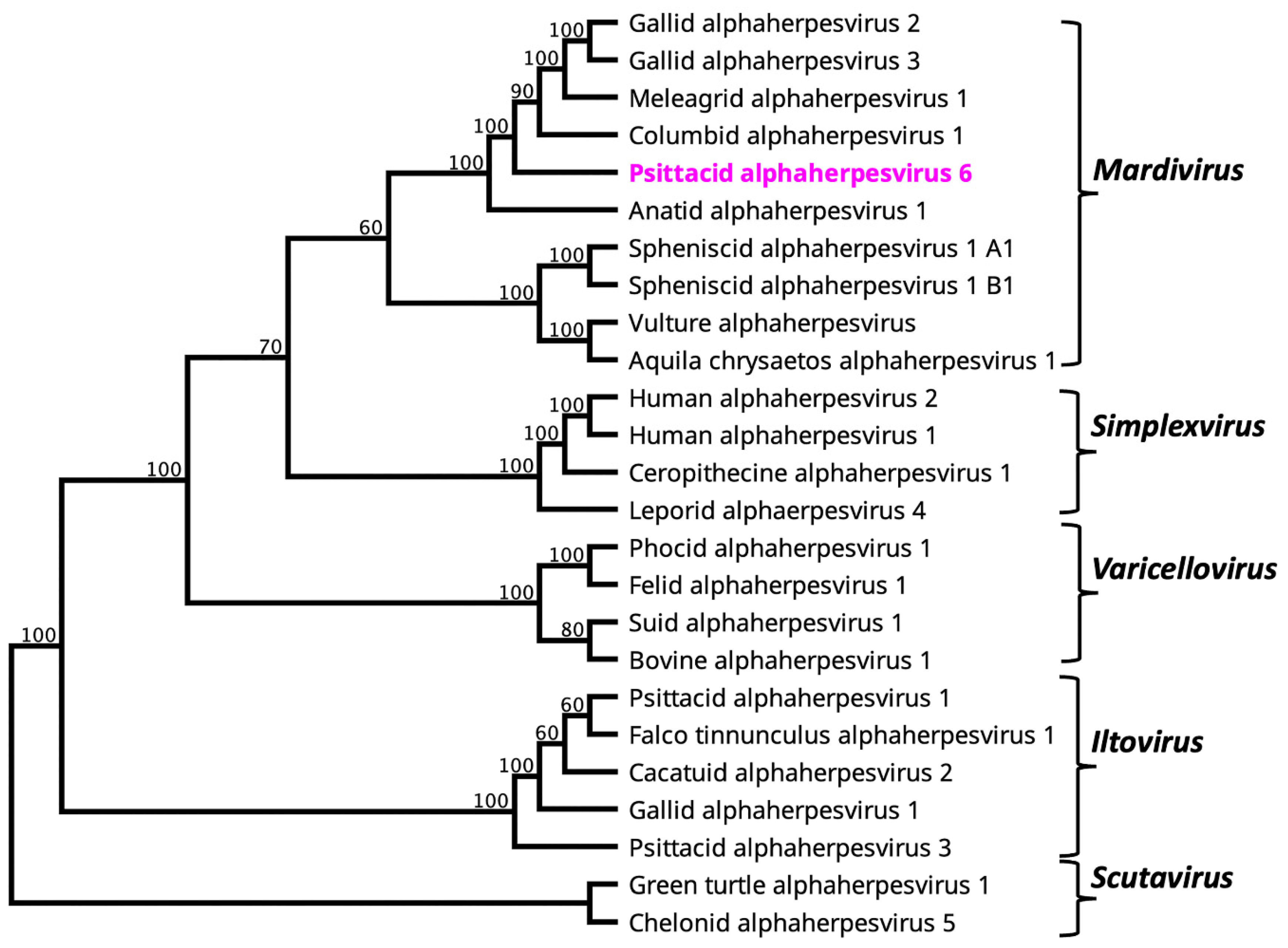
Figure 1. A maximum likelihood phylogenetic tree was constructed from the amino acid sequences of selected alphaherpesviruses using complete DNA polymerase genes except for Falco tinnunculus alphaherpesvirus 1 and Aquila chrysaetos alphaherpesvirus 1, for which only partial DNA polymerase sequences available. The numbers on the left show bootstrap values as percentages. The labels at branch tips refer to virus name. The novel PsAHV6 from rainbow lorikeet is shown in pink colour.
Viruses 15 02106 g001
Figure 2. An unrooted maximum likelihood phylogenetic tree was constructed from the amino acid sequences of selected avihepadnaviruses using complete DNA polymerase genes. The numbers on the left show bootstrap values as percentages. The labels at branch tips refer to GenBank accession numbers, followed by virus name. The novel parrot hepatitis B virus from rainbow lorikeet is shown in pink colour.
Viruses 15 02106 g002
Table 1. Psittacid alphaherpesvirus 6 (PsAHV6) genome annotation (partial) and comparative analysis of ORFs.
PsAHV6 SyntenyPsAHV6 Gene CoordinateNt LengthAA LengthBest Hit Gene ProductBest Hit (%Identity/Query Coverage/e-Value/PI/Organism)
PsAHV6-0011128-301828275envelope protein UL2038.43/85/6.00E-36/YP_009352926.1/CoAHV1
PsAHV6-0021628-188525885hypothetical proteinno significant BLAST hits
PsAHV6-0031959-37611803600tegument protein UL2133.28/92/7.00E-89/YP_009046517.1/FaAHV1
PsAHV6-0043847-4176330109hypothetical proteinno significant BLAST hits
PsAHV6-0056679-40102670889envelope glycoprotein H UL2233.87/88/1.00E-120/YP_009046519.1/FaAHV1
PsAHV6-0068196-70661131376thymidine kinase UL2336.47/85/7.00E-59/YP_009046521.1/FaAHV1
PsAHV6-0078112-91941083360nuclear protein UL2435.27/66/4.00E-26/ABK55352.1/AnAHV1
PsAHV6-0089374-114282055684DNA packaging tegument protein UL2554.55/82/5.00E-155/YP_009352933.1/CoAHV1
PsAHV6-00911370-1157020166hypothetical proteinno significant BLAST hits
PsAHV6-01011752-144002649882capsid maturation protease UL2656.57/28/2.00E-73/UJO49828.1/AnAHV1
PsAHV6-01117814-1473130841027envelope glycoprotein B UL260.91/80/0/CAA63039.1/GaAHV2
PsAHV6-01220681-1758630961031DNA packaging terminase subunit 2 UL2851.93/71/1.00E-151/NP_073322.1/MeAHV1
PsAHV6-01320801-21118318105hypothetical proteinno significant BLAST hits
PsAHV6-01425126-2109840291342single-stranded DNA-binding protein UL2951.90/99/0/YP_009352937.1/CoAHV1
PsAHV6-01525389-2512926186hypothetical proteinno significant BLAST hits
PsAHV6-01625677-2961239361311DNA polymerase UL3054.06/98/0/UOW62139.1/GaAHV2
PsAHV6-01730888-295361353450nuclear egress lamina protein UL3158.70/67/6.00E-111/YP_009046529.1/FaAHV1
PsAHV6-01833797-313232475824DNA packaging protein UL3251.12/84/2.00E-126/YP_009352940.1/CoAHV1
PsAHV6-01933808-34290483160DNA packaging protein UL3356.49/81/2.00E-34/YP_009046531.1/FaAHV1
PsAHV6-02034360-3455719865hypothetical proteinno significant BLAST hits
PsAHV6-02134643-357191077358nuclear egress membrane protein UL3458.62/48/5.00E-66/YP_009352942.1/CoAHV1
PsAHV6-02235815-36162348115small capsid protein UL3547.92/81/9.00E-12/YP_009046533.1/FaAHV1
PsAHV6-02346419-36400100203339large tegument protein UL3637.71/71/0/YP_009046534.1/FaAHV1
PsAHV6-02446742-46416327108hypothetical proteinno significant BLAST hits
PsAHV6-02550376-4696634111136tegument protein UL3736.23/98/0/YP_009046535.1/FaAHV1
PsAHV6-02650892-523971506501capsid triplex subunit 1 UL3854.68/93/1.00E-144/YP_009046536.1/FaAHV1
PsAHV6-02752766-555372772923ribonucleotide reductase subunit 1 UL3956.43/86/0/NP_073333.1/MeAHV1
PsAHV6-02856022-573981377458ribonucleotide reductase subunit 2 UL4067.17/71/1.00E-156/YP_009046538.1/FaAHV1
PsAHV6-02959232-576881545514tegument host shutoff protein UL4144.74/73/3.00E-79/YP_009352949.1/CoAHV1
PsAHV6-03059365-5955619263hypothetical proteinno significant BLAST hits
PsAHV6-03160266-616751410469DNA polymerase processivity subunit UL4242.02/73/5.00E-84/AUB50956.1/GaAHV2
PsAHV6-03262162-635201359452hypothetical proteinno significant BLAST hits
PsAHV6-03364113-6392219263hypothetical proteinno significant BLAST hits
PsAHV6-03464380-662931914637glycoprotein C UL4434.00/62/3.00E-78/AAM97710.1/GaAHV2
PsAHV6-03567702-66737966321hypothetical protein FaHV1S18_06034.68/90/5.00E-45/YP_009046544.1/FaAHV1
PsAHV6-03668228-6841618962hypothetical proteinno significant BLAST hits
PsAHV6-03768425-69114690229membrane protein UL4540.85/71/1.00E-40/YP_009352955.1/CoAHV1
PsAHV6-03872032-694922541846tegument protein VP11/12 UL4628.57/47/4.00E-40/YP_009046546.1/FaAHV1
PsAHV6-03971997-7227828293hypothetical proteinno significant BLAST hits
PsAHV6-04072552-7228327089hypothetical proteinno significant BLAST hits
PsAHV6-04172854-7304819564hypothetical proteinno significant BLAST hits
PsAHV6-04275190-732021989662tegument protein VP13/14 UL4733.16/81/3.00E-67/YP_009046547.1/FaAHV1
PsAHV6-04375135-75587453150hypothetical proteinno significant BLAST hits
PsAHV6-04475720-7547524681hypothetical proteinno significant BLAST hits
PsAHV6-04576074-7580826788hypothetical proteinno significant BLAST hits
PsAHV6-04676919-7672219865hypothetical proteinno significant BLAST hits
PsAHV6-04778218-770281191396transactivating tegument protein VP16 UL4845.56/89/9.00E-95/YP_009046548.1/FaAHV1
PsAHV6-04878299-7848118360hypothetical proteinno significant BLAST hits
PsAHV6-04978811-7853327992hypothetical proteinno significant BLAST hits
PsAHV6-05079856-79092765254tegument protein VP22 UL4938.36/28/7.00E-07/UOW62334.1/GaAHV2
PsAHV6-05179855-8005520166hypothetical proteinno significant BLAST hits
PsAHV6-05280468-8018128895envelope glycoprotein N UL49.551.04/98/8.00E-11/YP_009046550.1/FaAHV1
PsAHV6-05381124-825211398465deoxyuridine triphosphatase UL5040.74/98/2.00E-103/YP_009046551.1/FaAHV1
PsAHV6-05482780-8254124079hypothetical proteinno significant BLAST hits
PsAHV6-05583633-82671963320tegument protein UL5152.58/60/7.00E-59/YP_009046552.1/FaAHV1
PsAHV6-05683632-8730336721223helicase-primase primase subunit UL5242.05/94/0/YP_009352963.1/CoAHV1
PsAHV6-05787325-884941170389envelope glycoprotein K UL5349.29/89/2.00E-98/YP_009352964.1/CoAHV1
PsAHV6-05888959-903981440479multifunctional expression regulator UL5437.67/44/8.00E-30/YP_009046555.1/FaAHV1
PsAHV6-05991648-90716933310protein LORF436.59/87/3.00E-50/YP_009046556.1/FaAHV1
PsAHV6-06092305-92919615204nuclear protein UL5546.43/81/6.00E-46/YP_009352967.1/FaAHV1
PsAHV6-06194339-931101230409myristylated tegument protein CIRC33.19/55/5.00E-13/YP_009352969.1/CoAHV1
PsAHV6-06297772-9461131621053hypothetical protein LORF1128.14/86/4.00E-81/UOW65035.1/GaAHV2
PsAHV6-06399543-984491095364hypothetical proteinno significant BLAST hits
PsAHV6-06499943-10010716554hypothetical proteinno significant BLAST hits
PsAHV6-065100621-10046315952hypothetical proteinno significant BLAST hits
PsAHV6-066101828-1007581071356hypothetical proteinno significant BLAST hits
PsAHV6-067102213-101770444147hypothetical proteinno significant BLAST hits
PsAHV6-068102515-102832318105hypothetical proteinno significant BLAST hits
PsAHV6-069103442-103113330109hypothetical proteinno significant BLAST hits
Note: %, percentage, PI, Protein Identifier sequence identification number sourced by BLAST searches. The abbreviations for herpesviruses were used: PsAHV6, psittacid alphaherpesvirus 6; CoAHV1, columbid alphaherpesvirus 1; FaAHV1, falconid herpesvirus 1; GaAHV2, gallid alphaherpesvirus 2; AnAHV1, anatid alphaherpesvirus 1; MeAHV1, meleagrid alphaherpesvirus 1.
Disclaimer/Publisher’s Note: The statements, opinions and data contained in all publications are solely those of the individual author(s) and contributor(s) and not of MDPI and/or the editor(s). MDPI and/or the editor(s) disclaim responsibility for any injury to people or property resulting from any ideas, methods, instructions or products referred to in the content.