


Abstract
Sustainable development in wood technology demands eco-friendly alternatives to conventional preservatives, positioning nanotechnology as a promising frontier for green wood protection. The antifungal potential of silver nanoparticles (AgNPs) against the brown-rot agent Fomitopsis pinicola was investigated in vitro, alongside an assessment of their protective performance on Scots pine wood. AgNPs were tested at 5, 25, 50, and 100 ppm in vitro, with an additional 200 ppm concentration included in the wood-decay test. In the in vitro assays, lower concentrations (5 and 25 ppm) stimulated radial mycelial growth, 50 ppm showed no effect, and 100 ppm caused growth inhibition. In contrast, all tested concentrations enhanced the decay resistance of pine wood. Average wood mass loss ranged from 4.9% to 8.1% after 2 months and from 10.9% to 18.6% after 4 months, with the maximum protective effect observed at 50 ppm. While AgNPs did not entirely prevent F. pinicola-induced decay, they significantly mitigated mass loss. These findings highlight the potential of AgNPs for the short-term preservation of freshly harvested timber, particularly storm- or disaster-damaged wood stored in forests or log yards prior to processing.
1. Introduction
Scots pine (Pinus sylvestris L.) is an ecologically and economically vital tree species. Scots pine (Pinus sylvestris L.) serves as a cornerstone tree species, holding immense ecological and commercial value in Poland and across Europe, forming a dominant component of managed forests in boreal and temperate zones [1]. In Poland, it is the primary forest-forming species, covering 59.1% of the total forest area and accounting for 61.7% of the overall timber volume managed by the State Forests [2]. Scots pine wood has significant global commercial value and is widely used in the timber and paper industries [3]. Due to its favorable technological properties, it remains a preferred material for structural and construction purposes [4]. As a renewable resource with carbon sequestration capabilities, timber plays a pivotal role in sustainable development. However, both during storage and during structural use, wood is inherently vulnerable to biological degradation, which significantly limits its service life and results in substantial economic losses [5].
Among the biological agents responsible for timber deterioration, brown-rot fungi pose one of the most serious threats to coniferous wood. A prominent and widespread representative of these wood-decaying fungi in Poland and Central Europe is Fomitopsis pinicola (Sw.) P. Karst. [6]. As a primary saprotrophic species, it plays a key role in wood decomposition by rapidly degrading cellulose and hemicellulose while leaving the lignin matrix largely intact, resulting in a substantial loss of the mechanical strength of infected wood [7,8]. Although F. pinicola primarily colonizes dead or decaying coniferous trees of the Pinaceae family in natural forest ecosystems, its highly efficient wood-decaying activity also makes it a major cause of deterioration in harvested timber and in post-disaster timber stored in forests or log yards [9,10]. Today, nanotechnology is increasingly used across many industries, including wood protection [11,12]. Because nanoparticles are incredibly small (1–100 nm) and have a large surface area, they behave very differently and often perform better than standard materials [13]. These tiny particles can penetrate deeply into the wood’s cell walls and remain there longer, making them harder to leach than traditional treatments [14,15]. Research is now showing that nanoparticles could replace traditional chemical preservatives [16,17]. This shift would support sustainable development by providing a more effective, eco-friendly means of protecting wood from wood-decaying fungi [16].
To date, a relatively large number of studies have examined the potential of various types of nanoparticles to protect many wood species from fungal decay [15,16,17]. Numerous studies have demonstrated that AgNPs can substantially reduce fungal decay in wood; however, their efficacy varies considerably among experimental systems. High levels of protection have been reported for several wood species, including Acacia mangium, Vochysia guatemalensis, Cedrela odorata, Cordia alliodora, Populus spp., Paulownia fortunei, Pinus sylvestris, Fagus sylvatica, Gmelina arborea, and Goethalsia meiantha, particularly against the white-rot fungus Trametes versicolor [18,19,20,21,22,23]. In contrast, the same or similar nanoparticle formulations often provided only limited protection against other wood–fungus combinations. For example, AgNPs showed little or no efficacy against T. versicolor in protecting C. odorata [20] and brown brown-rot fungi Coniophora puteana, Poria placenta, or Lenzites acuta in protecting P. sylvestris, F. sylvatica, T. grandis [20,21,22].
These contrasting results suggest that nanoparticle performance cannot be attributed solely to particle characteristics such as size or concentration. Instead, antifungal efficacy appears to depend on the interaction between nanoparticle properties, wood characteristics, and the physiological traits of the target fungus. Wood anatomy influences nanoparticle penetration and retention [14,20], whereas fungal species differ in their decay strategies and physiological responses to nanoparticle-induced stress [21,22]. Consequently, results obtained for one wood–fungus combination cannot be readily extrapolated to others.
This issue is particularly evident when comparing white-rot and brown-rot fungi. While many studies report high efficacy of silver nanoparticles against white-rot fungi, especially T. versicolor [19,20,22], results for brown-rot fungi remain inconsistent. Silver nanoparticles have shown little or no protective effect against species such as C. puteana, P. placenta, and L. acuta [22], whereas they effectively protected the tropical woods Gmelina arborea and Goethalsia meiantha from decay caused by L. acuta [20]. These discrepancies indicate that susceptibility to silver nanoparticles cannot be generalized even within the same decay group and emphasize the need for species-specific evaluation of economically important brown-rot fungi.
Our earlier in vitro findings highlighted species-specific and type-dependent antifungal actions of AgNPs and CuNPs. Specifically, all tested concentrations of both nanoparticles successfully suppressed the growth of Meripilus giganteus, whereas Sparassis crispa displayed complete resistance. A selective response was noted for Fistulina hepatica and Grifola frondosa, which were vulnerable only to AgNPs [24]. Regarding the white-rot fungus Fomes fomentarius, CuNPs were found to stimulate mycelial growth, with the degree of stimulation inversely proportional to the concentration used, while AgNPs inhibited radial growth. Decay tests on beech wood treated with F. fomentarius correlated well with these in vitro findings: AgNPs enhanced the rot resistance of the wood, whereas CuNPs stimulated fungal decay [25].
Climate change is increasing the frequency of extreme weather events. Catastrophic winds cause large-scale windthrows and stem breakages in pine stands, resulting not only in immediate mechanical damage and timber losses but also in long-term ecological consequences [26,27]. These include heightened vulnerability to secondary pest outbreaks, increased wildfire risks, and the mass appearance of wood-decay and wood-staining fungi [28]. When the prompt removal of damaged timber is unfeasible, limiting fungal colonization during temporary storage becomes an important management objective. Metal nanoparticles have recently attracted attention as potential wood preservatives for such applications [25]. However, before their practical use can be considered, their antifungal activity must first be evaluated under controlled laboratory conditions using representative wood–fungus systems. Research to date indicates that fungal decay type alone does not explain susceptibility to silver nanoparticles. Considerable variation also exists among individual brown-rot species, highlighting the need to evaluate economically important taxa, such as F. pinicola, individually rather than extrapolating results from other brown-rot fungi.
The objective of this research was to assess how AgNPs influence the growth of F. pinicola in vitro and to concurrently evaluate their potential to protect pine wood from this brown-rot pathogen. Such experiments provide a necessary first step toward assessing their potential for protecting freshly harvested or storm-damaged timber during temporary storage.
2. Materials and Methods
2.1. Biological Materials
Test material consisted of Scots pine (Pinus sylvestris L.) sapwood, harvested from a 55-year-old tree stand located within a private forest area (Wola Uhruska commune, Włodawa County, Lublin Voivodeship, eastern Poland; coordinates: 23.551046, 51.343144). Defect-free specimens measuring 15 × 25 × 50 mm3 (tangential × radial × longitudinal) were prepared from the butt portion of the trunk at a height of 0.5–2 m, specifically targeted at the mid-radius zone, following the guidelines of the EN 113 standard [29]. Prior to testing, these blocks underwent a 2-week conditioning process under controlled environmental conditions (20 °C and 65% relative humidity). Mass measurements were performed post-conditioning, revealing an average air-dry wood density of 0.54 g/cm3.
Mycological testing was conducted using a Fomitopsis pinicola strain. This particular culture originated from the fungal collection maintained by the Department of Forest Protection at the Warsaw University of Life Sciences, having been previously isolated from wild fruiting bodies discovered on a Scots pine log within the Mińsk Forest District.
2.2. Nanoparticles
The selected metal nanoparticles were AgNPs. Commercial AgNP solutions utilized in this study were sourced from Nano-Koloid sp. z o. o. (Karolino 37, 05-140 Serock, Poland), operating under the license of Nano Technologies Group, Inc. (3772 Pinecone Court, Sarasota, FL 34238, USA). This product is manufactured in compliance with European patent EP2081672 A2. As specified by the supplier, the preparation involves a physical fabrication method yielding nanoparticles composed of approximately 100 atoms, with an average diameter of 5 nm. These particles are dispersed in a demineralized water matrix. The concentration of nanoparticles in the commercially available product is 50, but for the purposes of this study, the company prepared concentrations of 100 and 200 ppm.
2.3. In Vitro Assay
To evaluate the effects of the nanoparticles, F. pinicola was cultivated on malt extract agar (MEA) using 8 cm Petri dishes. The medium was amended with nanoparticles at four distinct concentrations: 5, 25, 50, and 100 ppm. To prevent thermal degradation, the non-sterilized nanoparticle solutions were incorporated into the agar only after it had cooled sufficiently to be handled comfortably by hand. MEA plates without any additives served as the control group. For each concentration, 10 replicates were prepared. Inoculation was performed using 5 mm mycelial discs obtained from 2-week-old fungal cultures. Subsequently, the plates were incubated under dark conditions at a constant temperature of 22 °C. Fungal growth was monitored daily between the third and tenth day of incubation, with the assessment period concluding once the mycelium reached the margins of the Petri dish. Colony diameter was determined by taking two perpendicular (“cross”) measurements from the underside of each dish, recorded with a precision of 1 mm. Based on the results obtained (after 10 days), an inhibition or stimulation index was calculated according to Formula (1):
where I/SI is the inhibition/stimulation index (%), DT is the diameter of the fungal colony on the test plate, and DC is the diameter of the fungal colony on the control plate.
I/SI = (DT − DC)/DC × 100%
According to this index, a negative result demonstrates fungal growth suppression, while a positive value denotes acceleration.
In contrast to the decay test, a 200 ppm concentration was not used in the in vitro experiment. This was due to the fact that metal nanoparticles undergo significant aggregation during autoclave sterilization, leading to a loss of their biological activity [30]. Therefore, they were added to the medium after it had been sterilized. Since the starting solution had a concentration of 200 ppm, it could not be diluted and added to the previously sterilized medium as with lower concentrations.
2.4. Decay Test
Since mass loss was used as the criterion for evaluating fungal activity, the samples’ dry mass was determined in accordance with EN 350-1 [31]. Prior to the experiment, all samples were conditioned at a constant temperature and relative humidity until they reached a constant weight, at which point they were weighed. Five randomly selected samples were then dried at 105 ± 2 °C to a constant weight to calculate the average moisture content (u, %) of the conditioned samples. Drying was performed in a Memmert UE500 laboratory dryer (Memmert GmbH & Co. KG, Schwabach, Germany). Based on these values, the initial dry mass of the remaining samples prior to the experiment was calculated using Formula (2). The remaining samples were not oven-dried at this stage, as exposure to high temperatures could cause structural damage and subsequently alter the wood decomposition process.
where: Mt—theoretical initial dry weight of the sample [g]; Mu—sample weight following conditioning at constant humidity [g]; and u—mean sample moisture content following conditioning at constant humidity [%].
Mt = (Mu/100 + u) × 100
Wood specimens were sterilized twice in an autoclave at 121 °C, with a 24 h interval between cycles. The first sterilization cycle lasted for 20 min, while the second was shortened to 10 min. Once sterilized, the specimens were left to cool in a laminar-flow chamber for 2–3 h. They were then treated by soaking in chilled, sterile, distilled water for 1 h for the control group, or in silver nanoparticle (AgNP) solutions at 5, 25, 50, 100, or 200 ppm. Nanoparticle solutions were not sterilized prior to use. The average retention of AgNPs at different nanoparticle concentrations was, respectively, 5.79 g/m3 (25 ppm), 11.59 g/m3 (50 ppm), 23.17 g/m3 (100 ppm), 46.32 g/m3 (200 ppm).
For the experiment, 192 samples were used, evenly distributed across 32 specimens per concentration and the control group. These treated and untreated wood samples were introduced into Kolle flasks on an MEA substrate previously inoculated with a 14-day-old F. pinicola culture. Incubation was carried out in a climate chamber (Q-Cell 700, Poll Lab, Wilkowice, Poland) maintained at 22 °C and 70% ± 5% relative humidity for 60 and 120 days, respectively. Following the initial 60-day period, half of the setup (16 replicates per group) was randomly selected and harvested, while the remaining samples were left until the 120-day mark. Once removed from the flasks, any surface mycelium was carefully cleaned off. The specimens were then oven-dried to a constant mass at 105 ± 2 °C before their final weight was recorded. To quantify wood degradation, mass loss was determined based on the difference between the theoretical initial dry weight and the post-exposure dry weight using the equation below:
where: Mloss—mass loss [%]; Mt—theoretical initial dry weight of the sample [g]; and Mf—final dry weight of the sample [g].
Mloss = [(Mt − Mf)/Mt] × 100
2.5. SEM Analysis
Morphological characteristics of both untreated (control) and nanoparticle-modified wood were investigated via scanning electron microscopy (SEM). For this purpose, small-sized specimens were prepared and subsequently analyzed using an FEI Quanta 200 SEM instrument (Thermo Fisher Scientific, Waltham, MA, USA). To assess fungal colonization at varying depths, the specimens were longitudinally sectioned (tangential plane) at a shallow angle to the external surface, using a fresh, high-grade carbon-steel razor blade for each sample to ensure a clean, artifact-free cut without crushing cellular structures. This technique exposed a gradient of wood layers, from the external surface down to a depth of approximately 5 mm into the wood interior. This approach enabled the direct comparison of hyphal density between the external surface and the underlying wood layers to characterize the spatial distribution of the F. pinicola mycelium.
2.6. Statistical Analysis
The effects of AgNP concentration and exposure duration on the antifungal response of F. pinicola grown on MEA medium were evaluated based on mycelial growth inhibition and mass loss of Scots pine wood specimens. The data were analyzed using a two-factor analysis of variance, with nanoparticle concentration and incubation time treated as fixed effects. When significant effects were detected, differences among treatment means were assessed using Tukey-adjusted pairwise comparisons. Before model interpretation, the assumptions of the analysis were checked by testing residual normality with the Shapiro–Wilk test and homogeneity of variance with Levene’s test. All statistical computations were performed in R version 4.4.2 (R Foundation for Statistical Computing, Vienna, Austria). Linear models were fitted and analyzed in R, while estimated marginal means and Tukey-adjusted comparisons were obtained using the emmeanspackage. Statistical significance was declared at p ≤ 0.05.
3. Results
3.1. In Vitro Assay
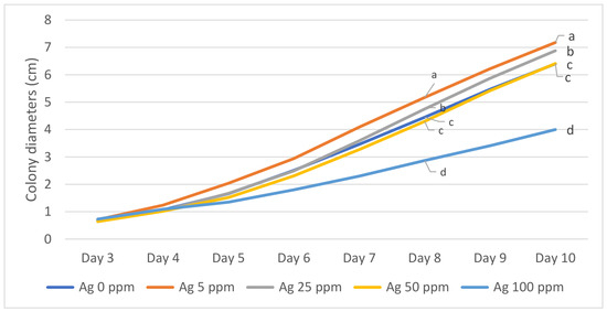
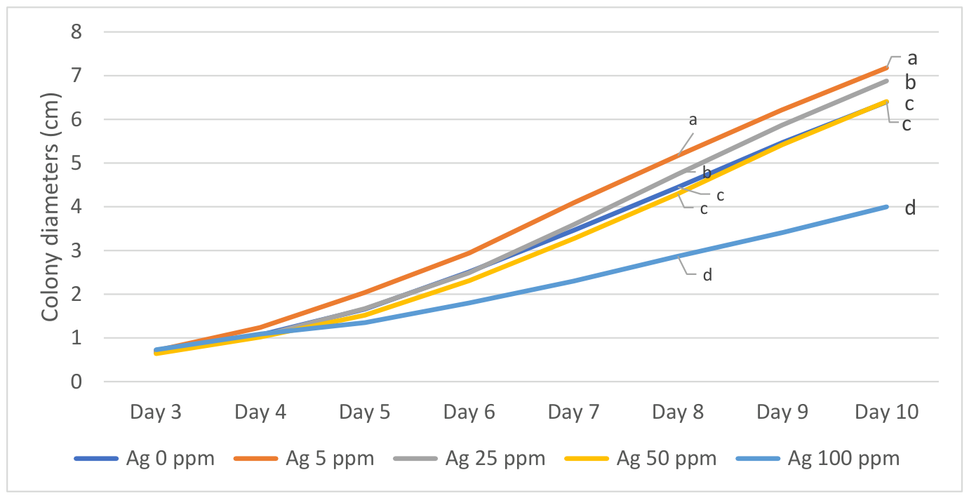
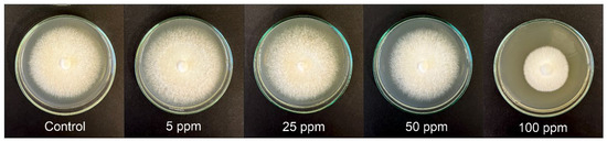
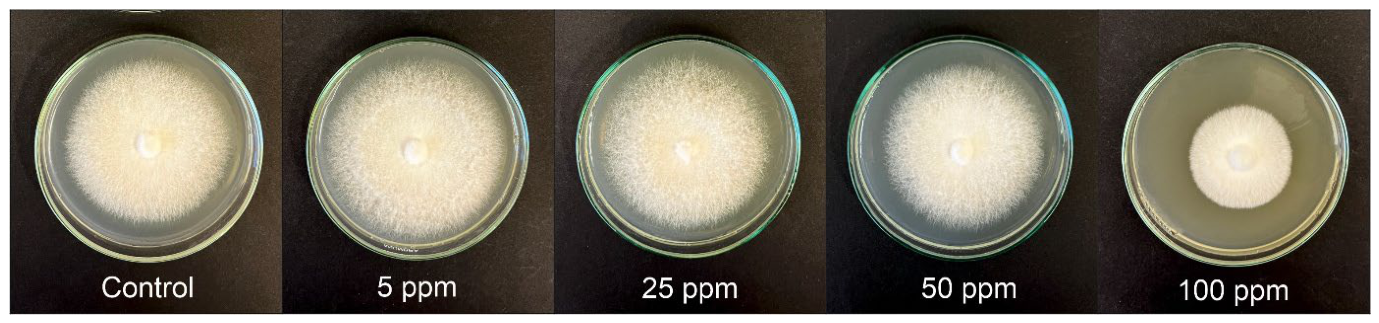
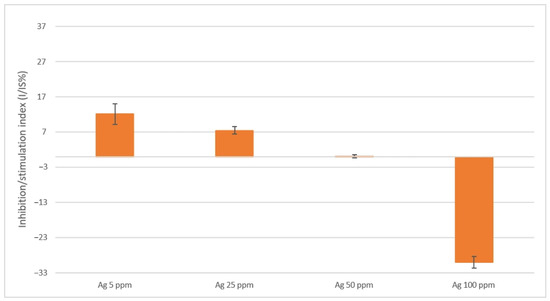
During the first four days of the experiment, no differences were observed in the growth rate of F. pinicola mycelium regardless of the nanoparticle concentration in the medium. From the 5th day until the end of the experiment, mycelial growth stimulation was observed at the lowest AgNPs concentration of 5 ppm. A beneficial effect on growth was also noted from the 8th day onwards for the 25 ppm concentration. Mycelium growing on the medium supplemented with 50 ppm AgNPs showed growth similar to that on the control plates (without NPs). AgNPs at 100 ppm significantly inhibited the growth of F. pinicola (Figure 1 and Figure 2).
Figure 1.
Inhibitory effect of various AgNP concentrations on the growth of F. pinicola cultured on MEA medium. Different letters indicate significant differences determined by the Tukey HSD test (p ≤ 0.05).
Figure 2.
F. pinicola colonies on MEA medium containing AgNPs at concentrations ranging from 5 to 100 ppm compared to the control (photograph by J. Piętka).
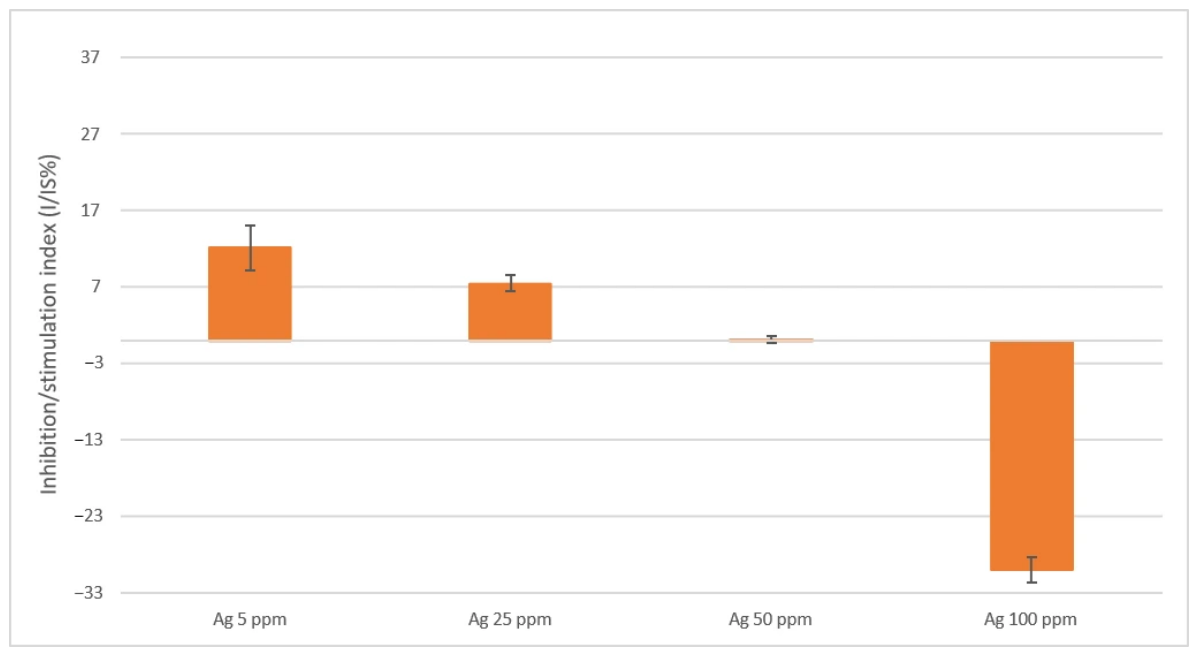
As illustrated by the stimulation/inhibition index at the end of the trial, F. pinicola mycelium experienced growth promotion when exposed to 5 and 25 ppm of AgNPs; contrastingly, the 100 ppm treatment resulted in clear inhibition (Figure 3).
Figure 3.
Antifungal activity expressed as index I/IS (%) of F. pinicola mycelia on MEA substrate containing AgNPs after a 10-day observation. Error bars indicate standard deviations.
3.2. Decay Test
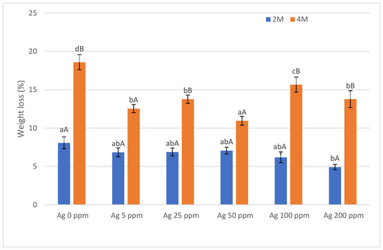
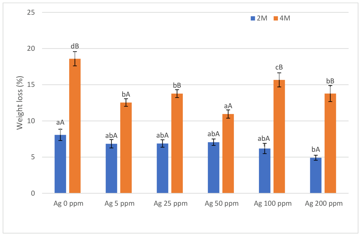
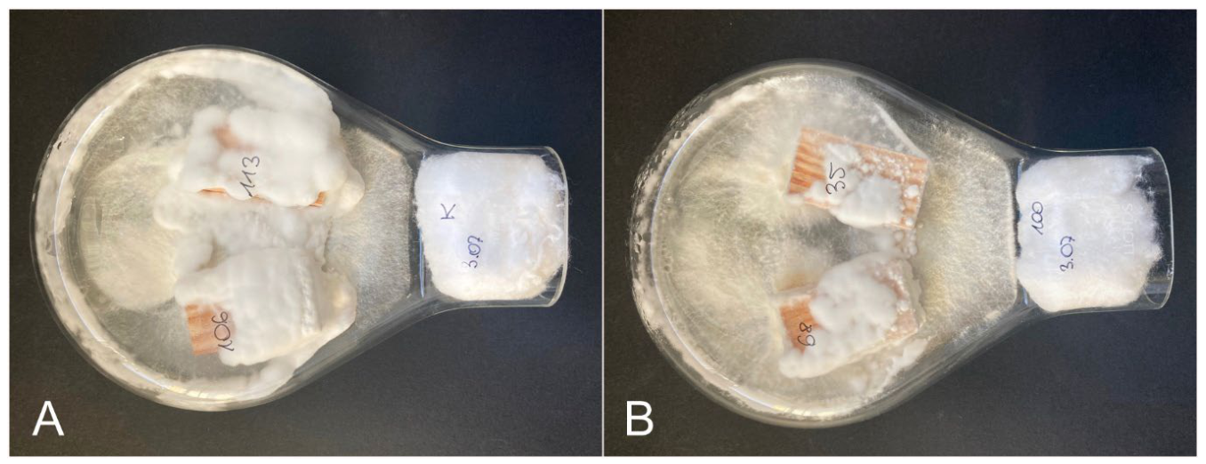
Depending on the experimental variant, the average wood mass loss ranged from 4.9% to 8.1% after two months and from 10.9% to 18.6% after four months. After 2 months of the experiment, a linear inhibition of wood decomposition by AgNPs was observed, but only at the highest concentration (200 ppm) was the difference in mass loss of the specimens statistically significant compared to the control. After 4 months, all tested concentrations significantly reduced wood degradation. The strongest inhibitory effect was observed at 50 ppm, while the weakest inhibition was observed at 100 ppm. The p-value indicates significant differences in the extent of weight loss between the specimens after 2 and 4 months at concentrations of 25, 100, and 200 ppm, and in the control group, but no differences at concentrations of 50 and 5 ppm (Figure 4 and Figure 5).
Figure 4.
Mass loss (%) of AgNP-treated pine wood after 60- and 120-day exposures. Statistically significant differences (Tukey’s HSD, p ≤ 0.05) among concentrations at a specific time point are marked by distinct lowercase letters. Variations between the two time periods for a single concentration are indicated by different uppercase letters. Error bars indicate standard deviations.
Figure 5.
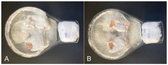
Kolle flasks with Scots pine wood specimens decomposed by F. pinicola: (A) control variant, (B) 100 ppm AgNP variant (photograph by M. Kędzierska).
3.3. SEM Analysis
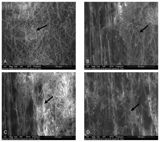
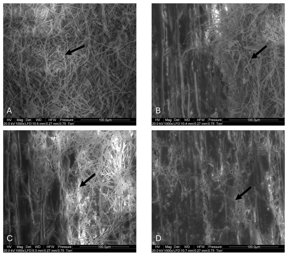
The surface of the control wood specimens exhibited substantially greater fungal mycelium coverage than the samples treated with AgNPs, particularly after four months of exposure (Figure 6A–D). SEM observations of specimens showing the highest weight loss within each treatment group indicated that mycelium predominantly colonized the wood surface. In internal longitudinal sections up to 5 mm in depth, the density of fungal hyphae was markedly lower than on the external surface in both control and AgNP-treated specimens (Figure 6B,C).
Figure 6.
SEM images of the longitudinally cut surface of Scots pine wood showing fungal mycelium (arrows) on the surface of control specimens (A,B) and specimens treated with silver nanoparticles at concentrations of 25 ppm (C), and 50 ppm (D) after four months of exposure. The images were selected from specimens exhibiting the greatest weight loss within every treatment group. In the control (B) and 25 ppm-treated (C) specimens, both the external surface and the underlying wood (approx. 5 mm deep) are visible, showing a lower density of fungal hyphae in the internal layers than on the external surface.
4. Discussion
The in vitro impact of metallic NPs on fungal growth is highly variable and is governed by several factors, including nanoparticle dimensions [32], their specific type and concentration, the targeted fungal species [24,33], and the formulation of the growth medium [34]. Our findings indicated a concentration-dependent response of F. pinicola to AgNPs; while the highest dose of 100 ppm suppressed mycelial development, the lowest concentrations (5 and 25 ppm) actually promoted radial growth, with no noticeable impact recorded at 50 ppm. Although many studies indicate that NPs exhibit high antifungal activity in vitro even at low concentrations (5–25 ppm) [32,35], our study does not confirm this. However, they are consistent with our earlier results [24,25]. The observed enhancement in mycelial growth at 5 and 25 ppm concentrations aligns with fungal hormesis, as documented by Spinoso-Castillo et al. [36] and Torres-Ruiz et al. [37] in the context of micropropagation. This biphasic dose–response relationship and the delayed response of the fungus to AgNP exposure (no changes in mycelial radial growth were observed until the fourth day of the experiment) are associated with mechanisms of nanoparticle action. Both AgNPs themselves and the silver ions released from their surface exert toxic effects, although through different pathways. Silver ions are largely responsible for the immediate toxicity of AgNPs [38], but their release occurs gradually [17,19]. One of the principal mechanisms of action of both AgNPs and silver ions involves the induction of oxidative stress by generating reactive oxygen species (ROS) [17,39]. The mycelium of F. pinicola is capable, to some extent, of neutralizing ROS through both enzymatic and non-enzymatic defense mechanisms. The fungus synthesizes antioxidant enzymes, including superoxide dismutase, which converts the highly reactive superoxide anion radical into the less reactive hydrogen peroxide, as well as catalase and glutathione peroxidase, which decompose hydrogen peroxide into water and oxygen, both of which are harmless to cellular structures [40,41]. In addition, low-molecular-weight antioxidants, including phenolic compounds, flavonoids, polysaccharides, and triterpenoids, neutralize free radicals [41,42]. During the first few days, these mechanisms may effectively protect the mycelium against the effects of nanoparticles. Only after several days might either accumulated cellular damage (in the case of growth inhibition) or potentially the attainment of an optimal level of stress signaling (in the case of growth stimulation) be associated with measurable changes in the mycelial radial growth rate.
In the case of mycelial growth stimulation induced by low concentrations of AgNPs, additional mechanisms may also play a potentially important role. The mycelium secretes organic acids into the agar medium, primarily oxalic acid, which chelates metal ions [22]. Furthermore, the fungal cell wall is reinforced through the synthesis of chitin and melanin. Melanin incorporated into the cell wall acts as a natural adsorbent for AgNPs, limiting their penetration into the cell and thereby protecting intracellular organelles from damage [43].
The decay test demonstrated that AgNPs inhibited the decomposition of Scots pine wood after both 2 and 4 months of incubation. In our earlier study on the effects of AgNPs on Fomes fomentarius in beech wood, the results of the in vitro assays were consistent with those obtained in wood-decay tests [25]. In contrast, the results obtained in the present study differed substantially between the in vitro and decay experiments. Such discrepancies are commonly observed in studies evaluating fungicides and biological plant protection agents (e.g., [44]). In our study, a direct comparison between the in vitro assay and the wood-decay experiment should be interpreted with caution. Besides the omission of the 200 ppm treatment from the in vitro assay due to nanoparticle aggregation during medium preparation, the two experiments evaluate different biological responses under different conditions. Fungal growth on agar reflects the direct response of the mycelium to silver nanoparticles, whereas wood decay additionally depends on nanoparticle penetration, distribution, and retention within wood tissues, as well as interactions between the fungus and the wood substrate. Therefore, discrepancies between the two assays should not be regarded as unexpected.
In the short term (2 months), a linear inhibition of wood decay by AgNPs was observed; however, only at the highest concentration (200 ppm) was the difference in sample mass loss statistically significant compared to the control. Surprising results were observed after 4 months: the lowest mass loss was recorded at 50 ppm, with no significant increase in degradation between the 2nd and 4th months of incubation (p = 0.0819). The application of 100 and 200 ppm doses resulted in mass losses similar to or higher than those observed at 5 and 25 ppm. The non-linear response of the fungus to increasing nanoparticle concentrations after 4 months may suggest the activation of alternative metabolic pathways or adaptive mechanisms at higher silver doses. However, since the decay test reflects the combined effects of nanoparticle retention, distribution, bioavailability, wood–nanoparticle interactions, and fungal activity, several alternative explanations remain equally plausible.
The higher resistance of F. pinicola to silver nanoparticles (AgNPs) may be closely linked to the physiology of this species, which is typical for brown-rot fungi. F. pinicola is characterized by the ability to intensively acidify the substrate through the secretion of large amounts of oxalic acid [45]. This process plays a dual role in the context of nanoparticle exposure: First, the low pH of the environment promotes the destabilization of the AgNP suspension, leading to their aggregation into larger structures with lower biological activity [46]. Second, silver ions released from the nanoparticles’ surfaces can be immediately bound by oxalate anions, forming insoluble, non-toxic complexes [47]. Furthermore, the specific wood-degrading mechanism employed by F. pinicola, based on the Fenton reaction, is associated with this species’s natural tolerance to high levels of oxidative stress [45]. Since one of the primary biocidal mechanisms of silver nanoparticles is the generation of ROS [39], the antioxidant systems of F. pinicola, which evolved to protect its own mycelium during the non-enzymatic degradation of the wood cell wall, may also effectively neutralize the toxic effect induced by nanosilver.
The absence of a linear relationship between AgNP concentration and the degree of wood protection after 4 months indicates that biocidal efficacy depends not only on nanoparticle concentration in the formulation but also on their retention, distribution, and bioavailability within the wood structure. It is possible that increasing the concentration of AgNPs did not result in a proportional increase in the amount of biologically active silver available to the fungus. This effect may be attributed to the limited number of binding (active) sites in wood cell walls and to nanoparticles’ tendency to aggregate at higher concentrations, thereby reducing their active surface area and potentially limiting their adhesion to wood [48]. Furthermore, the penetration of nanoparticles into wood is influenced by their physicochemical properties, and larger aggregates may be retained by the wood structure, leading to a heterogeneous distribution within the wood tissue [49]. Consequently, the protective efficacy may depend not only on the total silver content but also on its distribution and bioavailability within the wood. Our findings indicate that increasing AgNP concentration does not automatically enhance wood resistance to decay caused by F. pinicola, suggesting an optimal concentration range for maximum protective effectiveness.
The present data do not allow for discrimination between the potential mechanisms described above. Considering the factors discussed, the observed non-linear concentration response should be interpreted as the result of complex, multifaceted interactions among nanoparticle behavior within wood, wood–silver interactions, and fungal physiology, rather than evidence for a single underlying mechanism.
Similar to our study, Can [50] observed slight protection of Scots pine wood against brown rot caused by C. puteana following AgNP treatment. Conversely, Casado-Sanz et al. [23] achieved high antifungal efficacy with variable-sized AgNPs (10–30 nm) at concentrations as low as 5–20 ppm. In their review article, Slavin and Bach [43] emphasize that nanoparticle size is one of the most important factors influencing antifungal efficacy. Typically, smaller nanoparticles exhibit stronger antifungal activity than larger ones. The AgNPs used in our experiments had a size of 5 nm and were therefore considerably smaller than those employed in the studies cited above. Nevertheless, their antifungal activity was not particularly high, despite the concentrations applied being comparable to, or even higher than, those used by Casado-Sanz et al. [23].
These findings confirm that nanoparticles’ effectiveness depends on a wide range of factors. In addition to the type, shape, size, and concentration of nanoparticles, their efficacy is also influenced by the application method and the fungal and wood species involved [20,22,43].
Although AgNPs limit F. pinicola-induced wood decay, they do not provide absolute protection. Similarly, the limited effectiveness of silver nanocubes in protecting pine wood against brown-rot decay caused by the fungus C. puteana was demonstrated by Bak and Németh [22]. However, since nanoparticles show efficacy when applied by spraying or brushing methods [51], they may prove useful for the short-term protection of wood raw material, particularly disaster-damaged wood that remains in forests or timber yards for extended periods. The use of nanoparticles instead of traditional synthetic chemicals aligns with the principles of sustainable development, as it enables effective protection of the raw material while significantly reducing active-substance consumption and the release of toxic compounds into the forest ecosystem.
SEM observations revealed that F. pinicola mycelium was predominantly localized on the wood surface, whereas markedly fewer hyphae were observed approximately 5 mm beneath the surface in the examined samples. This pattern suggests a clear gradient of fungal colonization, with decreasing hyphal abundance toward the interior of the specimens. Although F. pinicola is a brown-rot fungus characterized by its ability to colonize tracheid lumens and traverse between cells via bordered pit apertures [52], the predominance of surface colonization observed after four months may reflect spatially heterogeneous decay development within the wood rather than uniform fungal penetration throughout the specimens. It is well established that brown-rot decay requires the diffusion of metabolites—including both enzymes and non-enzymatic agents—deep into the cell-wall matrix to initiate structural degradation [52,53].
Although bordered pit apertures in Pinus sylvestris (ca. 5.3–5.9 µm) [54] are substantially larger than the primary AgNPs used in this study (ca. 5 nm) and therefore would not be expected to impose strict size exclusion, nanoparticle mobility within wood is also likely influenced by aggregation phenomena and physicochemical interactions with wood cell-wall components. In addition, while bordered pit apertures may allow for the passage of primary nanoparticles under favorable conditions, transport within the wood structure is strongly dependent on its anatomy, in particular tracheid arrangement, bordered pit characteristics, and connectivity of axial tracheids [55], which collectively regulate the physical pathways available for nanoparticle movement. Furthermore, penetration into the wood cell wall is constrained by its inherently nanoporous and moisture-dependent architecture. The effective pore size of the hydrated cell wall is generally on the nanometer scale and depends strongly on moisture content, thereby limiting the accessibility and mobility of nanoparticles within the cell-wall matrix [56,57]. In addition, wood may act as a complex hierarchical porous system in which transport processes are affected by pore structure, pit membrane characteristics, and the tortuosity of the tracheid network [58]. Previous studies have reported that silver nanoparticles may undergo aggregation and become immobilized within wood microstructures, thereby influencing their transport behavior and spatial distribution [16]. It may therefore be hypothesized that AgNP aggregation and/or local accumulation occurred in pit chambers, pit membranes, or within the surrounding tracheid cell walls, potentially contributing to reduced mobility and heterogeneous spatial distribution of antifungal activity within the wood; however, direct evidence for such processes was not obtained in this study.
In addition, interactions between silver species and wood cell-wall polymers may also contribute to retention effects. The wood cell wall consists of a cellulose microfibril framework embedded in an amorphous matrix primarily composed of hemicelluloses and lignin, which together govern porosity, swelling behavior, and chemical reactivity, with minor contributions from other components such as proteins and bound water [22]. In particular, functional groups present in lignin, including phenolic and carboxylic groups, may provide potential coordination sites for silver species, which could contribute to their retention within the wood cell-wall matrix [59]. Such interactions may promote adsorption or binding of silver species to cell-wall surfaces, thereby influencing their mobility and the penetration of fungal decay agents; however, the relative contribution of chemical binding versus physical immobilization cannot be distinguished based on the present data.
It should also be acknowledged that the SEM observations represent only localized regions of the specimens and may not fully capture the three-dimensional extent of fungal colonization within the wood structure.
Collectively, these mechanisms provide a plausible explanation for the observed gradient of fungal colonization, characterized by a predominance of mycelium on the surface of wood specimens and a lower density of hyphae deeper within the wood structure. These factors may also contribute, at least in part, to the observed non-linear relationship between AgNP concentration and protective efficacy, as nanoparticle aggregation, retention within wood microstructures, and heterogeneous spatial distribution may reduce the effective bioavailability of silver despite increasing nominal concentrations. While our findings and existing literature confirm that AgNPs can effectively enhance wood resistance against decay fungi, establishing the precise conditions for optimal protection remains a challenge. The current body of research spans a broad range of variables, including nanoparticle morphology (e.g., spherical vs. nanocube), varying concentrations, diverse wood species, and different application methods. Given this complexity, further investigation is essential. Compared to traditional organic biocides, AgNPs offer superior advantages: they are non-volatile, do not degrade over time, remain odorless, and provide sustained long-term efficacy [20]. Further comprehensive research exploring a wider range of fungal species and diverse types of nanoparticles under varying environmental conditions is essential. Such studies are crucial not only for fully elucidating these complex interactions but also for optimizing the practical efficacy and ecological safety of nanotechnological wood preservatives.
5. Conclusions
This study demonstrates that 5 nm AgNPs can reduce wood decay caused by F. pinicola under controlled laboratory conditions, although their protective efficacy does not increase linearly with concentration. The results highlight the complexity of AgNP–wood–fungus interactions and indicate the existence of an apparent optimal concentration range for limiting fungal decay. They also demonstrate that antifungal activity observed in vitro does not predict performance in wood, emphasizing the importance of evaluating nanoparticle efficacy directly in wood substrates.
Because the present study was conducted using small, sterilized wood specimens under controlled laboratory conditions, the results should be regarded as a proof of concept rather than evidence of practical performance during timber storage. Further studies under conditions representative of freshly harvested or storm-damaged timber, including high moisture content, bark presence, natural microbial communities, fluctuating environmental conditions, and prolonged exposure, are required before the applicability of AgNP-based treatments for temporary timber preservation can be reliably assessed.
Author Contributions
Conceptualization, J.P. and M.A.-T.; Methodology, J.P.; Software, J.P.; Validation, J.P., M.A.-T. and M.T.; Formal Analysis, M.S.; Investigation, J.P., M.K., M.M. and M.T.; Resources, J.P. and M.T.; Data Curation, J.P. and M.S.; Writing—Original Draft Preparation, M.A.-T.; Writing—Review & Editing, M.A.-T., M.T. and J.P.; Visualization, M.S. and M.T.; Supervision, J.P. and M.A.-T.; Project Administration, J.P.; Funding Acquisition, J.P. and M.T. All authors have read and agreed to the published version of the manuscript.
Funding
This research received no external funding.
Data Availability Statement
The dataset generated for the study area is available from the corresponding author on reasonable request.
Conflicts of Interest
The authors declare no competing interests.
References
- Krakau, U.K.; Liesebach, M.; Aronen, T.; Lelu-Walter, M.A.; Schneck, V. Scots Pine (Pinus sylvestris L.). In Forest Tree Breeding in Europe; Pâques, L., Ed.; Managing Forest Ecosystems; Springer: Dordrecht, The Netherlands, 2013; Volume 25, pp. 267–323. ISBN 978-94-007-6145-2. [Google Scholar]
- Cierpiał-Wolan, M. Statistical Yearbook of Forestry, Statistics Poland, Statistical Office in Białystok; Zakład Wydawnictw Statystycznych: Warsaw, Poland, 2025; pp. 39–40. Available online: https://stat.gov.pl/obszary-tematyczne/roczniki-statystyczne/roczniki-statystyczne/rocznik-statystyczny-lesnictwa-2025,13,8.html.
- Brischke, C.; Alfredsen, G. Biological durability of pine wood. Wood Mater. Sci. Eng. 2023, 18, 1050–1064. [Google Scholar] [CrossRef] [Scilit]
- Mirski, R.; Dziurka, D.; Chuda-Kowalska, M.; Wieruszewski, M.; Kawalerczyk, J.; Trociński, A. The usefulness of pine timber (Pinus sylvestris L.) for the production of structural elements. Part I: Evaluation of the quality of the pine timber in the bending test. Materials 2020, 13, 3957. [Google Scholar] [CrossRef] [Scilit] [PubMed]
- Broda, M. Natural Compounds for Wood Protection against Fungi—A Review. Molecules 2020, 25, 3538. [Google Scholar] [CrossRef] [Scilit] [PubMed]
- Liu, S.; Han, M.-L.; Xu, T.-M.; Wang, Y.; Wu, D.-M.; Cui, B.-K. Taxonomy and Phylogeny of the Fomitopsis pinicola Complex with Descriptions of Six New Species From East Asia. Front. Microbiol. 2021, 12, 644979. [Google Scholar] [CrossRef] [Scilit] [PubMed]
- Bače, R.; Svoboda, M.; Pouška, V.; Janda, P.; Červenka, J. Natural regeneration in Central-European subalpine spruce forests: Which logs are suitable for seedling recruitment? For. Ecol. Manag. 2012, 266, 254–262. [Google Scholar] [CrossRef] [Scilit]
- Pouška, V.; Svoboda, M.; Lepš, J. Co-occurrence patterns of wood-decaying fungi on Picea abies logs: Does Fomitopsis pinicola influence the other species? Pol. J. Ecol. 2013, 61, 119–134. [Google Scholar]
- Butin, H. Tree Diseases and Disorders. Causes, Biology and Control in Forest and Amenity Trees; Oxford University Press: Oxford, NY, USA, 1995; pp. 165–166. [Google Scholar]
- Gáper, J.; Gáperová, S.; Pristaš, P.; Šebesta, M.; Kollárová, P.; Gallay, I.; Slobodník, B. The geographical distribution, trophic modes, and host preferences of Fomitopsis pinicola in Central Europe: A comprehensive review. Cent. Eur. For. J. 2025, 71, 73–82. [Google Scholar] [CrossRef] [Scilit]
- Evans, O.; Matsunaga, H.; Kiguchi, M. Large-scale application of nanotechnology for wood protection. Nat. Nanotechnol. 2008, 3, 577. [Google Scholar] [CrossRef] [Scilit] [PubMed]
- Pulit-Prociak, J.; Banach, M. Silver nanoparticles—A material of the future…? Open Chem. 2016, 14, 76–91. [Google Scholar] [CrossRef] [Scilit]
- Nel, A.; Xia, T.; Mädler, L.; Li, N. Toxic potential of materials at the nanolevel. Science 2006, 311, 622–627. [Google Scholar] [CrossRef] [Scilit] [PubMed]
- Kartal, S.N.; Green, F., III; Clausen, C.A. Do the unique properties of nanometals affect leachability or efficacy against fungi and termites? Int. Biodeterior. Biodegrad. 2009, 63, 490–495. [Google Scholar] [CrossRef] [Scilit]
- Borges, C.C.; Tonoli, G.H.D.; Cruz, T.M.; Duarte, P.J.; Junqueira, T.A. Nanoparticles-based wood preservatives: The next generation of wood protection? Cerne 2018, 24, 397–407. [Google Scholar] [CrossRef] [Scilit]
- Teng, T.-J.; Mat Arip, M.N.; Sudesh, K.; Nemoikina, A.; Jalaludin, Z.; Ng, E.-P.; Lee, H.-L. Conventional technology and nanotechnology in wood preservation: A Review. BioResources 2018, 13, 9220–9252. [Google Scholar] [CrossRef] [Scilit]
- Alsalamah, S.A.; Alghonaim, M.I.; Al Abboud, M.A. Role of nanoparticles for wood protection from decaying microorganisms and their enzymes: A review study. BioResources 2025, 20, 8119–8134. [Google Scholar] [CrossRef] [Scilit]
- Akhtari, M.; Arefkhani, M. Study of microscopy properties of wood impregnated with nanoparticles during exposed to white-rot fungus. Agric. Sci. Dev. 2013, 2, 116–119. [Google Scholar]
- Moya, R.; Berrocal, A.; Rodriguez-Zuniga, A.; Veja-Baudrit, J.; Noguera, S.C. Effect of silver nanoparticles on white rot wood decay and some physical properties of three tropical wood species. Wood Fiber Sci. 2014, 46, 527–538. [Google Scholar]
- Moya, R.; Rodriguez-Zuniga, A.; Berrocal, A.; Vega-Baudrit, J. Effect of silver nanoparticles synthesized with NPsAg-ethylene glycol (C2H6O2) on brown decay and white decay fungi of nine tropical woods. J. Nanosci. Nanotechnol. 2017, 17, 5233–5240. [Google Scholar] [CrossRef] [Scilit]
- Pařil, P.; Baar, J.; Čermák, P.; Rademacher, P.; Prucek, R.; Sivera, M.; Panáček, A. Antifungal effects of copper and silver nanoparticles against white and brown-rot fungi. J. Mater. Sci. 2017, 52, 2720–2729. [Google Scholar]
- Bak, M.; Németh, R. Effect of different nanoparticle treatments on the decay resistance of wood. BioResources 2018, 13, 7886–7899. [Google Scholar] [CrossRef] [Scilit]
- Casado-Sanz, M.M.; Silva-Castro, I.; Ponce-Herrero, L.; Martín-Ramos, P.; Martín-Gil, J.; Acuña-Rello, L. White-Rot Fungi Control on Populus spp. Wood by Pressure Treatments with Silver Nanoparticles, Chitosan Oligomers and Propolis. Forests 2019, 10, 885. [Google Scholar] [CrossRef] [Scilit]
- Aleksandrowicz-Trzcińska, M.; Szaniawski, A.; Olchowik, J.; Drozdowski, S. Effects of copper and silver nanoparticles on growth in selected species of pathogenic and wood-decay fungi in vitro. For. Chron. 2018, 93, 109–116. [Google Scholar] [CrossRef] [Scilit]
- Piętka, J.; Adamczuk, A.; Zarzycka, E.; Tulik, M.; Studnicki, M.; Oszako, T.; Aleksandrowicz-Trzcińska, M. The Application of Copper and Silver Nanoparticles in the Protection of Fagus sylvatica Wood against Decomposition by Fomes fomentarius. Forests 2022, 13, 1724. [Google Scholar] [CrossRef] [Scilit]
- Dale, V.H.; Joyce, L.A.; McNulty, S.; Neilson, R.P.; Ayres, M.P.; Flannigan, M.D.; Hanson, P.I.; Irland, L.C.; Lugo, A.E.; Peterson, C.J.; et al. Climate change and forest disturbances. BioScience 2001, 51, 723–734. [Google Scholar] [CrossRef] [Scilit]
- Seidl, R.; Thom, D.; Kautz, M.; Martin-Benito, D.; Peltoniemi, M.; Vacchiano, G.; Wild, J.; Ascoli, D.; Petr, M.; Honkaniemi, J.; et al. Forest disturbances under climate change. Nat. Clim. Change 2017, 7, 395–402. [Google Scholar] [CrossRef] [Scilit] [PubMed]
- Gardiner, B. Wind damage to forests and trees: A review with an emphasis on planted and managed forests. J. For. Res. 2021, 26, 248–266. [Google Scholar] [CrossRef] [Scilit]
- EN 113; Wood Preservatives—Test Method for Determining the Protective Effectiveness Against Wood Destroying Basidiomycetes—Determination of the Toxic Values. European Committee for Standardization: Brussels, Belgium, 1996.
- Bernal-Chávez, S.A.; Del Prado-Audelo, M.L.; Caballero-Florán, I.H.; Giraldo-Gomez, D.M.; Figueroa-Gonzalez, G.; Reyes-Hernandez, O.D.; González-Del Carmen, M.; González-Torres, M.; Cortés, H.; Leyva-Gómez, G. Insights into Terminal Sterilization Processes of Nanoparticles for Biomedical Applications. Molecules 2021, 26, 2068. [Google Scholar] [CrossRef] [Scilit] [PubMed]
- EN 350-1; Durability of Wood and Wood-Based Products—Natural Durability of Solid Wood—Part 1: Guide to the Principles of Testing and Classification of the Natural Durability of Wood. European Committee for Standardization: Brussels, Belgium, 1994.
- Akpinar, I.; Unal, M.; Sar, T. Potential antifungal effects of silver nanoparticles (AgNPs) of different sizes against phytopathogenic Fusarium oxysporum f. sp. radicis-lycopersici (FORL) strains. SN Appl. Sci. 2021, 3, 356. [Google Scholar] [CrossRef] [Scilit]
- Consolo, V.F.; Torres-Nicolini, A.; Alvarez, V.A. Mycosynthetized Ag, CuO, and ZnO nanoparticles from a promising Trichoderma harzianum strain and their antifungal potential against important phytopathogens. Sci. Rep. 2020, 10, 20499. [Google Scholar] [CrossRef] [Scilit] [PubMed]
- Kaur, A.; Kaur, J.; Kalia, A.; Singh, N. Effect of media composition on extent of antimycotic activity of silver nanoparticles against plant pathogenic fungus Fusarium moniliforme. Plant Dis. Res. 2016, 31, 1–5. [Google Scholar]
- Kim, S.W.; Kim, K.S.; Lamsal, K.; Kim, Y.J.; Kim, S.B.; Jung, M.; Sim, S.J.; Kim, H.S.; Chang, S.J.; Kim, J.K.; et al. An in vitro study of the antifungal effect of silver nanoparticles on oak wilt pathogen Raffaelea sp. J. Microbiol. Biotechnol. 2009, 19, 760–764. [Google Scholar] [CrossRef]
- Spinoso-Castillo, J.L.; Chávez-Santoscoy, R.A.; Bogdanchikova, N.E.; Pérez-Sato, J.A.; Morales-Ramos, V.; Bello-Bello, J.J. Antimicrobial and hormetic effects of silver nanoparticles on in vitro regeneration of vanilla (Vanilla planifolia Jacks. ex Andrews) using a temporary immersion system. Plant Cell Tissue Organ Cult. 2017, 129, 195–207. [Google Scholar] [CrossRef] [Scilit]
- Torres-Ruiz, J.R.; Silverio-Gómez, C.; Reyes-Zambrano, S.J.; Montes-Molina, J.A.; Rodríguez-Meneses, A.; Lecona-Guzmán, C.A. Hormetic effect of silver nanoparticles on the micropropagation of Creole pineapple (Ananas comosus). Folia Hortic. 2026, 37, 433–441. [Google Scholar] [CrossRef] [Scilit]
- Hsiao, I.L.; Hsieh, Y.K.; Wang, C.F.; Chen, I.C.; Huang, Y.J. Trojan-horse mechanism in the cellular uptake of silver nanoparticles verified by direct intra- and extracellular silver speciation analysis. Environ. Sci. Technol. 2015, 49, 3813–3821. [Google Scholar] [CrossRef] [Scilit] [PubMed]
- Cruz-Luna, A.R.; Cruz-Martínez, H.; Vásquez-López, A.; Medina, D.I. Metal Nanoparticles as Novel Antifungal Agents for Sustainable Agriculture: Current Advances and Future Directions. J. Fungi 2021, 7, 1033. [Google Scholar] [CrossRef] [Scilit] [PubMed]
- Choi, D.; Park, S.S.; Ding, J.L.; Cha, W.S. Effects of Fomitopsis pinicola extracts on antioxidant and antitumor activities. Biotechnol. Bioprocess Eng. 2007, 12, 516–524. [Google Scholar] [CrossRef] [Scilit]
- Onar, O.; Akata, I.; Celep, G.S.; Yildirim, O. Antioxidant Activity of Extracts from the Red-Belt Conk Medicinal Mushroom, Fomitopsis pinicola (Agaricomycetes), and Its Modulatory Effects on Antioxidant Enzymes. Int. J. Med. Mushrooms 2016, 18, 501–508. [Google Scholar] [CrossRef] [Scilit]
- Krupodorova, T.; Barshteyn, V.; Dzhagan, V.; Pluzhnyk, A.; Zaichenko, T.; Blume, Y. Enhancement of antioxidant activity and total phenolic content of Fomitopsis pinicola mycelium extract. Fungal Biol. Biotechnol. 2024, 11, 18. [Google Scholar] [CrossRef] [Scilit] [PubMed]
- Slavin, Y.N.; Bach, H. Mechanisms of Antifungal Properties of Metal Nanoparticles. Nanomaterials 2022, 12, 4470. [Google Scholar] [CrossRef] [Scilit] [PubMed]
- Mihajlović, M.; Rekanović, E.; Hrustić, J.; Grahovac, M.; Tanović, B. In vitro and in vivo toxicity of fungicides and biofungicides for the control of verticillium and fusarium wilt of pepper. Pestic. I Fitomedicina 2021, 36, 23–34. [Google Scholar] [CrossRef] [Scilit]
- Shah, F.; Mali, T.; Lundell, T.K. Polyporales Brown Rot Species Fomitopsis pinicola: Enzyme Activity Profiles, Oxalic Acid Production, and Fe3+-Reducing Metabolite Secretion. Appl. Environ. Microbiol. 2018, 84, e02662-17. [Google Scholar] [CrossRef] [Scilit] [PubMed]
- Bélteky, P.; Rónavári, A.; Igaz, N.; Szerencsés, B.; Tóth, I.Y.; Pfeiffer, I.; Kiricsi, M.; Kónya, Z. Silver nanoparticles: Aggregation behavior in biorelevant conditions and its impact on biological activity. Int. J. Nanomed. 2019, 14, 667–687. [Google Scholar] [CrossRef] [Scilit] [PubMed]
- Jarosz-Wilkolazka, A.; Gadd, G.M. Oxalate production by wood-rotting fungi growing in toxic metal-amended medium. Chemosphere 2003, 52, 541–547. [Google Scholar] [CrossRef] [Scilit] [PubMed]
- Dai, X.; Qi, Y.; Luo, H.; He, Z.; Wei, L.; Dong, X.; Ma, X.; Yang, D.-Q.; Li, Y. Leachability and Anti-Mold Efficiency of Nanosilver on Poplar Wood Surface. Polymers 2022, 14, 884. [Google Scholar] [CrossRef] [Scilit] [PubMed]
- Bossert, D.; Geers, C.; Placencia Peña, M.I.; Volkmer, T.; Rothen-Rutishauser, B.; Petri-Fink, A. Size and Surface Charge Dependent Impregnation of Nanoparticles in Soft- and Hardwood. Chemistry 2020, 2, 361–373. [Google Scholar] [CrossRef] [Scilit]
- Can, A.; Palanti, S.; Sivrikaya, H.; Hazer, B.; Stefanı, F. Physical, biological, and chemical characterization of wood treated with silver nanoparticles. Cellulose 2019, 26, 5075–5084. [Google Scholar] [CrossRef] [Scilit]
- De Filpo, G.; Palermo, A.M.; Rachiele, F.; Nicoletta, F.P. Preventing fungal growth in wood by titanium dioxide nanoparticles. Int. Biodeterior. Biodegrad. 2013, 85, 217–222. [Google Scholar] [CrossRef] [Scilit]
- Goodell, B.; Qian, Y.; Jellison, J. Brown-rot fungal degradation mechanisms. In Development of Commercial Wood Preservatives; ACS Sympposium Series; American Chemical Society: Washington, DC, USA, 2008. [Google Scholar]
- Arantes, V.; Goodell, B. Current Understanding of Brown-Rot Fungal Biodegradation Mechanisms: A Review. In Deterioration and Protection of Sustainable Biomaterials; Schultz, T.P., Goodell, B., Nicholas, D.D., Eds.; ACS Symp. Ser. 1158; American Chemical Society: Washington, DC, USA, 2014; pp. 3–22. [Google Scholar] [CrossRef] [Scilit]
- Held, M.; Ganthaler, A.; Lintunen, A.; Oberhuber, W.; Mayr, S. Tracheid and Pit Dimensions Hardly Vary in the Xylem of Pinus sylvestris Under Contrasting Growing Conditions. Front. Plant Sci. 2021, 12, 786593. [Google Scholar] [CrossRef] [Scilit] [PubMed]
- Choat, B.; Cobb, A.R.; Jansen, S. Structure and function of bordered pits: New discoveries and impacts on whole-plant hydraulic function. New Phytol. 2008, 177, 608–626. [Google Scholar] [CrossRef] [Scilit] [PubMed]
- Hill, C.A.S. Wood Modification: Chemical, Thermal and Other Processes; Wiley: Chichester, UK, 2006; ISBN 978-0-470-02172-9. [Google Scholar]
- Fredriksson, M.; Thybring, E.E. On sorption hysteresis in wood: Separating hysteresis in cell wall water and capillary water in the full moisture range. PLoS ONE 2019, 14, e0225111. [Google Scholar] [CrossRef] [Scilit] [PubMed]
- Lachenbruch, B.; McCulloh, K.A. Traits, properties, and performance: How woody plants combine hydraulic and mechanical functions in a cell, tissue, or whole plant. New Phytol. 2014, 204, 747–764. [Google Scholar] [CrossRef] [Scilit] [PubMed]
- Pajer, N.; Cestari, C.; Argyropoulos, D.S.; Crestini, C. From lignin self-assembly to nanoparticles nucleation and growth: A critical perspective. npj Mater. Sustain. 2024, 2, 31. [Google Scholar] [CrossRef] [Scilit]
Disclaimer/Publisher’s Note: The statements, opinions and data contained in all publications are solely those of the individual author(s) and contributor(s) and not of MDPI and/or the editor(s). MDPI and/or the editor(s) disclaim responsibility for any injury to people or property resulting from any ideas, methods, instructions or products referred to in the content. |
© 2026 by the authors. Licensee MDPI, Basel, Switzerland. This article is an open access article distributed under the terms and conditions of the Creative Commons Attribution (CC BY) license.