Next Issue
Volume 19, November
Previous Issue
Volume 19, September
 
 
ijms-logo

Journal Browser

Journal Browser

Int. J. Mol. Sci., Volume 19, Issue 10 (October 2018) – 439 articles

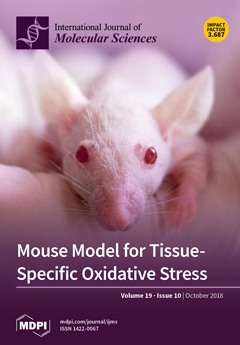
Cover Story (view full-size image): Tissue-specific responses to oxidative stressors reflect the organ-related variable vulnerability to chronic diseases, including cancer and age-related disorders. The current study defines the tissue-specific features of the oxidative stress-related transcriptome from 10 different tissues and organs of healthy mice. These results will serve as a reference for animal models and help to advance our understanding of tissue-specific variability in oxidative stress-associated pathogenesis. View this paper.
  • Issues are regarded as officially published after their release is announced to the table of contents alert mailing list.
  • You may sign up for e-mail alerts to receive table of contents of newly released issues.
  • PDF is the official format for papers published in both, html and pdf forms. To view the papers in pdf format, click on the "PDF Full-text" link, and use the free Adobe Reader to open them.
Order results
Result details
Section
Select all
Export citation of selected articles as:
14 pages, 863 KiB  
Review
Novel Transcriptional Mechanisms for Regulating Metabolism by Thyroid Hormone
by Brijesh Kumar Singh 1,*, Rohit Anthony Sinha 2 and Paul Michael Yen 1,3,*
1 Laboratory of Hormonal Regulation, Cardiovascular and Metabolic Disorders Program, Duke-NUS Medical School, Singapore 169857, Singapore
2 Department of Endocrinology, Sanjay Gandhi Post Graduate Institute of Medical Sciences, Raebareli Road, Lucknow 226014, Uttar Pradesh, India
3 Division of Endocrinology, Metabolism, and Nutrition, Department of Medicine, Duke Molecular Physiology Institute, Duke University School of Medicine, Durham, NC 27710, USA
Int. J. Mol. Sci. 2018, 19(10), 3284; https://doi.org/10.3390/ijms19103284 - 22 Oct 2018
Cited by 36 | Viewed by 6479
Abstract
The thyroid hormone plays a key role in energy and nutrient metabolisms in many tissues and regulates the transcription of key genes in metabolic pathways. It has long been believed that thyroid hormones (THs) exerted their effects primarily by binding to nuclear TH [...] Read more.
The thyroid hormone plays a key role in energy and nutrient metabolisms in many tissues and regulates the transcription of key genes in metabolic pathways. It has long been believed that thyroid hormones (THs) exerted their effects primarily by binding to nuclear TH receptors (THRs) that are associated with conserved thyroid hormone response elements (TREs) located on the promoters of target genes. However, recent transcriptome and ChIP-Seq studies have challenged this conventional view as discordance was observed between TH-responsive genes and THR binding to DNA. While THR association with other transcription factors bound to DNA, TH activation of THRs to mediate effects that do not involve DNA-binding, or TH binding to proteins other than THRs have been invoked as potential mechanisms to explain this discrepancy, it appears that additional novel mechanisms may enable TH to regulate the mRNA expression. These include activation of transcription factors by SIRT1 via metabolic actions by TH, the post-translational modification of THR, the THR co-regulation of transcription with other nuclear receptors and transcription factors, and the microRNA (miR) control of RNA transcript expression to encode proteins involved in the cellular metabolism. Together, these novel mechanisms enlarge and diversify the panoply of metabolic genes that can be regulated by TH. Full article
(This article belongs to the Special Issue Molecular Biology of Nuclear Receptors)
Show Figures

Graphical abstract

13 pages, 954 KiB  
Review
Crosstalk between Brassinosteroids and Ethylene during Plant Growth and under Abiotic Stress Conditions
by Petra Jiroutova, Jana Oklestkova * and Miroslav Strnad
Laboratory of Growth Regulators, Centre of the Region Haná for Biotechnological and Agricultural Research, Institute of Experimental Botany ASCR & Palacký University, Šlechtitelů 27, 78371 Olomouc, Czech Republic
Int. J. Mol. Sci. 2018, 19(10), 3283; https://doi.org/10.3390/ijms19103283 - 22 Oct 2018
Cited by 60 | Viewed by 11881
Abstract
Plant hormones through signaling networks mutually regulate several signaling and metabolic systems essential for both plant development and plant responses to different environmental stresses. Extensive research has enabled the main effects of all known phytohormones classes to be identified. Therefore, it is now [...] Read more.
Plant hormones through signaling networks mutually regulate several signaling and metabolic systems essential for both plant development and plant responses to different environmental stresses. Extensive research has enabled the main effects of all known phytohormones classes to be identified. Therefore, it is now possible to investigate the interesting topic of plant hormonal crosstalk more fully. In this review, we focus on the role of brassinosteroids and ethylene during plant growth and development especially flowering, ripening of fruits, apical hook development, and root and shoot growth. As well as it summarizes their interaction during various abiotic stress conditions. Full article
Show Figures

Graphical abstract

21 pages, 3546 KiB  
Review
The Importance of the Right Framework: Mitogen-Activated Protein Kinase Pathway and the Scaffolding Protein PTPIP51
by Eric Dietel 1,*, Alexander Brobeil 2, Stefan Gattenlöhner 2 and Monika Wimmer 1,3
1 Institute of Anatomy and Cell Biology, Justus-Liebig-University, 35392 Giessen, Germany
2 Institute of Pathology, Justus-Liebig-University, 35392 Giessen, Germany
3 Institute of Anatomy, Johannes-Kepler-University Linz, 4040 Linz, Austria
Int. J. Mol. Sci. 2018, 19(10), 3282; https://doi.org/10.3390/ijms19103282 - 22 Oct 2018
Cited by 7 | Viewed by 5530
Abstract
The protein tyrosine phosphatase interacting protein 51 (PTPIP51) regulates and interconnects signaling pathways, such as the mitogen-activated protein kinase (MAPK) pathway and an abundance of different others, e.g., Akt signaling, NF-κB signaling, and the communication between different cell organelles. PTPIP51 acts as a [...] Read more.
The protein tyrosine phosphatase interacting protein 51 (PTPIP51) regulates and interconnects signaling pathways, such as the mitogen-activated protein kinase (MAPK) pathway and an abundance of different others, e.g., Akt signaling, NF-κB signaling, and the communication between different cell organelles. PTPIP51 acts as a scaffold protein for signaling proteins, e.g., Raf-1, epidermal growth factor receptor (EGFR), human epidermal growth factor receptor 2 (Her2), as well as for other scaffold proteins, e.g., 14-3-3 proteins. These interactions are governed by the phosphorylation of serine and tyrosine residues of PTPIP51. The phosphorylation status is finely tuned by receptor tyrosine kinases (EGFR, Her2), non-receptor tyrosine kinases (c-Src) and the phosphatase protein tyrosine phosphatase 1B (PTP1B). This review addresses various diseases which display at least one alteration in these enzymes regulating PTPIP51-interactions. The objective of this review is to summarize the knowledge of the MAPK-related interactome of PTPIP51 for several tumor entities and metabolic disorders. Full article
Show Figures

Graphical abstract

16 pages, 4552 KiB  
Article
Dietary Isoliquiritigenin at a Low Dose Ameliorates Insulin Resistance and NAFLD in Diet-Induced Obesity in C57BL/6J Mice
by Youngmi Lee 1, Eun-Young Kwon 1,2 and Myung-Sook Choi 1,2,*
1 Department of Food Science and Nutrition, Kyungpook National University, 1370 San-Kyuk Dong Puk-Ku, Daegu 41566, Korea
2 Center for Food and Nutritional Genomics Research, Kyungpook National University, 1370 San-Kyuk Dong Puk-Ku, Daegu 41566, Korea
Int. J. Mol. Sci. 2018, 19(10), 3281; https://doi.org/10.3390/ijms19103281 - 22 Oct 2018
Cited by 21 | Viewed by 6115
Abstract
Isoliquiritigenin (ILG) is a flavonoid constituent of Glycyrrhizae plants. The current study investigated the effects of ILG on diet-induced obesity and metabolic diseases. C57BL/6J mice were fed a normal diet (AIN-76 purified diet), high-fat diet (40 kcal% fat), and high-fat diet +0.02% ( [...] Read more.
Isoliquiritigenin (ILG) is a flavonoid constituent of Glycyrrhizae plants. The current study investigated the effects of ILG on diet-induced obesity and metabolic diseases. C57BL/6J mice were fed a normal diet (AIN-76 purified diet), high-fat diet (40 kcal% fat), and high-fat diet +0.02% (w/w) ILG for 16 weeks. Supplementation of ILG resulted in decreased body fat mass and plasma cholesterol level. ILG ameliorated hepatic steatosis by suppressing the expression of hepatic lipogenesis genes and hepatic triglyceride and fatty acid contents, while enhancing β-oxidation in the liver. ILG improved insulin resistance by lowering plasma glucose and insulin levels. This was also demonstrated by the intraperitoneal glucose tolerance test (IPGTT). Additionally, ILG upregulated the expression of insulin signaling-related genes in the liver and muscle. Interestingly, ILG elevated energy expenditure by increasing the expression of thermogenesis genes, which is linked to stimulated mitochondrial biogenesis and uncoupled cellular respiration in brown adipose tissue. ILG also suppressed proinflammatory cytokine levels in the plasma. These results suggest that ILG supplemented at 0.02% in the diet can ameliorate body fat mass, plasma cholesterol, non-alcoholic fatty liver disease, and insulin resistance; these effects were partly mediated by increasing energy expenditure in high-fat fed mice. Full article
(This article belongs to the Section Molecular Endocrinology and Metabolism)
Show Figures

Graphical abstract

10 pages, 1785 KiB  
Article
Cepharanthine Enhances TRAIL-Mediated Apoptosis Through STAMBPL1-Mediated Downregulation of Survivin Expression in Renal Carcinoma Cells
by Sk Abrar Shahriyar, Seon Min Woo, Seung Un Seo, Kyoung-jin Min and Taeg Kyu Kwon *
Department of Immunology, School of Medicine, Keimyung University, Daegu 42601, Korea
Int. J. Mol. Sci. 2018, 19(10), 3280; https://doi.org/10.3390/ijms19103280 - 22 Oct 2018
Cited by 23 | Viewed by 5141
Abstract
Cepharanthine (CEP) is a natural plant alkaloid, and has anti-inflammatory, antineoplastic, antioxidative and anticancer properties. In this study, we investigated whether CEP could sensitize renal carcinoma Caki cells to tumor necrosis factor-related apoptosis-inducing ligand (TRAIL)-induced apoptosis. CEP alone and TRAIL alone had no [...] Read more.
Cepharanthine (CEP) is a natural plant alkaloid, and has anti-inflammatory, antineoplastic, antioxidative and anticancer properties. In this study, we investigated whether CEP could sensitize renal carcinoma Caki cells to tumor necrosis factor-related apoptosis-inducing ligand (TRAIL)-induced apoptosis. CEP alone and TRAIL alone had no effect on apoptosis. However, combined CEP and TRAIL treatment markedly enhanced apoptotic cell death in cancer cells, but not in normal cells. CEP induced downregulation of survivin and cellular-FLICE inhibitory protein (c-FLIP) expression at post-translational levels. Ectopic expression of survivin blocked apoptosis by combined treatment with CEP plus TRAIL, but not in c-FLIP overexpression. Interestingly, CEP induced survivin downregulation through downregulation of deubiquitin protein of STAM-binding protein-like 1 (STAMBPL1). Overexpression of STAMBPL1 markedly recovered CEP-mediated survivin downregulation. Taken together, our study suggests that CEP sensitizes TRAIL-mediated apoptosis through downregulation of survivin expression at the post-translational levels in renal carcinoma cells. Full article
(This article belongs to the Section Molecular Pathology, Diagnostics, and Therapeutics)
Show Figures

Graphical abstract

25 pages, 1200 KiB  
Review
The Dominant Role of Forkhead Box Proteins in Cancer
by Duc-Hiep Bach *,†,‡, Nguyen Phuoc Long, Thi-Thu-Trang Luu, Nguyen Hoang Anh, Sung Won Kwon and Sang Kook Lee *
1 College of Pharmacy, Seoul National University, Seoul 08826, Korea
These authors contributed equally to this work.
Present address: Department of Gynecologic Oncology and Reproductive Medicine, The University of Texas MD Anderson Cancer Center, Houston, TX 77030, USA.
Int. J. Mol. Sci. 2018, 19(10), 3279; https://doi.org/10.3390/ijms19103279 - 22 Oct 2018
Cited by 72 | Viewed by 11316
Abstract
Forkhead box (FOX) proteins are multifaceted transcription factors that are significantly implicated in cancer, with various critical roles in biological processes. Herein, we provide an overview of several key members of the FOXA, FOXC, FOXM1, FOXO and FOXP subfamilies. Important pathophysiological processes of [...] Read more.
Forkhead box (FOX) proteins are multifaceted transcription factors that are significantly implicated in cancer, with various critical roles in biological processes. Herein, we provide an overview of several key members of the FOXA, FOXC, FOXM1, FOXO and FOXP subfamilies. Important pathophysiological processes of FOX transcription factors at multiple levels in a context-dependent manner are discussed. We also specifically summarize some major aspects of FOX transcription factors in association with cancer research such as drug resistance, tumor growth, genomic alterations or drivers of initiation. Finally, we suggest that targeting FOX proteins may be a potential therapeutic strategy to combat cancer. Full article
(This article belongs to the Section Molecular Pathology, Diagnostics, and Therapeutics)
Show Figures

Graphical abstract

13 pages, 5387 KiB  
Article
Floral Scent Emission from Nectaries in the Adaxial Side of the Innermost and Middle Petals in Chimonanthus praecox
by Zhineng Li 1,2,3,†, Yingjie Jiang 1,2,3,†, Daofeng Liu 1,2,3, Jing Ma 1,2,3, Jing Li 1,2,3, Mingyang Li 1,2,3 and Shunzhao Sui 1,2,3,*
1 College of Horticulture and Landscape Achitecture, Southwest University, Chongqing 400715, China
2 Key Laboratory of Horticulture Science for Southern Mountains Regions, Ministry of Education, Chongqing 400715, China
3 Chongqing Engineering Research Center for Floriculture, Chongqing 400715, China
These authors contributed equally to this work.
Int. J. Mol. Sci. 2018, 19(10), 3278; https://doi.org/10.3390/ijms19103278 - 22 Oct 2018
Cited by 17 | Viewed by 4230
Abstract
Wintersweet (Chimonanthus praecox) is a well-known traditional fragrant plant and a winter-flowering deciduous shrub that originated in China. The five different developmental stages of wintersweet, namely, flower-bud period (FB), displayed petal stage (DP), open flower stage (OF), later blooming period (LB), [...] Read more.
Wintersweet (Chimonanthus praecox) is a well-known traditional fragrant plant and a winter-flowering deciduous shrub that originated in China. The five different developmental stages of wintersweet, namely, flower-bud period (FB), displayed petal stage (DP), open flower stage (OF), later blooming period (LB), and wilting period (WP) were studied using a scanning electron microscope (SEM) to determine the distribution characteristics of aroma-emitting nectaries. Results showed that the floral scent was probably emitted from nectaries distributed on the adaxial side of the innermost and middle petals, but almost none on the abaxial side. The nectaries in different developmental periods on the petals differ in numbers, sizes, and characteristics. Although the distribution of nectaries on different rounds of petals showed a diverse pattern at the same developmental periods, that of the nectaries on the same round of petals showed some of regularity. The nectary is concentrated on the adaxial side of the petals, especially in the region near the axis of the lower part of the petals. Based on transcriptional sequence and phylogenetic analysis, we report one nectary development related gene CpCRC (CRABS CLAW), and the other four YABBY family genes, CpFIL (FILAMENTOUS FLOWER), CpYABBY2, CpYABBY5-1, and CpYABBY5-2 in C. praecox (accession no. MH718960-MH718964). Quantitative RT-PCR (qRT-PCR) results showed that the expression characteristics of these YABBY family genes were similar to those of 11 floral scent genes, namely, CpSAMT, CpDMAPP, CpIPP, CpGPPS1, CpGPPS2, CpGPP, CpLIS, CpMYR1, CpFPPS, CpTER3, and CpTER5. The expression levels of these genes were generally higher in the lower part of the petals than in the upper halves in different rounds of petals, the highest being in the innermost petals, but the lowest in the outer petals. Relative expression level of CpFIL, CpCRC, CpYABBY5-1, and CpLIS in the innermost and middle petals in OF stages is significant higher than that of in outer petals, respectively. SEM and qRT-PCR results in C. praecox showed that floral scent emission is related to the distribution of nectaries. Full article
(This article belongs to the Special Issue Plant Genetics and Molecular Breeding)
Show Figures

Figure 1

12 pages, 1054 KiB  
Article
Dietary Supplementation with Trihexanoin Enhances Intestinal Function of Weaned Piglets
by Tao Wu 1, Kang Li 1, Dan Yi 1, Lei Wang 1, Di Zhao 1, Yang Lv 1, Lin Zhang 2, Hongbo Chen 1, Binying Ding 1, Yongqing Hou 1,* and Guoyao Wu 1,3
1 Engineering Research Center of Feed Protein Resources on Agricultural By-products, Ministry of Education, Hubei Key Laboratory of Animal Nutrition and Feed Science, Wuhan Polytechnic University, Wuhan 430023, China
2 Yangtze River Fisheries Research Institute, Chinese Academy of Fishery Sciences, Wuhan 430223, China
3 Department of Animal Science, Texas A&M University, College Station, TX 77843, USA
Int. J. Mol. Sci. 2018, 19(10), 3277; https://doi.org/10.3390/ijms19103277 - 22 Oct 2018
Cited by 12 | Viewed by 4175
Abstract
Trihexanoin is a short-chain triglyceride (SCT). Many studies have reported that SCTs play important roles in the maintenance of intestinal epithelial structure and function. The present work was to investigate the effects of trihexanoin on growth performance, carbohydrate and fat metabolism, as well [...] Read more.
Trihexanoin is a short-chain triglyceride (SCT). Many studies have reported that SCTs play important roles in the maintenance of intestinal epithelial structure and function. The present work was to investigate the effects of trihexanoin on growth performance, carbohydrate and fat metabolism, as well as intestinal morphology and function in weaned piglets. Twenty weaned piglets (21 ± 2 d) were randomly allocated to one of two treatment groups: The control group (basal diet supplemented with 0.5% soya oil); the TH group (basal diet supplemented with 0.5% trihexanoin). Dietary trihexanoin supplementation significantly reduced diarrhea rate; increased the concentrations of LDL, HDL and total protein in plasma; decreased cholesterol concentrations and glutamyl transpeptidase activity in plasma; improved intestinal morphologic structure; altered the mRNA levels and abundances of proteins related to glycogen and fat metabolism, mucosal barrier function, antioxidant capacity and water transport capacity; and altered the community of intestinal microflora. These results indicate that dietary trihexanoin supplementation could reduce diarrhea, regulate carbohydrate and fat metabolism, exert beneficial effects on the intestinal mucosal barrier, protect the intestinal mucosa from injuries, improve intestinal transport and absorption, and enhance antioxidant capacity. In conclusion, dietary supplementation with 0.5% trihexanoin improves the intestinal function and health of weaned piglets. Full article
(This article belongs to the Special Issue Nutrition and Gut Health)
Show Figures

Figure 1

18 pages, 1998 KiB  
Review
Endometrial Intracrinology: Oestrogens, Androgens and Endometrial Disorders
by Douglas A. Gibson, Ioannis Simitsidellis, Frances Collins and Philippa T.K. Saunders *
Centre for Inflammation Research, The University of Edinburgh, EH16 4TJ Edinburgh, UK
Int. J. Mol. Sci. 2018, 19(10), 3276; https://doi.org/10.3390/ijms19103276 - 22 Oct 2018
Cited by 46 | Viewed by 8339
Abstract
Peripheral tissue metabolism of steroids (intracrinology) is now accepted as a key way in which tissues, such as the endometrium, can utilise inactive steroids present in the blood to respond to local physiological demands and ‘fine-tune’ the activation or inhibition of steroid hormone [...] Read more.
Peripheral tissue metabolism of steroids (intracrinology) is now accepted as a key way in which tissues, such as the endometrium, can utilise inactive steroids present in the blood to respond to local physiological demands and ‘fine-tune’ the activation or inhibition of steroid hormone receptor-dependent processes. Expression of enzymes that play a critical role in the activation and inactivation of bioactive oestrogens (E1, E2) and androgens (A4, T, DHT), as well as expression of steroid hormone receptors, has been detected in endometrial tissues and cells recovered during the menstrual cycle. There is robust evidence that increased expression of aromatase is important for creating a local microenvironment that can support a pregnancy. Measurement of intra-tissue concentrations of steroids using liquid chromatography–tandem mass spectrometry has been important in advancing our understanding of a role for androgens in the endometrium, acting both as active ligands for the androgen receptor and as substrates for oestrogen biosynthesis. The emergence of intracrinology, associated with disordered expression of key enzymes such as aromatase, in the aetiology of common women’s health disorders such as endometriosis and endometrial cancer has prompted renewed interest in the development of drugs targeting these pathways, opening up new opportunities for targeted therapies and precision medicine. Full article
(This article belongs to the Special Issue Molecular Research of Endometrial Pathophysiology)
Show Figures

Figure 1

15 pages, 3307 KiB  
Article
Type-II tRNAs and Evolution of Translation Systems and the Genetic Code
by Yunsoo Kim 1, Bruce Kowiatek 2, Kristopher Opron 3 and Zachary F. Burton 4,*
1 University of Michigan, Ann Arbor, MI 48109, USA
2 Blue Ridge Community and Technical College, Martinsburg, WV 25403, USA
3 Bioinformatics Core, University of Michigan, Ann Arbor, MI 48109, USA
4 Department of Biochemistry and Molecular Biology, Michigan State University, E. Lansing, MI 48824, USA
Int. J. Mol. Sci. 2018, 19(10), 3275; https://doi.org/10.3390/ijms19103275 - 22 Oct 2018
Cited by 18 | Viewed by 4968
Abstract
Because tRNA is the core biological intellectual property that was necessary to evolve translation systems, tRNAomes, ribosomes, aminoacyl-tRNA synthetases, and the genetic code, the evolution of tRNA is the core story in evolution of life on earth. We have previously described the evolution [...] Read more.
Because tRNA is the core biological intellectual property that was necessary to evolve translation systems, tRNAomes, ribosomes, aminoacyl-tRNA synthetases, and the genetic code, the evolution of tRNA is the core story in evolution of life on earth. We have previously described the evolution of type-I tRNAs. Here, we use the same model to describe the evolution of type-II tRNAs, with expanded V loops. The models are strongly supported by inspection of typical tRNA diagrams, measuring lengths of V loop expansions, and analyzing the homology of V loop sequences to tRNA acceptor stems. Models for tRNA evolution provide a pathway for the inanimate-to-animate transition and for the evolution of translation systems, the genetic code, and cellular life. Full article
(This article belongs to the Special Issue Structure, Function and Evolution of the Ribosome)
Show Figures

Graphical abstract

20 pages, 5264 KiB  
Article
Neuroimmunological Implications of Subclinical Lipopolysaccharide from Salmonella Enteritidis
by Anita Mikołajczyk 1,* and Dagmara Złotkowska 2
1 Department of Public Health, Faculty of Health Sciences, Collegium Medicum, University of Warmia and Mazury in Olsztyn, 10-082 Olsztyn, Poland
2 Department of Food Immunology and Microbiology, Institute of Animal Reproduction and Food Research, Polish Academy of Sciences in Olsztyn, 10-748 Olsztyn, Poland
Int. J. Mol. Sci. 2018, 19(10), 3274; https://doi.org/10.3390/ijms19103274 - 22 Oct 2018
Cited by 12 | Viewed by 3945
Abstract
Mounting evidence has indicated that lipopolysaccharide (LPS) is implicated in neuroimmunological responses, but the body’s response to subclinical doses of bacterial endotoxin remains poorly understood. The influence of a low single dose of LPS from Salmonella Enteritidis, which does not result in any [...] Read more.
Mounting evidence has indicated that lipopolysaccharide (LPS) is implicated in neuroimmunological responses, but the body’s response to subclinical doses of bacterial endotoxin remains poorly understood. The influence of a low single dose of LPS from Salmonella Enteritidis, which does not result in any clinical symptoms of intoxication (subclinical lipopolysaccharide), on selected cells and signal molecules of the neuroimmune system was tested. Five juvenile crossbred female pigs were intravenously injected with LPS from S. Enteritidis (5 μg/kg body weight (b.w.)), while five pigs from the control group received sodium chloride in the same way. Our data demonstrated that subclinical LPS from S. Enteritidis increased levels of dopamine in the brain and neuropeptides such as substance P (SP), galanin (GAL), neuropeptide Y (NPY), and active intestinal peptide (VIP) in the cervical lymph nodes with serum hyperhaptoglobinaemia and reduction of plasma CD4 and CD8 T-lymphocytes seven days after lipopolysaccharide administration. CD4 and CD8 T-lymphocytes from the cervical lymph node and serum interleukin-6 and tumour necrosis factor α showed no significant differences between the control and lipopolysaccharide groups. Subclinical lipopolysaccharide from S. Enteritidis can affect cells and signal molecules of the neuroimmune system. The presence of subclinical lipopolysaccharide from S. Enteritidis is associated with unknown prolonged consequences and may require eradication and a deeper search into the asymptomatic carrier state of Salmonella spp. Full article
(This article belongs to the Special Issue Lipopolysaccharides (LPSs) 2018)
Show Figures

Graphical abstract

16 pages, 1198 KiB  
Article
Structural Analysis of Variability and Interaction of the N-terminal of the Oncogenic Effector CagA of Helicobacter pylori with Phosphatidylserine
by Cindy P. Ulloa-Guerrero, Maria Del Pilar Delgado * and Carlos A. Jaramillo
Laboratory of Molecular and Bioinformatic Diagnosis, Department of Biological Sciences, Universidad de los Andes, Bogotá 111711, Colombia
Int. J. Mol. Sci. 2018, 19(10), 3273; https://doi.org/10.3390/ijms19103273 - 22 Oct 2018
Cited by 5 | Viewed by 3371
Abstract
Helicobacter pylori cytotoxin-associated gene A protein (CagA) has been associated with the increase in virulence and risk of cancer. It has been demonstrated that CagA’s translocation is dependent on its interaction with phosphatidylserine. We evaluated the variability of the N-terminal CagA in 127 [...] Read more.
Helicobacter pylori cytotoxin-associated gene A protein (CagA) has been associated with the increase in virulence and risk of cancer. It has been demonstrated that CagA’s translocation is dependent on its interaction with phosphatidylserine. We evaluated the variability of the N-terminal CagA in 127 sequences reported in NCBI, by referring to molecular interaction forces with the phosphatidylserine and the docking of three mutations chosen from variations in specific positions. The major sites of conservation of the residues involved in CagA–Phosphatidylserine interaction were 617, 621 and 626 which had no amino acid variation. Position 636 had the lowest conservation score; mutations in this position were evaluated to observe the differences in intermolecular forces for the CagA–Phosphatidylserine complex. We evaluated the docking of three mutations: K636A, K636R and K636N. The crystal and mutation models presented a ΔG of −8.919907, −8.665261, −8.701923, −8.515097 Kcal/mol, respectively, while mutations K636A, K636R, K636N and the crystal structure presented 0, 3, 4 and 1 H-bonds, respectively. Likewise, the bulk effect of the ΔG and amount of H-bonds was estimated in all of the docking models. The type of mutation affected both the ΔG ( χ 2 ( 1 ) = 93.82 , p-value < 2.2 × 10 16 ) and the H-bonds ( χ 2 ( 1 ) = 91.93 , p-value < 2.2 × 10 16 ). Overall, 76.9% of the strains that exhibit the K636N mutation produced a severe pathology. The average H-bond count diminished when comparing the mutations with the crystal structure of all the docking models, which means that other molecular forces are involved in the CagA–Phosphatidylserine complex interaction. Full article
(This article belongs to the Section Molecular Pathology, Diagnostics, and Therapeutics)
Show Figures

Figure 1

22 pages, 4538 KiB  
Article
Transcription of the Human 5-Hydroxytryptamine Receptor 2B (HTR2B) Gene Is under the Regulatory Influence of the Transcription Factors NFI and RUNX1 in Human Uveal Melanoma
by Manel Benhassine 1 and Sylvain L. Guérin 1,2,*
1 Centre Universitaire d’Ophtalmologie-Recherche (CUO-Recherche), Axe médecine régénératrice, Hôpital du Saint-Sacrement, Centre de Recherche FRQS du CHU de Québec, Université Laval, Québec, QC G1S4L8, Canada
2 Département d’ophtalmologie, Faculté de Médecine, Université Laval, Québec, QC G1V0A6, Canada
Int. J. Mol. Sci. 2018, 19(10), 3272; https://doi.org/10.3390/ijms19103272 - 21 Oct 2018
Cited by 10 | Viewed by 4741
Abstract
Because it accounts for 70% of all eye cancers, uveal melanoma (UM) is therefore the most common primary ocular malignancy. In this study, we investigated the molecular mechanisms leading to the aberrant expression of the gene encoding the serotonin receptor 2B (HTR2B), one [...] Read more.
Because it accounts for 70% of all eye cancers, uveal melanoma (UM) is therefore the most common primary ocular malignancy. In this study, we investigated the molecular mechanisms leading to the aberrant expression of the gene encoding the serotonin receptor 2B (HTR2B), one of the most discriminating among the candidates from the class II gene signature, in metastatic and non-metastatic UM cell lines. Transfection analyses revealed that the upstream regulatory region of the HTR2B gene contains a combination of alternative positive and negative regulatory elements functional in HTR2B but not in HTR23B+ UM cells. We demonstrated that both the transcription factors nuclear factor I (NFI) and Runt-related transcription factor I (RUNX1) interact with regulatory elements from the HTR2B gene to either activate (NFI) or repress (RUNX1) HTR2B expression in UM cells. The results of this study will help understand better the molecular mechanisms accounting for the abnormal expression of the HTR2B gene in uveal melanoma. Full article
(This article belongs to the Section Biochemistry)
Show Figures

Graphical abstract

17 pages, 2580 KiB  
Article
Transcription Factor ANAC074 Binds to NRS1, NRS2, or MybSt1 Element in Addition to the NACRS to Regulate Gene Expression
by Lin He 1,†, Jingyu Xu 1,†, Yucheng Wang 2,* and Kejun Yang 1,*
1 Key Laboratory of Modern Agricultural Cultivation and Crop Germplasm Improvement of Heilongjiang Province, College of Agriculture, Heilongjiang Bayi Agricultural University, 5 Xinfeng Road, Daqing 163319, China
2 State Key Laboratory of Tree Genetics and Breeding, Northeast Forestry University, 26 Hexing Road, Harbin 150040, China
These authors contributed equally to this work.
Int. J. Mol. Sci. 2018, 19(10), 3271; https://doi.org/10.3390/ijms19103271 - 21 Oct 2018
Cited by 7 | Viewed by 5732
Abstract
NAC (NAM, ATAF1/2, and CUC2) transcription factors play important roles in many biological processes, and mainly bind to the NACRS with core sequences “CACG” or “CATGTG” to regulate gene expression. However, whether NAC proteins can bind to other motifs without these core sequences [...] Read more.
NAC (NAM, ATAF1/2, and CUC2) transcription factors play important roles in many biological processes, and mainly bind to the NACRS with core sequences “CACG” or “CATGTG” to regulate gene expression. However, whether NAC proteins can bind to other motifs without these core sequences remains unknown. In this study, we employed a Transcription Factor-Centered Yeast one Hybrid (TF-Centered Y1H) screen to study the motifs recognized by ANAC074. In addition to the NACRS core cis-element, we identified that ANAC074 could bind to MybSt1, NRS1, and NRS2. Y1H and GUS assays showed that ANAC074 could bind the promoters of ethylene responsive genes and stress responsive genes via the NRS1, NRS2, or MybSt1 element. ChIP study further confirmed that the bindings of ANAC074 to MybSt1, NRS1, and NRS2 actually occurred in Arabidopsis. Furthermore, ten NAC proteins from different NAC subfamilies in Arabidopsis thaliana were selected and confirmed to bind to the MybSt1, NRS1, and NRS2 motifs, indicating that they are recognized commonly by NACs. These findings will help us to further reveal the functions of NAC proteins. Full article
(This article belongs to the Section Biochemistry)
Show Figures

Graphical abstract

16 pages, 659 KiB  
Review
Anti-Cancer and Protective Effects of Royal Jelly for Therapy-Induced Toxicities in Malignancies
by Yasuyoshi Miyata * and Hideki Sakai
Department of Urology, Nagasaki University Graduate School of Biomedical Sciences, Nagasaki 852-8501, Japan
Int. J. Mol. Sci. 2018, 19(10), 3270; https://doi.org/10.3390/ijms19103270 - 21 Oct 2018
Cited by 64 | Viewed by 7456
Abstract
Royal jelly (RJ) is a glandular secretion produced by worker honeybees and is a special food for the queen honeybee. It results in a significant prolongation of the lifespan of the queen honeybee compared with the worker honeybees through anti-inflammatory, anti-oxidant and anti-microbial [...] Read more.
Royal jelly (RJ) is a glandular secretion produced by worker honeybees and is a special food for the queen honeybee. It results in a significant prolongation of the lifespan of the queen honeybee compared with the worker honeybees through anti-inflammatory, anti-oxidant and anti-microbial activities. Consequently, RJ is used as cosmetic and dietary supplement throughout the world. In addition, in vitro studies and animal experiments have demonstrated that RJ inhibits cell proliferation and stimulates apoptosis in various types of malignant cells and affects the production of various chemokines, anti-oxidants and growth factors and the expression of cancer-related molecules in patients with malignancies, especially in patients treated with anti-cancer agents. Therefore, RJ is thought to exert anti-cancer effects on tumor growth and exhibit protective functions against drug-induced toxicities. RJ has also been demonstrated to be useful for suppression of adverse events, the maintenance of the quality of life during treatment and the improvement of prognosis in animal models and patients with malignancies. To understand the mechanisms of the beneficial effects of RJ, knowledge of the changes induced at the molecular level by RJ with respect to cell survival, inflammation, oxidative stress and other cancer-related factors is essential. In addition, the effects of combination therapies of RJ and other anti-cancer agents or natural compounds are important to determine the future direction of RJ-based treatment strategies. Therefore, in this review, we have covered the following five issues: (1) the anti-cancer effects of RJ and its main component, 10-hydroxy-2-decenoic acid; (2) the protective effects of RJ against anti-cancer agent-induced toxicities; (3) the molecular mechanisms of such beneficial effects of RJ; (4) the safety and toxicity of RJ; and (5) the future directions of RJ-based treatment strategies, with a discussion on the limitations of the study of the biological activities of RJ. Full article
(This article belongs to the Special Issue The Effect of Dietary Factors on Cancer)
Show Figures

Figure 1

10 pages, 1512 KiB  
Communication
Routes of Clonal Evolution into Complex Karyotypes in Myelodysplastic Syndrome Patients with 5q Deletion
by Simone Feurstein 1,2, Kathrin Thomay 1, Winfried Hofmann 1, Guntram Buesche 3, Hans Kreipe 3, Felicitas Thol 4, Michael Heuser 4, Arnold Ganser 4, Brigitte Schlegelberger 1 and Gudrun Göhring 1,*
1 Department of Human Genetics, Hannover Medical School, Carl-Neuberg-Str.1, 30625 Hannover, Germany
2 Department of Medicine, Section of Hematology/Oncology, The University of Chicago, 900 E 57th street, Chicago, IL 60637, USA
3 Institute of Pathology, Hannover Medical School, Carl-Neuberg-Str.1, 30625 Hannover, Germany
4 Department of Hematology, Hemostasis, Oncology, and Stem Cell Transplantation, Hannover Medical School, Carl-Neuberg-Str.1, 30625 Hannover, Germany
Int. J. Mol. Sci. 2018, 19(10), 3269; https://doi.org/10.3390/ijms19103269 - 21 Oct 2018
Cited by 6 | Viewed by 4218
Abstract
Myelodysplastic syndrome (MDS) can easily transform into acute myeloid leukemia (AML), a process which is often associated with clonal evolution and development of complex karyotypes. Deletion of 5q (del(5q)) is the most frequent aberration in complex karyotypes. This prompted us to analyze clonal [...] Read more.
Myelodysplastic syndrome (MDS) can easily transform into acute myeloid leukemia (AML), a process which is often associated with clonal evolution and development of complex karyotypes. Deletion of 5q (del(5q)) is the most frequent aberration in complex karyotypes. This prompted us to analyze clonal evolution in MDS patients with del(5q). There were 1684 patients with low and intermediate-risk MDS and del(5q) with or without one additional cytogenetic abnormality, who were investigated cytogenetically in our department, involving standard karyotyping, fluorescence in situ hybridization (FISH) and multicolor FISH. We identified 134 patients (8%) with aspects of clonal evolution. There are two main routes of cytogenetic clonal evolution: a stepwise accumulation of cytogenetic events over time and a catastrophic event, which we defined as the occurrence of two or more aberrations present at the same time, leading to a sudden development of highly complex clones. Of the 134 patients, 61% underwent a stepwise accumulation of events whereas 39% displayed a catastrophic event. Patients with isolated del(5q) showed significantly more often a stepwise accumulation of events rather than a catastrophic event. The most frequent aberrations in the group of stepwise accumulation were trisomy 8 and trisomy 21 which were significantly more frequent in this group compared to the catastrophic event group. In the group with catastrophic events, del(7q)/-7 and del(17p)/-17 were the most common aberrations. A loss of 17p, containing the tumor suppressor gene TP53, was found significantly more frequent in this group compared to the group of stepwise accumulation. This leads to the assumption that the loss of TP53 is the driving force in patients with del(5q) who undergo a sudden catastrophic event and evolve into complex karyotypes. Full article
(This article belongs to the Special Issue Genetics, Biology, and Treatment of Acute Myeloid Leukemia)
Show Figures

Figure 1

19 pages, 3061 KiB  
Article
Construction of A High-Density Genetic Map and Mapping of Fruit Traits in Watermelon (Citrullus Lanatus L.) Based on Whole-Genome Resequencing
by Bingbing Li, Xuqiang Lu, Junling Dou, Ali Aslam, Lei Gao, Shengjie Zhao, Nan He and Wenge Liu *
1 Zhengzhou Fruit Research Institute, Chinese Academy of Agricultural Sciences, Zhengzhou 450009, China
These authors contributed equally to this work.
Int. J. Mol. Sci. 2018, 19(10), 3268; https://doi.org/10.3390/ijms19103268 - 21 Oct 2018
Cited by 38 | Viewed by 7719
Abstract
Watermelon (Citrullus lanatus L.) is an important horticultural crop that is grown worldwide and has a high economic value. To dissect the loci associated with important horticultural traits and to analyze the genetic and genomic information of this species, a high-density genetic [...] Read more.
Watermelon (Citrullus lanatus L.) is an important horticultural crop that is grown worldwide and has a high economic value. To dissect the loci associated with important horticultural traits and to analyze the genetic and genomic information of this species, a high-density genetic map was constructed based on whole-genome resequencing (WGR), a powerful high-resolution method for single-nucleotide polymorphism (SNP) marker development, genetic map construction, and gene mapping. Resequencing of both parental lines and 126 recombinant inbred lines (RIL) resulted in the detection of 178,762 single-nucleotide polymorphism (SNP) markers in the parental lines at a sequencing depth greater than four-fold. Additionally, 2132 recombination bin markers comprising 103,029 SNP markers were mapped onto 11 linkage groups (LGs). Substantially more SNP markers were mapped to the genetic map compared with other recent studies. The total length of the linkage map was 1508.94 cM, with an average distance of 0.74 cM between adjacent bin markers. Based on this genetic map, one locus for fruit bitterness, one locus for rind color, and one locus for seed coat color with high LOD scores (58.361, 18.353, 26.852) were identified on chromosome 1, chromosome 8, and chromosome 3, respectively. These prominent loci were identified in a region of 6.16 Mb, 2.07 Mb, and 0.37 Mb, respectively. On the basis of current research, the high-density map and mapping results will provide a valuable tool for identifying candidate genes, map-based gene cloning, comparative mapping, and marker-assisted selection (MAS) in watermelon breeding. Full article
(This article belongs to the Section Molecular Genetics and Genomics)
Show Figures

Figure 1

15 pages, 2039 KiB  
Review
mTOR Complexes as a Nutrient Sensor for Driving Cancer Progression
by Mio Harachi 1, Kenta Masui 1,*, Yukinori Okamura 1, Ryota Tsukui 1, Paul S. Mischel 2 and Noriyuki Shibata 1
1 Department of Pathology, Division of Pathological Neuroscience, Tokyo Women’s Medical University, Tokyo 162-8666, Japan
2 Ludwig Institute for Cancer Research, University of California San Diego, La Jolla, CA 92093, USA
Int. J. Mol. Sci. 2018, 19(10), 3267; https://doi.org/10.3390/ijms19103267 - 21 Oct 2018
Cited by 31 | Viewed by 7866
Abstract
Recent advancement in the field of molecular cancer research has clearly revealed that abnormality of oncogenes or tumor suppressor genes causes tumor progression thorough the promotion of intracellular metabolism. Metabolic reprogramming is one of the strategies for cancer cells to ensure their survival [...] Read more.
Recent advancement in the field of molecular cancer research has clearly revealed that abnormality of oncogenes or tumor suppressor genes causes tumor progression thorough the promotion of intracellular metabolism. Metabolic reprogramming is one of the strategies for cancer cells to ensure their survival by enabling cancer cells to obtain the macromolecular precursors and energy needed for the rapid growth. However, an orchestration of appropriate metabolic reactions for the cancer cell survival requires the precise mechanism to sense and harness the nutrient in the microenvironment. Mammalian/mechanistic target of rapamycin (mTOR) complexes are known downstream effectors of many cancer-causing mutations, which are thought to regulate cancer cell survival and growth. Recent studies demonstrate the intriguing role of mTOR to achieve the feat through metabolic reprogramming in cancer. Importantly, not only mTORC1, a well-known regulator of metabolism both in normal and cancer cell, but mTORC2, an essential partner of mTORC1 downstream of growth factor receptor signaling, controls cooperatively specific metabolism, which nominates them as an essential regulator of cancer metabolism as well as a promising candidate to garner and convey the nutrient information from the surrounding environment. In this article, we depict the recent findings on the role of mTOR complexes in cancer as a master regulator of cancer metabolism and a potential sensor of nutrients, especially focusing on glucose and amino acid sensing in cancer. Novel and detailed molecular mechanisms that amino acids activate mTOR complexes signaling have been identified. We would also like to mention the intricate crosstalk between glucose and amino acid metabolism that ensures the survival of cancer cells, but at the same time it could be exploitable for the novel intervention to target the metabolic vulnerabilities of cancer cells. Full article
(This article belongs to the Special Issue mTOR in Human Diseases)
Show Figures

Graphical abstract

23 pages, 956 KiB  
Review
Traditional Chinese Medicine as a Potential Source for HSV-1 Therapy by Acting on Virus or the Susceptibility of Host
by Wen Li 1,2, Xiao-Hua Wang 1,2, Zhuo Luo 1,2, Li-Fang Liu 1,2, Chang Yan 1,2, Chang-Yu Yan 1,2, Guo-Dong Chen 1,2, Hao Gao 1,2, Wen-Jun Duan 1,2, Hiroshi Kurihara 1,2, Yi-Fang Li 1,2,* and Rong-Rong He 1,2,*
1 Guangdong Engineering Research Center of Chinese Medicine & Disease Susceptibility, Jinan University, Guangzhou 510632, China
2 Institute of Traditional Chinese Medicine and Natural Products, College of Pharmacy, Jinan University, Guangzhou 510632, China
Int. J. Mol. Sci. 2018, 19(10), 3266; https://doi.org/10.3390/ijms19103266 - 20 Oct 2018
Cited by 42 | Viewed by 9715
Abstract
Herpes simplex virus type 1 (HSV-1) is the most common virus, with an estimated infection rate of 60–95% among the adult population. Once infected, HSV-1 can remain latent in the host for a lifetime and be reactivated in patients with a compromised immune [...] Read more.
Herpes simplex virus type 1 (HSV-1) is the most common virus, with an estimated infection rate of 60–95% among the adult population. Once infected, HSV-1 can remain latent in the host for a lifetime and be reactivated in patients with a compromised immune system. Reactivation of latent HSV-1 can also be achieved by other stimuli. Though acyclovir (ACV) is a classic drug for HSV-1 infection, ACV-resistant strains have been found in immune-compromised patients and drug toxicity has also been commonly reported. Therefore, there is an urge to search for new anti-HSV-1 agents. Natural products with potential anti-HSV-1 activity have the advantages of minimal side effects, reduced toxicity, and they exert their effect by various mechanisms. This paper will not only provide a reference for the safe dose of these agents if they are to be used in humans, referring to the interrelated data obtained from in vitro experiments, but also introduce the main pharmacodynamic mechanisms of traditional Chinese medicine (TCM) against HSV-1. Taken together, TCM functions as a potential source for HSV-1 therapy by direct (blocking viral attachment/absorption/penetration/replication) or indirect (reducing the susceptibility to HSV-1 or regulating autophagy) antiviral activities. The potential of these active components in the development of anti-HSV-1 drugs will also be described. Full article
(This article belongs to the Special Issue Natural Products against Viral Infections)
Show Figures

Graphical abstract

33 pages, 5026 KiB  
Article
Distinct Signatures of Host Defense Suppression by Plant-Feeding Mites
by Bernardus C. J. Schimmel 1,†,‡, Juan M. Alba 1,†, Nicky Wybouw 1,2, Joris J. Glas 1,§, Tomas T. Meijer 1, Robert C. Schuurink 3 and Merijn R. Kant 1,*
1 Department of Evolutionary and Population Biology, Institute for Biodiversity and Ecosystem Dynamics, University of Amsterdam, P.O. Box 94240, 1090 GE Amsterdam, The Netherlands
2 Department of Plants and Crops, Faculty of Bioscience Engineering, Ghent University, B-9000 Ghent, Belgium
3 Department of Plant Physiology, Swammerdam Institute for Life Sciences, University of Amsterdam, P.O. Box 94215, 1090 GE Amsterdam, The Netherlands
These authors contributed equally to this work.
Current address: Institute of Plant Sciences, University of Bern, 3013 Bern, Switzerland.
§ Current address: Rijk Zwaan Breeding BV, P.O. Box 40, 2678 ZG De Lier, The Netherlands.
Int. J. Mol. Sci. 2018, 19(10), 3265; https://doi.org/10.3390/ijms19103265 - 20 Oct 2018
Cited by 20 | Viewed by 6670
Abstract
Tomato plants are attacked by diverse herbivorous arthropods, including by cell-content-feeding mites, such as the extreme generalist Tetranychus urticae and specialists like Tetranychus evansi and Aculops lycopersici. Mite feeding induces plant defense responses that reduce mite performance. However, T. evansi and A. [...] Read more.
Tomato plants are attacked by diverse herbivorous arthropods, including by cell-content-feeding mites, such as the extreme generalist Tetranychus urticae and specialists like Tetranychus evansi and Aculops lycopersici. Mite feeding induces plant defense responses that reduce mite performance. However, T. evansi and A. lycopersici suppress plant defenses via poorly understood mechanisms and, consequently, maintain a high performance on tomato. On a shared host, T. urticae can be facilitated by either of the specialist mites, likely due to the suppression of plant defenses. To better understand defense suppression and indirect plant-mediated interactions between herbivorous mites, we used gene-expression microarrays to analyze the transcriptomic changes in tomato after attack by either a single mite species (T. urticae, T. evansi, A. lycopersici) or two species simultaneously (T. urticae plus T. evansi or T. urticae plus A. lycopersici). Additionally, we assessed mite-induced changes in defense-associated phytohormones using LC-MS/MS. Compared to non-infested controls, jasmonates (JAs) and salicylate (SA) accumulated to higher amounts upon all mite-infestation treatments, but the response was attenuated after single infestations with defense-suppressors. Strikingly, whereas 8 to 10% of tomato genes were differentially expressed upon single infestations with T. urticae or A. lycopersici, respectively, only 0.1% was altered in T. evansi-infested plants. Transcriptome analysis of dual-infested leaves revealed that A. lycopersici primarily suppressed T. urticae-induced JA defenses, while T. evansi dampened T. urticae-triggered host responses on a transcriptome-wide scale. The latter suggests that T. evansi not solely down-regulates plant gene expression, but rather directs it back towards housekeeping levels. Our results provide valuable new insights into the mechanisms underlying host defense suppression and the plant-mediated facilitation of competing herbivores. Full article
(This article belongs to the Special Issue Plant Defense Genes Against Biotic Stresses)
Show Figures

Graphical abstract

37 pages, 7324 KiB  
Review
Nanoparticle-Mediated Combination Therapy: Two-in-One Approach for Cancer
by Sangiliyandi Gurunathan *,†, Min-Hee Kang, Muhammad Qasim and Jin-Hoi Kim *
1 Department of Stem Cell and Regenerative Biotechnology, Konkuk University, Seoul 05029, Korea
These authors contributed equally to this work.
Int. J. Mol. Sci. 2018, 19(10), 3264; https://doi.org/10.3390/ijms19103264 - 20 Oct 2018
Cited by 247 | Viewed by 13611
Abstract
Cancer represents a group of heterogeneous diseases characterized by uncontrolled growth and spread of abnormal cells, ultimately leading to death. Nanomedicine plays a significant role in the development of nanodrugs, nanodevices, drug delivery systems and nanocarriers. Some of the major issues in the [...] Read more.
Cancer represents a group of heterogeneous diseases characterized by uncontrolled growth and spread of abnormal cells, ultimately leading to death. Nanomedicine plays a significant role in the development of nanodrugs, nanodevices, drug delivery systems and nanocarriers. Some of the major issues in the treatment of cancer are multidrug resistance (MDR), narrow therapeutic window and undesired side effects of available anticancer drugs and the limitations of anticancer drugs. Several nanosystems being utilized for detection, diagnosis and treatment such as theranostic carriers, liposomes, carbon nanotubes, quantum dots, polymeric micelles, dendrimers and metallic nanoparticles. However, nonbiodegradable nanoparticles causes high tissue accumulation and leads to toxicity. MDR is considered a major impediment to cancer treatment due to metastatic tumors that develop resistance to chemotherapy. MDR contributes to the failure of chemotherapies in various cancers, including breast, ovarian, lung, gastrointestinal and hematological malignancies. Moreover, the therapeutic efficiency of anticancer drugs or nanoparticles (NPs) used alone is less than that of the combination of NPs and anticancer drugs. Combination therapy has long been adopted as the standard first-line treatment of several malignancies to improve the clinical outcome. Combination therapy with anticancer drugs has been shown to generally induce synergistic drug actions and deter the onset of drug resistance. Therefore, this review is designed to report and analyze the recent progress made to address combination therapy using NPs and anticancer drugs. We first provide a comprehensive overview of the angiogenesis and of the different types of NPs currently used in treatments of cancer; those emphasized in this review are liposomes, polymeric NPs, polymeric micelles (PMs), dendrimers, carbon NPs, nanodiamond (ND), fullerenes, carbon nanotubes (CNTs), graphene oxide (GO), GO nanocomposites and metallic NPs used for combination therapy with various anticancer agents. Nanotechnology has provided the convenient tools for combination therapy. However, for clinical translation, we need continued improvements in the field of nanotechnology. Full article
(This article belongs to the Section Materials Science)
Show Figures

Figure 1

18 pages, 6502 KiB  
Article
Integrative Analysis of lncRNAs, miRNAs, and mRNA-Associated ceRNA Network in an Atopic Dermatitis Recurrence Model
by Xiaoyu Wang, Kaifan Bao, Peng Wu, Xi Yu, Can Wang, Lv Ji and Min Hong *
1 Jiangsu Key Laboratory for Pharmacology and Safety Evaluation of Chinese Materia Medica, School of Pharmacy, Nanjing University of Chinese Medicine, Nanjing 210023, China
These authors contributed equally to this work.
Present address: Nanjing OGPharmaceutical Co., LTD, Nanjing 210023, China.
Int. J. Mol. Sci. 2018, 19(10), 3263; https://doi.org/10.3390/ijms19103263 - 20 Oct 2018
Cited by 19 | Viewed by 5211
Abstract
Atopic dermatitis (AD) is a prevalent inflammatory skin disease characterized by its chronic nature and relapse. Ample evidence suggests that non-coding RNAs play a major role in AD pathogenesis. However, the mechanism remains unknown, particularly in AD recurrence. Dynamic morphological and cytokine changes [...] Read more.
Atopic dermatitis (AD) is a prevalent inflammatory skin disease characterized by its chronic nature and relapse. Ample evidence suggests that non-coding RNAs play a major role in AD pathogenesis. However, the mechanism remains unknown, particularly in AD recurrence. Dynamic morphological and cytokine changes were measured throughout the whole course of an FITC-induced AD recurrence murine model. Microarray assay and integrative analysis were performed to comprehensively explore long non-coding RNA (lncRNA), messenger RNA (mRNA), and microRNA (miRNA) networks. Our results showed that an AD recurrence model was established. Overall, 5766 lncRNAs, 4025 mRNAs, and 202 miRNAs changed after elicitation, whereas, 419 lncRNAs, 349 mRNAs, and more notably, only 23 miRNAs, were dysregulated in the remission phase. Gene ontology (GO) and KEGG pathway enrichment analyses were used to investigate the potential functions of the dysregulated genes. The altered regulation of seven miRNAs and seven lncRNAs were validated in different stages of the model. The competing endogenous RNA (ceRNA) network inferred that lncRNA humanlincRNA0490+ could compete for miR-155-5p binding, through which it might affect Pkiα expression. Altogether, our findings have provided a novel perspective on the potential roles of non-coding RNAs in AD, and suggest that specific non-coding RNAs could be new therapeutic targets against AD recurrence. Full article
(This article belongs to the Special Issue Inflammatory Skin Conditions 2018)
Show Figures

Figure 1

19 pages, 4869 KiB  
Article
Structural and Comparative Analysis of the Complete Chloroplast Genome of Pyrus hopeiensis—“Wild Plants with a Tiny Population”—and Three Other Pyrus Species
by Yongtan Li 1,2,†, Jun Zhang 1,2,†, Longfei Li 3, Lijuan Gao 3, Jintao Xu 3 and Minsheng Yang 1,2,*
1 Institute of Forest Biotechnology, Forestry College, Agricultural University of Hebei, Baoding 071000, China
2 Hebei Key Laboratory for Tree Genetic Resources and Forest Protection, Baoding 071000, China
3 Changli Institute for Pomology, Hebei Academy of Agricultural and Forestry Science, Changli 066600, China
These authors contributed to the work equally and should be regarded as co-first authors.
Int. J. Mol. Sci. 2018, 19(10), 3262; https://doi.org/10.3390/ijms19103262 - 20 Oct 2018
Cited by 30 | Viewed by 5086
Abstract
Pyrus hopeiensis is a valuable wild resource of Pyrus in the Rosaceae. Due to its limited distribution and population decline, it has been listed as one of the “wild plants with a tiny population” in China. To date, few studies have been conducted [...] Read more.
Pyrus hopeiensis is a valuable wild resource of Pyrus in the Rosaceae. Due to its limited distribution and population decline, it has been listed as one of the “wild plants with a tiny population” in China. To date, few studies have been conducted on P. hopeiensis. This paper offers a systematic review of P. hopeiensis, providing a basis for the conservation and restoration of P. hopeiensis resources. In this study, the chloroplast genomes of two different genotypes of P. hopeiensis, P. ussuriensis Maxin. cv. Jingbaili, P. communis L. cv. Early Red Comice, and P. betulifolia were sequenced, compared and analyzed. The two P. hopeiensis genotypes showed a typical tetrad chloroplast genome, including a pair of inverted repeats encoding the same but opposite direction sequences, a large single copy (LSC) region, and a small single copy (SSC) region. The length of the chloroplast genome of P. hopeiensis HB-1 was 159,935 bp, 46 bp longer than that of the chloroplast genome of P. hopeiensis HB-2. The lengths of the SSC and IR regions of the two Pyrus genotypes were identical, with the only difference present in the LSC region. The GC content was only 0.02% higher in P. hopeiensis HB-1. The structure and size of the chloroplast genome, the gene species, gene number, and GC content of P. hopeiensis were similar to those of the other three Pyrus species. The IR boundary of the two genotypes of P. hopeiensis showed a similar degree of expansion. To determine the evolutionary history of P. hopeiensis within the genus Pyrus and the Rosaceae, 57 common protein-coding genes from 36 Rosaceae species were analyzed. The phylogenetic tree showed a close relationship between the genera Pyrus and Malus, and the relationship between P. hopeiensis HB-1 and P. hopeiensis HB-2 was the closest. Full article
(This article belongs to the Special Issue Plant Genomics)
Show Figures

Graphical abstract

31 pages, 8023 KiB  
Article
Lipid Identification and Transcriptional Analysis of Controlling Enzymes in Bovine Ovarian Follicle
by Priscila Silvana Bertevello 1, Ana-Paula Teixeira-Gomes 2,4, Alexandre Seyer 5, Anaïs Vitorino Carvalho 1,3, Valérie Labas 1,4, Marie-Claire Blache 1, Charles Banliat 1, Luiz Augusto Vieira Cordeiro 1, Veronique Duranthon 3, Pascal Papillier 1, Virginie Maillard 1, Sebastien Elis 1 and Svetlana Uzbekova 1,4,*
1 UMR PRC, INRA 85, CNRS 7247, Université de Tours, IFCE, 37380 Nouzilly, France
2 UMR ISP, INRA 1282, Université de Tours, 37380 Nouzilly, France
3 UMR BDR, ENVA, INRA, Université Paris-Saclay, 78350 Jouy-en-Josas, France
4 PAIB (Pôle d’Analyse et d’Imagerie des Biomolécules), Plate-forme CIRE (Chirurgie et Imagerie pour la Recherche et l’Enseignement, INRA, Université de Tours, CHRU de Tours, 37380 Nouzilly, France
5 Profilomic SA, F-92100 Boulogne-Billancourt, France
Int. J. Mol. Sci. 2018, 19(10), 3261; https://doi.org/10.3390/ijms19103261 - 20 Oct 2018
Cited by 31 | Viewed by 6354
Abstract
Ovarian follicle provides a favorable environment for enclosed oocytes, which acquire their competence in supporting embryo development in tight communications with somatic follicular cells and follicular fluid (FF). Although steroidogenesis in theca (TH) and granulosa cells (GC) is largely studied, and the molecular [...] Read more.
Ovarian follicle provides a favorable environment for enclosed oocytes, which acquire their competence in supporting embryo development in tight communications with somatic follicular cells and follicular fluid (FF). Although steroidogenesis in theca (TH) and granulosa cells (GC) is largely studied, and the molecular mechanisms of fatty acid (FA) metabolism in cumulus cells (CC) and oocytes are emerging, little data is available regarding lipid metabolism regulation within ovarian follicles. In this study, we investigated lipid composition and the transcriptional regulation of FA metabolism in 3–8 mm ovarian follicles in bovine. Using liquid chromatography and mass spectrometry (MS), 438 and 439 lipids were identified in FF and follicular cells, respectively. From the MALDI-TOF MS lipid fingerprints of FF, TH, GC, CC, and oocytes, and the MS imaging of ovarian sections, we identified 197 peaks and determined more abundant lipids in each compartment. Transcriptomics revealed lipid metabolism-related genes, which were expressed constitutively or more specifically in TH, GC, CC, or oocytes. Coupled with differential lipid composition, these data suggest that the ovarian follicle contains the metabolic machinery that is potentially capable of metabolizing FA from nutrient uptake, degrading and producing lipoproteins, performing de novo lipogenesis, and accumulating lipid reserves, thus assuring oocyte energy supply, membrane synthesis, and lipid-mediated signaling to maintain follicular homeostasis. Full article
(This article belongs to the Special Issue Transcriptional Regulation in Lipid Metabolism)
Show Figures

Figure 1

22 pages, 5360 KiB  
Article
Oncotoxic Properties of Serotonin Transporter Inhibitors and 5-HT1A Receptor Ligands
by Jarosław Walory 1,*, Lidia Mielczarek 1,2, Małgorzata Jarończyk 1, Mirosława Koronkiewicz 1, Jerzy Kossakowski 2, Ryszard Bugno 3, Andrzej J. Bojarski 3 and Zdzisław Chilmonczyk 1
1 Department of Drug Biotechnology and Bioinformatics, National Medicines Institute, Chełmska 30/34, 00-725 Warsaw, Poland
2 Warsaw University of Medicine, Banacha 1, 02-097 Warsaw, Poland
3 Department of Medicinal Chemistry, Institute of Pharmacology, Polish Academy of Sciences, Smętna 12, 31-343 Cracow, Poland
Int. J. Mol. Sci. 2018, 19(10), 3260; https://doi.org/10.3390/ijms19103260 - 20 Oct 2018
Cited by 3 | Viewed by 5326
Abstract
The cytotoxic activity of several serotonin transporter (SERT) inhibitors and subtype of serotonin receptor 1A (5-HT1A receptor) ligands have been examined in androgen-insensitive human PC-3 prostate and neuroblastoma SH-SY5Y cancer cells. Almost all of the studied compounds (except 5-HT1A receptor agonist [...] Read more.
The cytotoxic activity of several serotonin transporter (SERT) inhibitors and subtype of serotonin receptor 1A (5-HT1A receptor) ligands have been examined in androgen-insensitive human PC-3 prostate and neuroblastoma SH-SY5Y cancer cells. Almost all of the studied compounds (except 5-HT1A receptor agonist (2R)-(+)-8-Hydroxy-2-(di-n-propylamino)tetralin hydrobromide (8-OH-DPAT)) exhibited absolute cytotoxic activity against the examined cancer cells. The compound 4-Fluoro-N-[2-[4-(7-methoxy-1-naphthalenyl)-1-piperazinyl]ethyl]benzamide hydrochloride (S14506) that showed highest activity against neuroblastoma tumors was the 5-HT1A receptor agonist (although not alike other 5-HT1A receptor agonists). On the other hand, the compound 6-nitro-2-(4-undecylpiperazin-1-yl)quinoline hydrochloride (AZ07) that had the highest activity against PC-3 prostate cancer cells was a compound exhibiting antagonistic activity against the 5-HT1A receptor. Thus, compounds of oncotoxic properties S14506 and AZ07 should be evaluated further for their potential use in the prevention and treatment of cancer. Most of the 15 compounds tested exhibited either agonistic or antagonistic activity for both the cyclic adenosine monophosphate (cAMP) and extracellular signal-regulated kinase 1 and 2 (ERK1/2) pathways in human embryonic kidney 293 (HEK293) cells that overexpress the 5HT1AR gene. However, compounds paroxetine, N-Ac-paroxetine and 2-[4-(cyclobutylmethyl)piperazin-1-yl]-6-nitroquinoline hydrochloride (AB22) simultaneously exhibited antagonistic activity on the cAMP pathway and agonistic activity on the ERK1/2 pathway. Fluoxetine relative to compound AZ07 had almost three times lower cytotoxic activity against PC-3 prostate cancer cells. However, the proapoptotic activity of fluoxetine compared to compound AZ07 is almost two times higher which would suggest that the cytotoxic activity of both compounds may be dependent on different cell death mechanisms. Compound S14506 was found to be an antagonist of the serine-threonine protein kinase B (Akt) pathway. Prosurvival Akt activity may be reversed by Akt antagonists. Therefore, the antagonistic activity of S14506 on the Akt pathway may evoke caspase-3 expression and cytotoxicity. It appears that one should not expect a straightforward relationship between the activation of particular serotonergic pathways by selective serotonin reuptake inhibitors (SSRIs) and 5-HT1A receptor ligands and their cytotoxic or cytoprotective activity. Additionally, nuclear transcription factor κB (NF-κB), which may be involved in 5-HT-dependent biochemical pathways by coordinating different subunits in the formation of a dimer, may regulate the transcription of different transduction pathways. Therefore, it can be suggested that the mechanism of the cytotoxic activity of certain compounds (serotonergic against nonserotonergic) may depend on the compound and cancer type being examined. Docking studies showed that S14506, buspirone and spiperone bind in similar ways in the 5-HT1A receptor model and interacted with similar 5-HT1A receptor residues. S14506 and spiperone were found to be located closer to both phenylalanines in TM6 than buspirone, thus exhibiting more antagonist binding modes. Full article
(This article belongs to the Section Molecular Pharmacology)
Show Figures

Graphical abstract

17 pages, 2487 KiB  
Article
Type I Interferon Signaling Is Required for Dacryoadenitis in the Nonobese Diabetic Mouse Model of Sjögren Syndrome
by Yury Chaly 1, Jennifer Y. Barr 1, David A. Sullivan 2, Helen E. Thomas 3, Thomas C. Brodnicki 3 and Scott M. Lieberman 1,4,*
1 Stead Family Department of Pediatrics, Carver College of Medicine, University of Iowa, Iowa City, IA 52242, USA
2 Department of Ophthalmology, Schepens Eye Research Institute of Massachusetts Eye and Ear, Harvard Medical School, Boston, MA 02114, USA
3 Department of Medicine, St. Vincent’s Hospital, St. Vincent’s Institute, The University of Melbourne, Fitzroy, Victoria 3065, Australia
4 Interdisciplinary Graduate Program in Immunology, University of Iowa, Iowa City, IA 52242, USA
Int. J. Mol. Sci. 2018, 19(10), 3259; https://doi.org/10.3390/ijms19103259 - 20 Oct 2018
Cited by 11 | Viewed by 5029
Abstract
Nonobese diabetic (NOD) mice spontaneously develop lacrimal and salivary gland autoimmunity similar to human Sjögren syndrome. In both humans and NOD mice, the early immune response that drives T-cell infiltration into lacrimal and salivary glands is poorly understood. In NOD mice, lacrimal gland [...] Read more.
Nonobese diabetic (NOD) mice spontaneously develop lacrimal and salivary gland autoimmunity similar to human Sjögren syndrome. In both humans and NOD mice, the early immune response that drives T-cell infiltration into lacrimal and salivary glands is poorly understood. In NOD mice, lacrimal gland autoimmunity spontaneously occurs only in males with testosterone playing a role in promoting lacrimal gland inflammation, while female lacrimal glands are protected by regulatory T cells (Tregs). The mechanisms of this male-specific lacrimal gland autoimmunity are not known. Here, we studied the effects of Treg depletion in hormone-manipulated NOD mice and lacrimal gland gene expression to determine early signals required for lacrimal gland inflammation. While Treg-depletion was not sufficient to drive dacryoadenitis in castrated male NOD mice, chemokines (Cxcl9, Ccl19) and other potentially disease-relevant genes (Epsti1, Ubd) were upregulated in male lacrimal glands. Expression of Cxcl9 and Ccl19, in particular, remained significantly upregulated in the lacrimal glands of lymphocyte-deficient NOD-severe combined immunodeficiency (SCID) mice and their expression was modulated by type I interferon signaling. Notably, Ifnar1-deficient NOD mice did not develop dacryoadenitis. Together these data identify disease-relevant genes upregulated in the context of male-specific dacryoadenitis and demonstrate a requisite role for type I interferon signaling in lacrimal gland autoimmunity in NOD mice. Full article
(This article belongs to the Special Issue Mechanisms of Disease in Sjögren Syndrome)
Show Figures

Figure 1

15 pages, 1596 KiB  
Review
Pazopanib, Cabozantinib, and Vandetanib in the Treatment of Progressive Medullary Thyroid Cancer with a Special Focus on the Adverse Effects on Hypertension
by Rikke Vilsbøll Milling 1, Daniela Grimm 1,2,*, Marcus Krüger 2, Jirka Grosse 3, Sascha Kopp 2, Johann Bauer 4, Manfred Infanger 2 and Markus Wehland 2
1 Department of Biomedicine, Aarhus University, 8000 Aarhus C, Denmark
2 Clinic and Policlinic for Plastic, Aesthetic and Hand Surgery, Otto-von-Guericke-University, 39120 Magdeburg, Germany
3 Department of Nuclear Medicine, University of Regensburg, 95053 Regensburg, Germany
4 Max-Planck Institute of Biochemistry, 82152 Martinsried, Germany
Int. J. Mol. Sci. 2018, 19(10), 3258; https://doi.org/10.3390/ijms19103258 - 20 Oct 2018
Cited by 34 | Viewed by 9920
Abstract
Medullary thyroid cancer (MTC) is a rare malignancy with a poor prognosis. First line therapy is surgery, which is the only curative method of the disease. However, in non-operable cases or with tumor progression and metastases, a systemic treatment is necessary. This form [...] Read more.
Medullary thyroid cancer (MTC) is a rare malignancy with a poor prognosis. First line therapy is surgery, which is the only curative method of the disease. However, in non-operable cases or with tumor progression and metastases, a systemic treatment is necessary. This form of cancer is often insensitive to conventional chemotherapy, but the use of tyrosine kinase inhibitors (TKIs), such as pazopanib, cabozantinib, and vandetanib, has shown promising results with an increase in progression-free survival and prolonged lifetime. Therefore, we focused on the pharmacological characteristics of TKIs, their mechanism of action, their application as a secondary treatment option for MTC, their efficacy as a cancer drug treatment, and reviewed the ongoing clinical trials. TKIs also act systemically causing various adverse events (AEs). One common AE of this treatment is hypertension, known to be associated with cardiovascular disease and can therefore potentially worsen the well-being of the treated patients. The available treatment strategies of drug-induced hypertension were discussed. The mechanism behind the development of hypertension is still unclear. Therefore, the treatment of this AE remains symptomatic. Thus, future studies are necessary to investigate the link between tumor growth inhibition and hypertension. In addition, optimized, individual treatment strategies should be implemented. Full article
(This article belongs to the Special Issue Cell and Molecular Biology of Thyroid Disorders)
Show Figures

Graphical abstract

14 pages, 1293 KiB  
Article
C-C Motif Ligand 20 (CCL20) and C-C Motif Chemokine Receptor 6 (CCR6) in Human Peripheral Blood Mononuclear Cells: Dysregulated in Ulcerative Colitis and a Potential Role for CCL20 in IL-1β Release
by Helene Kolstad Skovdahl 1,2, Jan Kristian Damås 1,2,3, Atle Van Beelen Granlund 1,2, Ann Elisabet Østvik 1,2,4, Berit Doseth 1,2,5, Torunn Bruland 2,5, Tom Eirik Mollnes 1,6,7,8 and Arne Kristian Sandvik 1,2,4,*
1 Centre of Molecular Inflammation Research, Norwegian University of Science and Technology (NTNU), 7030 Trondheim, Norway
2 Department of Clinical and Molecular Medicine, NTNU, 7030 Trondheim, Norway
3 Department of Infectious Diseases, St. Olav’s University Hospital, 7030 Trondheim, Norway
4 Department of Gastroenterology and Hepatology, St. Olav’s University Hospital, 7030 Trondheim, Norway
5 Clinic of Medicine, St. Olav’s University Hospital, 7030 Trondheim, Norway
6 Department of Immunology, Oslo University Hospital and University of Oslo, 0372 Oslo, Norway
7 Research Laboratory, Department of Laboratory Medicine, Nordland Hospital, 8005 Bodo, Norway
8 K.G. Jebsen TREC, University of Tromsø, 9037 Tromsø, Norway
Int. J. Mol. Sci. 2018, 19(10), 3257; https://doi.org/10.3390/ijms19103257 - 20 Oct 2018
Cited by 21 | Viewed by 4926
Abstract
The chemokine C-C motif ligand 20 (CCL20) is increased in the colonic mucosa during active inflammatory bowel disease (IBD) and can be found both in the epithelium and immune cells in the lamina propria. The present study investigated CCL20 and C-C motif Chemokine [...] Read more.
The chemokine C-C motif ligand 20 (CCL20) is increased in the colonic mucosa during active inflammatory bowel disease (IBD) and can be found both in the epithelium and immune cells in the lamina propria. The present study investigated CCL20 and C-C motif Chemokine Receptor 6 (CCR6) in peripheral blood mononuclear cells (PBMCs) (n = 40) from IBD patients and healthy controls, to identify inductors of CCL20 release encountered in a local proinflammatory environment. CCL20 release from PBMCs was increased when activating TLR2/1 or NOD2, suggesting that CCL20 is part of a first line response to danger-associated molecular patterns also in immune cells. Overall, ulcerative colitis (UC) had a significantly stronger CCL20 release than Crohn’s disease (CD) (+242%, p < 0.01), indicating that the CCL20-CCR6 axis may be more involved in UC. The CCL20 receptor CCR6 is essential for the chemotactic function of CCL20. UC with active inflammation had significantly decreased CCR6 expression and a reduction in CCR6+ cells in circulation, indicating chemoattraction of CCR6+ cells from circulation towards peripheral tissues. We further examined CCL20 induced release of cytokines from PBMCs. Stimulation with CCL20 combined with TNF increased IL-1β release from PBMCs. By attracting additional immune cells, as well as inducing proinflammatory IL-1β release from immune cells, CCL20 may protract the inflammatory response in ulcerative colitis. Full article
(This article belongs to the Special Issue Update on Basic and Molecular Research in Inflammatory Bowel Disease)
Show Figures

Figure 1

12 pages, 4661 KiB  
Article
How to Process Sputum Samples and Extract Bacterial DNA for Microbiota Analysis
by Leonardo Terranova 1,2,†, Martina Oriano 3,4,†, Antonio Teri 5, Luca Ruggiero 1, Camilla Tafuro 1, Paola Marchisio 1, Andrea Gramegna 3, Martina Contarini 3, Elisa Franceschi 3, Samantha Sottotetti 5, Lisa Cariani 5, Annamaria Bevivino 6, James D. Chalmers 7, Stefano Aliberti 3,* and Francesco Blasi 3
1 Fondazione IRCCS Cà Granda Ospedale Maggiore Policlinico, Pediatric Highly Intensive Care Unit, 20100 Milan, Italy
2 Department of Clinical Sciences and Community Health, University of Milan, 20100 Milan, Italy
3 Department of Pathophysiology and Transplantation, University of Milan, Internal Medicine Department, Respiratory unit and Adult Cystic Fibrosis Center, Fondazione IRCCS Ca’ Granda Ospedale Maggiore Policlinico, 20100 Milan, Italy
4 Department of Molecular Medicine, University of Pavia, 27100 Pavia, Italy
5 Cystic Fibrosis Microbiology Laboratory, Fondazione IRCCS Ca’ Granda, 20100 Milan, Italy
6 Department for Sustainability, Italian National Agency for New Technologies, Energy and Sustainable Economic Development (ENEA), 00123 Rome, Italy
7 Scottish Centre for Respiratory Research, University of Dundee, Ninewells Hospital and Medical School, DD1 9SY Dundee, UK
These two authors contributed equally and are considered first authors for this manuscript.
Int. J. Mol. Sci. 2018, 19(10), 3256; https://doi.org/10.3390/ijms19103256 - 20 Oct 2018
Cited by 25 | Viewed by 8025
Abstract
Different steps and conditions for DNA extraction for microbiota analysis in sputum have been reported in the literature. We aimed at testing both dithiothreitol (DTT) and enzymatic treatments of sputum samples and identifying the most suitable DNA extraction technique for the microbiota analysis [...] Read more.
Different steps and conditions for DNA extraction for microbiota analysis in sputum have been reported in the literature. We aimed at testing both dithiothreitol (DTT) and enzymatic treatments of sputum samples and identifying the most suitable DNA extraction technique for the microbiota analysis of sputum. Sputum treatments with and without DTT were compared in terms of their median levels and the coefficient of variation between replicates of both DNA extraction yield and real-time PCR for the 16S rRNA gene. Treatments with and without lysozyme and lysostaphin were compared in terms of their median levels of real-time PCR for S. aureus. Two enzyme-based and three beads-based techniques for DNA extraction were compared in terms of their DNA extraction yield, real-time PCR for the 16S rRNA gene and microbiota analysis. DTT treatment decreased the coefficient of variation between replicates of both DNA extraction yield and real-time PCR. Lysostaphin (either 0.18 or 0.36 mg/mL) and lysozyme treatments increased S. aureus detection. One enzyme-based kit offered the highest DNA yield and 16S rRNA gene real-time PCR with no significant differences in terms of alpha-diversity indexes. A condition using both DTT and lysostaphin/lysozyme treatments along with an enzymatic kit seems to be preferred for the microbiota analysis of sputum samples. Full article
(This article belongs to the Special Issue Lung Infections: From Molecular Biology to Therapy)
Show Figures

Graphical abstract

25 pages, 1828 KiB  
Review
Detours to Replication: Functions of Specialized DNA Polymerases during Oncogene-induced Replication Stress
by Wei-Chung Tsao and Kristin A. Eckert *
Department of Pathology, The Jake Gittlen Laboratories for Cancer Research, Hershey, PA 17033, USA
Int. J. Mol. Sci. 2018, 19(10), 3255; https://doi.org/10.3390/ijms19103255 - 20 Oct 2018
Cited by 24 | Viewed by 5857
Abstract
Incomplete and low-fidelity genome duplication contribute to genomic instability and cancer development. Difficult-to-Replicate Sequences, or DiToRS, are natural impediments in the genome that require specialized DNA polymerases and repair pathways to complete and maintain faithful DNA synthesis. DiToRS include non B-DNA secondary structures [...] Read more.
Incomplete and low-fidelity genome duplication contribute to genomic instability and cancer development. Difficult-to-Replicate Sequences, or DiToRS, are natural impediments in the genome that require specialized DNA polymerases and repair pathways to complete and maintain faithful DNA synthesis. DiToRS include non B-DNA secondary structures formed by repetitive sequences, for example within chromosomal fragile sites and telomeres, which inhibit DNA replication under endogenous stress conditions. Oncogene activation alters DNA replication dynamics and creates oncogenic replication stress, resulting in persistent activation of the DNA damage and replication stress responses, cell cycle arrest, and cell death. The response to oncogenic replication stress is highly complex and must be tightly regulated to prevent mutations and tumorigenesis. In this review, we summarize types of known DiToRS and the experimental evidence supporting replication inhibition, with a focus on the specialized DNA polymerases utilized to cope with these obstacles. In addition, we discuss different causes of oncogenic replication stress and its impact on DiToRS stability. We highlight recent findings regarding the regulation of DNA polymerases during oncogenic replication stress and the implications for cancer development. Full article
(This article belongs to the Special Issue DNA Replication Stress)
Show Figures

Figure 1

Previous Issue
Next Issue
Back to TopTop