Next Issue
Volume 18, October
Previous Issue
Volume 18, August
 
 
ijms-logo

Journal Browser

Journal Browser

Int. J. Mol. Sci., Volume 18, Issue 9 (September 2017) – 196 articles

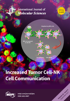
Cover Story (view full-size image): MM cells show basal levels of intercellular communication with lipids involvement. Bodipy503 stains neutral lipids, and GC stains species carrying Glucosylceramide. CMAC is a cell-tracker living reagent which stains MM cells. When MM cells and cord blood derived NK cells (CB-NK) come into contact, there is an increased transfer of material between cells in both directions. As a consequence, NK cell receptors, and tumor cell proteins will be transferred between cells. CB-NK are stained with cell tracker living reagent Bodipy, which binds to lipid–protein structures. View this paper
  • Issues are regarded as officially published after their release is announced to the table of contents alert mailing list.
  • You may sign up for e-mail alerts to receive table of contents of newly released issues.
  • PDF is the official format for papers published in both, html and pdf forms. To view the papers in pdf format, click on the "PDF Full-text" link, and use the free Adobe Reader to open them.
Order results
Result details
Section
Select all
Export citation of selected articles as:
3308 KiB  
Article
Rhein Induces Oxidative Stress and Apoptosis in Mouse Blastocysts and Has Immunotoxic Effects during Embryonic Development
by Chien-Hsun Huang and Wen-Hsiung Chan
Int. J. Mol. Sci. 2017, 18(9), 2018; https://doi.org/10.3390/ijms18092018 - 20 Sep 2017
Cited by 20 | Viewed by 7656
Abstract
Rhein, a glucoside chemical compound found in a traditional Chinese medicine derived from the roots of rhubarb, induces cell apoptosis and is considered to have high potential as an antitumor drug. Several previous studies showed that rhein can inhibit cell proliferation and trigger [...] Read more.
Rhein, a glucoside chemical compound found in a traditional Chinese medicine derived from the roots of rhubarb, induces cell apoptosis and is considered to have high potential as an antitumor drug. Several previous studies showed that rhein can inhibit cell proliferation and trigger mitochondria-related or endoplasmic reticulum (ER) stress-dependent apoptotic processes. However, the side effects of rhein on pre- and post-implantation embryonic development remain unclear. Here, we show that rhein has cytotoxic effects on blastocyst-stage mouse embryos and induces oxidative stress and immunotoxicity in mouse fetuses. Blastocysts incubated with 5–20 μM rhein showed significant cell apoptosis, as well as decreases in their inner cell mass cell numbers and total cell numbers. An in vitro development assay showed that rhein affected the developmental potentials of both pre- and post-implantation embryos. Incubation of blastocysts with 5–20 μM rhein was associated with increased resorption of post-implantation embryos and decreased fetal weight in an embryo transfer assay. Importantly, in an in vivo model, intravenous injection of dams with rhein (1, 3, and 5 mg/kg body weight/day) for four days resulted in apoptosis of blastocyst-stage embryos, early embryonic developmental injury, and decreased fetal weight. Intravenous injection of dams with 5 mg/kg body weight/day rhein significantly increased the total reactive oxygen species (ROS) content of fetuses and the transcription levels of antioxidant proteins in fetal livers. Additional work showed that rhein induced apoptosis through ROS generation, and that prevention of apoptotic processes effectively rescued the rhein-induced injury effects on embryonic development. Finally, the transcription levels of the innate-immunity related genes, CXCL1, IL-1 β and IL-8, were down-regulated in the fetuses of dams that received intravenous injections of rhein. These results collectively show that rhein has the potential to induce embryonic cytotoxicity and induce oxidative stress and immunotoxicity during the development of mouse embryos. Full article
(This article belongs to the Special Issue Natural Anti-Inflammatory Agents)
Show Figures

Figure 1

970 KiB  
Article
Comprehensive Evaluation of TFF3 Promoter Hypomethylation and Molecular Biomarker Potential for Prostate Cancer Diagnosis and Prognosis
by Maibritt Nørgaard, Christa Haldrup, Tine Maj Storebjerg, Else Marie Vestergaard, Peter J. Wild, Søren Høyer, Michael Borre, Torben Falck Ørntoft and Karina Dalsgaard Sørensen
Int. J. Mol. Sci. 2017, 18(9), 2017; https://doi.org/10.3390/ijms18092017 - 20 Sep 2017
Cited by 14 | Viewed by 4921
Abstract
Overdiagnosis and overtreatment of clinically insignificant tumors remains a major problem in prostate cancer (PC) due to suboptimal diagnostic and prognostic tools. Thus, novel biomarkers are urgently needed. In this study, we investigated the biomarker potential of Trefoil factor 3 (TFF3) [...] Read more.
Overdiagnosis and overtreatment of clinically insignificant tumors remains a major problem in prostate cancer (PC) due to suboptimal diagnostic and prognostic tools. Thus, novel biomarkers are urgently needed. In this study, we investigated the biomarker potential of Trefoil factor 3 (TFF3) promoter methylation and RNA expression levels for PC. Initially, by quantitative methylation specific PCR (qMSP) analysis of a large radical prostatectomy (RP) cohort (n = 292), we found that the TFF3 promoter was significantly hypomethylated in PC compared to non-malignant (NM) prostate tissue samples (p < 0.001) with an AUC (area under the curve) of 0.908 by receiver operating characteristics (ROC) curve analysis. Moreover, significant TFF3 promoter hypomethylation (p ≤ 0.010) as well as overexpression (p < 0.001) was found in PC samples from another large independent patient sample set (498 PC vs. 67 NM) analyzed by Illumina 450K DNA methylation arrays and/or RNA sequencing. TFF3 promoter methylation and transcriptional expression levels were inversely correlated, suggesting that epigenetic mechanisms contribute to the regulation of gene activity. Furthermore, low TFF3 expression was significantly associated with high ERG, ETS transcription factor (ERG) expression (p < 0.001), as well as with high Gleason score (p < 0.001), advanced pathological T-stage (p < 0.001), and prostate-specific antigen (PSA) recurrence after RP (p = 0.013; univariate Cox regression analysis). There were no significant associations between TFF3 promoter methylation levels, ERG status, or PSA recurrence in these RP cohorts. In conclusion, our results demonstrated diagnostic biomarker potential of TFF3 promoter hypomethylation for PC as well as prognostic biomarker potential of TFF3 RNA expression. To the best of our knowledge, this is the most comprehensive study of TFF3 promoter methylation and transcriptional expression in PC to date. Full article
(This article belongs to the Special Issue Cancer Epigenetics)
Show Figures

Graphical abstract

1719 KiB  
Review
Autophagy and Inflammatory Response in the Tumor Microenvironment
by Daniel Ngabire and Gun-Do Kim
Int. J. Mol. Sci. 2017, 18(9), 2016; https://doi.org/10.3390/ijms18092016 - 20 Sep 2017
Cited by 60 | Viewed by 9155
Abstract
Cell death is the last fate of the life cycle of cells. Different pathways involved in cell death are known to date, and are mostly represented by apoptosis, necrosis, and autophagy. Autophagy is one of the most preserved cell death pathways, characterized by [...] Read more.
Cell death is the last fate of the life cycle of cells. Different pathways involved in cell death are known to date, and are mostly represented by apoptosis, necrosis, and autophagy. Autophagy is one of the most preserved cell death pathways, characterized by the elimination of large parts of cytoplasmic components after being consumed by a double-membraned vesicle called an autophagosome. The formed autophagosome then fuses with a lysosome containing degrading enzymes and leads to the digestion of the autophagosome content. Autophagy is triggered by stress-related inducers, and is partially dependent on apoptotic proteins. It plays a major role in cancer, particularly in the tumor microenvironment where it has a paradoxical function in acting as a tumor suppressor and also as a tumor promoter. In the tumor microenvironment, autophagy regulates the differentiation of macrophages into tumor-associated macrophages (TAMs) and fibroblasts into cancer-associated fibroblasts (CAFs). TAMs and CAFs are abundantly present in the tumor microenvironment, and participate actively in tumor growth, tumor invasiveness, and tumor resistance to chemotherapy. Full article
(This article belongs to the Special Issue Autophagy at the Intersection of the Immune System and Cancer)
Show Figures

Graphical abstract

3207 KiB  
Article
Reduced Fluorescent Protein Switching Fatigue by Binding-Induced Emissive State Stabilization
by Thijs Roebroek, Sam Duwé, Wim Vandenberg and Peter Dedecker
Int. J. Mol. Sci. 2017, 18(9), 2015; https://doi.org/10.3390/ijms18092015 - 20 Sep 2017
Cited by 14 | Viewed by 6953
Abstract
Reversibly switchable fluorescent proteins (RSFPs) enable advanced fluorescence imaging, though the performance of this imaging crucially depends on the properties of the labels. We report on the use of an existing small binding peptide, named Enhancer, to modulate the spectroscopic properties of the [...] Read more.
Reversibly switchable fluorescent proteins (RSFPs) enable advanced fluorescence imaging, though the performance of this imaging crucially depends on the properties of the labels. We report on the use of an existing small binding peptide, named Enhancer, to modulate the spectroscopic properties of the recently developed rsGreen series of RSFPs. Fusion constructs of Enhancer with rsGreen1 and rsGreenF revealed an increased molecular brightness and pH stability, although expression in living E. coli or HeLa cells resulted in a decrease of the overall emission. Surprisingly, Enhancer binding also increased off-switching speed and resistance to switching fatigue. Further investigation suggested that the RSFPs can interconvert between fast- and slow-switching emissive states, with the overall protein population gradually converting to the slow-switching state through irradiation. The Enhancer modulates the spectroscopic properties of both states, but also preferentially stabilizes the fast-switching state, supporting the increased fatigue resistance. This work demonstrates how the photo-physical properties of RSFPs can be influenced by their binding to other small proteins, which opens up new horizons for applications that may require such modulation. Furthermore, we provide new insights into the photoswitching kinetics that should be of general consideration when developing new RSFPs with improved or different photochromic properties. Full article
(This article belongs to the Special Issue Fluorescent Proteins)
Show Figures

Graphical abstract

1720 KiB  
Review
Human Chorionic Gonadotrophin as a Possible Mediator of Leiomyoma Growth during Pregnancy: Molecular Mechanisms
by Veronica Sarais, Greta Chiara Cermisoni, Matteo Schimberni, Alessandra Alteri, Enrico Papaleo, Edgardo Somigliana and Paola Vigano’
Int. J. Mol. Sci. 2017, 18(9), 2014; https://doi.org/10.3390/ijms18092014 - 20 Sep 2017
Cited by 21 | Viewed by 7440
Abstract
Uterine fibroids are the most common gynecologic benign tumors. Studies supporting a strong pregnancy-related growth of leiomyomas generally claimed a crucial role of sex steroid hormones. However, sex steroids are unlikely the unique actors involved as estrogen and progesterone achieve a pick serum [...] Read more.
Uterine fibroids are the most common gynecologic benign tumors. Studies supporting a strong pregnancy-related growth of leiomyomas generally claimed a crucial role of sex steroid hormones. However, sex steroids are unlikely the unique actors involved as estrogen and progesterone achieve a pick serum concentration in the last trimester while leiomyomas show a typical increase during the first trimester. Given the rapid exponential raise in serum human Chorionic Gonadotrophin (hCG) at the beginning of gestation, we conducted a review to assess the potential role of hCG in the striking growth of leiomyomas during initial pregnancy. Fibroid growth during initial pregnancy seems to correlate to the similar increase of serum hCG levels until 12 weeks of gestation. The presence of functional Luteinizing Hormone/human Chorionic Gonadotropin (LH/hCG) receptors was demonstrated on leiomyomas. In vitro treatment of leiomyoma cells with hCG determines an up to 500% increase in cell number after three days. Expression of cyclin E and cyclin-dependent kinase 1 was significantly increased in leiomyoma cells by hCG treatment. Moreover, upon binding to the receptor, hCG stimulates prolactin secretion in leiomyoma cells, promoting cell proliferation via the mitogen-activated protein kinase cascade. Fibroid enlargement during initial pregnancy may be regulated by serum hCG. Full article
(This article belongs to the Special Issue hCG—An Endocrine, Regulator of Gestation and Cancer)
Show Figures

Graphical abstract

7171 KiB  
Article
Elongation of Axon Extension for Human iPSC-Derived Retinal Ganglion Cells by a Nano-Imprinted Scaffold
by Tien-Chun Yang, Jen-Hua Chuang, Waradee Buddhakosai, Wen-Ju Wu, Chen-Ju Lee, Wun-Syuan Chen, Yi-Ping Yang, Ming-Chia Li, Chi-Hsien Peng and Shih-Jen Chen
Int. J. Mol. Sci. 2017, 18(9), 2013; https://doi.org/10.3390/ijms18092013 - 20 Sep 2017
Cited by 30 | Viewed by 8472
Abstract
Optic neuropathies, such as glaucoma and Leber’s hereditary optic neuropathy (LHON) lead to retinal ganglion cell (RGC) loss and therefore motivate the application of transplantation technique into disease therapy. However, it is a challenge to direct the transplanted optic nerve axons to the [...] Read more.
Optic neuropathies, such as glaucoma and Leber’s hereditary optic neuropathy (LHON) lead to retinal ganglion cell (RGC) loss and therefore motivate the application of transplantation technique into disease therapy. However, it is a challenge to direct the transplanted optic nerve axons to the correct location of the retina. The use of appropriate scaffold can promote the proper axon growth. Recently, biocompatible materials have been integrated into the medical field, such as tissue engineering and reconstruction of damaged tissues or organs. We, herein, utilized nano-imprinting to create a scaffold mimicking the in vitro tissue microarchitecture, and guiding the axonal growth and orientation of the RGCs. We observed that the robust, long, and organized axons of human induced pluripotent stem cell (iPSC)-derived RGCs projected axially along the scaffold grooves. The RGCs grown on the scaffold expressed the specific neuronal biomarkers indicating their proper functionality. Thus, based on our in vitro culture system, this device can be useful for the neurophysiological analysis and transplantation for ophthalmic neuropathy treatment. Full article
(This article belongs to the Special Issue Disease Modeling Using Human Induced Pluripotent Stem Cells)
Show Figures

Graphical abstract

8239 KiB  
Article
Selective HDL-Raising Human Apo A-I Gene Therapy Counteracts Cardiac Hypertrophy, Reduces Myocardial Fibrosis, and Improves Cardiac Function in Mice with Chronic Pressure Overload
by Ruhul Amin, Ilayaraja Muthuramu, Joseph Pierre Aboumsallem, Mudit Mishra, Frank Jacobs and Bart De Geest
Int. J. Mol. Sci. 2017, 18(9), 2012; https://doi.org/10.3390/ijms18092012 - 20 Sep 2017
Cited by 30 | Viewed by 5194
Abstract
Epidemiological studies support an independent inverse association between high-density lipoprotein (HDL) cholesterol levels and heart failure incidence. The effect of selective HDL-raising adeno-associated viral serotype 8-human apolipoprotein (apo) A-I (AAV8-A-I) gene transfer on cardiac remodeling induced by transverse aortic constriction (TAC) was evaluated [...] Read more.
Epidemiological studies support an independent inverse association between high-density lipoprotein (HDL) cholesterol levels and heart failure incidence. The effect of selective HDL-raising adeno-associated viral serotype 8-human apolipoprotein (apo) A-I (AAV8-A-I) gene transfer on cardiac remodeling induced by transverse aortic constriction (TAC) was evaluated in C57BL/6 low-density lipoprotein receptor-deficient mice. Septal wall thickness and cardiomyocyte cross-sectional area were reduced by 16.5% (p < 0.001) and by 13.8% (p < 0.01), respectively, eight weeks after TAC in AAV8-A-I mice (n = 24) compared to control mice (n = 39). Myocardial capillary density was 1.11-fold (p < 0.05) higher and interstitial cardiac fibrosis was 45.3% (p < 0.001) lower in AAV8-A-I TAC mice than in control TAC mice. Lung weight and atrial weight were significantly increased in control TAC mice compared to control sham mice, but were not increased in AAV8-A-I TAC mice. The peak rate of isovolumetric contraction was 1.19-fold (p < 0.01) higher in AAV8-A-I TAC mice (n = 17) than in control TAC mice (n = 29). Diastolic function was also significantly enhanced in AAV8-A-I TAC mice compared to control TAC mice. Nitro-oxidative stress and apoptosis were significantly reduced in the myocardium of AAV8-A-I TAC mice compared to control TAC mice. In conclusion, selective HDL-raising human apo A-I gene transfer potently counteracts the development of pressure overload-induced cardiomyopathy. Full article
(This article belongs to the Special Issue Gene Therapy)
Show Figures

Graphical abstract

3527 KiB  
Article
Detection of Bacterial Pathogens from Broncho-Alveolar Lavage by Next-Generation Sequencing
by Stefano Leo, Nadia Gaïa, Etienne Ruppé, Stephane Emonet, Myriam Girard, Vladimir Lazarevic and Jacques Schrenzel
Int. J. Mol. Sci. 2017, 18(9), 2011; https://doi.org/10.3390/ijms18092011 - 20 Sep 2017
Cited by 39 | Viewed by 7516
Abstract
The applications of whole-metagenome shotgun sequencing (WMGS) in routine clinical analysis are still limited. A combination of a DNA extraction procedure, sequencing, and bioinformatics tools is essential for the removal of human DNA and for improving bacterial species identification in a timely manner. [...] Read more.
The applications of whole-metagenome shotgun sequencing (WMGS) in routine clinical analysis are still limited. A combination of a DNA extraction procedure, sequencing, and bioinformatics tools is essential for the removal of human DNA and for improving bacterial species identification in a timely manner. We tackled these issues with a broncho-alveolar lavage (BAL) sample from an immunocompromised patient who had developed severe chronic pneumonia. We extracted DNA from the BAL sample with protocols based either on sequential lysis of human and bacterial cells or on the mechanical disruption of all cells. Metagenomic libraries were sequenced on Illumina HiSeq platforms. Microbial community composition was determined by k-mer analysis or by mapping to taxonomic markers. Results were compared to those obtained by conventional clinical culture and molecular methods. Compared to mechanical cell disruption, a sequential lysis protocol resulted in a significantly increased proportion of bacterial DNA over human DNA and higher sequence coverage of Mycobacterium abscessus, Corynebacterium jeikeium and Rothia dentocariosa, the bacteria reported by clinical microbiology tests. In addition, we identified anaerobic bacteria not searched for by the clinical laboratory. Our results further support the implementation of WMGS in clinical routine diagnosis for bacterial identification. Full article
(This article belongs to the Special Issue Deciphering the Human Microbiota: Methods and Impact on Human Health)
Show Figures

Graphical abstract

857 KiB  
Review
To Wnt or Lose: The Missing Non-Coding Linc in Colorectal Cancer
by Peng Shen, Martin Pichler, Meng Chen, George A. Calin and Hui Ling
Int. J. Mol. Sci. 2017, 18(9), 2003; https://doi.org/10.3390/ijms18092003 - 20 Sep 2017
Cited by 44 | Viewed by 9076
Abstract
Colorectal cancer (CRC) is the third most frequent cancer and one of the leading causes for cancer-related mortality. Aberrant activation of the Wnt signaling is an essential initiating factor in colon carcinogenesis, and a driving force of CRC progression. Recently, long non-coding RNAs [...] Read more.
Colorectal cancer (CRC) is the third most frequent cancer and one of the leading causes for cancer-related mortality. Aberrant activation of the Wnt signaling is an essential initiating factor in colon carcinogenesis, and a driving force of CRC progression. Recently, long non-coding RNAs (lncRNAs) have emerged as significant players in CRC pathogenesis through diversified mechanisms. Although both Wnt signaling and lncRNAs represent interesting research areas for CRC, an effort of directly connecting these two areas is lacking. To fill in the knowledge gap, we focus on the reported findings of lncRNAs that regulate Wnt signaling or essential Wnt signaling targets. These include several newly discovered lncRNAs originated from the amplified cancer-associated chromosome 8q24 region that surrounds the essential Wnt target MYC gene, lncRNAs reported to be involved in CRC stem cells, and several individual lncRNAs connected to Wnt signaling through other mechanisms. This review will provide essential information that assists in understanding the missing link of lncRNAs to the classical Wnt signaling in CRC. Full article
(This article belongs to the Collection Regulation by Non-coding RNAs)
Show Figures

Figure 1

870 KiB  
Correction
Correction: Mariko Nishizaki, et al. Bioactivity of NANOZR Induced by Alkali Treatment. Int. J. Mol. Sci. 2017, 18, 780
by Mariko Nishizaki, Satoshi Komasa, Yoichiro Taguchi, Hiroshi Nishizaki and Joji Okazaki
Int. J. Mol. Sci. 2017, 18(9), 2009; https://doi.org/10.3390/ijms18092009 - 19 Sep 2017
Cited by 1 | Viewed by 3769
Abstract
We would like to submit the following correction to the published paper [1].[...] Full article
(This article belongs to the Special Issue Bioactive Nanoparticles)
Show Figures

Figure 1

4306 KiB  
Article
Osmolyte-Like Stabilizing Effects of Low GdnHCl Concentrations on d-Glucose/d-Galactose-Binding Protein
by Alexander V. Fonin, Alexandra D. Golikova, Irina A. Zvereva, Sabato D’Auria, Maria Staiano, Vladimir N. Uversky, Irina M. Kuznetsova and Konstantin K. Turoverov
Int. J. Mol. Sci. 2017, 18(9), 2008; https://doi.org/10.3390/ijms18092008 - 19 Sep 2017
Cited by 2 | Viewed by 6288
Abstract
The ability of d-glucose/d-galactose-binding protein (GGBP) to reversibly interact with its ligands, glucose and galactose, makes this protein an attractive candidate for sensing elements of glucose biosensors. This potential is largely responsible for attracting researchers to study the conformational properties [...] Read more.
The ability of d-glucose/d-galactose-binding protein (GGBP) to reversibly interact with its ligands, glucose and galactose, makes this protein an attractive candidate for sensing elements of glucose biosensors. This potential is largely responsible for attracting researchers to study the conformational properties of this protein. Previously, we showed that an increase in the fluorescence intensity of the fluorescent dye 6-bromoacetyl-2-dimetylaminonaphtalene (BADAN) is linked to the holo-form of the GGBP/H152C mutant in solutions containing sub-denaturing concentrations of guanidine hydrochloride (GdnHCl). It was hypothesized that low GdnHCl concentrations might lead to compaction of the protein, thereby facilitating ligand binding. In this work, we utilize BADAN fluorescence spectroscopy, intrinsic protein UV fluorescence spectroscopy, and isothermal titration calorimetry (ITC) to show that the sub-denaturing GdnHCl concentrations possess osmolyte-like stabilizing effects on the structural dynamics, conformational stability, and functional activity of GGBP/H152C and the wild type of this protein (wtGGBP). Our data are consistent with the model where low GdnHCl concentrations promote a shift in the dynamic distribution of the protein molecules toward a conformational ensemble enriched in molecules with a tighter structure and a more closed conformation. This promotes the increase in the configurational complementarity between the protein and glucose molecules that leads to the increase in glucose affinity in both GGBP/H152C and wtGGBP. Full article
(This article belongs to the Section Molecular Biophysics)
Show Figures

Graphical abstract

3602 KiB  
Article
Interference of Paraben Compounds with Estrogen Metabolism by Inhibition of 17β-Hydroxysteroid Dehydrogenases
by Roger T. Engeli, Simona R. Rohrer, Anna Vuorinen, Sonja Herdlinger, Teresa Kaserer, Susanne Leugger, Daniela Schuster and Alex Odermatt
Int. J. Mol. Sci. 2017, 18(9), 2007; https://doi.org/10.3390/ijms18092007 - 19 Sep 2017
Cited by 46 | Viewed by 12663
Abstract
Parabens are effective preservatives widely used in cosmetic products and processed food, with high human exposure. Recent evidence suggests that parabens exert estrogenic effects. This work investigated the potential interference of parabens with the estrogen-activating enzyme 17β-hydroxysteroid dehydrogenase (17β-HSD) 1 and the estrogen-inactivating [...] Read more.
Parabens are effective preservatives widely used in cosmetic products and processed food, with high human exposure. Recent evidence suggests that parabens exert estrogenic effects. This work investigated the potential interference of parabens with the estrogen-activating enzyme 17β-hydroxysteroid dehydrogenase (17β-HSD) 1 and the estrogen-inactivating 17β-HSD2. A ligand-based 17β-HSD2 pharmacophore model was applied to screen a cosmetic chemicals database, followed by in vitro testing of selected paraben compounds for inhibition of 17β-HSD1 and 17β-HSD2 activities. All tested parabens and paraben-like compounds, except their common metabolite p-hydroxybenzoic acid, inhibited 17β-HSD2. Ethylparaben and ethyl vanillate inhibited 17β-HSD2 with IC50 values of 4.6 ± 0.8 and 1.3 ± 0.3 µM, respectively. Additionally, parabens size-dependently inhibited 17β-HSD1, whereby hexyl- and heptylparaben were most active with IC50 values of 2.6 ± 0.6 and 1.8 ± 0.3 µM. Low micromolar concentrations of hexyl- and heptylparaben decreased 17β-HSD1 activity, and ethylparaben and ethyl vanillate decreased 17β-HSD2 activity. However, regarding the very rapid metabolism of these compounds to the inactive p-hydroxybenzoic acid by esterases, it needs to be determined under which conditions low micromolar concentrations of these parabens or their mixtures can occur in target cells to effectively disturb estrogen effects in vivo. Full article
(This article belongs to the Special Issue Advances in the Research of Endocrine Disrupting Chemicals)
Show Figures

Graphical abstract

4813 KiB  
Article
Behind Resveratrol Stabilization by Carboxymethylated (1,3/1,6)-β-d-Glucan: Does the Polyphenol Play a Role in Polymer Structural Organization?
by Antonio Francioso, Simone Dinarelli, Marco Girasole, Laura Cervoni, Maria D’Erme, Francesco Mura, Alberto Boffi, Elita Montanari and Luciana Mosca
Int. J. Mol. Sci. 2017, 18(9), 2006; https://doi.org/10.3390/ijms18092006 - 19 Sep 2017
Cited by 3 | Viewed by 5168
Abstract
Resveratrol stability in solution can be improved by combining the polyphenol with carboxymethylated (1,3/1,6)-β-d-glucan (CM-glucan), a carbohydrate polymer widely used in the food and pharmaceutical industries. The present work was undertaken to elucidate the mechanism behind this stabilizing effect. The supramolecular [...] Read more.
Resveratrol stability in solution can be improved by combining the polyphenol with carboxymethylated (1,3/1,6)-β-d-glucan (CM-glucan), a carbohydrate polymer widely used in the food and pharmaceutical industries. The present work was undertaken to elucidate the mechanism behind this stabilizing effect. The supramolecular structural, physico-chemical and morphological features of the CM-glucan/resveratrol complex have been studied under different physical and chemical stimuli by means of spectroscopic techniques, microscopy and physical methods such as UV-Visible spectroscopy (UV-Vis), spectrofluorimetry, Circular Dichroism (CD), Infrared spectroscopy (FT-IR), Differential Scanning Calorimetry (DSC), Atomic Force Microscopy (AFM) and Scanning Electron Microscopy (SEM). Our experimental data indicate that CM-glucan conformational organized architecture in aqueous solution is enhanced in the presence of resveratrol, suggesting that the polyphenol is able to confer a high degree of order to the polymer by a probable cooperative structural organization that results in a long term stabilization for the polyphenol. Full article
(This article belongs to the Special Issue Glucan: New Perspectives on Biochemistry and Application)
Show Figures

Graphical abstract

8397 KiB  
Article
Genome-Wide Identification of the PHD-Finger Family Genes and Their Responses to Environmental Stresses in Oryza sativa L.
by Mingzhe Sun, Bowei Jia, Junkai Yang, Na Cui, Yanming Zhu and Xiaoli Sun
Int. J. Mol. Sci. 2017, 18(9), 2005; https://doi.org/10.3390/ijms18092005 - 19 Sep 2017
Cited by 24 | Viewed by 5638
Abstract
The PHD-finger family has been demonstrated to be involved in regulating plant growth and development. However, little information is given for its role in environmental stress responses. Here, we identified a total of 59 PHD family genes in the rice genome. These OsPHDs [...] Read more.
The PHD-finger family has been demonstrated to be involved in regulating plant growth and development. However, little information is given for its role in environmental stress responses. Here, we identified a total of 59 PHD family genes in the rice genome. These OsPHDs genes were located on eleven chromosomes and synteny analysis only revealed nine duplicated pairs within the rice PHD family. Phylogenetic analysis of all OsPHDs and PHDs from other species revealed that they could be grouped into two major clusters. Furthermore, OsPHDs were clustered into eight groups and members from different groups displayed a great divergence in terms of gene structure, functional domains and conserved motifs. We also found that with the exception of OsPHD6, all OsPHDs were expressed in at least one of the ten tested tissues and OsPHDs from certain groups were expressed in specific tissues. Moreover, our results also uncovered differential responses of OsPHDs expression to environmental stresses, including ABA (abscisic acid), water deficit, cold and high Cd. By using quantitative real-time PCR, we further confirmed the differential expression of OsPHDs under these stresses. OsPHD1/7/8/13/33 were differentially expressed under water deficit and Cd stresses, while OsPHD5/17 showed altered expression under water deficit and cold stresses. Moreover, OsPHD3/44/28 displayed differential expression under ABA and Cd stresses. In conclusion, our results provide valuable information on the rice PHD family in plant responses to environmental stress, which will be helpful for further characterizing their biological roles in responding to environmental stresses. Full article
(This article belongs to the Special Issue Selected Papers from the 6th National Plant Protein Research Congress)
Show Figures

Graphical abstract

1054 KiB  
Review
Cold Atmospheric Plasma in the Treatment of Osteosarcoma
by Denis Gümbel, Sander Bekeschus, Nadine Gelbrich, Matthias Napp, Axel Ekkernkamp, Axel Kramer and Matthias B. Stope
Int. J. Mol. Sci. 2017, 18(9), 2004; https://doi.org/10.3390/ijms18092004 - 19 Sep 2017
Cited by 45 | Viewed by 8552
Abstract
Human osteosarcoma (OS) is the most common primary malignant bone tumor occurring most commonly in adolescents and young adults. Major improvements in disease-free survival have been achieved by implementing a combination therapy consisting of radical surgical resection of the tumor and systemic multi-agent [...] Read more.
Human osteosarcoma (OS) is the most common primary malignant bone tumor occurring most commonly in adolescents and young adults. Major improvements in disease-free survival have been achieved by implementing a combination therapy consisting of radical surgical resection of the tumor and systemic multi-agent chemotherapy. However, long-term survival remains poor, so novel targeted therapies to improve outcomes for patients with osteosarcoma remains an area of active research. This includes immunotherapy, photodynamic therapy, or treatment with nanoparticles. Cold atmospheric plasma (CAP), a highly reactive (partially) ionized physical state, has been shown to inherit a significant anticancer capacity, leading to a new field in medicine called “plasma oncology.” The current article summarizes the potential of CAP in the treatment of human OS and reviews the underlying molecular mode of action. Full article
(This article belongs to the Special Issue Current Advances in Soft Tissue and Bone Sarcoma)
Show Figures

Graphical abstract

197 KiB  
Review
Zebrafish as a Model for the Study of Microvascular Complications of Diabetes and Their Mechanisms
by Karl Heckler and Jens Kroll
Int. J. Mol. Sci. 2017, 18(9), 2002; https://doi.org/10.3390/ijms18092002 - 19 Sep 2017
Cited by 49 | Viewed by 10664
Abstract
Diabetes mellitus (DM) is a crucial metabolic disease that leads to severe disorders. These include macrovascular complications such as myocardial infarction, stroke, and peripheral artery disease and microvascular complications including diabetic nephropathy, neuropathy, and retinopathy. Diabetes mellitus, along with its associated organ pathologies, [...] Read more.
Diabetes mellitus (DM) is a crucial metabolic disease that leads to severe disorders. These include macrovascular complications such as myocardial infarction, stroke, and peripheral artery disease and microvascular complications including diabetic nephropathy, neuropathy, and retinopathy. Diabetes mellitus, along with its associated organ pathologies, is one of the key problems in today’s medicine. Zebrafish is an upcoming disease model organism in diabetes research. Its glucose metabolism and the pathways of reactive metabolite formation are very similar to those of humans. Moreover, several physiological and pathophysiological pathways that also exist in humans and other mammals have been identified in this species or are currently under intense investigation. Zebrafish offer sophisticated imaging techniques and allow simple and fast genetic and pharmacological approaches with a high throughput. In this review, we highlight achievements and mechanisms concerning microvascular complications discovered in zebrafish, and we discuss the advantages and disadvantages of zebrafish as a model for studying diabetic complications. Full article
(This article belongs to the Special Issue Glyoxalase System in Health and Disease 2017)
Show Figures

Graphical abstract

2383 KiB  
Article
In Vitro Glucuronidation of Wushanicaritin by Liver Microsomes, Intestine Microsomes and Expressed Human UDP-Glucuronosyltransferase Enzymes
by Xiaodan Hong, Yuanru Zheng, Zifei Qin, Baojian Wu, Yi Dai, Hao Gao, Zhihong Yao, Frank J. Gonzalez and Xinsheng Yao
Int. J. Mol. Sci. 2017, 18(9), 1983; https://doi.org/10.3390/ijms18091983 - 19 Sep 2017
Cited by 17 | Viewed by 7199
Abstract
Wushanicaritin, a natural polyphenol compound, exerts many biological activities. This study aimed to characterize wushanicaritin glucuronidation by pooled human liver microsomes (HLM), human intestine microsomes and individual uridine diphosphate-glucuronosyltransferase (UGT) enzyme. Glucuronidation rates were determined by incubating wushanicaritin with uridine diphosphoglucuronic acid-supplemented microsomes. [...] Read more.
Wushanicaritin, a natural polyphenol compound, exerts many biological activities. This study aimed to characterize wushanicaritin glucuronidation by pooled human liver microsomes (HLM), human intestine microsomes and individual uridine diphosphate-glucuronosyltransferase (UGT) enzyme. Glucuronidation rates were determined by incubating wushanicaritin with uridine diphosphoglucuronic acid-supplemented microsomes. Kinetic parameters were derived by appropriate model fitting. Reaction phenotyping, the relative activity factor (RAF) and activity correlation analysis were performed to identify the main UGT isoforms. Wushanicaritin glucuronidation in HLM was efficient with a high CLint (intrinsic clearance) value of 1.25 and 0.69 mL/min/mg for G1 and G2, respectively. UGT1A1 and 1A7 showed the highest activities with the intrinsic clearance (CLint) values of 1.16 and 0.38 mL/min/mg for G1 and G2, respectively. In addition, G1 was significantly correlated with β-estradiol glucuronidation (r = 0.847; p = 0.0005), while G2 was also correlated with chenodeoxycholic acid glucuronidation (r = 0.638, p = 0.026) in a bank of individual HLMs (n = 12). Based on the RAF approach, UGT1A1 contributed 51.2% for G1, and UGT1A3 contributed 26.0% for G2 in HLM. Moreover, glucuronidation of wushanicaritin by liver microsomes showed marked species difference. Taken together, UGT1A1, 1A3, 1A7, 1A8, 1A9 and 2B7 were identified as the main UGT contributors responsible for wushanicaritin glucuronidation. Full article
(This article belongs to the Section Biochemistry)
Show Figures

Graphical abstract

4938 KiB  
Article
The Impact of Non-Lethal Single-Dose Radiation on Tumor Invasion and Cytoskeletal Properties
by Tim Hohmann, Urszula Grabiec, Carolin Vogel, Chalid Ghadban, Stephan Ensminger, Matthias Bache, Dirk Vordermark and Faramarz Dehghani
Int. J. Mol. Sci. 2017, 18(9), 2001; https://doi.org/10.3390/ijms18092001 - 18 Sep 2017
Cited by 11 | Viewed by 5559
Abstract
Irradiation is the standard therapy for glioblastoma multiforme. Glioblastoma are highly resistant to radiotherapy and the underlying mechanisms remain unclear. To better understand the biological effects of irradiation on glioblastoma cells, we tested whether nonlethal irradiation influences the invasiveness, cell stiffness, and actin [...] Read more.
Irradiation is the standard therapy for glioblastoma multiforme. Glioblastoma are highly resistant to radiotherapy and the underlying mechanisms remain unclear. To better understand the biological effects of irradiation on glioblastoma cells, we tested whether nonlethal irradiation influences the invasiveness, cell stiffness, and actin cytoskeleton properties. Two different glioblastoma cell lines were irradiated with 2 Gy and changes in mechanical and migratory properties and alterations in the actin structure were measured. The invasiveness of cell lines was determined using a co-culture model with organotypic hippocampal slice cultures. Irradiation led to changes in motility and a less invasive phenotype in both investigated cell lines that were associated with an increase in a ”generalized stiffness” and changes in the actin structure. In this study we demonstrate that irradiation can induce changes in the actin cytoskeleton and motility, which probably results in reduced invasiveness of glioblastoma cell lines. Furthermore, “generalized stiffness” was shown to be a profound marker of the invasiveness of a tumor cell population in our model. Full article
(This article belongs to the Special Issue Glioma Cell Invasion)
Show Figures

Graphical abstract

4602 KiB  
Article
Expression of the Grape VaSTS19 Gene in Arabidopsis Improves Resistance to Powdery Mildew and Botrytis cinerea but Increases Susceptibility to Pseudomonas syringe pv Tomato DC3000
by Yaqiong Wang, Dejun Wang, Fan Wang, Li Huang, Xiaomin Tian, Steve Van Nocker, Hua Gao and Xiping Wang
Int. J. Mol. Sci. 2017, 18(9), 2000; https://doi.org/10.3390/ijms18092000 - 17 Sep 2017
Cited by 13 | Viewed by 6532
Abstract
Stilbene synthase (STS) is a key enzyme that catalyzes the biosynthesis of resveratrol compounds and plays an important role in disease resistance. The molecular pathways linking STS with pathogen responses and their regulation are not known. We isolated an STS gene, VaSTS19, [...] Read more.
Stilbene synthase (STS) is a key enzyme that catalyzes the biosynthesis of resveratrol compounds and plays an important role in disease resistance. The molecular pathways linking STS with pathogen responses and their regulation are not known. We isolated an STS gene, VaSTS19, from a Chinese wild grape, Vitis amurensis Rupr. cv. “Tonghua-3”, and transferred this gene to Arabidopsis. We then generated VaSTS19-expressing Arabidopsis lines and evaluated the functions of VaSTS19 in various pathogen stresses, including powdery mildew, B. cinerea and Pseudomonas syringae pv. tomato DC3000 (PstDC3000). VaSTS19 enhanced resistance to powdery mildew and B. cinerea, but increased susceptibility to PstDC3000. Aniline blue staining revealed that VaSTS19 transgenic lines accumulated more callose compared to nontransgenic control plants, and showed smaller stomatal apertures when exposed to pathogen-associated molecular patterns (flagellin fragment (flg22) or lipopolysaccharides (LPS)). Analysis of the expression of several disease-related genes suggested that VaSTS19 expression enhanced defense responses though salicylic acid (SA) and/or jasmonic acid (JA) signaling pathways. These findings provide a deeper insight into the function of STS genes in defense against pathogens, and a better understanding of the regulatory cross talk between SA and JA pathways. Full article
(This article belongs to the Section Molecular Plant Sciences)
Show Figures

Graphical abstract

2260 KiB  
Article
Mouse Monoclonal Antibodies Generated from Full Length Human Cereblon: Detection of Cereblon Protein in Patients with Multiple Myeloma
by Xiubao Chang, Qinqin Xu, Yuexian Hou, Cynthia Li, Ye Xu and A. Keith Stewart
Int. J. Mol. Sci. 2017, 18(9), 1999; https://doi.org/10.3390/ijms18091999 - 17 Sep 2017
Cited by 6 | Viewed by 4337
Abstract
Immunomodulatory drugs (IMiDs) are profoundly active compounds in the treatment of patients with multiple myeloma (MM). However, despite the fact that treatment with IMiDs has dramatically improved survival for patients with MM, the majority of MM patients develop IMiDs resistance over time. We [...] Read more.
Immunomodulatory drugs (IMiDs) are profoundly active compounds in the treatment of patients with multiple myeloma (MM). However, despite the fact that treatment with IMiDs has dramatically improved survival for patients with MM, the majority of MM patients develop IMiDs resistance over time. We have found that expression of functional cereblon is required for IMiDs′ action. In addition, it has been reported that cells expressing high levels of cereblon are resistant to proteasome inhibitor, implying that patients with high levels of cereblon should be resistant to proteasome inhibitor. If the above conclusions are correct, cereblon could be considered as a biomarker to determine which standard regimens should be used to treat patients with MM. Unfortunately, the conclusions mentioned above have not been clinically confirmed. In order to confirm these conclusions, we have generated three highly specific mouse monoclonal antibodies (mAbs) against full-length human cereblon. These mAbs can be used to do western blot, immunoprecipitation and immunohistochemistry staining. In addition, their epitopes have been precisely determined and the peptides covering their epitopes completely blocked the antibody binding to cereblon in western blot analysis or in immunohistochemistry staining of MM patients′ specimens. Full article
(This article belongs to the Section Biochemistry)
Show Figures

Figure 1

1445 KiB  
Case Report
Juvenile Moyamoya and Craniosynostosis in a Child with Deletion 1p32p31: Expanding the Clinical Spectrum of 1p32p31 Deletion Syndrome and a Review of the Literature
by Paolo Prontera, Daniela Rogaia, Amedea Mencarelli, Valentina Ottaviani, Ester Sallicandro, Giorgio Guercini, Susanna Esposito, Anna Bersano, Giuseppe Merla and Gabriela Stangoni
Int. J. Mol. Sci. 2017, 18(9), 1998; https://doi.org/10.3390/ijms18091998 - 17 Sep 2017
Cited by 8 | Viewed by 6005
Abstract
Moyamoya angiopathy (MA) is a rare cerebrovascular disorder characterised by the progressive occlusion of the internal carotid artery. Its aetiology is uncertain, but a genetic background seems likely, given the high MA familial rate. To investigate the aetiology of craniosynostosis and juvenile moyamoya [...] Read more.
Moyamoya angiopathy (MA) is a rare cerebrovascular disorder characterised by the progressive occlusion of the internal carotid artery. Its aetiology is uncertain, but a genetic background seems likely, given the high MA familial rate. To investigate the aetiology of craniosynostosis and juvenile moyamoya in a 14-year-old male patient, we performed an array-comparative genomic hybridisation revealing a de novo interstitial deletion of 8.5 Mb in chromosome region 1p32p31. The deletion involved 34 protein coding genes, including NF1A, whose haploinsufficiency is indicated as being mainly responsible for the 1p32-p31 chromosome deletion syndrome phenotype (OMIM 613735). Our patient also has a deleted FOXD3 of the FOX gene family of transcription factors, which plays an important role in neural crest cell growth and differentiation. As the murine FOXD3−/− model shows craniofacial anomalies and abnormal common carotid artery morphology, it can be hypothesised that FOXD3 is involved in the pathogenesis of the craniofacial and vascular defects observed in our patient. In support of our assumption, we found in the literature another patient with a syndromic form of MA who had a deletion involving another FOX gene (FOXC1). In addition to describing the clinical history of our patient, we have reviewed all of the available literature concerning other patients with a 1p32p31 deletion, including cases from the Decipher database, and we have also reviewed the genetic disorders associated with MA, which is a useful guide for the diagnosis of syndromic form of MA. Full article
(This article belongs to the Section Molecular Pathology, Diagnostics, and Therapeutics)
Show Figures

Graphical abstract

1904 KiB  
Review
Anti-NKG2D mAb: A New Treatment for Crohn’s Disease?
by Kasper Vadstrup and Flemming Bendtsen
Int. J. Mol. Sci. 2017, 18(9), 1997; https://doi.org/10.3390/ijms18091997 - 16 Sep 2017
Cited by 19 | Viewed by 8619
Abstract
Crohn’s disease (CD) and ulcerative colitis (UC) are immunologically-mediated, debilitating conditions resulting from destructive inflammation of the gastrointestinal tract. The pathogenesis of IBD is incompletely understood, but is considered to be the result of an abnormal immune response with a wide range of [...] Read more.
Crohn’s disease (CD) and ulcerative colitis (UC) are immunologically-mediated, debilitating conditions resulting from destructive inflammation of the gastrointestinal tract. The pathogenesis of IBD is incompletely understood, but is considered to be the result of an abnormal immune response with a wide range of cell types and proteins involved. Natural Killer Group 2D (NKG2D) is an activating receptor constitutively expressed on human Natural Killer (NK), γδ T, mucosal-associated invariant T (MAIT), CD56+ T, and CD8+ T cells. Activation of NKG2D triggers cellular proliferation, cytokine production, and target cell killing. Research into the NKG2D mechanism of action has primarily been focused on cancer and viral infections where cytotoxicity evasion is a concern. In human inflammatory bowel disease (IBD) this system is less characterized, but the ligands have been shown to be highly expressed during intestinal inflammation and the following receptor activation may contribute to tissue degeneration. A recent phase II clinical trial showed that an antibody against NKG2D induced clinical remission of CD in some patients, suggesting NKG2D and its ligands to be of importance in the pathogenesis of CD. This review will describe the receptor and its ligands in intestinal tissues and the clinical potential of blocking NKG2D in Crohn’s disease. Full article
Show Figures

Graphical abstract

4156 KiB  
Article
Effects of Metal Ions, Temperature, and a Denaturant on the Oxidative Folding Pathways of Bovine α-Lactalbumin
by Reina Shinozaki and Michio Iwaoka
Int. J. Mol. Sci. 2017, 18(9), 1996; https://doi.org/10.3390/ijms18091996 - 16 Sep 2017
Cited by 12 | Viewed by 4502
Abstract
Bovine α-lactalbumin (αLA) has four disulfide (SS) bonds in the native form (N). On the oxidative folding pathways of this protein, two specific SS folding intermediates, i.e., (61–77, 73–91) and des[6–120], which have two and three native SS bonds, respectively, accumulate predominantly in [...] Read more.
Bovine α-lactalbumin (αLA) has four disulfide (SS) bonds in the native form (N). On the oxidative folding pathways of this protein, two specific SS folding intermediates, i.e., (61–77, 73–91) and des[6–120], which have two and three native SS bonds, respectively, accumulate predominantly in the presence of Ca2+. In this study, we reinvestigated the pathways using a water-soluble cyclic selenoxide reagent, trans-3,4-dihydroxyselenolane oxide (DHSox), as a strong and quantitative oxidant to oxidize the fully reduced form (R). In the presence of ethylenediaminetetraacetic acid (EDTA) (under a metal-free condition), SS formation randomly proceeded, and N did not regenerate. On the other hand, two specific SS intermediates transiently generated in the presence of Ca2+. These intermediates could be assigned to (61–77, 73–91) and des[6–120] having two common SS bonds, i.e., Cys61-Cys77 and Cys73-Cys91, near the calcium binding pocket of the β-sheet domain. Much faster folding to N was observed in the presence of Mn2+, whereas Na+, K+, Mg2+, and Zn2+ did not affect the pathways. The two key intermediates were susceptible to temperature and a denaturant. The oxidative folding pathways revealed were significantly different from those of hen egg white lysozyme, which has the same SS-bonding pattern as αLA, suggesting that the folding pathways of SS-containing proteins can alter depending on the amino acid sequence and other factors, even when the SS-bond topologies are similar to each other. Full article
(This article belongs to the Special Issue Protein Folding)
Show Figures

Graphical abstract

1149 KiB  
Article
Why Ubiquitin Has Not Evolved
by Douglas C. Allan and James C. Phillips
Int. J. Mol. Sci. 2017, 18(9), 1995; https://doi.org/10.3390/ijms18091995 - 16 Sep 2017
Cited by 4 | Viewed by 4596
Abstract
Ubiquitin, discovered less than 50 years ago, tags thousands of diseased proteins for destruction. It is small (only 76 amino acids), and is found unchanged in mammals, birds, fish, and even worms, indicating that ubiquitin is perfect. Key features of its functionality are [...] Read more.
Ubiquitin, discovered less than 50 years ago, tags thousands of diseased proteins for destruction. It is small (only 76 amino acids), and is found unchanged in mammals, birds, fish, and even worms, indicating that ubiquitin is perfect. Key features of its functionality are identified here using critical point thermodynamic scaling theory. These include synchronized pivots and hinges, a stabilizing central pivot, and Fano interference between first- and second-order elements of correlated long-range (allosteric) globular surface shape transitions. Comparison with its closest relative, 76 amino acid Nedd8, shows that the latter lacks all these features. A cracked elastic network model is proposed for the common target shared by many diseased proteins. Full article
(This article belongs to the Special Issue Ubiquitin System)
Show Figures

Figure 1

3169 KiB  
Article
The Semi-Synthetic Peptide Lin-SB056-1 in Combination with EDTA Exerts Strong Antimicrobial and Antibiofilm Activity against Pseudomonas aeruginosa in Conditions Mimicking Cystic Fibrosis Sputum
by Giuseppantonio Maisetta, Lucia Grassi, Semih Esin, Ilaria Serra, Mariano A. Scorciapino, Andrea C. Rinaldi and Giovanna Batoni
Int. J. Mol. Sci. 2017, 18(9), 1994; https://doi.org/10.3390/ijms18091994 - 16 Sep 2017
Cited by 23 | Viewed by 5612
Abstract
Pseudomonas aeruginosa is a major cause of chronic lung infections in cystic fibrosis (CF) patients. The ability of the bacterium to form biofilms and the presence of a thick and stagnant mucus in the airways of CF patients largely contribute to antibiotic therapy [...] Read more.
Pseudomonas aeruginosa is a major cause of chronic lung infections in cystic fibrosis (CF) patients. The ability of the bacterium to form biofilms and the presence of a thick and stagnant mucus in the airways of CF patients largely contribute to antibiotic therapy failure and demand for new antimicrobial agents able to act in the CF environment. The present study investigated the anti-P. aeruginosa activity of lin-SB056-1, a recently described semi-synthetic antimicrobial peptide, used alone and in combination with the cation chelator ethylenediaminetetraacetic acid (EDTA). Bactericidal assays were carried out in standard culture conditions and in an artificial sputum medium (ASM) closely resembling the CF environment. Peptide’s structure and interaction with large unilamellar vesicles in media with different ionic strengths were also investigated through infrared spectroscopy. Lin-SB056-1 demonstrated fast and strong bactericidal activity against both mucoid and non-mucoid strains of P. aeruginosa in planktonic form and, in combination with EDTA, caused significant reduction of the biomass of P. aeruginosa mature biofilms. In ASM, the peptide/EDTA combination exerted a strong bactericidal effect and inhibited the formation of biofilm-like structures of P. aeruginosa. Overall, the results obtained highlight the potential of the lin-SB056-1/EDTA combination for the treatment of P. aeruginosa lung infections in CF patients. Full article
(This article belongs to the Special Issue Biofilm Formation)
Show Figures

Graphical abstract

831 KiB  
Review
Genetic and Hormonal Regulation of Chlorophyll Degradation during Maturation of Seeds with Green Embryos
by Galina Smolikova, Elena Dolgikh, Maria Vikhnina, Andrej Frolov and Sergei Medvedev
Int. J. Mol. Sci. 2017, 18(9), 1993; https://doi.org/10.3390/ijms18091993 - 16 Sep 2017
Cited by 34 | Viewed by 9237
Abstract
The embryos of some angiosperms (usually referred to as chloroembryos) contain chlorophylls during the whole period of embryogenesis. Developing embryos have photochemically active chloroplasts and are able to produce assimilates, further converted in reserve biopolymers, whereas at the late steps of embryogenesis, seeds [...] Read more.
The embryos of some angiosperms (usually referred to as chloroembryos) contain chlorophylls during the whole period of embryogenesis. Developing embryos have photochemically active chloroplasts and are able to produce assimilates, further converted in reserve biopolymers, whereas at the late steps of embryogenesis, seeds undergo dehydration, degradation of chlorophylls, transformation of chloroplast in storage plastids, and enter the dormancy period. However, in some seeds, the process of chlorophyll degradation remains incomplete. These residual chlorophylls compromise the quality of seed material in terms of viability, nutritional value, and shelf life, and represent a serious challenge for breeders and farmers. The mechanisms of chlorophyll degradation during seed maturation are still not completely understood, and only during the recent decades the main pathways and corresponding enzymes could be characterized. Among the identified players, the enzymes of pheophorbide a oxygenase pathway and the proteins encoded by STAY GREEN (SGR) genes are the principle ones. On the biochemical level, abscisic acid (ABA) is the main regulator of seed chlorophyll degradation, mediating activity of corresponding catabolic enzymes on the transcriptional level. In general, a deep insight in the mechanisms of chlorophyll degradation is required to develop the approaches for production of chlorophyll-free high quality seeds. Full article
(This article belongs to the Special Issue Photosynthesis)
Show Figures

Graphical abstract

5668 KiB  
Review
Monoclonal Antibodies in Preclinical EAE Models of Multiple Sclerosis: A Systematic Review
by Katja Schmitz, Gerd Geisslinger and Irmgard Tegeder
Int. J. Mol. Sci. 2017, 18(9), 1992; https://doi.org/10.3390/ijms18091992 - 16 Sep 2017
Cited by 6 | Viewed by 7955
Abstract
Monoclonal antibodies (mAb) are promising therapeutics in multiple sclerosis and multiple new candidates have been developed, hence increasing the need for some agreement for preclinical mAb studies. We systematically analyzed publications of experimental autoimmune encephalomyelitis (EAE) studies showing effects of monoclonal antibodies. A [...] Read more.
Monoclonal antibodies (mAb) are promising therapeutics in multiple sclerosis and multiple new candidates have been developed, hence increasing the need for some agreement for preclinical mAb studies. We systematically analyzed publications of experimental autoimmune encephalomyelitis (EAE) studies showing effects of monoclonal antibodies. A PubMed search retrieved 570 records, out of which 122 studies with 253 experiments were eligible based on experimental design, number of animals and presentation of time courses of EAE scores. Analysis of EAE models, treatment schedules, single and total doses, routes of administration, and onset of treatment from pre-immunization up to 35 days after immunization revealed high heterogeneity. Total doses ranged from 0.1 to 360 mg/kg for observation times of up to 35 days after immunization. About half of experiments (142/253) used total doses of 10–70 mg/kg. Employing this range, we tested anti-Itga4 as a reference mAb at varying schedules and got no, mild or substantial EAE-score reductions, depending on the mouse strain and onset of the treatment. The result agrees with the range of outcomes achieved in 10 reported anti-Itga4 experiments. Studies comparing low and high doses of various mAbs or early vs. late onset of treatment did not reveal dose-effect or timing-effect associations, with a tendency towards better outcomes with preventive treatments starting within the first week after immunization. The systematic comparison allows for extraction of some “common” design characteristics, which may be helpful to further assess the efficacy of mAbs and role of specific targets in preclinical models of multiple sclerosis. Full article
(This article belongs to the Special Issue Advances in Multiple Sclerosis 2017)
Show Figures

Graphical abstract

3431 KiB  
Article
Identification of Circulating miRNAs Differentially Regulated by Opioid Treatment
by Kaoru Toyama, Naoki Kiyosawa, Kenji Watanabe and Hitoshi Ishizuka
Int. J. Mol. Sci. 2017, 18(9), 1991; https://doi.org/10.3390/ijms18091991 - 16 Sep 2017
Cited by 40 | Viewed by 6026
Abstract
Emerging evidence demonstrates functional contributions of microRNAs (miRNAs) to μ-opioid receptor (MOR) signaling, but the information so far has been mostly limited to their intracellular regulatory mechanisms. The present study aimed to investigate changes in plasma miRNA profiles elicited by opioid treatment in [...] Read more.
Emerging evidence demonstrates functional contributions of microRNAs (miRNAs) to μ-opioid receptor (MOR) signaling, but the information so far has been mostly limited to their intracellular regulatory mechanisms. The present study aimed to investigate changes in plasma miRNA profiles elicited by opioid treatment in blood samples collected from clinical studies. Healthy male subjects were orally administered with hydromorphone or oxycodone and blood samples were collected at a specified time after the drug treatment. A total of 179 plasma miRNAs were measured using multiplex qRT-PCR. Nine and seventeen miRNAs were commonly upregulated (let-7a-5p, miR-423-3p, miR-199a-3p, miR-146a-5p, miR-23b-3p, miR-24-3p, miR-221-3p, miR-223-3p, and miR-146b-5p) and downregulated (miR-144-3p, miR-215, miR-363-3p, etc.), respectively, following opioid treatment. The MOR signaling-associated miRNAs, namely let-7 family miRNAs (i.e., let-7d-5p, let-7f-5p, let-7c, let-7e-5p), miR-103a-3p, miR-339-3p, miR-146a-5p, miR-23b-3p, miR-23a-3p, and miR-181a-5p, were differentially expressed following drug treatment. These differentially expressed miRNAs are circulating biomarker candidates that can be used to evaluate MOR stimulation and serve as novel clinical diagnostic tools for improving clinical outcomes. Full article
(This article belongs to the Special Issue Precision Medicine—From Bench to Bedside)
Show Figures

Graphical abstract

5265 KiB  
Article
iTRAQ-Based Quantitative Proteomic Analysis Reveals Cold Responsive Proteins Involved in Leaf Senescence in Upland Cotton (Gossypium hirsutum L.)
by Xuewei Zheng, Shuli Fan, Hengling Wei, Chengcheng Tao, Qiang Ma, Qifeng Ma, Siping Zhang, Hongbin Li, Chaoyou Pang and Shuxun Yu
Int. J. Mol. Sci. 2017, 18(9), 1984; https://doi.org/10.3390/ijms18091984 - 16 Sep 2017
Cited by 16 | Viewed by 5331
Abstract
Premature leaf senescence occurs in the ultimate phase of the plant, and it occurs through a complex series of actions regulated by stress, hormones and genes. In this study, a proteomic analysis was performed to analyze the factors that could induce premature leaf [...] Read more.
Premature leaf senescence occurs in the ultimate phase of the plant, and it occurs through a complex series of actions regulated by stress, hormones and genes. In this study, a proteomic analysis was performed to analyze the factors that could induce premature leaf senescence in two cotton cultivars. We successfully identified 443 differential abundant proteins (DAPs) from 7388 high-confidence proteins at four stages between non-premature senescence (NS) and premature senescence (PS), among which 158 proteins were over-accumulated, 238 proteins were down-accumulated at four stages, and 47 proteins displayed overlapped accumulation. All the DAPs were mapped onto 21 different categories on the basis of a Clusters of Orthologous Groups (COG) analysis, and 9 clusters were based on accumulation. Gene Ontology (GO) enrichment results show that processes related to stress responses, including responses to cold temperatures and responses to hormones, are significantly differentially accumulated. More importantly, the enriched proteins were mapped in The Arabidopsis Information Resource (TAIR), showing that 58 proteins play an active role in abiotic stress, hormone signaling and leaf senescence. Among these proteins, 26 cold-responsive proteins (CRPs) are significantly differentially accumulated. The meteorological data showed that the median temperatures declined at approximately 15 days before the onset of aging, suggesting that a decrease in temperature is tightly linked to an onset of cotton leaf senescence. Because accumulations of H2O2 and increased jasmonic acid (JA) were detected during PS, we speculate that two pathways associated with JA and H2O2 are closely related to premature leaf senescence in cotton. Full article
(This article belongs to the Section Molecular Plant Sciences)
Show Figures

Graphical abstract

4848 KiB  
Article
The Beneficial Effects of Allicin in Chronic Kidney Disease Are Comparable to Losartan
by Ehécatl Miguel Ángel García Trejo, Abraham Said Arellano Buendía, Omegar Sánchez Reyes, Fernando Enrique García Arroyo, Raúl Arguello García, María Lilia Loredo Mendoza, Edilia Tapia, Laura Gabriela Sánchez Lozada and Horacio Osorio Alonso
Int. J. Mol. Sci. 2017, 18(9), 1980; https://doi.org/10.3390/ijms18091980 - 16 Sep 2017
Cited by 39 | Viewed by 8543
Abstract
Recent studies suggest that allicin may play a role in chronic kidney disease (CKD), reducing hypertension and oxidative stress and improving renal dysfunction. In the present study, CKD was induced by 5/6 nephrectomy and the animals were divided into four treatment groups as [...] Read more.
Recent studies suggest that allicin may play a role in chronic kidney disease (CKD), reducing hypertension and oxidative stress and improving renal dysfunction. In the present study, CKD was induced by 5/6 nephrectomy and the animals were divided into four treatment groups as follows: control (C), CKD, CKD+allicin (40 mg/kg pathway oral) (CKDA), and CKD+Losartan (20 mg/kg) (CKDL). After CKD induction, the rats developed hypertension from week 3 to the end of the study. This was associated with increased creatinine and blood urea nitrogen (BUN) levels in serum, increased albuminuria, increased urinary excretion of N-acetyl-β-d-glucosaminidase (NAG), increased nephrin expression, and incrased histological alterations in the cortex. The levels of angiotensin receptors and endothelial nitric oxide synthase (eNOS) were decreased in the renal cortex from the CKD group. Otherwise, lipid and protein oxidation were higher in the CKD group than in the control group. A disturbance was observed in the expression levels of the nuclear factor erythroid 2-related factor 2/Kelch ECH associating protein 1 system (Nrf2/keap1) and the antioxidant enzymes catalase, superoxide dismutase, and heme oxygenase-1. Allicin or losartan treatments relieved renal dysfunction, hypertension, and oxidative stress. In addition, both treatments showed the same efficacy on the expression of angiotensin receptors, the nephrin, Nrf2/keap1 pathway, and eNOS. Further in silico analyses suggest that allicin and losartan could have a common mechanism involving interaction with AT1 receptors. Allicin showed antihypertensive, antioxidant, and nephroprotective effects. The beneficial effects showed by allicin are similar, or even better, than those of losartan. In fact, the effect of allicin on blood pressure and renal function is comparable to reductions seen with losartan, a prescription drug commonly used as a first-line therapy. Full article
(This article belongs to the Special Issue Nutraceuticals in Human Health and Disease)
Show Figures

Graphical abstract

Previous Issue
Next Issue
Back to TopTop