


![Metabolic Differences in Neuroimaging with [<sup>18</sup>F]FDG in Rats Under Isoflurane and Hypnorm–Dormicum](https://pub.mdpi-res.com/title_story/title_story_17473672685397.jpg?1750315394)

Tomography 2025, 11(6), 71; https://doi.org/10.3390/tomography11060071 - 19 Jun 2025
Abstract
Background: Pediatric high-grade glioma (pHGG) is a highly aggressive cancer with unique biology distinct from adult high-grade glioma, limiting the effectiveness of standard treatment protocols derived from adult research. Objective: The purpose of this report is to present preliminary results from an ongoing
[...] Read more.
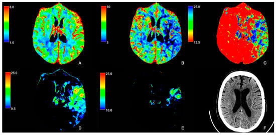
Background: Pediatric high-grade glioma (pHGG) is a highly aggressive cancer with unique biology distinct from adult high-grade glioma, limiting the effectiveness of standard treatment protocols derived from adult research. Objective: The purpose of this report is to present preliminary results from an ongoing pilot study integrating spectroscopic magnetic resonance imaging (sMRI) to guide proton beam therapy and longitudinal imaging analysis in pediatric patients with high-grade glioma (pHGG). Methods: Thirteen pediatric patients under 21 years old with supratentorial WHO grade III-IV glioma underwent baseline and serial whole-brain spectroscopic MRI alongside standard structural MRIs. Radiation targets were defined using T1-weighted contrast enhanced, T2-FLAIR, and Cho/NAA ≥ 2X maps. Longitudinal analyses included voxel-level metabolic change maps and spatial overlap metrics comparing pre-proton therapy and post-. Results: Six patients had sufficient longitudinal data; five received sMRI-guided PBT. Significant positive correlation (R2 = 0.89, p < 0.0001) was observed between T2-FLAIR and Cho/NAA ≥ 2X volumes. Voxel-level difference maps of Cho/NAA and Choline revealed dynamic metabolic changes across follow-up scans. Analyzing Cho/NAA and Cho changes over time allowed differentiation between true progression and pseudoprogression, which conventional MRI alone struggles to achieve. Conclusions: Longitudinal sMRI enhanced metabolic tracking in pHGG, detects early tumor changes, and refines RT targeting beyond structural imaging. This first in-kind study highlights the potential of sMRI biomarkers in tracking treatment effects and emphasizes the complementary roles of metabolic and radiographic metrics in evaluating therapy response in pHGG.
Full article
(This article belongs to the Section Cancer Imaging)
►
Show Figures

Figure 1


