


Tomography, Volume 11, Issue 5 (May 2025) – 10 articles
Cover Story (view full-size image):
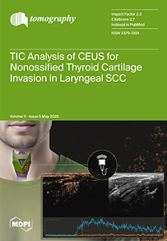
The accurate assessment of thyroid cartilage invasion is essential for the planning of effective treatment in laryngeal cancer. This study investigates the diagnostic value of contrast-enhanced ultrasound (CEUS) time–intensity curve (TIC) parameters in distinguishing between invaded and non-invaded non-ossified thyroid cartilage. Given that non-ossified thyroid cartilage is composed of avascular hyaline cartilage, it was hypothesized that CEUS would show no enhancement unless the tissue was invaded by a tumor, with corresponding differences in TIC parameters. By comparing these TIC-derived quantitative metrics with histopathological findings, this study shows the potential of CEUS as a complementary imaging modality to computed tomography or/and magnetic resonance imaging. View this paper
- Issues are regarded as officially published after their release is announced to the table of contents alert mailing list.
- You may sign up for e-mail alerts to receive table of contents of newly released issues.
- PDF is the official format for papers published in both, html and pdf forms. To view the papers in pdf format, click on the "PDF Full-text" link, and use the free Adobe Reader to open them.
Previous Issue
Next Issue