


Tomography, Volume 11, Issue 6 (June 2025) – 13 articles
Cover Story (view full-size image):
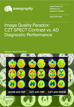
This study investigates whether advanced CZT SPECT technology’s superior image quality translates to improved diagnostic accuracy for Alzheimer’s disease. We compared conventional SPECT (eCAM) with CZT SPECT in 29 patients with suspected neurodegenerative diseases. Despite CZT SPECT showing significantly higher gray matter/white matter contrast (1.615 vs. 1.458, p < 0.001), diagnostic accuracy for AD did not improve. Both participating clinicians experienced decreased sensitivity or specificity with CZT images. These findings reveal an important paradox: enhanced image clarity does not automatically enhance diagnostic performance, highlighting the need for specialized training to fully leverage advanced imaging technology in neurodegenerative disease diagnosis. View this paper
- Issues are regarded as officially published after their release is announced to the table of contents alert mailing list.
- You may sign up for e-mail alerts to receive table of contents of newly released issues.
- PDF is the official format for papers published in both, html and pdf forms. To view the papers in pdf format, click on the "PDF Full-text" link, and use the free Adobe Reader to open them.
Previous Issue
Next Issue