


Int. J. Mol. Sci. 2026, 27(2), 965; https://doi.org/10.3390/ijms27020965 - 18 Jan 2026
Cited by 1 | Viewed by 990
Abstract
►
Show Figures
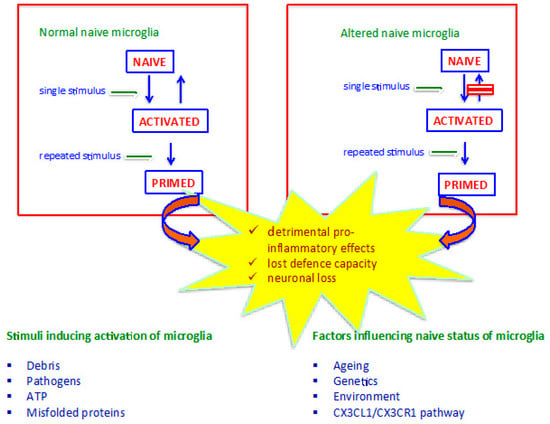
Parkinson’s disease (PD) is a progressive neurodegenerative disorder characterized by the selective loss of dopaminergic (DAergic) neurons in the substantia nigra (SN) and the accumulation of misfolded α-synuclein (aSyn). In addition to neuronal pathology, activated microglia are recognized as key mediators of the
[...] Read more.
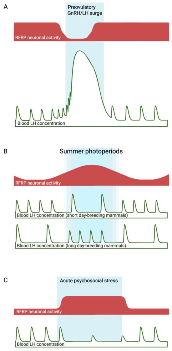
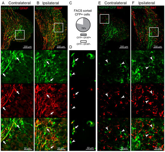
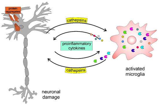
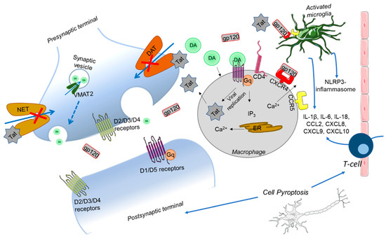
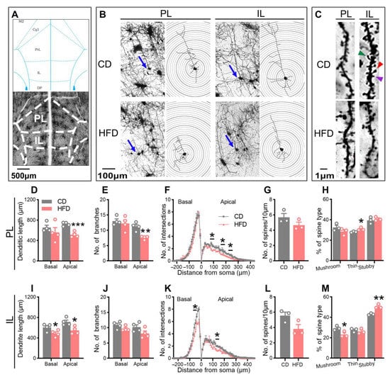
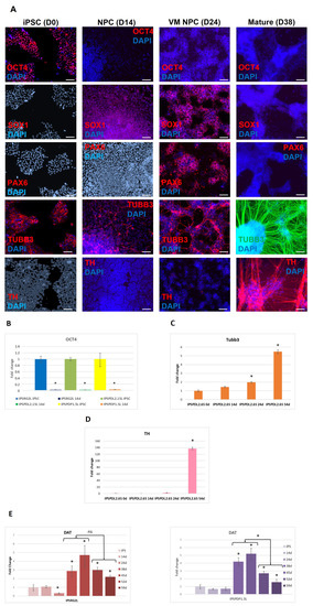
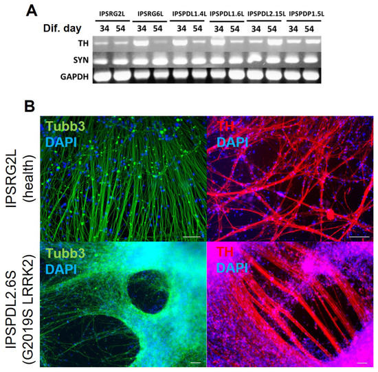
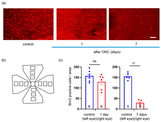
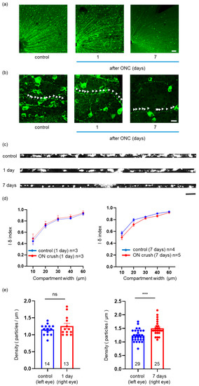
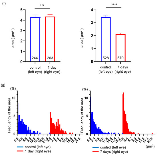
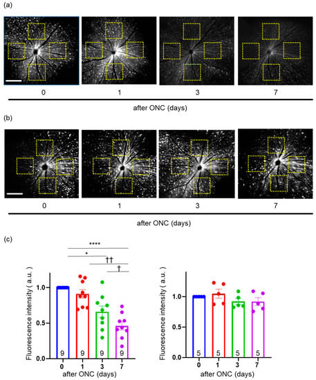
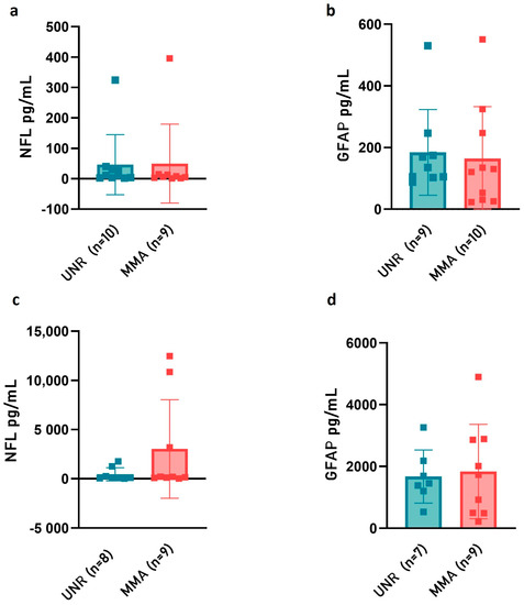
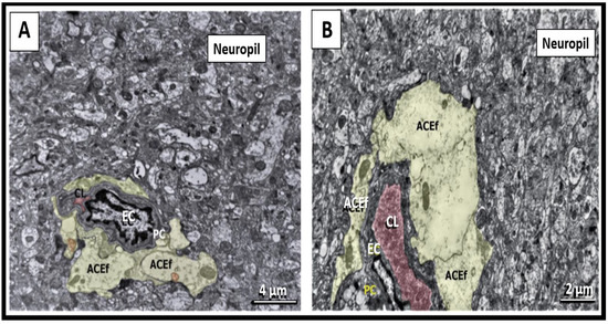
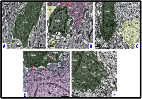
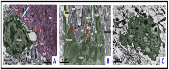
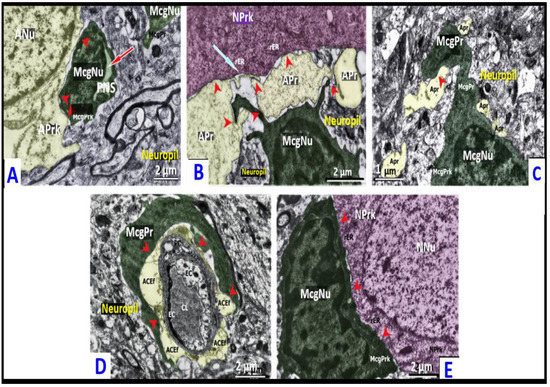
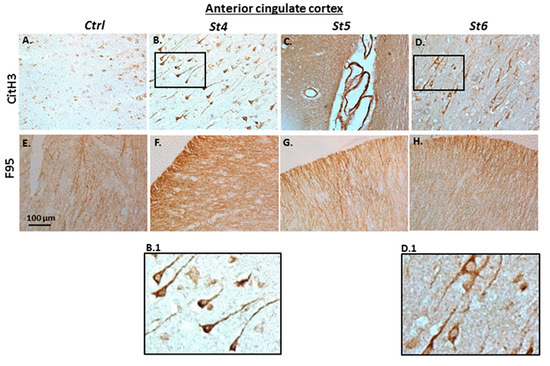
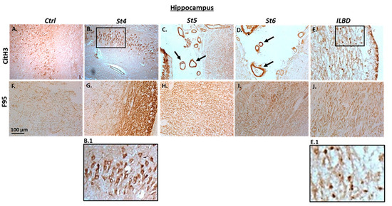
Parkinson’s disease (PD) is a progressive neurodegenerative disorder characterized by the selective loss of dopaminergic (DAergic) neurons in the substantia nigra (SN) and the accumulation of misfolded α-synuclein (aSyn). In addition to neuronal pathology, activated microglia are recognized as key mediators of the neuroinflammatory milieu in PD, contributing to DAergic neuron vulnerability. Emerging evidence suggests that the immune system, particularly T-cell-mediated responses, plays a key role in the pathogenesis of PD. However, the heterogeneity of these immune responses across species and preclinical models with varying regenerative capacities remains poorly understood. A comparative analysis of T-cell infiltration, astroglial reactivity, and DAergic neuronal loss across multiple models and species was performed. These included acute DAergic degeneration induced by 1-methyl-4-phenyl-1,2,3,6-tetrahydropyridine (MPTP), genetically modified mice with accumulation of aSyn (Thy1-aSyn L61 model), adult zebrafish exposed to MPTP-induced neurotoxicity and human post-mortem midbrain tissue obtained from PD patients. Zebrafish exhibited transient DAergic neurodegeneration, followed by neuronal regeneration and temporary CD4+ T-cell infiltration accompanied by an astroglial response and activation of microglia. In contrast, MPTP-treated mice showed a permanent neuronal loss, marked microglial activation, increased astrogliosis and CD8+ T-cell infiltration that was negatively correlated with neuronal survival. By contrast, L61 mice exhibited progressive aSyn accumulation with chronic astrogliosis, mild activation of microglia and CD4+ T-cell infiltration not directly linked to neuronal loss. Unlike age-matched controls, the SN from PD brains exhibited DAergic degeneration, aSyn aggregation, and elevated CD3+ T-cell infiltration, and increased microglial activation. These changes correlated with neuronal loss and aSyn burden. These findings emphasize the species- and model-specific immune profiles underlying PD pathology. Our results reveal that CD4+ T-cells contribute to neuronal regeneration following injury in zebrafish. This process is absent in the MPTP and L61 mouse models, which are instead driven by CD8+ or CD4+, respectively. This work underscores the potential of targeted immunomodulation aimed at T cell–glial interactions to slow neurodegeneration and promote repair in PD.
Full article

Figure 1


