


Cells 2024, 13(10), 820; https://doi.org/10.3390/cells13100820 - 10 May 2024
Cited by 12 | Viewed by 8602
Abstract
►
Show Figures
This review addresses the need for innovative co-culture systems integrating the enteric nervous system (ENS) with intestinal organoids. The breakthroughs achieved through these techniques will pave the way for a transformative era in gastrointestinal (GI) disease modeling and treatment strategies. This review serves
[...] Read more.
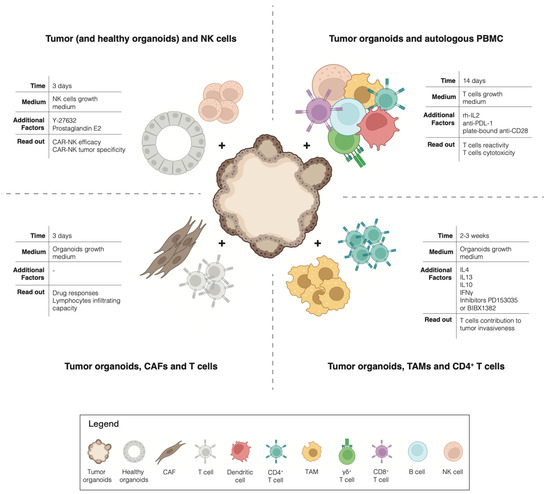
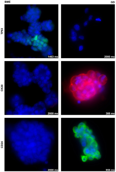
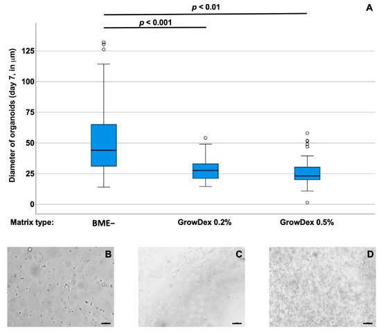
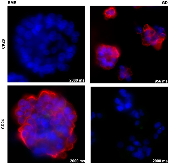
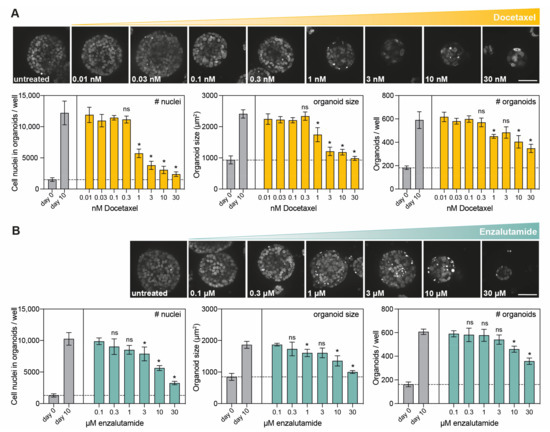
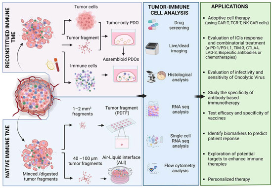
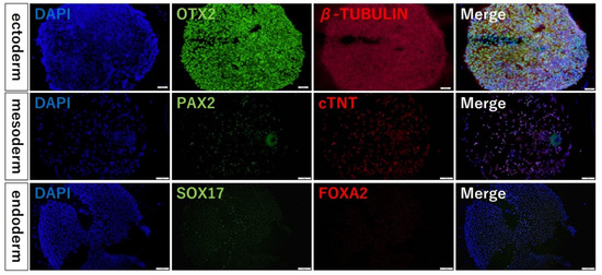
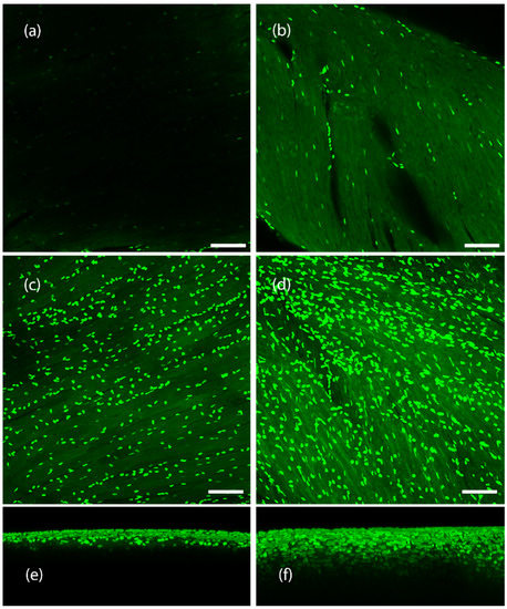
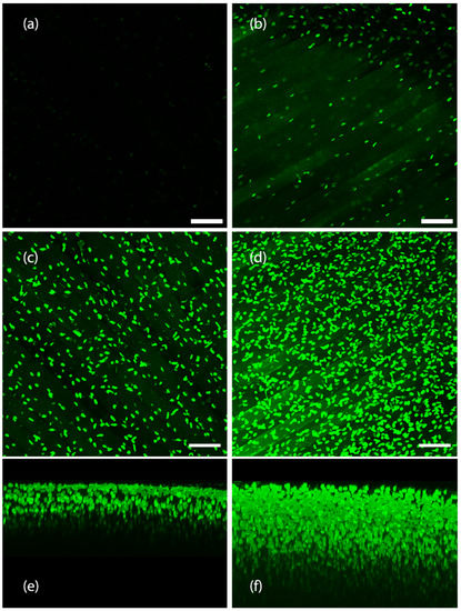
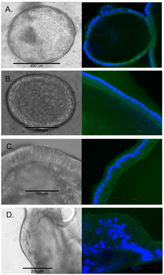
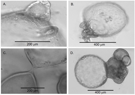
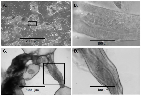
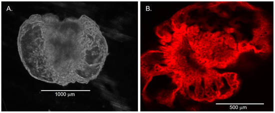
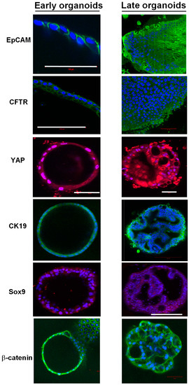
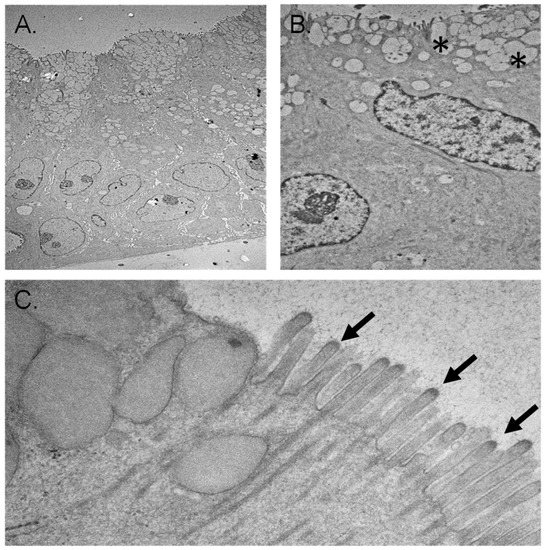
This review addresses the need for innovative co-culture systems integrating the enteric nervous system (ENS) with intestinal organoids. The breakthroughs achieved through these techniques will pave the way for a transformative era in gastrointestinal (GI) disease modeling and treatment strategies. This review serves as an introduction to the companion protocol paper featured in this journal. The protocol outlines the isolation and co-culture of myenteric and submucosal neurons with small intestinal organoids. This review provides an overview of the intestinal organoid culture field to establish a solid foundation for effective protocol application. Remarkably, the ENS surpasses the number of neurons in the spinal cord. Referred to as the “second brain”, the ENS orchestrates pivotal roles in GI functions, including motility, blood flow, and secretion. The ENS is organized into myenteric and submucosal plexuses. These plexuses house diverse subtypes of neurons. Due to its proximity to the gut musculature and its cell type complexity, there are methodological intricacies in studying the ENS. Diverse approaches such as primary cell cultures, three-dimensional (3D) neurospheres, and induced ENS cells offer diverse insights into the multifaceted functionality of the ENS. The ENS exhibits dynamic interactions with the intestinal epithelium, the muscle layer, and the immune system, influencing epithelial physiology, motility, immune responses, and the microbiome. Neurotransmitters, including acetylcholine (ACh), serotonin (5-HT), and vasoactive intestinal peptide (VIP), play pivotal roles in these intricate interactions. Understanding these dynamics is imperative, as the ENS is implicated in various diseases, ranging from neuropathies to GI disorders and neurodegenerative diseases. The emergence of organoid technology presents an unprecedented opportunity to study ENS interactions within the complex milieu of the small and large intestines. This manuscript underscores the urgent need for standardized protocols and advanced techniques to unravel the complexities of the ENS and its dynamic relationship with the gut ecosystem. The insights gleaned from such endeavors hold the potential to revolutionize GI disease modeling and treatment paradigms.
Full article

Figure 1


