- Review

The Role of Cold-Inducible RNA-Binding Protein (CIRP) in Neurological Disorders
- Xueqi Lai and
- Peng Zhong
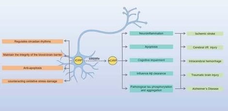
Cold-inducible RNA-binding protein (CIRP) is a critical molecule in the central nervous system (CNS) with functions that depend on its subcellular localization, exhibiting biphasic regulatory roles in both physiological and pathological processes. Under physiological conditions, intracellular cold-inducible RNA-binding protein (iCIRP) contributes to the maintenance of circadian rhythms by regulating the stability of core clock gene mRNAs and exerts neuroprotective effects during mild hypothermia by preserving the blood–brain barrier and inhibiting apoptosis. Pathologically, extracellular cold-inducible RNA-binding protein (eCIRP) functions as a damage-associated molecular pattern (DAMP) that drives neuroinflammation and brain injury. In ischemic stroke (IS), eCIRP promotes neutrophil extracellular trap (NET) formation and increases microglial activity via the Toll-like receptor 4 (TLR4) pathway. In cerebral ischemia–reperfusion (I/R) injury, eCIRP activates oxidative stress and the NOD-like receptor thermal protein domain associated protein 3 (NLRP3) inflammasome through the TLR4 axis, exacerbating mitochondrial damage. In intracerebral hemorrhage (ICH), eCIRP further amplifies inflammation via the interleukin-6 receptor (IL-6R)/signal transducer and activator of transcription 3 (STAT3) signaling pathway. In traumatic brain injury (TBI), eCIRP activates the endoplasmic reticulum stress pathway, intensifying apoptosis. In Alzheimer’s disease (AD), eCIRP regulates tau phosphorylation and β-amyloid (Aβ) metabolism and may mediate the link between alcohol exposure and AD pathology. Preclinical studies indicate that serum eCIRP levels correlate with IS and ICH severity, highlighting its potential as a biomarker. This systematic review elucidates the mechanisms of CIRP in CNS diseases, providing insights for understanding and preventing conditions such as IS, cerebral I/R injury, ICH, TBI, and AD.
9 February 2026