- Review

Vinpocetine—An “Old” Drug with a New Face: Moving Toward a Better Understanding of Its Neuroprotective Mechanism of Action
- E. Sylvester Vizi and
- Béla Kiss
Synthesized more than 60 years ago, vinpocetine—the active ingredient of Cavinton®, with over five decades of clinical use—has remained the subject of extensive investigation, particularly during the past 15 years. During this time, a large body of experimental preclinical evidence has accumulated demonstrating its neuroprotective potential and complex mechanisms of action in cerebral ischemia–hypoxia. Comprehensive in vitro studies and animal experiments have significantly elucidated the molecular basis of vinpocetine and the signaling pathways through which it prevents or mitigates ischemic injury. In this review, we summarize earlier and more recent experimental results that highlight the multifaceted nature of vinpocetine’s neuroprotective actions, which include inhibition of phosphodiesterase type 1, blockade of voltage-dependent NaV1.8 channels, reduction of oxidative stress, and suppression of neuroinflammatory processes triggered by cerebral ischemia–hypoxia. Taken together, it can be hypothesized that, under in vivo conditions, vinpocetine’s individual actions are additive or synergistic, thereby contributing in a combined manner to recovery from cerebral ischemic insult.
17 March 2026
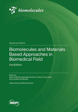
![Structure of vinpocetine ([(3α,16α)-eburnamenine-14-carboxylic acid ethyl ester]) (Mw: 350.5).](https://mdpi-res.com/cdn-cgi/image/w=470,h=317/https://mdpi-res.com/biomolecules/biomolecules-16-00454/article_deploy/html/images/biomolecules-16-00454-g001-550.jpg)