


Abstract
Acidophilic kinases are serine/threonine protein kinases that preferentially phosphorylate substrates bearing negatively charged determinants near the phosphoacceptor site. These determinants may be encoded by aspartate or glutamate, or introduced by prior phosphorylation of serine, threonine, or tyrosine residues. CK2 is the most representative of this historically small group of kinases because of its high basal activity, broad localization, and extensive substrate repertoire. Kinome-wide specificity profiling covering more than 84% of the active human S/T kinome has now provided a near-comprehensive view of acidophilic kinases across several families. Here, we compare these enzymes through three interconnected features: subcellular localization, substratome size, and consensus-motif specificity. We broadly categorize them as D/E-directed kinases or phosphate-directed/primed kinases, while recognizing that these preferences are not mutually exclusive. We then compare intrinsic preferences measured with peptide libraries with those observed in bona fide substrates. Despite their shared preference for negative charge, acidophilic kinases display markedly different positional requirements for acidic or phosphorylated determinants. For kinases that rely strongly on consensus-sequence recognition, these preferences can generate distinctive kinase-specific fingerprints that can support the attribution of experimentally validated phosphosites, particularly when combined with subcellular localization.
1. Introduction
Reversible phosphorylation of serine, threonine and tyrosine residues is a widespread mechanism of protein regulation and frequently occurs at multiple sites within the same protein [1]. In humans, these reactions are catalyzed by more than 500 protein kinases, collectively referred to as the human kinome [2]. The scale of the process is reflected in the annotated phosphoproteome: PhosphoSitePlus currently lists approximately 300,000 non-redundant phosphorylation sites, yet the upstream kinase remains unknown for most of them [3]. Protein kinases vary greatly in substrate selectivity, from enzymes acting on a restricted set of targets to highly pleiotropic kinases that phosphorylate hundreds of proteins. Substrate recognition depends on the local sequence surrounding the phosphoacceptor, distal docking interactions, subcellular localization, and scaffolding proteins [4,5]. Among these determinants, the kinase consensus sequence is particularly informative and, when sufficiently distinctive, can provide a molecular fingerprint of the activity of a specific kinase [6,7]. S/T kinases are commonly grouped according to their preferred sequence features into proline-directed, basophilic, and acidophilic classes. Proline-directed kinases, including many MAPKs and CDKs, are characterized by a strong requirement for a proline residue immediately downstream of the phosphoacceptor site, generating the canonical S/T-P motif. Basophilic kinases, such as PKA, AKT, and PKC, preferentially recognize basic residues around the target serine or threonine, where the requirement of arginine at the −3 position is shared by multiple basophilic kinases. Acidophilic kinases, exemplified by CK2, select negatively charged environments generated by aspartate, glutamate, or pre-existing phosphoserine, phosphothreonine, and phosphotyrosine [5,8].
The most common determinants of kinase substrate recognition in the phosphoproteome can be readily visualized by comparing residues surrounding annotated phosphoserine and phosphothreonine sites with a background of random S/T-containing peptides. The two-sample logo in Figure 1 was generated from the −7 to +7 window surrounding more than 200,000 non-redundant human phospho-S/T sites. This logo highlights residues that are enriched or depleted at each position relative to the phosphoacceptor site, with symbol size reflecting the relative contribution of each amino acid [9]. Its major features are consistent with an earlier analysis of approximately 35,000 sites [8], indicating that the principal global signatures have remained stable as phosphosite coverage has increased. The most prominent and conserved feature is the strong enrichment of proline at the +1 position, highlighting the major contribution of proline-directed kinases to the cellular phosphoproteome. A second major feature is the enrichment of basic residues upstream of the phosphoacceptor site, particularly arginine at the −3 position, which represents a hallmark of many basophilic kinases [8]. A third pattern, but less conserved, is represented by acidophilic phosphorylation sites, characterized by enrichment of acidic residues, mainly downstream of the phosphoacceptor residue. Another recurrent feature emerging from the global phosphosite analysis is the enrichment of serine residues at multiple positions around the target site, likely reflecting the prevalence of multisite phosphorylation clusters. Unlike proline-directed motifs, in which proline at +1 can be incompatible with phosphorylation by most other kinase classes, such as CK2 [10], AGC, and CAMK kinases [11], the distinction between basophilic and acidophilic motifs is less sharply defined. Basic residues, although preferentially selected by basophilic kinases, can in some cases be tolerated in substrates of acidophilic kinases, and acidic residues may occur in substrates of kinases not primarily classified as acidophilic [12]. This partial overlap has two important implications. First, although the linear motif remains a major determinant of kinase recognition, similar sequence features may be compatible with more than one kinase and therefore may not allow an unambiguous kinase assignment. Second, the same phosphoacceptor residue can be targeted by different kinases, providing functional redundancy or enabling its regulation by distinct signaling pathways and under different cellular conditions. In such cases, kinase activation, localization, and substrate availability determine which enzyme predominates, as illustrated by α-synuclein Ser129 phosphorylation (see below).
Figure 1.
Global sequence determinants of human S/T phosphorylation sites. Two-sample logo showing amino acid enrichment and depletion in the −7 to +7 sequence window surrounding annotated human phosphoserine and phosphothreonine sites (PhosphoSitePlus database [3]), compared with randomly selected S/T-containing peptides from the human proteome. To generate a background dataset, random human peptides of the same length (positions −7 to +7) were extracted from the Swiss-Prot database using a custom BioPerl script and UNIX text processing commands. Non-redundant sequences were then randomized using the UNIX shuf command as described in [8]. Residues above the central axis are enriched, whereas residues below the axis are depleted.
Historically, acidophilic S/T phosphorylation was associated mainly with a limited number of kinases, most notably CK2, CK1, and GSK3 kinases. This view progressively changed with the recognition that other kinases, including PLK1, PLK2, PLK3 [13,14], and FAM20C [15] also exploit negatively charged determinants for substrate recognition. A major advance came from the peptide-library profiling of 303 human S/T kinases by Johnson and colleagues, covering more than 84% of the active human S/T kinome. This study provided a near-comprehensive specificity landscape and identified acidophilic features in CK1, CK2, GSK3, GRK, FAM20C, PLK, and TGFβ/BMP receptors, as well as ALPK1 and STK32 kinases [16].
In this review, we discuss acidophilic kinases through three interconnected parameters: subcellular localization, substratome size, and consensus-motif specificity. Localization defines the accessible substrate space; the number of experimentally annotated substrates provides a first approximation of kinase pleiotropy; and finally, the consensus motif determines how confidently a phosphorylation event can be assigned to a given kinase.
2. Localization and Substratome Size Define the Functional Space of Acidophilic Kinases
Subcellular localization and substratome size provide complementary information on acidophilic kinase biology. Localization defines the accessible substrate pool, whereas the number of experimentally annotated phosphosites offers a first, although biased, estimate of kinase pleiotropy. Table 1 reports the predominant localization and number of experimentally annotated human phosphosites assigned to each acidophilic kinase, based on PhosphoSitePlus entries in which both kinase and substrate were human. These values are not absolute measures of biological importance, but help distinguish broadly acting, compartment-restricted, and poorly characterized kinases. The first group comprises broadly distributed nuclear and/or cytoplasmic kinases, including CK2, CK1α/δ/ε, GSK3α/β, PLK1/2/3, ALPK1, and STK32B/STK32C. CK2 represents the most prominent example of pleiotropy. Its ubiquitous expression, high basal activity, and distribution between cytoplasm and nucleus are mirrored by the exceptionally large number of CK2-dependent phosphorylation events. CK2α is associated with 590 human annotated phosphosites in Table 1, the highest number among the acidophilic kinases. Although PhosphoSitePlus assigned a lower number of substrates to CK2α′, this difference is unlikely to reflect a true isoform-specific disparity, because most CK2-dependent phosphorylation events cannot be unambiguously attributed to either catalytic isoform (Table 1). This ambiguity reflects CK2 biology: the enzyme commonly forms tetrameric holoenzymes containing two catalytic subunits, CK2α and/or CK2α′, and a CK2β dimer; the catalytic domains of CK2α and CK2α′ are highly conserved, and mixed holoenzymes may coexist [17,18]. The CK2β subunit should not be viewed as a classical regulatory subunit that simply switches on the catalytic activity of CK2. CK2α and CK2α′ are active as isolated enzymes, whereas CK2β modulates the biological output of CK2 [17,18]. How CK2β directs CK2 toward specific substrates remains incompletely understood and is unlikely to rely on a single universal docking motif. CK2β may act as an adaptor or scaffold, linking the CK2 catalytic subunits to selected substrates and protein complexes [18]. Accordingly, CK2β-deficient cells retain substantial catalytic activity but show reduced phosphorylation of several endogenous CK2 substrates [19].
Table 1.
Acidophilic kinases: subcellular localization and experimentally annotated phosphosites. UniProt accession numbers and subcellular localization were obtained from UniProt for human proteins; when localization information was unavailable, it was retrieved from the literature. Localization is abbreviated as N, nucleus; C, cytoplasm; PM, plasma membrane; and GL, Golgi lumen. Annotated phosphosites were obtained from the PhosphoSitePlus kinase-substrate dataset [3] (June 2026 release), considering only experimentally supported phosphorylation events in which both the kinase and substrate were human. Abbreviations: Sub. Loc.: Subcellular localization, and Ann. P-sites: annotated phosphosites. * the numbers assigned to individual CK2 and GSK3 isoforms should be interpreted cautiously because overlapping substrate specificity and annotation bias may prevent unambiguous attribution to a single isoform.
Functional enrichment analysis shows that CK2 substrates are enriched in processes related to transcriptional regulation, chromatin remodeling, gene-expression control, protein stabilization, apoptosis, and circadian rhythm regulation (Table S1A). This broad profile is consistent with the involvement of CK2 in numerous cellular pathways [6,18,20,21,22,23]. Not surprisingly, CK2 deregulation has been implicated in several pathological conditions, including inflammation [24,25,26], neurodegenerative disorders [27,28], viral infections [29,30,31], and cancer [28,32,33,34,35]. Its role in oncology is particularly well established: CK2 supports multiple hallmarks of tumor progression, including sustained proliferation, resistance to apoptosis, DNA-damage tolerance, angiogenesis, and metastatic behavior, making it an attractive pharmacological target [28,32,33,34,35,36,37].
PLKs provide a different model. The human family comprises PLK1-PLK5. PLK1-4 contain an N-terminal S/T kinase domain and C-terminal polo-box motifs (PBDs), although the polo-box architecture of PLK4 is markedly divergent from that of PLK1-3. Human PLK5 retains a polo-box domain but lacks a functional kinase domain [38]. Within this family, only PLK1, PLK2, and PLK3 display a clear acidophilic substrate preference [13,39,40]. PLK1 has by far the largest annotated substratome among PLK acidophilic kinases in Table 1, but its substrates are not broadly distributed across cellular functions. Gene Ontology analysis shows a strong enrichment in cell-cycle and mitotic processes, including chromosome segregation, sister chromatid segregation, and chromosome organization (Table S1B). This focused substrate repertoire is explained by the modular structure of PLKs. Their catalytic domain determines which site can be phosphorylated, while the PBDs recruit the kinase to substrates and to specific cellular structures [41]. Importantly, the PBDs of different PLK isoforms are not functionally equivalent and can confer distinct target specificities within the family [41]. Thus, whereas CK2 exemplifies pleiotropy, PLKs illustrate a more spatially and temporally constrained form of substrate selection, driven by catalytic specificity, PBD-mediated recruitment and cell-state-dependent localization.
CK1 and GSK3 represent another mode of broad but regulated substrate access.
Human cells express CK1α, CK1α-like, CK1δ, CK1ε and the three membrane-associated CK1γ isoforms, whereas CK1β is absent from the human kinome. Their catalytic domains are highly conserved, whereas divergent terminal regions influence localization, interactions, and pathway-specific functions [42,43]. CK1α, CK1δ, and CK1ε show broad nucleo-cytoplasmic distribution and substantial substrate annotation. CK1α, the most substrate-rich CK1 isoform in Table 1, shows enrichment in regulation of apoptosis, protein stabilization, protein ubiquitination, NF-κB signaling, and transcriptional regulation (Table S1C), suggesting a substrate repertoire centered on stress-response and cell-fate control.
GSK3α and GSK3β are both broadly distributed kinases with nearly identical catalytic domains and largely overlapping substrate specificities [44]. GSK3 was originally identified through its ability to phosphorylate and inhibit glycogen synthase, but it is now recognized as a regulator of Wnt/β-catenin signaling, PI3K-AKT pathways, inflammatory responses, and neuronal homeostasis [45]. Functional enrichment analysis of annotated GSK3β substrates reveals a broad profile that includes transcriptional regulation, DNA-damage responses, miRNA transcription, hypoxia responses, rhythmic processes, and apoptotic regulation (Table S1D). This profile resembles that of CK2 in functional breadth, but the underlying regulation is different. CK2 has high basal activity and broad pleiotropicity, whereas GSK3 acts in a more context-dependent manner and preferentially phosphorylates primed substrates, a feature shared with CK1 (see below).
ALPK1 represents a distinct example of a predominantly cytoplasmic acidophilic kinase with a still limited substrate repertoire. ALPK1 belongs to the atypical α-kinase family, a small group of protein kinases structurally distinct from conventional eukaryotic protein kinases that includes ALPK1, ALPK2, ALPK3, eEF2K, and the kinase domains of the TRPM6 and TRPM7 channels [46]. ALPK1 functions as an intracellular sensor of bacterial ADP-heptose, and ligand binding activates its kinase domain, leading to phosphorylation of TIFA, its best-characterized physiological substrate, at Thr9. This event promotes TIFA oligomerization and downstream activation of NF-κB signaling [47,48]. Thus, in contrast to broadly acting kinases such as CK2 or GSK3, ALPK1 appears to operate within a more restricted signaling context. However, the small number of currently validated substrates should be interpreted primarily as reflecting the still incomplete characterization of the ALPK1 substratome rather than a necessarily narrow biological role.
The STK32 family comprises three poorly characterized members of the AGC kinase group: the nucleo-cytoplasmic kinases STK32B and STK32C, and the plasma-membrane associated STK32A. Their catalytic domains are closely related, with STK32A sharing approximately 70% and 67% sequence identity with STK32B and STK32C, respectively, but only about 36% identity with the closest kinases outside the family [49]. Despite this conserved catalytic core, the three paralogues appear to have distinct biological roles. STK32A regulates planar cell polarity and stereociliary-bundle orientation in inner-ear hair cells [50], whereas STK32B has been genetically associated with essential tremor [51] and reported to promote glioma growth [52]. STK32C has mainly been investigated in cancer, where it has been linked to tumor-cell proliferation, migration, invasion, and angiogenesis [53]. Nevertheless, the physiological functions and bona fide substrates of all three kinases remain largely undefined.
A second major group is defined by predominant association with the plasma membrane. This includes receptor kinases, whose function is intrinsically linked to ligand-dependent signaling at the cell surface, and membrane-associated kinases, which are recruited to, or enriched at, the plasma membrane but may also operate elsewhere. TGFβ/BMP receptor kinases, including TGFBR1, TGFBR2, BMPR1A, BMPR1B, BMPR2, ACVR1, ACVR1B, ACVR2A, ACVR2B, belong to the first category [54]. For these enzymes, membrane localization is not simply a spatial feature but a functional requirement: phosphorylation occurs within ligand-dependent receptor complexes. Their curated substratome is comparatively small, with several members having few or no kinase-specific human substrates annotated in Table 1. Their small substratome does not imply limited biological importance; rather, they reflect a restricted mode of substrate selection. In these pathways, signaling is transmitted mainly through SMAD proteins, although additional non-SMAD proteins can also be phosphorylated when recruited close to an activated receptor complex [54].
By contrast, CK1γ isoforms, STK32A, and GRKs represent membrane-associated kinases whose localization is more dynamic and does not necessarily restrict their activity to the plasma membrane. Among CK1 family members, the CK1γ isoforms have the smallest annotated substratomes (Table 1), consistent with their more restricted cellular distribution. Indeed, CK1γ kinases are targeted predominantly to the plasma membrane through lipid modifications [55], distinguishing them from the nucleo-cytoplasmic CK1α/δ/ε isoforms.
STK32A is also subject to lipid-dependent membrane targeting. Although this kinase remains poorly characterized, it undergoes N-terminal myristoylation, which promotes its association with the plasma membrane [56]. This modification may restrict STK32A to a more confined cellular environment and thereby limit the pool of accessible substrates.
GRKs, by contrast, are recruited specifically to activated G protein-coupled receptors and phosphorylate receptor intracellular domains to regulate desensitization and arrestin-dependent signaling [57]. GRK isoforms associate with membranes through distinct mechanisms. GRK2 and GRK3 are recruited through interactions with Gβγ and membrane phospholipids. GRK1, GRK4, GRK6, and GRK7 are membrane-associated through lipidation, whereas GRK5 binds membrane phospholipids through a C-terminal basic region [58]. GRK2 is the most extensively annotated GRK in Table 1. Gene Ontology functional enrichment analysis of its substrates identified biological processes strongly related to GPCR signaling, including G protein-coupled receptor signaling, cyclic nucleotide-coupled GPCR pathways, adenylate cyclase-inhibiting GPCR signaling and neuropeptide signaling, smooth muscle contraction and vasoconstriction (Table S1E). Thus, GRK2 does not display broad pleiotropy in the CK2 sense; rather, it has a receptor-centered substratome shaped by ligand activation and membrane recruitment. Notably, GRK localization is not limited to the plasma membrane. Several family members remain partly cytosolic, while GRK5 can also enter the nucleus, allowing selected GRKs to act on substrates outside activated GPCR complexes [57].
Finally, FAM20C defines a unique spatial model. It localizes predominantly to the Golgi and can also be secreted, although the biological relevance of extracellular FAM20C activity remains incompletely understood [15,59]. The limited number of FAM20C substrates listed in PhosphoSitePlus largely reflects the way kinase–substrate relationships are curated, rather than a restricted biological role. Indeed, phosphoproteomic studies have shown that FAM20C is the principal kinase responsible for Ser phosphorylation of secreted proteins [7,60]. Because its catalytic domain faces the lumen of the secretory pathway, FAM20C may act on proteins that reside in topologically equivalent spaces. These include luminal ER-Golgi proteins, membrane-protein domains facing the luminal compartments or exposed to the cell surface, and secreted proteins. Thus, despite its restricted localization, FAM20C acts on a broad but topologically defined substratome [7].
Overall, these comparisons show that acidophilic kinases occupy very different cellular and functional spaces. CK2 combines broad nucleo-cytoplasmic distribution with the largest annotated substratome, whereas PLK1 has many substrates concentrated mainly in mitotic processes. CK1 and GSK3 also have broad substrate repertoires, but their activity is more dependent on signaling context. TGFβ/BMP receptors and GRKs act predominantly within membrane-associated signaling systems, while FAM20C is confined to the luminal space of the secretory pathway. By contrast, the very limited substratome of ALPK1 and STK32 kinases reflects the fact that their physiological substrates remain largely unidentified.
3. Consensus Specificity: From Acidic Motifs to Kinase-Specific Fingerprints
Although acidophilic kinases are commonly grouped according to their preference for negatively charged determinants, this classification should not imply that they recognize identical motifs. They can be divided broadly into D/E-directed kinases, which primarily read constitutive acidic residues, and phosphate-directed or primed kinases, which recognize pre-existing phosphoresidues. According to kinome-wide profiling [16], the first group includes CK2, FAM20C, PLK, and TGFβ/BMP receptor kinases, whereas the second includes CK1, GSK3, GRK, ALPK1, and STK32 kinases. This classification is not absolute but reflects the predominant mode of substrate recognition observed in the kinase peptide-profiling assays of Johnson et al. [16]: D/E-directed acidophilic kinases can, in some contexts, also use phosphorylated residues as positive determinants, whereas phosphate-directed kinases may tolerate acidic residues in place of a priming phosphosite. At the molecular level, recognition of negative charge generally relies on electrostatic complementarity between negatively charged substrate determinants and basic residues in the kinase substrate-binding region. Depending on the kinase, these residues may form an extended electropositive surface able to interact with several acidic residues at different positions, or a more localized pocket that recognizes a pre-existing phosphate group or negatively charged determinants at a defined distance from the phosphoacceptor site. The geometry of these interactions can also contribute to discrimination among Asp, Glu, and phosphorylated residues. Importantly, these negatively charged determinants differ in size, geometry, and ionization state and may therefore be recognized with different efficiencies. For some kinases, the molecular basis of negative-charge recognition has been defined by mutagenesis and structures containing peptide substrates [61,62,63], whereas for others it remains largely inferred from sequence specificity. Acidophilic recognition therefore represents a common physicochemical principle rather than a single conserved structural mechanism.
3.1. D/E-Directed Acidophilic Kinases
Figure 2 compares D/E-directed kinase profiles generated from the peptide-library dataset of Johnson and colleagues [16]. The figure includes CK2α, CK2α′, FAM20C, PLK1-3, and TGFBR1 profiles (the other TGFβ/BMP receptor kinases are shown in Figure S1). The most evident feature emerging from this comparison is that all these kinases prefer negatively charged residues, although at different positions relative to the phosphoacceptor site. CK2 displays a broad preference for acidic residues across the entire −5 to +4 window, with the strongest determinants located downstream of the target residue, particularly at positions +1 and +3. As expected, the specificity profiles of CK2α and CK2α′ are almost identical (Figure 2), consistent with the high conservation of their kinase domains, and indicating that isoform-specific substrates are more likely to be selected through interactions involving non-catalytic regions. Moreover, in the in vitro phosphorylation assays, acidic determinants can, in several positions, be replaced by phosphorylated residues. CK2 can indeed act on primed substrates, and several examples of phosphorylation-dependent hierarchical recognition have been reported [64,65,66]. This further illustrates that the distinction between D/E-directed and phosphate-directed kinases should not be regarded as absolute, but rather as a description of their predominant recognition mode. When the kinase specificity inferred from peptide phosphorylation assays is compared with the specificity observed analyzing the bona fide CK2 substrate dataset (Figure 3), a strong similarity is apparent. In both cases, acidic residues are preferred throughout the analyzed sequence window, with the most prominent enrichment occurring downstream of the phosphoacceptor site. Notably, the +1 and +3 acidic determinants identified by kinome-wide specificity profiling are also evident in bona fide CK2 substrates, although the +3 position emerges as the dominant contributor in the experimentally annotated CK2 substrate dataset. In contrast, no clear enrichment of phosphorylated residues is evident at +1 or +3 in the bona fide substrate profile, suggesting that, although CK2 can recognize primed substrates, this mode of recognition likely accounts for only a limited fraction of CK2 phosphorylation events in cells.
Figure 2.
Positional preferences of D/E-directed acidophilic kinases. Sequence logos showing the substrate-recognition profiles of representative D/E-directed acidophilic kinases. Data from the peptide-library specificity dataset of Johnson et al. [16] were rescaled and plotted using Logomaker [67]. The logos display amino acid preferences from −5 to +4 relative to the phosphoacceptor residue, with the phosphoacceptor S/T fixed at position 0. TGFBR1 is shown as a representative TGFβ/BMP receptor kinase (the specificity profiles of the other members of this receptor kinase group are shown in Figure S1).
Figure 3.
Sequence signatures of bona fide substrates of representative acidophilic kinases. pLogo analysis [68] of experimentally validated phosphorylation sites annotated in PhosphoSitePlus. Sequences surrounding bona fide phosphosites were compared with the same background dataset described in Figure 1, consisting of randomly selected S/T-containing peptides from the human proteome. At each position, amino acid enrichment or depletion was evaluated by pLogo using binomial probabilities based on residue frequencies in the background dataset. Default pLogo statistical settings were used, including Bonferroni correction for multiple testing with an overall significance threshold of p = 0.05. Residues above the central axis are enriched, whereas residues below the axis are depleted; red horizontal lines indicate the statistical significance threshold.
FAM20C shows a different behavior. Rather than displaying a broad preference for acidic residues, it is characterized by a highly restricted and dominant preference for glutamate at the +2 position (Figure 2). Consistent with the peptide phosphorylation data, analysis of the bona fide FAM20C substrate set revealed an almost identical specificity profile, with a marked and highly selective enrichment of glutamate at the +2 position (Figure 3).
Compared with CK2 and FAM20C, PLK-family members and TGFβ/BMP receptor kinases rely more strongly on upstream acidic determinants, although residues downstream of the phosphoacceptor site can also contribute to substrate recognition (Figure 2). Within this general trend, PLK isoforms display partially distinct positional preferences together with a shared preference for a hydrophobic residue at +1. In peptide-library profiles, PLK1 preferentially recognizes acidic residues at −2, PLK2 shows a stronger preference at −3 with an additional contribution at +2, whereas PLK3 displays a broader pattern involving −3, −2, −1 and +2, +3. These differences are supported by studies using protein substrates and peptides derived from cellular proteins [39,40]. Analysis of bona fide substrates further confirms these isoform-specific signatures, giving a more precise preference (Figure 3): PLK1 major determinants are the acidic residue at −2, with an additional contribution of an acidic residue at −1. For PLK2 and PLK3, the limited number of annotated substrates does not allow a statistically robust definition of their dominant determinants. Nevertheless, the observed trends are consistent with the peptide-library profiles: PLK2 substrates show a prominent acidic determinant at −3, with a weaker contribution at +2, whereas PLK3 substrates are enriched in glutamate at −3 and −2, together with an additional acidic contribution at +2, indicating partial overlap with the PLK2 recognition pattern.
Similar to PLKs, TGFβ/BMP receptor kinases, including TGFBR1, TGFBR2, BMPR1A, BMPR1B, BMPR2, ACVR1, ACVR1B, ACVR2A and ACVR2B, prefer acidic residues upstream of the phosphoacceptor site, particularly around −2, with variable and generally weaker downstream contributions, including at +1 (Figure 2 and Figure S1). The catalytic domains of TGFβ/BMP receptor kinases are structurally highly related, consistent with their common evolutionary origin and with the similar acidophilic-like preferences observed in peptide-based specificity profiling (Figure 2 and Figure S1). However, their physiological specificity is not dictated solely by the catalytic cleft. TGFβ/BMP receptor kinases can be further divided into type I and type II receptors, which have distinct roles within the signaling complex. Type II receptors, including TGFBR2, BMPR2, ACVR2A and ACVR2B, are ligand-binding receptor kinases that phosphorylate and activate type I receptors. Type I receptors, including TGFBR1, ACVR1, ACVR1B, BMPR1A and BMPR1B, contain a conserved glycine- and serine-rich (GS) region immediately N-terminal to the kinase domain. Phosphorylation of this GS region by type II receptors activates the type I receptor kinase, which then phosphorylates receptor-regulated SMADs (R-SMAD) [69,70]. Thus, although both receptor classes possess structurally related S/T kinase domains, type II receptors primarily act as upstream activators, whereas type I receptors function as the main SMAD-phosphorylating kinases. In addition, substrate selection is strongly influenced by docking surfaces within the kinase domain, particularly the L45 loop, a short exposed region between kinase subdomains IV and V that differs among type I receptors and determines their ability to phosphorylate specific SMAD proteins [71]. Therefore, receptor S/T kinases represent a distinctive class of membrane-confined acidophilic-like kinases in which intrinsic catalytic-site preference is subordinated to receptor activation and substrate recruitment.
The distinct positional preferences of the D/E directed acidophilic kinases raise an important question: can these kinase-specific signatures be used to identify the enzyme responsible for a phosphorylation event? Although, as noted above, consensus motifs alone are rarely sufficient for definitive assignment, highly distinctive determinants can provide strong predictive information, particularly when combined with biochemical validation and subcellular context. FAM20C provides a particularly notable example. Before its molecular identification, biochemical studies had already revealed a Golgi-associated casein kinase activity (G-CK) distinct from CK2 [72]. This G-CK activity phosphorylated secretory-pathway proteins at sites conforming to an S-x-E motif, a feature particularly evident in caseins and other secreted phosphoproteins [73,74]. This positional requirement differed from the broader downstream acidic preference of CK2, which relies more strongly on acidic determinants at +1 and +3 and does not display a dominant requirement for glutamate at +2 [72]. Thus, the motif itself already suggested the existence of a distinct Golgi casein kinase. This idea was further strengthened by sequence analysis of early secreted and plasma phosphoproteomes, which revealed a high frequency of S-x-E phosphosites leading to the hypothesis that a single kinase, the Golgi casein kinase, could be responsible for the serine phosphorylation of most of the secreted proteins [75]. This prediction, initially based only on substrate-sequence analysis, was subsequently validated by the identification of the Golgi casein kinase as FAM20C [15,76], and by phosphoproteomic studies in FAM20C-deficient cells [60], which established FAM20C as the principal Ser kinase of the secreted proteins. More recently, by combining this distinctive S-x-E specificity with the restricted localization of FAM20C in the Golgi lumen and secretory pathway, analysis of experimentally validated phosphosites annotated in PhosphoSitePlus database identified a high-confidence putative FAM20C substratome consisting of 443 phosphosites across 256 secretory-pathway proteins [7]. Thus, FAM20C illustrates how a highly distinctive consensus motif, when interpreted together with subcellular localization, can move from predicting the existence of a kinase to defining its biologically coherent substratome.
A second example is provided by α-synuclein Ser129, where consensus-sequence analysis helped discriminate among several candidate acidophilic kinases. This phosphorylation event has been extensively investigated because of its close association with synucleinopathies, including Parkinson’s disease and dementia with Lewy bodies, although recent evidence also supports a physiological role of this phosphorylation in fine-tuning α-synuclein function and synaptic transmission [77]. Several acidophilic kinases, including CK2, CK1, and GRKs, were initially proposed as candidate Ser129 kinases, largely on the basis of the acidic sequence surrounding the target site [78,79]. Subsequent studies pointed instead to a predominant role for PLKs, particularly PLK2 [80,81]. Importantly, the greater efficiency of PLK2/3 relative to CK2 could be predicted from the local sequence and was confirmed by in vitro kinase assays, using recombinant kinases, full-length α-synuclein and Ser129-containing peptides [82]. Although the region surrounding S129 is rich in acidic residues (DNEAYEMPSEEGYQD), it lacks an acidic residue at +3, the dominant determinant of CK2 specificity [83]. Instead, E126 at −3 and E131 at +2 generate an E-x-x-S-x-E motif that is compatible with both PLK2 and PLK3, but more closely matches the preferred determinants of PLK2 (Figure 2). Mutational analysis showed that E126 at −3 is the major determinant, as its substitution completely abolished PLK2-mediated phosphorylation. Replacement of E131 at +2 strongly reduced phosphorylation, whereas substitution of E130 at +1 had only a minor effect [82]. The same acidic determinants also contributed to recognition by PLK3, although this kinase phosphorylated Ser129 in vitro less efficiently than PLK2 [82]. Thus, the −3/+2 configuration supports phosphorylation by both isoforms but preferentially favors PLK2, explaining its greater catalytic efficiency relative to CK2 and PLK3 [82]. In this case, consensus-sequence analysis identified the most likely kinase and explained its greater catalytic efficiency.
Again, the presence of an acidic residue at +3 has been successfully used to predict and validate the CK2 substratome through the analysis of experimentally validated phosphosites. This was illustrated by phosphoproteome-based analyses revealing the extraordinary pleiotropy of CK2 [6] and by subsequent studies identifying PDCD5 and ESCRT-III complex components as CK2 substrates in vitro and in cells [84,85]. The diagnostic value of the +3 acidic determinant must nevertheless be interpreted with caution. A global analysis of phosphorylated motifs in the human phosphoproteome showed that the CK2-like motif pS/pT-x-x-D/E is highly prevalent and relatively promiscuous, being found not only in CK2 substrates but also in phosphosites assigned to many other kinases [12]. Thus, the +3 acidic residue is an important determinant of CK2 recognition, but it is not sufficiently stringent on its own for confident kinase assignment. More confident prediction of the CK2 substratome therefore requires the integration of a more stringent consensus sequence (S/T-E/D-x-E/D) and subcellular-context filtering.
3.2. Phosphate-Directed or Primed Acidophilic Kinases
The second major category of acidophilic kinases comprises phosphate-directed or primed kinases, which preferentially recognize substrates containing a pre-existing phosphorylated residue near the target site. Their consensus motifs are therefore often conditional, with efficient phosphorylation frequently depending on the prior action of another kinase, creating hierarchical phosphorylation cascades. GSK3 represents a paradigmatic example of phosphate-directed kinases. Its optimal substrates contain a priming phosphoserine or phosphothreonine four residues C-terminal to the target site, generating the consensus S/T-x-x-x-pS/pT [86] (Figure 4A and Figure S2). Structural studies showed that the priming phosphate is recognized by a positively charged pocket in GSK3 [87]. This phosphate-binding pocket explains both the strong preference of GSK3 for primed substrates and the mechanism of inhibitory phosphorylation at the N-terminus of GSK3, in which phosphorylated S9 in GSK3β and S21 in GSK3α can act as an intramolecular pseudosubstrate [88]. The β-catenin destruction complex provides one of the notable biological examples of phosphate-directed substrate recognition. In the absence of Wnt signaling, CK1α first phosphorylates β-catenin at S45, thereby generating a priming determinant for GSK3. GSK3 then phosphorylates β-catenin sequentially at Thr41, Ser37, and Ser33 [89]. Notably, although GSK3 preferentially phosphorylates primed substrates, prior phosphorylation is not an absolute requirement. GSK3 can directly phosphorylate selected unprimed proteins, including PP1 inhibitor-2 [90], cyclin D1 [91] and tau at several sites [92]. GSK3 can also initiate Axin phosphorylation without an exogenous priming kinase, although subsequent phosphorylation within its multisite region may proceed hierarchically [93].
Figure 4.
Peptide-library and bona fide substrate signatures of CK1 and GSK3 kinases. Sequence logos comparing the intrinsic substrate preferences of representative phosphate-directed kinases with the sequence signatures of their experimentally annotated substrates. The upper panels (A) show peptide-library specificity profiles for GSK3α and CK1α. Data from the peptide-library specificity dataset of Johnson et al. [16] were rescaled and plotted using Logomaker [67]. The logos display amino acid preferences from −5 to +4 relative to the phosphoacceptor residue, with S/T fixed at position 0. GSK3A and CK1α are shown as representatives of GSK3 and CK1 families, respectively; the specificity profiles of the other members of these two kinase groups are shown in Figures S2 and S3, respectively. The lower panels (B) show pLogo analyses [68] of bona fide phosphosites assigned to GSK3A and CK1α. pLogo analysis and statistical significance were calculated as described in Figure 3.
CK1 family kinases provide a related but distinct example. CK1 proteins are evolutionarily conserved, widely expressed monomeric S/T kinases that, like CK2, are basally active [74], although CK1δ and CK1ε are additionally restrained by inhibitory autophosphorylation of their C-terminal tails [94]. CK1 kinases typically prefer phosphorylated residues upstream of the phosphoacceptor site, in particular in the pS/pT-x-x-S/T configuration (Figure 4A and Figure S3). Also CK1 can phosphorylate both primed and unprimed substrates as in the previous example on β-catenin phosphorylation by CK1α [89]. On selected substrates, CK1 can itself phosphorylate a non-canonical priming site and then sequentially phosphorylate downstream sites [95]. Thus, CK1 can function both as a priming kinase and as a phosphate-directed kinase, depending on the substrate context. Substrate access may also be controlled by specific docking interactions. Some proteins contain CK1-binding domains (CK1BDs) that establish relatively stable complexes with the kinase, increase its local concentration, and facilitate efficient multisite phosphorylation [96]. This mechanism is particularly well characterized in circadian clock proteins. Mammalian PER1 and PER2 recruit CK1δ/ε through a conserved CK1BD [95,96]. CK1BDs are not a general feature of all CK1 substrates, but rather a specialized mechanism that supports efficient and spatially restricted phosphorylation within specific protein complexes.
Analysis of bona fide substrates of CK1α, CK1δ, CK1ε, and GSK3α/β reveals the expected signatures of phosphate-directed kinase activity but also highlights the clustered organization of their phosphorylation sites (Figure 4B and Figure S4). CK1 substrates show enrichment of S at positions compatible with sequential phosphorylation, including the canonical upstream −3 position and additional S upstream (−6) and downstream (+3, +6) of the centered phosphoacceptor site (Figure 4B). Conversely, GSK3 substrates show the expected enrichment of S/T at +4, consistent with its C-terminal priming mechanism, but also an additional S/T enrichment at −4. This pattern likely reflects the organization of many CK1 and GSK3 substrates into multisite phosphorylation clusters, in which neighboring S/T residues act sequentially as phosphorylation targets and priming sites. As a result, bona fide substrate logos capture not only intrinsic kinase specificity but also the clustered organization of hierarchical phosphorylation sites. A striking feature of the GSK3 substrate logo is the strong enrichment of proline at +1, together with repeated S/T-P pairs separated by four residues. This recurring SP-x-x-SP arrangement indicates that bona fide GSK3 substrates frequently contain clusters of sites sharing the same proline-directed specificity. Within these clusters, phosphorylation of a downstream S/T-P site can provide the +4 priming determinant required for phosphorylation of the preceding site. A preference for proline at +1 is also detectable in the peptide-library profile reported by Johnson and colleagues [16], although it is much weaker than the dominant preference for a phosphorylated residue at +4 (Figure 4A). The two analyses are therefore complementary: peptide-library profiling identifies the intrinsic catalytic preference of GSK3 for a priming phosphate, whereas bona fide substrate analysis reveals that physiological phosphorylation sites are frequently embedded in proline-rich, multisite clusters. Together, these observations explain why GSK3 is commonly classified as a proline-directed kinase [12,97].
GRKs represent a distinctive group of acidophilic-like kinases. Their main substrates are activated G protein-coupled receptors (GPCRs), which contain clusters of S/T residues in their intracellular regions. GRKs substrate selection depends mainly on receptor activation, kinase recruitment to the membrane and accessibility of the phosphorylation sites. Different GRK isoforms can phosphorylate different residues on the same receptor, generating phosphorylation “barcodes” that influence β-arrestin binding, conformation, and downstream signaling [98]. As mentioned above, GRK subfamilies also use different mechanisms to reach membrane-bound receptors. Local sequence nevertheless contributes to site selection. Early peptide studies showed that acidic residues upstream of the target site favor GRK phosphorylation [99]. Kinome-wide peptide-library profiling [16] adds a further layer to this view by showing that GRKs do not simply prefer acidic residues, but display a marked preference for pre-existing phosphorylated residues, particularly phosphotyrosine, at defined positions within substrate peptides (Figure 5 and Figure S5). This finding suggests that phospho-priming may contribute to GRK substrate selection in specific contexts. Supporting this view, recent time-resolved NMR analysis showed that GRK1 and GRK2 phosphorylate GPCR C-terminal peptides in a defined order. Some sites were phosphorylated only after neighboring sites, indicating that previous phosphorylation events can influence subsequent GRK activity [100]. GRK specificity therefore results from the combination of receptor activation, membrane recruitment, local negative charge, and phosphorylation history. However, the cellular relevance of this pY-directed preference remains to be established, and it should currently be regarded as an in vitro specificity feature rather than a general mechanism of GRK targeting.
Figure 5.
Peptide-library and bona fide substrate signatures of GRK2 kinase. The left panel shows peptide-library specificity profiles for GRK2. Data from the peptide-library specificity dataset of Johnson et al. [16] were rescaled and plotted using Logomaker [67]. The logos display amino acid preferences from −5 to +4 relative to the phosphoacceptor residue, with S/T fixed at position 0. The right panel shows pLogo analyses [68] of bona fide phosphosites assigned to GRK2. pLogo analysis and statistical significance were calculated as described in Figure 3.
ALPK1 shows a complex acidophilic profile that lies between D/E-directed and phosphate-directed recognition. In kinome-wide peptide-library profiling, the strongest individual determinant is phosphotyrosine at the −1 position, supporting its classification among phosphate-directed kinases. However, unlike the highly defined priming requirements of CK1 and GSK3, ALPK1 also shows a broad preference for acidic residues at several positions, particularly upstream of the phosphoacceptor site. Its specificity therefore appears to reflect recognition of a generally negatively charged environment, with an additional strong preference for pY at −1.
ALPK1 therefore provides a clear example of the continuum between D/E-directed and phosphate-directed recognition and illustrates why these categories should be regarded as predominant rather than mutually exclusive modes of substrate selection. The limited number of currently validated ALPK1 substrates does not yet allow an assessment of whether the pY preference observed in vitro is retained in cellular substrates. Whether acidophilic recognition is shared across the α-kinase family remains unclear. Peptide-library profiling is available for eEF2K, TRPM6, and TRPM7, which display predominantly basophilic rather than acidophilic sequence preferences [16], whereas comparable specificity data are still lacking for ALPK2 and ALPK3. In addition, the limited number of bona fide substrates currently characterized for these kinases does not allow an assessment of their cellular consensus preferences.
STK32 proteins represent a final, poorly characterized branch of phosphate-directed kinases. Kinome-wide peptide-library profiling of STK32B and STK32C revealed a strong preference for pre-existing phosphorylated residues at defined positions, with the most prominent determinant being phosphotyrosine at +2 (Figure 6). As with GRKs, the pY preference observed in peptide-library assays is an important in vitro finding, but its role in cellular substrate selection remains to be demonstrated [16]. STK32A was not included in the kinome-wide peptide-library dataset and is therefore not represented in Figure 6. Nevertheless, an independent study showed that STK32A is also an acidophilic kinase with dual Ser/Thr and Tyr specificity [49]. Kinase specificity was analyzed using a proteolytically digested and dephosphorylated cell lysate as a complex source of peptide substrates. This approach revealed a preference for sequences enriched in acidic residues, but the removal of pre-existing phosphate groups meant that a possible requirement for phospho-primed substrates could not be detected [49]. Structural analysis identified a cluster of basic residues in the substrate-recognition region, including Arg109, Arg221, and Arg304, which may account for the preference for negatively charged substrates. These residues are conserved in STK32B and STK32C, suggesting that acidophilic recognition is a shared feature of the STK32 family [49]. However, other regions potentially involved in substrate recognition are less conserved, indicating that the three family members may differ in their precise sequence preferences.
Figure 6.
Peptide-library substrate signatures of ALPK1 and STK32B/STK32C kinases. Peptide-library specificity profiles for ALPK1 and STK32B/STK32C kinases. Data from the peptide-library specificity dataset of Johnson et al. [16] were rescaled and plotted using Logomaker [67]. The logos display amino acid preferences from −5 to +4 relative to the phosphoacceptor residue, with S/T fixed at position 0.
4. Discussion
Peptide-library profiling provides a systematic measure of the intrinsic sequence preferences of kinase catalytic domains. However, these in vitro profiles do not necessarily reproduce the effective selection of protein substrates in cells. Peptide libraries isolate the contribution of the catalytic domain and define its intrinsic sequence preferences, whereas analysis of bona fide substrates reflects phosphorylation sites embedded within full-length proteins and that may be selected in cells under additional constraints imposed by protein conformation, substrate accessibility, localization, kinase activation, docking interactions, and complex formation. Close correspondence between the two analyses therefore supports a major contribution of the local sequence to substrate selection. Limited correspondence does not necessarily invalidate the in vitro specificity profile but may indicate that other determinants have a greater influence on substrate recognition. A kinase may therefore phosphorylate a non-canonical site when these additional interactions compensate for a suboptimal sequence.
Comparison with bona fide substrates is therefore essential to determine which intrinsic preferences are retained in experimentally characterized substrates. For well-characterized kinases such as CK2 and FAM20C, the available substrate datasets show a strong correspondence with peptide-library profiles. For CK1 and GSK3, bona fide substrates reveal a clustered organization of phosphorylation sites that is not fully captured by peptide-library assays. For several acidophilic kinases, however, the number of confidently assigned substrates or phosphosites remains too small to generate statistically robust sequence logos. This limitation is particularly relevant for PLK2 and PLK3, TGFβ/BMP receptor kinases, GRKs, ALPK1, and STK32 kinases. GRKs, ALPK1, and STK32 kinases illustrate this limitation particularly clearly. Peptide-library assays reveal a strong preference for phosphorylated determinants, including pY, but current substrate datasets do not yet allow a rigorous evaluation of this feature. The pY preference, or a general preference for phosphorylated residues, is not evident in the bona fide GRK2 substrate logo, but this absence cannot yet be interpreted as evidence against its cellular relevance, because the available substrate set may lack sufficient size or representation of the appropriate signaling contexts. Expanding these substratomes will be necessary to establish whether the pY preferences observed in vitro contribute to physiological substrate selection and, if so, to identify the Tyr kinases and signaling conditions responsible for generating the priming sites.
More generally, expanding the experimentally validated substratomes of poorly characterized acidophilic kinases should be a major priority. Larger bona fide substrate datasets will permit statistically meaningful comparisons with peptide-library profiles and clarify the relative contributions of sequence specificity, docking, localization, substrate accessibility, and isoform-specific recognition.
5. Conclusions
Kinome-wide profiling of more than 84% of the active human S/T kinome captures most of the acidophilic kinases that can currently be identified from their intrinsic substrate preferences, although additional members may still emerge. This analysis highlights that acidophilic preference does not simply reflect a generic response to negative charge and, at the same time, cannot be reduced to a single consensus rule. Sequence preference can be highly informative, particularly when combined with subcellular localization, but its predictive value varies substantially among kinases and depends on how strongly substrate selection relies on the local sequence rather than on recruitment, docking, or complex formation.
Supplementary Materials
The following supporting information can be downloaded at: https://www.mdpi.com/article/10.3390/kinasesphosphatases4030026/s1, Table S1: Biological Process (BP) annotation of kinase substrates. Biological Process (BP) annotations have been performed using DAVID [101]. A. CK2. B. PLK1. C. CK1α. D. GSK3β. E. GRK2; Figure S1: Substrate-recognition profiles of TGFβ/BMP receptor kinases. Sequence logos showing the peptide-library substrate preferences of TGFβ/BMP receptor kinases, generated from the specificity dataset reported in [16], rescaled and plotted using Logomaker [67]. The logos display amino acid preferences from −5 to +4 relative to the phosphoacceptor residue, with S/T fixed at position 0; Figure S2: Substrate-recognition profiles of GSK3 family kinases. Sequence logos showing the peptide-library substrate preferences of human GSK3 isoforms, generated from the specificity dataset reported in [16], rescaled and plotted using Logomaker [67]. The logos display amino acid preferences from −5 to +4 relative to the phosphoacceptor residue, with S/T fixed at position 0; Figure S3: Substrate-recognition profiles of CK1 family kinases. Sequence logos showing the peptide-library substrate preferences of human CK1 isoforms, generated from the specificity dataset reported in [16], rescaled and plotted using Logomaker [67]. The logos display amino acid preferences from −5 to +4 relative to the phosphoacceptor residue, with S/T fixed at position 0; Figure S4: Sequence signatures of bona fide substrates of CK1α/δ/ε and GSK3α/β. pLogo analysis [68] of experimentally validated phosphorylation sites annotated in PhosphoSitePlus. Sequences surrounding bona fide phosphosites were compared with the same background dataset described in Figure 1, consisting of randomly selected S/T-containing peptides from the human proteome. At each position, amino acid enrichment or depletion was evaluated by pLogo using binomial probabilities based on residue frequencies in the background dataset. Default pLogo statistical settings were used, including Bonferroni correction for multiple testing with an overall significance threshold of p = 0.05. Residues above the central axis are enriched, whereas residues below the axis are depleted; red horizontal lines indicate the statistical significance threshold; Figure S5: Substrate-recognition profiles of GRK family kinases. Sequence logos showing the peptide-library substrate preferences of human GRK isoforms, generated from the specificity dataset reported in [16], rescaled, and plotted using Logomaker [67]. The logos display amino acid preferences from −5 to +4 relative to the phosphoacceptor residue, with S/T fixed at position 0.
Author Contributions
Conceptualization, M.S.; methodology, L.C. and M.S.; formal analysis, L.C. and M.S.; data curation, L.C. and M.S.; visualization, L.C. and M.S.; writing—original draft preparation, M.S.; writing—review and editing, L.C. and M.S. All authors have read and agreed to the published version of the manuscript.
Funding
This research received no external funding.
Data Availability Statement
No new data were created in this study. All data analyzed in this study were obtained from publicly available sources. Data sharing is not applicable to this article.
Acknowledgments
The authors would like to thank Peter S. Klein (University of Pennsylvania) for critical reading of the manuscript. The authors used ChatGPT Edu (GPT-5.6 Sol; OpenAI) to improve language and readability throughout the manuscript. After using this tool, the authors reviewed and edited the content as needed and take full responsibility for the content of the publication.
Conflicts of Interest
The authors declare no conflicts of interest.
References
- Cohen, P. The Regulation of Protein Function by Multisite Phosphorylation—A 25 Year Update. Trends Biochem. Sci. 2000, 25, 596–601. [Google Scholar] [CrossRef] [Scilit]
- Manning, G.; Whyte, D.B.; Martinez, R.; Hunter, T.; Sudarsanam, S. The Protein Kinase Complement of the Human Genome. Science 2002, 298, 1912–1934. [Google Scholar] [CrossRef] [Scilit]
- Hornbeck, P.V.; Kornhauser, J.M.; Latham, V.; Murray, B.; Nandhikonda, V.; Nord, A.; Skrzypek, E.; Wheeler, T.; Zhang, B.; Gnad, F. 15 Years of PhosphoSitePlus®: Integrating Post-Translationally Modified Sites, Disease Variants and Isoforms. Nucleic Acids Res. 2019, 47, D433–D441. [Google Scholar] [CrossRef] [Scilit]
- Miller, C.J.; Turk, B.E. Homing in: Mechanisms of Substrate Targeting by Protein Kinases. Trends Biochem. Sci. 2018, 43, 380–394. [Google Scholar] [CrossRef] [Scilit]
- Pinna, L.A.; Ruzzene, M. How Do Protein Kinases Recognize Their Substrates? Biochim. Biophys. Acta 1996, 1314, 191–225. [Google Scholar] [CrossRef] [Scilit]
- Salvi, M.; Sarno, S.; Cesaro, L.; Nakamura, H.; Pinna, L.A. Extraordinary Pleiotropy of Protein Kinase CK2 Revealed by Weblogo Phosphoproteome Analysis. Biochim. Biophys. Acta 2009, 1793, 847–859. [Google Scholar] [CrossRef] [Scilit]
- Cesaro, L.; Noventa, F.; De Los Angeles Cordero, T.; Molon, B.; Bosello Travain, V.; Aspromonte, M.C.; Salvi, M. Comprehensive Analysis of the Putative Substratome of FAM20C, the Master Serine Kinase of the Secretory Pathway. Biomolecules 2025, 15, 1582. [Google Scholar] [CrossRef] [Scilit]
- Salvi, M.; Cesaro, L.; Pinna, L.A. Variable Contribution of Protein Kinases to the Generation of the Human Phosphoproteome: A Global Weblogo Analysis. Biomol. Concepts 2010, 1, 185–195. [Google Scholar] [CrossRef] [Scilit]
- Vacic, V.; Iakoucheva, L.M.; Radivojac, P. Two Sample Logo: A Graphical Representation of the Differences between Two Sets of Sequence Alignments. Bioinformatics 2006, 22, 1536–1537. [Google Scholar] [CrossRef] [Scilit]
- Marin, O.; Meggio, F.; Draetta, G.; Pinna, L.A. The Consensus Sequences for Cdc2 Kinase and for Casein Kinase-2 Are Mutually Incompatible. A Study with Peptides Derived from the Beta-Subunit of Casein Kinase-2. FEBS Lett. 1992, 301, 111–114. [Google Scholar] [CrossRef] [Scilit]
- Zhu, G.; Fujii, K.; Belkina, N.; Liu, Y.; James, M.; Herrero, J.; Shaw, S. Exceptional Disfavor for Proline at the P+1 Position among AGC and CAMK Kinases Establishes Reciprocal Specificity between Them and the Proline-Directed Kinases. J. Biol. Chem. 2005, 280, 10743–10748. [Google Scholar] [CrossRef] [Scilit]
- Cesaro, L.; Pinna, L.A. Prevalence and Significance of the Commonest Phosphorylated Motifs in the Human Proteome: A Global Analysis. Cell. Mol. Life Sci. 2020, 77, 5281–5298. [Google Scholar] [CrossRef] [Scilit]
- Johnson, E.F.; Stewart, K.D.; Woods, K.W.; Giranda, V.L.; Luo, Y. Pharmacological and Functional Comparison of the Polo-like Kinase Family: Insight into Inhibitor and Substrate Specificity. Biochemistry 2007, 46, 9551–9563. [Google Scholar] [CrossRef] [Scilit]
- Cozza, G.; Salvi, M. The Acidophilic Kinases PLK2 and PLK3: Structure, Substrate Targeting and Inhibition. Curr. Protein Pept. Sci. 2018, 19, 728–745. [Google Scholar] [CrossRef] [Scilit]
- Tagliabracci, V.S.; Engel, J.L.; Wen, J.; Wiley, S.E.; Worby, C.A.; Kinch, L.N.; Xiao, J.; Grishin, N.V.; Dixon, J.E. Secreted Kinase Phosphorylates Extracellular Proteins That Regulate Biomineralization. Science 2012, 336, 1150–1153. [Google Scholar] [CrossRef] [Scilit]
- Johnson, J.L.; Yaron, T.M.; Huntsman, E.M.; Kerelsky, A.; Song, J.; Regev, A.; Lin, T.-Y.; Liberatore, K.; Cizin, D.M.; Cohen, B.M.; et al. An Atlas of Substrate Specificities for the Human Serine/Threonine Kinome. Nature 2023, 613, 759–766. [Google Scholar] [CrossRef] [Scilit]
- Litchfield, D.W. Protein Kinase CK2: Structure, Regulation and Role in Cellular Decisions of Life and Death. Biochem. J. 2003, 369, 1–15. [Google Scholar] [CrossRef] [Scilit]
- Borgo, C.; D’Amore, C.; Cesaro, L.; Sarno, S.; Pinna, L.A.; Ruzzene, M.; Salvi, M. How Can a Traffic Light Properly Work If It Is Always Green? The Paradox of CK2 Signaling. Crit. Rev. Biochem. Mol. Biol. 2021, 56, 321–359. [Google Scholar] [CrossRef] [Scilit]
- Borgo, C.; Franchin, C.; Scalco, S.; Bosello-Travain, V.; Donella-Deana, A.; Arrigoni, G.; Salvi, M.; Pinna, L.A. Generation and Quantitative Proteomics Analysis of CK2α/α’(-/-) Cells. Sci. Rep. 2017, 7, 42409. [Google Scholar] [CrossRef] [Scilit]
- Nuñez de Villavicencio-Diaz, T.; Rabalski, A.J.; Litchfield, D.W. Protein Kinase CK2: Intricate Relationships within Regulatory Cellular Networks. Pharmaceuticals 2017, 10, 27. [Google Scholar] [CrossRef] [Scilit]
- Götz, C.; Montenarh, M. Protein Kinase CK2 in Development and Differentiation. Biomed. Rep. 2017, 6, 127–133. [Google Scholar] [CrossRef] [Scilit]
- D’Amore, C.; Salizzato, V.; Borgo, C.; Cesaro, L.; Pinna, L.A.; Salvi, M. A Journey through the Cytoskeleton with Protein Kinase CK2. Curr. Protein Pept. Sci. 2019, 20, 547–562. [Google Scholar] [CrossRef] [Scilit]
- Filhol, O.; Cochet, C. Protein Kinase CK2 in Health and Disease: Cellular Functions of Protein Kinase CK2: A Dynamic Affair. Cell. Mol. Life Sci. 2009, 66, 1830–1839. [Google Scholar] [CrossRef] [Scilit]
- Da Silva, I.I.N.; Ramirez, D.; Parylak, S.L.; Wallace, L.A.; Tucker, J.K.; Erberich, J.M.; Katariya, R.; McDonald, A.H.; Gallina, I.S.; Tucker, A.L.; et al. CK2 Inhibition Suppresses Glial Inflammation in Models of Neuroinflammation and Neurodegeneration. Nat. Commun. 2026, 17, 5518. [Google Scholar] [CrossRef] [Scilit]
- Porchietto, E.; Aimaretti, E.; Einaudi, G.; Alves, G.F.; Collotta, D.; Marzani, E.; Camillò, L.; Rubeo, C.; Mastrocola, R.; Irrera, N.; et al. Pharmacological Inhibition of Casein Kinase II Attenuates Metaflammation in a Murine Model of Diet-induced Metabolic Dysfunction. Int. J. Mol. Med. 2025, 56, 175. [Google Scholar] [CrossRef] [Scilit]
- Hong, H.; Benveniste, E.N. The Immune Regulatory Role of Protein Kinase CK2 and Its Implications for Treatment of Cancer. Biomedicines 2021, 9, 1932. [Google Scholar] [CrossRef] [Scilit]
- White, A.; McGlone, A.; Gomez-Pastor, R. Protein Kinase CK2 and Its Potential Role as a Therapeutic Target in Huntington’s Disease. Biomedicines 2022, 10, 1979. [Google Scholar] [CrossRef] [Scilit]
- Borgo, C.; D’Amore, C.; Sarno, S.; Salvi, M.; Ruzzene, M. Protein Kinase CK2: A Potential Therapeutic Target for Diverse Human Diseases. Signal Transduct. Target. Ther. 2021, 6, 183. [Google Scholar] [CrossRef] [Scilit]
- Quezada Meza, C.P.; Ruzzene, M. Protein Kinase CK2 and SARS-CoV-2: An Expected Interplay Story. Kinases Phosphatases 2023, 1, 141–150. [Google Scholar] [CrossRef] [Scilit]
- Piirsoo, A.; Piirsoo, M.; Kala, M.; Sankovski, E.; Lototskaja, E.; Levin, V.; Salvi, M.; Ustav, M. Activity of CK2α Protein Kinase Is Required for Efficient Replication of Some HPV Types. PLoS Pathog. 2019, 15, e1007788. [Google Scholar] [CrossRef] [Scilit]
- Montenarh, M.; Grässer, F.A.; Götz, C. Protein Kinase CK2 and Epstein-Barr Virus. Biomedicines 2023, 11, 358. [Google Scholar] [CrossRef] [Scilit]
- Strum, S.W.; Gyenis, L.; Litchfield, D.W. CSNK2 in Cancer: Pathophysiology and Translational Applications. Br. J. Cancer 2022, 126, 994–1003. [Google Scholar] [CrossRef] [Scilit]
- Trembley, J.H.; Kren, B.T.; Afzal, M.; Scaria, G.A.; Klein, M.A.; Ahmed, K. Protein Kinase CK2—Diverse Roles in Cancer Cell Biology and Therapeutic Promise. Mol. Cell. Biochem. 2023, 478, 899–926, Erratum in Mol. Cell. Biochem. 2023, 478, 1659. [Google Scholar] [CrossRef] [Scilit]
- Ong, H.W.; Drewry, D.H.; Axtman, A.D. CK2 Chemical Probes: Past, Present, and Future. Kinases Phosphatases 2023, 1, 288–305. [Google Scholar] [CrossRef] [Scilit]
- D’Amore, C.; Borgo, C.; Sarno, S.; Salvi, M. Role of CK2 Inhibitor CX-4945 in Anti-Cancer Combination Therapy—Potential Clinical Relevance. Cell. Oncol. 2020, 43, 1003–1016. [Google Scholar] [CrossRef] [Scilit]
- Day-Riley, S.; West, R.M.; Brear, P.D.; Hyvönen, M.; Spring, D.R. CK2 Inhibitors Targeting Inside and Outside the Catalytic Box. Kinases Phosphatases 2024, 2, 110–135. [Google Scholar] [CrossRef] [Scilit]
- Pandit, V.; DeGeorge, K.; Nohe, A. Scoping Pleiotropy of CK2 in Musculoskeletal Disorders for a Novel Targeting Approach. Kinases Phosphatases 2024, 2, 43–66. [Google Scholar] [CrossRef] [Scilit]
- Zitouni, S.; Nabais, C.; Jana, S.C.; Guerrero, A.; Bettencourt-Dias, M. Polo-like Kinases: Structural Variations Lead to Multiple Functions. Nat. Rev. Mol. Cell Biol. 2014, 15, 433–452. [Google Scholar] [CrossRef] [Scilit]
- Salvi, M.; Trashi, E.; Cozza, G.; Franchin, C.; Arrigoni, G.; Pinna, L.A. Investigation on PLK2 and PLK3 Substrate Recognition. Biochim. Biophys. Acta 2012, 1824, 1366–1373. [Google Scholar] [CrossRef] [Scilit]
- Franchin, C.; Cesaro, L.; Pinna, L.A.; Arrigoni, G.; Salvi, M. Identification of the PLK2-Dependent Phosphopeptidome by Quantitative Proteomics. PLoS ONE 2014, 9, e111018, Erratum in PLoS ONE 2014, 9, e114202. [Google Scholar] [CrossRef] [Scilit]
- van de Weerdt, B.C.M.; Littler, D.R.; Klompmaker, R.; Huseinovic, A.; Fish, A.; Perrakis, A.; Medema, R.H. Polo-Box Domains Confer Target Specificity to the Polo-like Kinase Family. Biochim. ET Biophys. Acta (BBA) Mol. Cell Res. 2008, 1783, 1015–1022. [Google Scholar] [CrossRef] [Scilit]
- Fulcher, L.J.; Sapkota, G.P. Functions and Regulation of the Serine/Threonine Protein Kinase CK1 Family: Moving beyond Promiscuity. Biochem. J. 2020, 477, 4603–4621. [Google Scholar] [CrossRef] [Scilit]
- Knippschild, U.; Wolff, S.; Giamas, G.; Brockschmidt, C.; Wittau, M.; Würl, P.U.; Eismann, T.; Stöter, M. The Role of the Casein Kinase 1 (CK1) Family in Different Signaling Pathways Linked to Cancer Development. Onkologie 2005, 28, 508–514. [Google Scholar] [CrossRef] [Scilit]
- Cormier, K.W.; Larsen, B.; Gingras, A.-C.; Woodgett, J.R. Interactomes of Glycogen Synthase Kinase-3 Isoforms. J. Proteome Res. 2023, 22, 977–989. [Google Scholar] [CrossRef] [Scilit]
- Frame, S.; Cohen, P. GSK3 Takes Centre Stage More than 20 Years after Its Discovery. Biochem. J. 2001, 359, 1–16. [Google Scholar] [CrossRef] [Scilit]
- Middelbeek, J.; Clark, K.; Venselaar, H.; Huynen, M.A.; van Leeuwen, F.N. The Alpha-Kinase Family: An Exceptional Branch on the Protein Kinase Tree. Cell. Mol. Life Sci. 2010, 67, 875–890. [Google Scholar] [CrossRef] [Scilit]
- Milivojevic, M.; Dangeard, A.-S.; Kasper, C.A.; Tschon, T.; Emmenlauer, M.; Pique, C.; Schnupf, P.; Guignot, J.; Arrieumerlou, C. ALPK1 Controls TIFA/TRAF6-Dependent Innate Immunity against Heptose-1,7-Bisphosphate of Gram-Negative Bacteria. PLoS Pathog. 2017, 13, e1006224. [Google Scholar] [CrossRef] [Scilit]
- Zhou, P.; She, Y.; Dong, N.; Li, P.; He, H.; Borio, A.; Wu, Q.; Lu, S.; Ding, X.; Cao, Y.; et al. Alpha-Kinase 1 Is a Cytosolic Innate Immune Receptor for Bacterial ADP-Heptose. Nature 2018, 561, 122–126. [Google Scholar] [CrossRef] [Scilit]
- Sorrell, F.J.; Miranda, F.; Azeez, K.R.A.; Chaikuad, A.; Kettenbach, A.N.; Gerber, S.A.; Knapp, S.; Ahmed, A.A.; Elkins, J.M. STK32A Is a Dual-Specificity AGC Kinase with a Preference for Acidic Substrates. bioRxiv 2020. [Google Scholar] [CrossRef] [Scilit]
- Jia, S.; Ratzan, E.M.; Goodrich, E.J.; Abrar, R.; Heiland, L.; Tarchini, B.; Deans, M.R. The Dark Kinase STK32A Regulates Hair Cell Planar Polarity Opposite of EMX2 in the Developing Mouse Inner Ear. eLife 2023, 12, e84910. [Google Scholar] [CrossRef] [Scilit]
- Liao, C.; Sarayloo, F.; Vuokila, V.; Rochefort, D.; Akçimen, F.; Diamond, S.; Houle, G.; Laporte, A.D.; Spiegelman, D.; He, Q.; et al. Transcriptomic Changes Resulting From STK32B Overexpression Identify Pathways Potentially Relevant to Essential Tremor. Front. Genet. 2020, 11, 813. [Google Scholar] [CrossRef] [Scilit]
- Shi, Y.; Cheng, Y.; Wang, W.; Tang, L.; Li, W.; Zhang, L.; Yuan, Z.; Zhu, F.; Duan, Q. YANK2 Activated by Fyn Promotes Glioma Tumorigenesis via the mTOR-Independent p70S6K Activation Pathway. Sci. Rep. 2024, 14, 10507. [Google Scholar] [CrossRef] [Scilit]
- Liu, L.; Feng, Y.; Liu, Y.; Cheng, L.; Cheng, P. Serine/Threonine Kinase 32 Family Proteins: The Potential Multifaceted Regulators in Cancer. Transl. Oncol. 2026, 63, 102608. [Google Scholar] [CrossRef] [Scilit]
- Heldin, C.-H.; Moustakas, A. Signaling Receptors for TGF-β Family Members. Cold Spring Harb. Perspect. Biol. 2016, 8, a022053. [Google Scholar] [CrossRef] [Scilit]
- Goto, A.; Sakai, S.; Mizuike, A.; Yamaji, T.; Hanada, K. Compartmentalization of Casein Kinase 1 γ CSNK1G Controls the Intracellular Trafficking of Ceramide. iScience 2022, 25, 104624. [Google Scholar] [CrossRef] [Scilit]
- Takamitsu, E.; Fukunaga, K.; Iio, Y.; Moriya, K.; Utsumi, T. Cell-Free Identification of Novel N-Myristoylated Proteins from Complementary DNA Resources Using Bioorthogonal Myristic Acid Analogues. Anal. Biochem. 2014, 464, 83–93. [Google Scholar] [CrossRef] [Scilit]
- Komolov, K.E.; Benovic, J.L. G Protein-Coupled Receptor Kinases: Past, Present and Future. Cell. Signal. 2018, 41, 17–24. [Google Scholar] [CrossRef] [Scilit]
- Gurevich, E.V.; Tesmer, J.J.G.; Mushegian, A.; Gurevich, V.V. G Protein-Coupled Receptor Kinases: More than Just Kinases and Not Only for GPCRs. Pharmacol. Ther. 2012, 133, 40–69. [Google Scholar] [CrossRef] [Scilit]
- Noventa, F.; Salvi, M. Protein Kinase FAM20C-When Subcellular Localization Matters. FEBS Lett. 2025, 599, 3281–3283. [Google Scholar] [CrossRef] [Scilit]
- Tagliabracci, V.S.; Wiley, S.E.; Guo, X.; Kinch, L.N.; Durrant, E.; Wen, J.; Xiao, J.; Cui, J.; Nguyen, K.B.; Engel, J.L.; et al. A Single Kinase Generates the Majority of the Secreted Phosphoproteome. Cell 2015, 161, 1619–1632. [Google Scholar] [CrossRef] [Scilit]
- Sarno, S.; Vaglio, P.; Cesaro, L.; Marin, O.; Pinna, L.A. A Multifunctional Network of Basic Residues Confers Unique Properties to Protein Kinase CK2. Mol. Cell. Biochem. 1999, 191, 13–19. [Google Scholar] [CrossRef] [Scilit]
- Enos, M.D.; Gavagan, M.; Jameson, N.; Zalatan, J.G.; Weis, W.I. Structural and Functional Effects of Phosphopriming and Scaffolding in the Kinase GSK-3β. Sci. Signal. 2024, 17, eado0881. [Google Scholar] [CrossRef] [Scilit]
- Philpott, J.M.; Freeberg, A.M.; Park, J.; Lee, K.; Ricci, C.G.; Hunt, S.R.; Narasimamurthy, R.; Segal, D.H.; Robles, R.; Cai, Y.; et al. PERIOD Phosphorylation Leads to Feedback Inhibition of CK1 Activity to Control Circadian Period. Mol. Cell 2023, 83, 1677–1692.e8. [Google Scholar] [CrossRef] [Scilit]
- Top, D.; Harms, E.; Syed, S.; Adams, E.L.; Saez, L. GSK-3 and CK2 Kinases Converge on Timeless to Regulate the Master Clock. Cell Rep. 2016, 16, 357–367. [Google Scholar] [CrossRef] [Scilit]
- Ranganathan, P.; Vasquez-Del Carpio, R.; Kaplan, F.M.; Wang, H.; Gupta, A.; VanWye, J.D.; Capobianco, A.J. Hierarchical Phosphorylation within the Ankyrin Repeat Domain Defines a Phosphoregulatory Loop That Regulates Notch Transcriptional Activity. J. Biol. Chem. 2011, 286, 28844–28857. [Google Scholar] [CrossRef] [Scilit]
- Ottaviani, D.; Marin, O.; Arrigoni, G.; Franchin, C.; Vilardell, J.; Sandre, M.; Li, W.; Parfitt, D.A.; Pinna, L.A.; Cheetham, M.E.; et al. Protein Kinase CK2 Modulates HSJ1 Function through Phosphorylation of the UIM2 Domain. Hum. Mol. Genet. 2017, 26, 611–623. [Google Scholar] [CrossRef] [Scilit]
- Tareen, A.; Kinney, J.B. Logomaker: Beautiful Sequence Logos in Python. Bioinformatics 2020, 36, 2272–2274. [Google Scholar] [CrossRef] [Scilit]
- O’Shea, J.P.; Chou, M.F.; Quader, S.A.; Ryan, J.K.; Church, G.M.; Schwartz, D. pLogo: A Probabilistic Approach to Visualizing Sequence Motifs. Nat. Methods 2013, 10, 1211–1212. [Google Scholar] [CrossRef] [Scilit]
- Dituri, F.; Cossu, C.; Mancarella, S.; Giannelli, G. The Interactivity between TGFβ and BMP Signaling in Organogenesis, Fibrosis, and Cancer. Cells 2019, 8, 1130. [Google Scholar] [CrossRef] [Scilit]
- Goebel, E.J.; Hart, K.N.; McCoy, J.C.; Thompson, T.B. Structural Biology of the TGFβ Family. Exp. Biol. Med. 2019, 244, 1530–1546. [Google Scholar] [CrossRef] [Scilit]
- Persson, U.; Izumi, H.; Souchelnytskyi, S.; Itoh, S.; Grimsby, S.; Engström, U.; Heldin, C.H.; Funa, K.; ten Dijke, P. The L45 Loop in Type I Receptors for TGF-Beta Family Members Is a Critical Determinant in Specifying Smad Isoform Activation. FEBS Lett. 1998, 434, 83–87. [Google Scholar] [CrossRef] [Scilit]
- Lasa-Benito, M.; Marin, O.; Meggio, F.; Pinna, L.A. Golgi Apparatus Mammary Gland Casein Kinase: Monitoring by a Specific Peptide Substrate and Definition of Specificity Determinants. FEBS Lett. 1996, 382, 149–152. [Google Scholar] [CrossRef] [Scilit]
- Lasa, M.; Chang, P.L.; Prince, C.W.; Pinna, L.A. Phosphorylation of Osteopontin by Golgi Apparatus Casein Kinase. Biochem. Biophys. Res. Commun. 1997, 240, 602–605. [Google Scholar] [CrossRef] [Scilit]
- Venerando, A.; Ruzzene, M.; Pinna, L.A. Casein Kinase: The Triple Meaning of a Misnomer. Biochem. J. 2014, 460, 141–156. [Google Scholar] [CrossRef] [Scilit]
- Salvi, M.; Cesaro, L.; Tibaldi, E.; Pinna, L.A. Motif Analysis of Phosphosites Discloses a Potential Prominent Role of the Golgi Casein Kinase (GCK) in the Generation of Human Plasma Phospho-Proteome. J. Proteome Res. 2010, 9, 3335–3338. [Google Scholar] [CrossRef] [Scilit]
- Ishikawa, H.O.; Xu, A.; Ogura, E.; Manning, G.; Irvine, K.D. The Raine Syndrome Protein FAM20C Is a Golgi Kinase That Phosphorylates Bio-Mineralization Proteins. PLoS ONE 2012, 7, e42988. [Google Scholar] [CrossRef] [Scilit]
- Ramalingam, N.; Haass, C.; Dettmer, U. Physiological Roles of α-Synuclein Serine-129 Phosphorylation—Not an Oxymoron. Trends Neurosci. 2024, 47, 480–490. [Google Scholar] [CrossRef] [Scilit]
- Okochi, M.; Walter, J.; Koyama, A.; Nakajo, S.; Baba, M.; Iwatsubo, T.; Meijer, L.; Kahle, P.J.; Haass, C. Constitutive Phosphorylation of the Parkinson’s Disease Associated Alpha-Synuclein. J. Biol. Chem. 2000, 275, 390–397. [Google Scholar] [CrossRef] [Scilit]
- Pronin, A.N.; Morris, A.J.; Surguchov, A.; Benovic, J.L. Synucleins Are a Novel Class of Substrates for G Protein-Coupled Receptor Kinases. J. Biol. Chem. 2000, 275, 26515–26522. [Google Scholar] [CrossRef] [Scilit]
- Mbefo, M.K.; Paleologou, K.E.; Boucharaba, A.; Oueslati, A.; Schell, H.; Fournier, M.; Olschewski, D.; Yin, G.; Zweckstetter, M.; Masliah, E.; et al. Phosphorylation of Synucleins by Members of the Polo-like Kinase Family. J. Biol. Chem. 2010, 285, 2807–2822. [Google Scholar] [CrossRef] [Scilit]
- Inglis, K.J.; Chereau, D.; Brigham, E.F.; Chiou, S.-S.; Schöbel, S.; Frigon, N.L.; Yu, M.; Caccavello, R.J.; Nelson, S.; Motter, R.; et al. Polo-like Kinase 2 (PLK2) Phosphorylates Alpha-Synuclein at Serine 129 in Central Nervous System. J. Biol. Chem. 2009, 284, 2598–2602. [Google Scholar] [CrossRef] [Scilit]
- Salvi, M.; Trashi, E.; Marin, O.; Negro, A.; Sarno, S.; Pinna, L.A. Superiority of PLK-2 as α-Synuclein Phosphorylating Agent Relies on Unique Specificity Determinants. Biochem. Biophys. Res. Commun. 2012, 418, 156–160. [Google Scholar] [CrossRef] [Scilit]
- Cesaro, L.; Zuliani, A.M.; Bosello Travain, V.; Salvi, M. Exploring Protein Kinase CK2 Substrate Recognition and the Dynamic Response of Substrate Phosphorylation to Kinase Modulation. Kinases Phosphatases 2023, 1, 251–264. [Google Scholar] [CrossRef] [Scilit]
- Salvi, M.; Xu, D.; Chen, Y.; Cabrelle, A.; Sarno, S.; Pinna, L.A. Programmed Cell Death Protein 5 (PDCD5) Is Phosphorylated by CK2 in Vitro and in 293T Cells. Biochem. Biophys. Res. Commun. 2009, 387, 606–610. [Google Scholar] [CrossRef] [Scilit]
- Salvi, M.; Raiborg, C.; Hanson, P.I.; Campsteijn, C.; Stenmark, H.; Pinna, L.A. CK2 Involvement in ESCRT-III Complex Phosphorylation. Arch. Biochem. Biophys. 2014, 545, 83–91. [Google Scholar] [CrossRef] [Scilit]
- Fiol, C.J.; Mahrenholz, A.M.; Wang, Y.; Roeske, R.W.; Roach, P.J. Formation of Protein Kinase Recognition Sites by Covalent Modification of the Substrate. Molecular Mechanism for the Synergistic Action of Casein Kinase II and Glycogen Synthase Kinase 3. J. Biol. Chem. 1987, 262, 14042–14048. [Google Scholar] [CrossRef] [Scilit]
- ter Haar, E.; Coll, J.T.; Austen, D.A.; Hsiao, H.M.; Swenson, L.; Jain, J. Structure of GSK3beta Reveals a Primed Phosphorylation Mechanism. Nat. Struct. Mol. Biol. 2001, 8, 593–596. [Google Scholar] [CrossRef] [Scilit]
- Frame, S.; Cohen, P.; Biondi, R.M. A Common Phosphate Binding Site Explains the Unique Substrate Specificity of GSK3 and Its Inactivation by Phosphorylation. Mol. Cell 2001, 7, 1321–1327. [Google Scholar] [CrossRef] [Scilit]
- Cruciat, C.-M. Casein Kinase 1 and Wnt/β-Catenin Signaling. Curr. Opin. Cell Biol. 2014, 31, 46–55. [Google Scholar] [CrossRef] [Scilit]
- Park, I.K.; Roach, P.; Bondor, J.; Fox, S.P.; DePaoli-Roach, A.A. Molecular Mechanism of the Synergistic Phosphorylation of Phosphatase Inhibitor-2. Cloning, Expression, and Site-Directed Mutagenesis of Inhibitor-2. J. Biol. Chem. 1994, 269, 944–954. [Google Scholar] [CrossRef] [Scilit]
- Diehl, J.A.; Cheng, M.; Roussel, M.F.; Sherr, C.J. Glycogen Synthase Kinase-3beta Regulates Cyclin D1 Proteolysis and Subcellular Localization. Genes Dev. 1998, 12, 3499–3511. [Google Scholar] [CrossRef] [Scilit]
- Wang, J.Z.; Wu, Q.; Smith, A.; Grundke-Iqbal, I.; Iqbal, K. Tau Is Phosphorylated by GSK-3 at Several Sites Found in Alzheimer Disease and Its Biological Activity Markedly Inhibited Only after It Is Prephosphorylated by A-Kinase. FEBS Lett. 1998, 436, 28–34. [Google Scholar] [CrossRef] [Scilit]
- Yamamoto, H.; Kishida, S.; Kishida, M.; Ikeda, S.; Takada, S.; Kikuchi, A. Phosphorylation of Axin, a Wnt Signal Negative Regulator, by Glycogen Synthase Kinase-3beta Regulates Its Stability. J. Biol. Chem. 1999, 274, 10681–10684. [Google Scholar] [CrossRef] [Scilit]
- Gietzen, K.F.; Virshup, D.M. Identification of Inhibitory Autophosphorylation Sites in Casein Kinase I ε. J. Biol. Chem. 1999, 274, 32063–32070. [Google Scholar] [CrossRef] [Scilit]
- Narasimamurthy, R.; Hunt, S.R.; Lu, Y.; Fustin, J.-M.; Okamura, H.; Partch, C.L.; Forger, D.B.; Kim, J.K.; Virshup, D.M. CK1δ/ε Protein Kinase Primes the PER2 Circadian Phosphoswitch. Proc. Natl. Acad. Sci. USA 2018, 115, 5986–5991. [Google Scholar] [CrossRef] [Scilit]
- Lee, C.; Weaver, D.R.; Reppert, S.M. Direct Association between Mouse PERIOD and CKIε Is Critical for a Functioning Circadian Clock. Mol. Cell. Biol. 2004, 24, 584–594. [Google Scholar] [CrossRef] [Scilit]
- Leroy, A.; Landrieu, I.; Huvent, I.; Legrand, D.; Codeville, B.; Wieruszeski, J.-M.; Lippens, G. Spectroscopic Studies of GSK3{beta} Phosphorylation of the Neuronal Tau Protein and Its Interaction with the N-Terminal Domain of Apolipoprotein E. J. Biol. Chem. 2010, 285, 33435–33444. [Google Scholar] [CrossRef] [Scilit]
- Liggett, S.B. Phosphorylation Barcoding as a Mechanism of Directing GPCR Signaling. Sci. Signal. 2011, 4, pe36. [Google Scholar] [CrossRef] [Scilit]
- Onorato, J.J.; Palczewski, K.; Regan, J.W.; Caron, M.G.; Lefkowitz, R.J.; Benovic, J.L. Role of Acidic Amino Acids in Peptide Substrates of the Beta-Adrenergic Receptor Kinase and Rhodopsin Kinase. Biochemistry 1991, 30, 5118–5125. [Google Scholar] [CrossRef] [Scilit]
- Löbbert, A.; Lorz, N.; Matthees, E.S.F.; Rößler, P.; Hoffmann, C.; Gossert, A.D. GPCR Kinases Phosphorylate GPCR C-Terminal Peptides in a Hierarchical Manner. Commun. Biol. 2025, 8, 899. [Google Scholar] [CrossRef] [Scilit]
- Sherman, B.T.; Hao, M.; Qiu, J.; Jiao, X.; Baseler, M.W.; Lane, H.C.; Imamichi, T.; Chang, W. DAVID: A Web Server for Functional Enrichment Analysis and Functional Annotation of Gene Lists (2021 Update). Nucleic Acids Res. 2022, 50, W216–W221. [Google Scholar] [CrossRef] [Scilit]
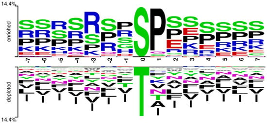
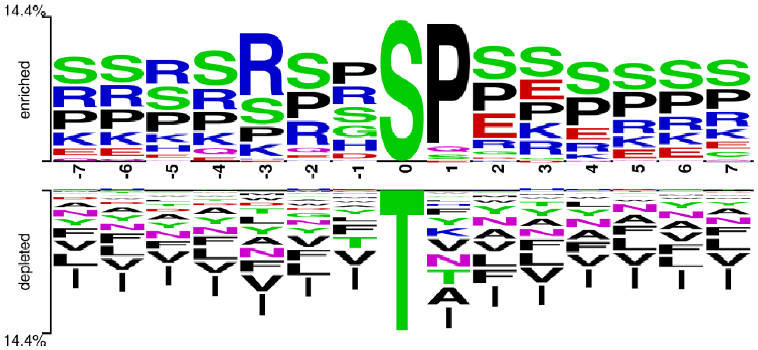
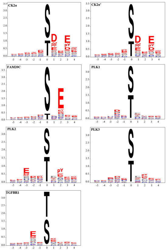
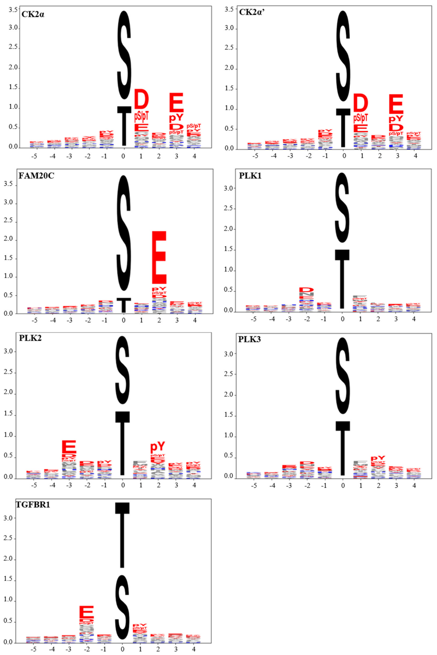
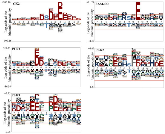
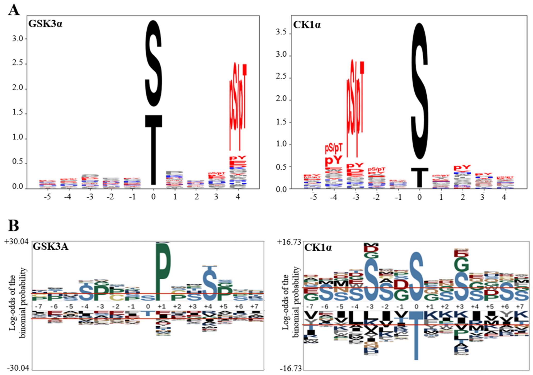
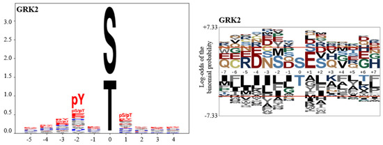
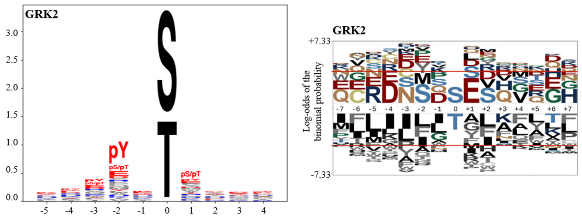
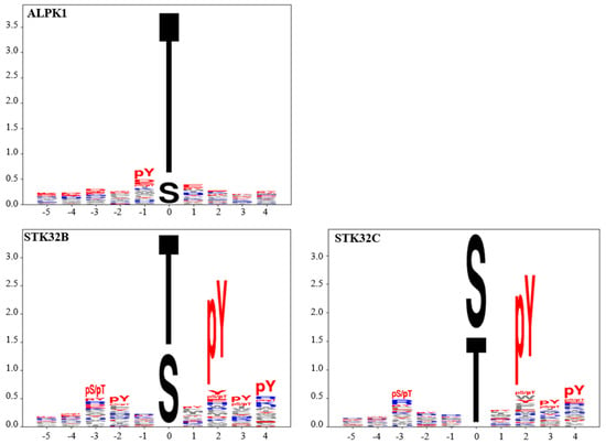
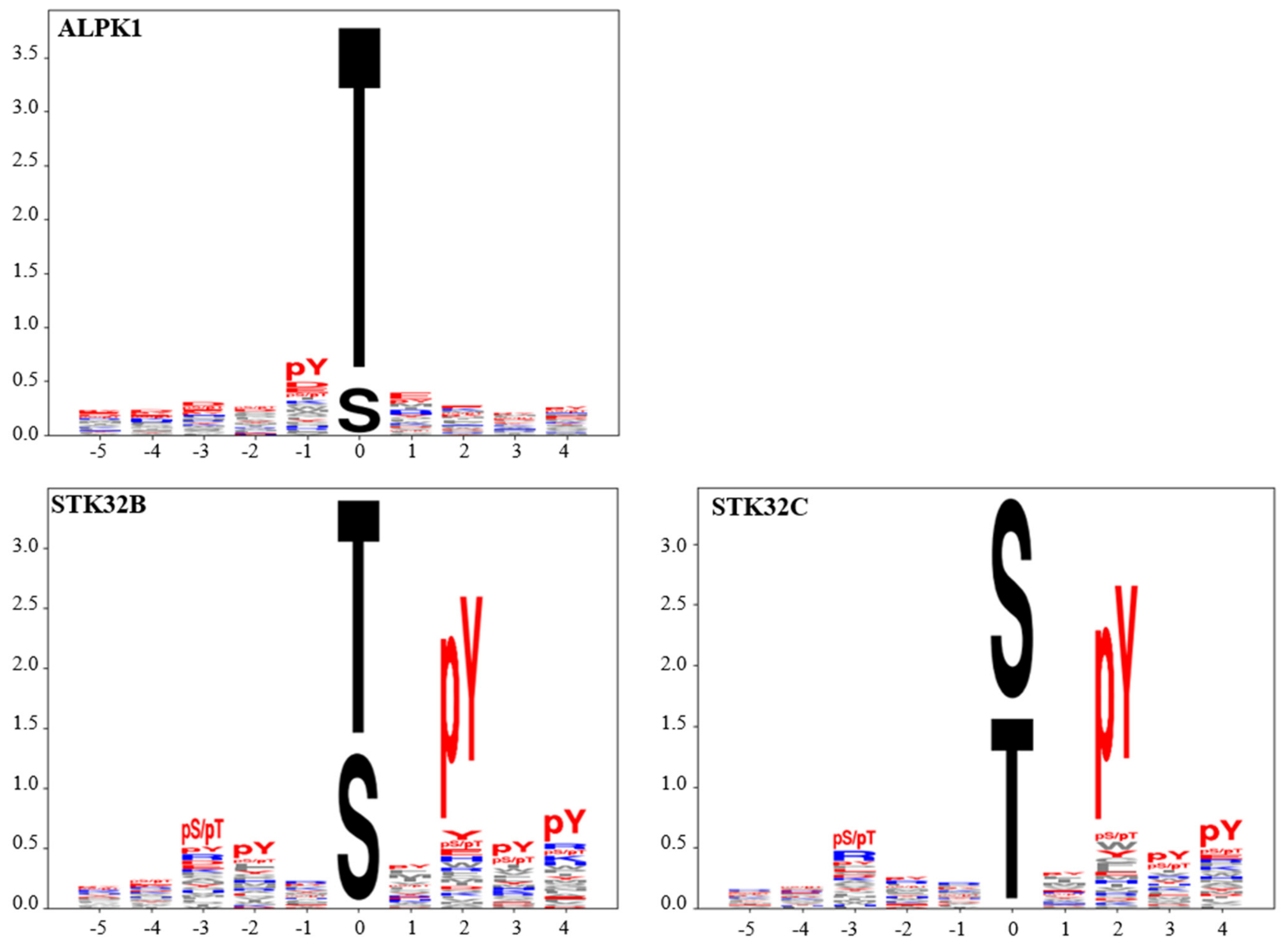

Disclaimer/Publisher’s Note: The statements, opinions and data contained in all publications are solely those of the individual author(s) and contributor(s) and not of MDPI and/or the editor(s). MDPI and/or the editor(s) disclaim responsibility for any injury to people or property resulting from any ideas, methods, instructions or products referred to in the content. |
© 2026 by the authors. Licensee MDPI, Basel, Switzerland. This article is an open access article distributed under the terms and conditions of the Creative Commons Attribution (CC BY) license.