


Abstract
One mechanism contributing to insulin resistance is the serine/threonine phosphorylation of critical proteins involved in insulin signaling, particularly the insulin receptor, its downstream substrates, and the activation of protein tyrosine phosphatases. These modifications are associated with decreased insulin signaling, leading to insulin resistance. Protein kinase C (PKC), a family of serine/threonine kinases activated by lipids and/or Ca2+, is essential for regulating metabolism and maintaining cellular homeostasis. These kinases regulate glucose uptake and lipid storage, thereby influencing energy balance through tissue-specific mechanisms. However, dysregulation of PKC activity is closely associated with the development of metabolic syndrome (MetS), a cluster of interconnected physiological and biochemical alterations, including insulin resistance, lipid dysregulation, chronic inflammation, hypertension, and obesity, all of which increase the risk of cardiovascular disease (CVD) and type 2 diabetes (T2D). Notably, alterations in PKC signaling can impair insulin’s ability to facilitate glucose uptake and storage and disrupt lipid storage mechanisms, thereby exacerbating insulin resistance. These dysfunctions significantly contribute to the pathophysiology of MetS. This review aims to examine PKC activation under normal physiological conditions, with particular emphasis on its role in insulin signaling, and to explore how PKC dysregulation participates in the pathophysiology of insulin resistance and MetS.
1. Introduction
In recent decades, metabolic disorders have become progressively prevalent, with insulin resistance playing a central role in the pathophysiology of metabolic syndrome (MetS) [1]. Insulin resistance is characterized by excessive insulin production, which is required to maintain glucose homeostasis. At the molecular level, it involves the impairment of signaling pathways downstream of the insulin receptor (InsR) [2,3]. This disruption of signal transduction from the cell surface to the interior alters physiological responses by affecting the molecular processes at effector sites. Among the most common alterations observed in insulin resistance are a reduction in both the number and catalytic activity of InsRs [4]; an increase in serine/threonine (Ser/Thr) phosphorylation on InsR [5] and its substrate (IRS) [6]; an increase in the activity of tyrosine phosphatases, mainly protein tyrosine phosphatase 1B (PTP1B) [7], which contributes to the dephosphorylation of the receptor and IRS; a decrease in the activity of phosphatidylinositol 3-kinase (PI3K) and Akt [8]; and defects in the expression and function of the insulin-dependent glucose transporter type 4 (GLUT4) [9]. These alterations collectively reduce glucose uptake in the muscle, liver, and adipose tissue, thereby altering metabolism [8,10].
In the context of physiological regulation of the insulin signaling pathway, the activation of various isoforms of the protein kinase C (PKC) family has been reported in different cell types, including insulin-sensitive tissues [11]. Some PKC isoforms positively regulate insulin action by promoting the translocation of glucose transporters (GLUTs) [12] or negatively regulating them through Ser/Thr phosphorylation of InsR/IRS [13]. Ser/Thr phosphorylation is the most common post-translational modification of signal transduction proteins, particularly those involved in desensitization processes [14]. Phosphorylation of Ser/Thr residues in key proteins involved in insulin signaling has emerged as a critical mechanism underlying insulin resistance. Ser/Thr hyperphosphorylation of InsR and IRS proteins disrupts Tyr phosphorylation and reduces their interaction with essential protein substrates, thereby impairing insulin signaling [15]. Furthermore, studies have shown that phosphorylation of Ser/Thr residues in InsR and IRS may accelerate their degradation [16]. Although several protein kinases (e.g., c-Jun N-terminal kinase (JNK), inhibitor of k-B kinase (IKK), PKC, and Akt/mammalian target of rapamycin (mTOR)/ribosomal S6 kinase (S6K)) [13] have been identified as key regulators of insulin signaling under the influence of multiple insulin resistance inducers, PKC has been consistently implicated in the development of metabolic disorders, including insulin resistance, MetS, and type 2 diabetes (T2D). In this review, we examine the key molecular aspects of PKC’s involvement in the development of insulin resistance and MetS. We also examine current therapeutic strategies targeting PKC-related pathways and summarize the available evidence from preclinical studies and clinical trials, highlighting both their potential and limitations as therapeutic approaches for MetS-related diseases and insulin resistance.
2. PKC Overview
PKC is a member of the Ser/Thr kinase family, which is activated by external signals and subsequently phosphorylates various target molecules. This PKC-mediated phosphorylation is crucial for maintaining cellular homeostasis and regulates processes such as proliferation, survival, differentiation, apoptosis, and metabolism [17]. PKC was first identified in the late 1970s by Nishizuka et al. [18], coinciding with the discovery of other significant protein kinases, including protein kinase A (PKA) and protein kinase G (PKG). These kinases were later classified as members of the AGC family of kinases [19]. Members of the AGC kinase family share key structural features, including a highly conserved catalytic domain-containing motif required for ATP/substrate binding and catalysis, as well as a regulatory domain that keeps the enzymes in an inactive state [17,20].
In mammals, the PKC family consists of 9 genes encoding 10 distinct isoforms. These isoforms share a common structural organization, featuring a poorly conserved N-terminal regulatory domain and a highly conserved catalytic domain at the C-terminus, which is connected by a hinge region [17,21]. They can be classified into three subfamilies based on their structure, particularly their N-terminal regulatory domains, and functional characteristics [22]: classical or conventional PKCs (cPKCs), novel PKCs (nPKCs), and atypical PKCs (aPKCs). cPKCs (α, alternatively spliced βI, βII, and γ) are activated by diacylglycerol (DAG) and Ca2+ and contain four conserved domains: C1, C2, C3, and C4. The C1 domain consists of two tandem subdomains rich in cysteines, known as C1A and C1B, which bind DAG, phorbol esters, and phosphatidylserine. The C2 domain is responsible for Ca2+ binding, whereas the C3 domain contains a sequence that binds ATP. Finally, the C4 domain contains a substrate-binding site and exhibits phosphotransferase activity (Figure 1) [17,23]. nPKC isoforms (δ, ε, η, and θ) are lipid-dependent kinases that require DAG and phosphatidylserine and respond to phorbol esters [17,23]. Furthermore, these isoforms possess a non-Ca2+-binding C2-like domain at the N-terminus of their C1 domains, enabling them to function independently of Ca2+ (Figure 1). aPCKs, which include ζ and ι in humans (also known as λ in rodents), exhibit the most substantial differences among PKC isoforms, with a single atypical cysteine-rich C1 domain that contains phosphatidylinositol 3,4,5-trisphosphate (PIP3) and ceramide-binding domains, thereby facilitating their interaction with the plasma membrane (PM). Moreover, their C2 domains are substituted with the PB1 (Phox/Bem1) domain, facilitating interactions with specific scaffold proteins to enhance kinase activation (Figure 1) [17,23,24].
Figure 1.
Classification of the structural characteristics of PKC isoforms. The regulatory domains of PKC differ among the three subfamilies, while their catalytic domains are largely conserved. Classical or conventional PKCs (cPKCs) contain an autoinhibitory pseudosubstrate (PS) motif, two tandem C1 domains (C1A and C1B) that bind diacylglycerol (DAG), and a C2 domain that binds Ca2+. In contrast, novel PKC isoforms (nPKCs) lack the Ca2+-binding motif and instead include an expanded N-terminal domain that enables them to receive regulatory signals; however, their activity remains dependent on DAG levels. Atypical PKC isoforms (aPKCs) exhibit catalytic activity independent of DAG or Ca2+. Rather, their regulation primarily depends on intracellular localization mediated by interactions with regulatory proteins and the production of phosphoinositides, such as PIP2 and PIP3, which recruit aPKCs to specific cellular membranes. V1–V5 = variable regions. Created with BioRender. Olivares-Reyes, J. A. (2026) (https://www.biorender.com/d00n302).
All PKCs possess a pseudosubstrate (PS) domain located immediately before the C1 domain, which participates in the autoinhibitory mechanism by interacting with the catalytic site, thereby suppressing kinase activity and substrate phosphorylation [25]. Furthermore, PKCs undergo maturation before activation, a process that requires three critical phosphorylation steps. Newly synthesized PKCs translocate to the membrane, where phosphoinositide-dependent kinase 1 (PDK1) phosphorylates the activation loop of the catalytic domain [17,22]. This is followed by two sequential autophosphorylations in the turn and hydrophobic motifs of the carboxy-terminal region of the protein. These phosphorylations are highly conserved within the PKC family, except for aPKC isoforms, in which the C-terminal hydrophobic site is substituted with a glutamic acid residue [17,22]. After maturation, PKCs remain inactive in the cytosol and are ready for activation. Although PKC maturation was initially considered constitutive, subsequent research identified several stimuli that can trigger this process [17].
PKC activation is regulated by complex interactions involving second messengers, scaffold proteins, upstream kinases, and phosphatases at different cellular locations [26]. According to the classical model, upon activation of Gαq-coupled GPCRs, phospholipase C-β (PLC-β) is stimulated at the PM, which leads to the hydrolysis of phosphatidylinositol 4, 5-bisphosphate (PIP2) to generate DAG and inositol-1,4,5-trisphosphate (IP3). IP3 subsequently induces Ca2+ release from the endoplasmic reticulum (ER) through activation of IP3 receptors. The resulting presence of Ca2+ and/or DAG then facilitates the recruitment of both cPKCs and nPKCs to the PM, which serves as the primary site of PKC activation [26]. PKC activation through this mechanism is generally transient because enzymes that degrade DAG, such as DAG kinase, terminate lipid signaling [27]. Consequently, PKC returns to an autoinhibited conformation, thereby terminating the signaling cascade after reducing the second-messenger levels. Moreover, once active, PKCs are susceptible to ubiquitination and undergo activation-induced degradation [17].
Receptor tyrosine kinases (RTKs), including receptors for epidermal growth factor (EGFR), platelet-derived growth factor (PDGFR), vascular endothelial growth factor (VEGFR), and insulin (InsRs), have been shown to activate both cPKCs and nPKCs [28]. This process involves the activation of phospholipase C-γ1 (PLC-γ1) isozymes by RTKs, which depends on the physical interaction between them via the SH2 domains of PLC-γ1 that recognize phosphotyrosine residues in RTKs. Subsequently, RTKs phosphorylate and activate PLC-γ1, enabling it to hydrolyze PIP2 into DAG and IP3 [28].
3. PKC in Metabolism
3.1. PKC and Insulin Signaling
Insulin is an anabolic hormone essential for metabolic regulation by promoting nutrient uptake, storage, and synthesis [29]. Its biological actions are initiated upon binding to InsR, an integral membrane glycoprotein that belongs to the family of receptors with intrinsic tyrosine kinase activity. InsR is a tetrameric protein comprising two pairs of αβ-subunits linked by sulfhydryl bonds (Figure 2). The α-subunits are exclusively extracellular and include domains for insulin-binding, a region rich in cysteines (CRs), and fibronectin type III (FnIII) domains. In contrast, the β-subunits are composed of an extracellular domain containing a FnIII domain, a transmembrane domain, a juxtamembrane (JM) domain, an intracellular tyrosine kinase (TK) domain, and a C-terminal tail [30,31]. Upon insulin binding to the InsR α-subunits, the receptor first undergoes a conformational change in the α-subunit, which is transmitted to the β-subunit. This change activates the receptor’s intrinsic kinase activity, leading to trans-autophosphorylation of Tyr1146, Tyr1150, and Tyr1151 within the kinase regulatory region of the adjacent β-subunit. The increased kinase activity then enables phosphorylation of additional Tyr residues: Tyr953 and Tyr960 in the JM domain and Tyr1316 and Tyr1322 in the carboxy-terminal (CT) domain [32,33] (Figure 2).
Figure 2.
Structure and domains of the insulin receptor. The receptor consists of two α-subunits and two β-subunits, which are linked by disulfide bonds that stabilize the dimeric structure. The α-subunits contain a cysteine-rich region (CR) and fibronectin type III domains (FnIII-1 and FnIII-2, shown in green). The β-subunits contain fibronectin type III domains (FnIII-2 and FnIII-3, shown in blue), a single transmembrane (TM) domain, a juxtamembrane (JM) region, a tyrosine kinase (TK) domain, and a carboxy-terminal (CT) domain. Critical Tyr residues are located within the JM, TK, and CT domains, where they undergo autophosphorylation and facilitate protein interactions and signal propagation. Created with BioRender. Olivares-Reyes, J. A. (2026) (https://www.biorender.com/p24w715).
Phosphorylation sites function as protein recognition motifs that enable high-affinity interactions with other protein substrates, which subsequently undergo Tyr phosphorylation to propagate the signal [30]. Among InsR substrates, IRS-1 and IRS-2 are recognized as the principal mediators of metabolic actions, whereas Shc is primarily associated with the growth and proliferative effects of insulin. Both IRS and Shc proteins act as adaptor molecules, organizing molecular complexes that trigger intracellular signaling cascades [29].
The primary metabolic pathway activated by the InsR is the IRS/PI3K/Akt cascade [34]. Activation of this pathway begins with InsR-mediated Tyr phosphorylation of IRS, which promotes IRS association with and activation of PI3K. PI3K subsequently phosphorylates PIP2 to generate PIP3 [35], which recruits Akt and PDK1 [34]. PDK1 phosphorylates Akt at Thr308, while mTORC2 phosphorylates Akt at Ser473 [36]. This dual phosphorylation fully activates Akt, which enhances glucose transport by phosphorylating AS160 (Rab-GAP) and promotes glucose uptake by translocating GLUT4 vesicles to the plasma membrane [37,38].
The mitogen-activated protein kinase (MAPK) represents the second major branch of insulin signaling and mediates the mitogenic effects of insulin [39]. This pathway is initiated when activated InsR interacts with the Shc protein, leading to recruitment of the GRB2-SOS complex. This interaction activates the small G-protein Ras, which acts as a molecular switch. Ras subsequently transduces upstream tyrosine phosphorylation signals into a serine kinase cascade by sequentially activating MAPKs, including Raf, MEK, and extracellular signal-regulated kinase 1/2 (ERK1/2). This signaling cascade is critical for regulating cellular proliferation, division, migration, and apoptosis [29,34].
PKC is another important Ser/Thr kinase that is differentially activated by insulin. Activated InsR can stimulate multiple PKC isoforms across all three PKC subfamilies through mechanisms involving the activation of PLC-γ1 and PI3K [40]. The SH2 domains of PLC-γ1 enhance its interaction with the active InsR, resulting in the hydrolysis of PIP2 into DAG and IP3. IP3-mediated Ca2+ release and DAG promote the activation of conventional PKC isoforms, whereas novel PKC isoforms are activated by DAG in a Ca2+-independent manner (Figure 3A) [40,41].
Figure 3.
Molecular mechanisms of PKC activation by insulin. (A) Activation of the insulin receptor (InsR) stimulates phospholipase C-γ (PLC-γ), leading to the production of inositol trisphosphate (IP3), which releases Ca2+ from the endoplasmic reticulum (ER), and diacylglycerol (DAG). While DAG and Ca2+ are the main activators of classical PKC (cPKC) isoforms, DAG activates novel PKC (nPKC) isoforms. (B) Atypical PKCs (aPKCs) are activated by increased PIP3, which is generated via PI3K activation following insulin pathway activation. PIP3 binds to the basic arginine residues in the pseudosubstrate (PS) sequence of the aPKC regulatory domain, facilitating dissociation of the PS from the catalytic domain. Furthermore, phosphorylation of the aPKC regulatory domain by 3-phosphoinositide-dependent protein kinase-1 (PDK1) further contributes to PKC isoform activation. Created with BioRender. Cruz-Villarreal, D. E. (2026) (https://www.biorender.com/okm51lh).
It has been proposed that, via a mechanism requiring the βγ subunit of the activated heterotrimeric G protein, InsR activates PI3Kγ to generate PIP3 from PIP2 [40]. PIP3 acts as a key signal for recruiting the pleckstrin homology (PH) domain of PLC, which binds to the intracellular surface of the PM. Subsequently, PLC catalyzes the production of IP3 and DAG from PIP3. IP3 activates its receptor, resulting in the release of Ca2+ from the ER [42]. The resulting increase in Ca2+ contributes to GLUT4 vesicle trafficking and fusion with the PM, thereby promoting glucose uptake [42].
Alternatively, aPKC can be triggered downstream of the PI3K/Akt pathway [43]. Upon insulin stimulation, PI3K activation increases PIP3 levels, which bind to basic arginine residues in the PS domain of the aPKC regulatory domain and dissociate the PS from the catalytic domain (Figure 3B) [44,45]. This dissociation enables target proteins to access the active sites. Furthermore, PDK1 phosphorylates aPKC at Ser/Thr residues within the activation loop of the kinase domain, while mTORC2 phosphorylates aPKC at the turn motif and hydrophobic sites in the C-terminal tail [20]. These modifications stabilize the active conformation of aPKCs, thereby facilitating their function in GLUT4 translocation and increasing glucose uptake in response to insulin (Figure 3B) [46].
3.2. PKC and Glucose Transport
One of the most important actions of insulin is the stimulation of glucose uptake into adipocytes and skeletal muscle (SkM), a process that requires GLUT4 translocation to the cell membrane and involves two distinct signaling pathways [47]. The former depends on activation of the IRS/PI3K/Akt pathway, whereas the latter operates through the CAP/Cbl/TC10 protein complex (Figure 4) [37,41,47]. The PI3K/Akt signaling cascade facilitates glucose uptake by inactivating TBC1D1 and AS160 (Rab GAPs) through Akt-mediated phosphorylation, thereby promoting the translocation of GLUT4-containing vesicles to the PM (Figure 4) [47,48,49]. The second mechanism involves the phosphorylation of Cbl, which recruits the adaptor protein CrkII and guanine nucleotide exchange factor (GEF) C3G. Upon phosphorylation of Cbl by InsR, the Cbl–CAP–CrkII–C3G complex translocates to the lipid raft microdomain, where TC10 is located. Once recruited, C3G functions as a GEF for TC10, catalyzing the exchange of GDP for GTP (Figure 4) [47]. Together, these two insulin-activated pathways promote GLUT4 vesicle translocation, a process mediated by membrane trafficking systems [47,48].
Figure 4.
Insulin signaling pathways involved in GLUT4 translocation and the regulatory roles of PKC isoforms. Insulin stimulates GLUT4 translocation to the plasma membrane through the IRS/PI3K/Akt and CAP/Cbl/TC10 pathways. Atypical PKCs (aPKCs), particularly PKCζ and PKCλ, act as positive regulators of insulin-stimulated GLUT4 translocation. In contrast, conventional (cPKCs) and novel (nPKCs) PKCs have been implicated as negative regulators of insulin signaling and glucose uptake. The isoform-specific roles of PKCs contribute to the regulation of insulin sensitivity and glucose transport in skeletal muscle and adipocytes. cPKCs and nPKCs negatively regulate GLUT4 translocation and glucose uptake by inhibiting IRS and Akt signaling. Green arrows represent activation or positive regulation, while red blunt-ended lines denote inhibition mediated by PKC isoforms. Black arrows indicate the canonical processes of each pathway, whereas dashed lines indicate GLUT4 translocation. Created with BioRender. Hernandez-Gonzalez, K. D. (2026) (https://www.biorender.com/bf686lv).
PKC isoforms play key roles in regulating GLUT4 translocation and glucose uptake in insulin-sensitive tissues, thereby influencing metabolic homeostasis. Of these, aPKC isoforms are downstream effectors of the PI3K pathway and are consistently associated with insulin-stimulated GLUT4 translocation to the PM, particularly in SkM and adipocytes (Figure 4) [50,51,52,53,54]. Despite extensive research, the molecular mechanisms underlying aPKC activation, GLUT4 vesicle trafficking, and PM fusion remain unclear [50,55]. Nonetheless, emerging evidence suggests that aPKCs may require TC10 activation for insulin-stimulated GLUT4 translocation, likely because they contribute to microtubule and cytoskeletal remodeling [56]. Supporting this, overexpression of constitutively active PKCλ in adipocytes [52,56] or wild-type PKCζ in muscle in vivo [57] increases both basal and insulin-stimulated glucose transport. Conversely, overexpression of a dominant-negative mutant of PKCλ or PKCζ abolishes insulin-stimulated glucose transport and GLUT4 translocation in adipose and muscle cells [52,58,59]. Of note, experimental evidence suggests that PKCλ and PKCζ isoforms may function interchangeably, with the overexpression of one isoform compensating for the inhibitory effects of a dominant-negative mutant of the other on insulin-stimulated GLUT4 translocation [60]. This functional redundancy suggests a coordinated regulatory mechanism in which both PKCζ and PKCλ contribute significantly to insulin-mediated glucose transport [50]. Collectively, these findings suggest that impaired activation of PKCζ and PKCλ contributes to insulin resistance, highlighting their importance in metabolic regulation. In contrast, cPKC isoforms generally act as negative regulators of the insulin signaling pathway, which promotes GLUT4 translocation to the cell surface in muscle and fat cells. For instance, global deletion of PKCα has been shown to enhance insulin-stimulated glucose transport in SkM, whereas PKCβ knockout increases both basal and insulin-stimulated glucose uptake in isolated soleus muscle, indicating the inhibitory roles of these classical isoforms [61]. Mechanistically, PKCα and PKCβ can phosphorylate IRS-1 at inhibitory sites, thereby attenuating insulin signaling, while PKCα may additionally promote PP2A activation and stability, favoring Akt dephosphorylation and further suppressing downstream signaling [62,63,64]. This implies that cPKC isoforms, particularly PKCα and PKCβ, may act as negative regulators in the complex interplay of insulin-mediated glucose metabolism [11].
In the case of nPKC isoforms, particularly PKCε and PKCη, they have also been implicated in insulin resistance by phosphorylating IRS proteins, thereby impeding downstream signaling events necessary for proper glucose uptake [65]. This differential involvement highlights the complex isoform-specific roles of PKC in the regulation of insulin sensitivity and glucose transport [55,66].
3.3. PKC and Lipid Metabolism
PKC isoforms play significant roles in lipid metabolism, including adipogenesis, lipolysis, and lipogenesis [67]. The predominant PKC isoforms identified in adipocytes are PKCβ, PKCγ, and PKCε, and a significant association between insulin signaling and these enzymes has been documented in adipose cells (Figure 5A) [68]. In the context of adipogenesis, reports indicate that PKCβ is one of the most highly expressed genes during 3T3-L1 adipocyte differentiation [69]. Studies using a dominant-negative form of PKCβI have shown that it inhibits adipogenesis [70]. Other isoforms, such as PKCγ and PKCε, are essential for preadipocyte differentiation [71]. PKCε knockdown inhibits 3T3-F442A cell differentiation without affecting clonal expansion, maintenance, or phenotype [72]. Interestingly, inhibition of PKCε reduces adipocyte differentiation, supporting its role in cell growth regulation. In contrast, overexpression of PKCε (but not other isoforms) drives multipotent NIH-3T3 cells toward adipogenic differentiation in response to hormonal inducers [72]. Additionally, PKCε has been implicated in several metabolic functions, including short-term hepatic lipid accumulation, β-cell lipid partitioning, and regulation of fatty acid (FA) release during fasting [73].
Figure 5.
Insulin-dependent PKC activation regulates lipid metabolism. (A) PKCβ, PKCγ, and PKCε are involved in adipocyte differentiation. (B) PKCβI mediates antilipolytic effects by negatively regulating β-AR/cAMP/PKA signaling. (C) In skeletal muscle, PKCβII promotes lipid accumulation by increasing SREBP-1c activity and reducing the activity of regulators of fatty acid oxidation. (D) In the liver, PKCδ promotes lipogenesis through SREBP-1c activity and upregulation of FAS and ACC expression. AC, adenylyl cyclase; ACC, acetyl-CoA carboxylase; ACOX, acyl-CoA oxidase; β-AR, β-adrenergic receptor; cAMP, cyclic adenosine monophosphate; FA, fatty acid; FAS, fatty acid synthase; HSL, hormone-sensitive lipase; InsR, insulin receptor; PGC-1β, peroxisome proliferator-activated receptor γ coactivator 1β; PKA, protein kinase A; PPARδ, peroxisome proliferator-activated receptor delta; SREBP-1c, sterol regulatory element-binding protein 1c. Black dashed lines indicate different stages of the biological differentiation process. Black dashed arrows indicate intermediate steps of activation/stimulation, whereas red dashed blunt-ended lines indicate intermediate steps of regulation/inhibition. Green upward arrows indicate an increase in the corresponding process or protein expression, whereas red downward arrows indicate a decrease in protein expression. Created with BioRender. Cruz-Villarreal, D. E. (2026) (https://www.biorender.com/n18f1u9).
PKC has also been implicated in the regulation of lipolysis in mature adipocytes. For instance, phorbol ester-mediated PKCβI activation in rat adipocytes inhibits the canonical lipolytic pathway (AMPc/PKA/hormone-sensitive lipase (HSL)/perilipin), presumably through negative regulation of β-adrenergic receptor signaling [74,75]. Similarly, stimulation of InsRs induces sustained activation of cPKC-dependent antilipolytic effects in rat adipocytes (Figure 5B) [74]. Contrary to expectations, PKC activation induces lipolytic effects independent of PKA, potentially via MAPK/ERK1/2-mediated activation of HSL through a PKCε-dependent mechanism [76,77,78]. This hypothesis is supported by the observation that PKCε can mediate phorbol ester-dependent inhibition of the antilipolytic effect of insulin [79]. Additionally, the participation of PKC in the activation of PKA-dependent lipolysis cannot be ruled out, as PKC can induce adenylate cyclase (AC) activity in rat adipocytes, with a subsequent increase in cAMP- and PKA-dependent activation of lipolysis [80,81].
Studies have demonstrated that insulin-induced PKCβII activation, both in vitro and in vivo, maintains de novo lipogenesis by regulating the expression and activity of sterol regulatory element-binding protein 1c (SREBP-1c), a key modulator of lipid biosynthesis. Specific overexpression of a constitutively active PKCβII mutant in mouse SkM revealed its effects on the regulation of several genes implicated in lipid metabolism [67,82], decreasing the expression of transcriptional regulators of genes involved in FA oxidation (such as peroxisome proliferator-activated receptor-δ, PGC-1β, and acyl-CoA oxidase) and lipolysis (including HSL), but enhancing the expression of SREBP-1c in SkM. Both effects are associated with reduced lipid oxidation and increased lipid accumulation in the muscle cells (Figure 5C) [82].
PKCδ is another isoform that is associated with lipogenesis. Studies conducted in PKCδ-deficient mice showed reduced expression of SREBP1c and its downstream targets, including FA synthase and acetyl-CoA carboxylase [83,84]. In contrast, overexpression of PKCδ in the liver increased the expression of these proteins, consistent with the previously reported lipogenic actions of PKCδ [83]. Previous research has demonstrated that insulin increases PKCδ activity and promotes its Tyr phosphorylation (Figure 5D) [85].
4. Regulation of Insulin Signaling
Two regulatory mechanisms control insulin’s metabolic effects and its ability to promote cell growth and proliferation. The first is homologous regulation, where enzymes activated by insulin pathways suppress the activity of key signaling proteins. The second is heterologous regulation, which involves homeostatic molecular processes unrelated to insulin activation that inhibit insulin signaling pathways [31]. Together, these mechanisms play an important role in maintaining cellular balance, thereby influencing the extent and duration of insulin signaling and its effects [31,86].
The key mechanisms identified in insulin regulation include: (a) Ser/Thr phosphorylation of receptors and IRS proteins, which disrupt protein interactions or promote their degradation [5,32,87,88]; (b) endocytic trafficking of receptors, resulting in a reduced number of InsRs on the cell surface [89]; (c) InsR/IRS association with specific proteins, such as SOCSs and PC-1 [90,91], which inhibit the formation of insulin signaling complexes; (d) dephosphorylation of phospho-Tyr and phospho-Ser/-Thr residues on activated proteins in the insulin signaling pathway, including InsR, IRS, and Akt [92,93]; and (e) genetic regulation of proteins involved in insulin signaling, such as GLUT4 and InsRs [94,95].
Among the mechanisms described above, phosphorylation at Ser/Thr residues constitutes the triggering mechanism for pathway regulation. For instance, through Akt-mediated activation of the mTORC1 complex, homologous desensitization is achieved by phosphorylation of Ser/Thr residues in IRS proteins, thereby decreasing Tyr phosphorylation by InsR [96].
Another mechanism in which phosphorylation of Ser/Thr residues plays a key role is receptor internalization, or endocytosis. Endocytosis is essential for maintaining InsR availability on the cell surface and for modulating downstream signaling pathways [97]. The endocytic trafficking of InsR determines whether the receptor is degraded or recycled back to the plasma membrane, thereby influencing both receptor availability and the duration of signaling. It has been well established that the kinase activity of InsR is essential for receptor internalization, indicating that InsR endocytosis usually occurs after receptor activation [89]. InsR endocytosis can occur via both clathrin-dependent and clathrin-independent mechanisms. In clathrin-dependent endocytosis, ERK1/2 activation leads to its subsequent recruitment to IRS-1 through specific Tyr-phosphorylated residues (Tyr612, Tyr632, and Tyr662 within YXXΦ motifs, where X denotes any amino acid and Φ represents hydrophobic amino acids). This process facilitates subsequent Ser phosphorylation of IRS-1 by ERK1/2 (at Ser616, Ser636, and Ser666, which are located immediately after the YXXΦ motifs). These phosphorylations generate sites for the Tyr phosphatase SHP2. SHP2-mediated dephosphorylation of the pYXXΦ motifs results in a switch from phospho-Tyr to phospho-Ser/Thr. Only IRS proteins containing phospho-Ser/Thr can interact with AP2 and trigger clathrin-dependent IR endocytosis (Figure 6A) [97,98,99].
Figure 6.
Regulation of insulin signaling by feedback mechanisms. (A) Activated insulin receptors (InsRs) undergo internalization via clathrin-coated pits mediated by adaptor proteins such as AP2 and dynamin. Once the receptor–ligand complex is internalized into early endosomes, it can follow the recycling pathway, in which the receptor is dephosphorylated and returned to the plasma membrane, allowing further insulin binding and sustained signaling. Alternatively, in the degradation pathway, the receptor is directed to lysosomes, where it undergoes degradation, thereby reducing receptor availability and attenuating insulin signaling. (B) Besides these trafficking events, Ser/Thr phosphorylation of key proteins, such as IRS, plays a crucial role in modulating the signaling pathway. Activation of protein phosphatases (PHLPP and PP2A) and lipid phosphatases (PTEN and SHIP1), which dephosphorylate Akt and PIP3, respectively, further modulate the insulin signaling cascade. Black solid arrows indicate a series of steps, whereas black dashed arrows indicate intermediate steps or events not shown in the activation/stimulation pathway. Red blunt-ended lines indicate inhibition, whereas red solid arrows indicate Ser/Thr phosphorylation. Created with BioRender. Cruz-Villarreal, D. E. (2026) (https://www.biorender.com/r74a597).
Clathrin-independent endocytosis is characterized by the phosphorylation of caveolin at Tyr14 by activated InsR, which promotes internalization through a clathrin-independent pathway. Caveolin, functioning as a scaffold protein, interacts with InsR and induces membrane curvature within microdomains composed of lipid rafts enriched in cholesterol and sphingolipids, thereby facilitating caveolar endocytosis [89].
Alternative mechanisms involve protein tyrosine phosphatases (PTPs), which modulate insulin signaling by dephosphorylating the active form of InsR during endocytosis [100]. Furthermore, Ser/Thr phosphatases, such as protein phosphatase 2A (PP2A) and PH domain and leucine-rich repeat protein phosphatases (PHLLP), are involved in the dephosphorylation of Akt, thereby diminishing its activity [101]. Lipid phosphatases, including PTEN and SHIP2, act on PIP3, converting it back to PIP2 (Figure 6B) [102,103].
Signaling pathways activated by insulin are regulated through feedback loops, with Ser/Thr kinases playing crucial roles in this process. IRS-1 and IRS-2 are key proteins in insulin signaling and primary targets of regulation [87]. Phosphorylation of IRS-1 at Ser residues, mediated by kinases such as Akt (Ser265, Ser267, Ser302, Ser325, Ser358, Ser522, Ser612) [104,105], PKCζ (Ser498, Ser570) [105,106], ERK1/2 (Ser267, Ser612, Ser632) [87,105], mTOR (Ser267, Ser307, Ser318, Ser612, Ser632, Ser662, Ser731) [104,105], and S6K (Ser265, Ser302, Ser307, Ser522) [87,107], attenuates IRS signaling and promotes its proteasomal degradation (Figure 6B).
5. PKC in Metabolic Syndrome
MetS comprises a cluster of conditions that includes insulin resistance, hypertension, dyslipidemia, and central obesity [1]. MetS is associated with an increased risk of developing T2D, cardiovascular diseases (CVDs), and other adverse health outcomes [108,109]. The increasing prevalence of MetS is driven by multiple factors, particularly a sedentary lifestyle and a high-calorie diet. Several studies have shown a significant role of PKC in the pathophysiology of MetS, particularly through its involvement in insulin resistance, obesity, and inflammation [110,111,112,113]. PKC isoforms can detect the DAG generated in different cellular compartments during various physiological processes [67]. DAG accumulates in multiple organs of individuals with obesity, thereby disrupting metabolic homeostasis and contributing to the development of MetS and associated disorders [60]. Research indicates that abnormal PKC activation plays a significant role in the onset of metabolic disorders. Thus, c/nPKC isoforms, which respond to Ca2+/DAG and DAG, respectively, may represent potential therapeutic targets for treating several of the conditions associated with MetS [61].
5.1. PKC in Insulin Resistance
Insulin resistance is defined as the diminished responsiveness of cells to insulin. It is characterized by three main effects: (a) reduced insulin-stimulated glucose transport and metabolism in the SkM, (b) reduced insulin-mediated suppression of lipolysis in adipose tissue, and (c) impaired ability of insulin to store glucose and inhibit its production in the liver [31,114,115]. At the molecular level, insulin-deficient signaling results from several alterations, including mutations and/or post-translational modifications in the InsR, IRS, and downstream effectors. Insulin resistance is commonly associated with several alterations, including a reduction in both the number and catalytic activity of InsRs, increased Ser/Thr phosphorylation in InsR and IRS, and elevated Tyr phosphatase activity, particularly PTP1B, which contributes to the dephosphorylation of receptors and IRS [7,31,60]. Additionally, there is a decrease in PI3K and Akt kinase activity, as well as impairment in GLUT4 expression and function [31]. These modifications diminish insulin-mediated metabolic responses, affecting glucose, amino acid, and lipid metabolism and resulting in significant metabolic alterations [114]. Several insulin resistance inducers have been identified, including hyperinsulinemia, pro-inflammatory cytokines [116], free fatty acids (FFAs) [117], ER stress, oxidative stress [118,119], and obesity [114]. In every instance, Ser/Thr phosphorylation of critical insulin signaling proteins, particularly the InsR and IRS proteins, has been implicated as a common mechanism that negatively regulates signaling, thereby contributing to the development of insulin resistance [120].
In the 1990s, multiple research groups conducted comprehensive analyses of potential phosphorylation sites on InsR, identifying several Ser/Thr residues within the cytosolic region of the receptor β-subunit. Most of these sites were implicated in receptor regulation and were found to influence insulin signaling [121,122,123,124,125,126,127]. In this context, evidence indicates that Ser/Thr phosphorylation of InsR modulates its autophosphorylation and kinase activity following insulin binding, a modification commonly observed in obesity and glucose toxicity in both rodents and humans. While several Ser/Thr kinases, such as PKA, JNK, and p38 MAPK, have been shown to phosphorylate InsR and reduce its activity, studies have identified PKC as the primary kinase responsible for receptor phosphorylation, targeting various intracellular regions of the β-subunit [32,121,122,127,128].
IRS-1 and IRS-2 have been identified as principal targets of multiple Ser/Thr kinases implicated in insulin resistance. Ser/Thr phosphorylation of IRS proteins is a critical regulatory mechanism in insulin signaling in both physiological and pathological contexts [129]. More than 50 potential Ser/Thr phosphorylation sites have been proposed for these proteins, and phosphorylation at these sites functions as a negative feedback mechanism in insulin signaling [130,131]. In this context, previous studies revealed that the IRS-1 protein specifically contains 40 Ser/Thr residues; in contrast, the IRS-2 protein contains 19 Ser/Thr residues that can be phosphorylated by the action of several Ser/Thr kinases, including mTOR/S6K1, IKKβ, MAP kinases, JNK, GSK3, AMPK, and several PKC isoforms [129,131]. Increased phosphorylation of IRS proteins at specific Ser/Thr residues has been observed in models of insulin resistance and T2D [132,133]. From these studies, it has been established that elevated Ser/Thr IRS phosphorylation may inhibit insulin signaling by several mechanisms: (1) downregulation of IRS protein levels via ubiquitination [86], (2) inhibition of the interaction between Tyr-phosphorylated InsR and the phosphotyrosine-binding (PTB) domain of IRS-1 [129], or (3) interference with the insulin-stimulated interaction between IRS-1 and the p85 regulatory subunit of PI3K [106,134].
Given the distinct roles of PKC isoforms in cellular regulation and their established association with metabolic dysregulation, it is crucial to elucidate their specific actions in major metabolic tissues. Table 1 summarizes the effects of classical, novel, and atypical PKC isoforms on SkM, liver, and adipose tissue. It delineates their mechanisms of action in these tissues, emphasizing their contributions to impaired insulin signaling, altered lipid metabolism, and disrupted glucose uptake, thereby providing insights into their roles in metabolic homeostasis and the pathogenesis of insulin resistance and T2D.
Table 1.
Roles of PKC isoforms in insulin resistance across specific tissues.
5.1.1. Regulation of the Insulin Receptor and IRS by PKC
Numerous protein targets, including those involved in insulin signaling, have been identified as substrates for PKC phosphorylation. Studies have shown that c/nPKCs are associated with InsR and IRS Ser/Thr phosphorylation under insulin resistance, leading to decreased insulin activity (Figure 7) [83,122,152,160].
Figure 7.
PKC isoforms and phosphorylation sites involved in the regulation of insulin signaling. Isoforms from the conventional, novel, and atypical PKC families phosphorylate key components of the insulin signaling pathway, including the InsR and IRS-1. The figure displays selected phosphorylation sites and their corresponding PKC isoforms, highlighting major inhibitory mechanisms, including PKCε-mediated phosphorylation of InsR at Thr1160/1150 and PKCζ-mediated phosphorylation of IRS-1 at Ser323/318. Overall, these modifications predominantly impair insulin signaling and contribute to insulin resistance. The region within human IRS-1, located immediately after the phosphotyrosine binding (PTB) domain (Ser307, Ser312, Ser315, and Ser323), is phosphorylated by kinases associated with inflammation, stress, and obesity in models of insulin resistance and T2D. Atypical PKCζ, a downstream target of PI3K, inhibits the interaction between InsR and IRS-1 during prolonged insulin stimulation by phosphorylating human/mouse IRS-1 at Ser323/318, which affects its downstream interaction with PI3K. Red blunt-ended lines indicate inhibition mediated by PKC isoforms, and dashed arrows indicate indirect regulation. Black phosphorylation sites indicate potential PKC phosphorylation sites identified in all three domains of InsR. Created with BioRender. Hernandez-Gonzalez, K. D. (2026) (https://www.biorender.com/iz7mbav).
Additionally, research indicates that PKC isoforms can regulate protein transcription, thereby affecting the expression and subcellular localization of critical proteins, including InsR and GLUT4 [55,56,113]. In most cases, PKC activation under insulin resistance depends on DAG generation, which is triggered by increased FA levels in obese and insulin-resistant models [161]. Upon activation by FA, PKCα, PKCβ, and PKCδ directly participate in the inhibitory phosphorylation of InsR/IRS or activate JNK or IKK, leading to the subsequent Ser/Thr phosphorylation of IRS, while FA-activated PKCε translocates to the nucleus, where it suppresses the transcription of InsRs by phosphorylating and inhibiting HMGA1 [113]. Conversely, excessive activation of aPKCs in the liver promotes hepatic glucose production, lipid synthesis, and the expression of inflammatory mediators, which can lead to insulin resistance [162]. In this context, research has demonstrated that under T2D conditions, PKC becomes hyperactivated, and hyperglycemia-induced intracellular and extracellular alterations result in modifications in signaling pathways [162].
Regulation of Insulin Receptor
Multiple research groups reported initial evidence of PKC involvement in InsR phosphorylation and regulation of insulin signaling in the 1980s [163,164,165,166]. Their investigations demonstrated that phorbol esters triggered InsR phosphorylation of Ser/Thr residues while simultaneously inhibiting insulin-induced receptor autophosphorylation, InsR Tyr kinase activity, and insulin action [163,164,165]. To date, several potential PKC phosphorylation sites have been identified in all three domains of InsR: JM (Ser944, Ser967, and Ser968), catalytic (Ser994, Ser1006, Ser1035, Ser1037, and Thr1160), and C-terminal domains (Ser1288, Ser1305, Ser1306, Ser1321, Ser1327, and Ser1348) (Figure 7) [32,167,168].
Research suggests that DAG-induced activation of PKCε and the subsequent inhibition of InsR kinase activity contribute to hepatic insulin resistance. However, the specific molecular interaction between PKCε and InsR remains unclear [169,170]. Petersen et al. [171] demonstrated that DAG-dependent activation of PKCε leads to phosphorylation of human InsR at Thr1160 within the kinase activation loop, thereby destabilizing its active conformation and inhibiting InsR kinase activity (Figure 7). In vitro and cellular studies have established that phosphorylation at Thr1160 is both necessary and sufficient for PKCε-mediated inhibition of InsR kinase activity. The physiological significance of this mechanism is further supported by the finding that phosphorylation of InsR Thr1150, the corresponding residue in mice, is essential for acute lipid-induced hepatic insulin resistance [171]. These results offer mechanistic insights into the controversial relationship between hepatic lipids and insulin resistance [171,172].
Furthermore, a recent study by Xu et al. reported that inhibition of de novo ceramide synthesis, either through myriocin treatment (a potent, non-specific inhibitor of serine palmitoyltransferase) or knockdown of dihydroceramide desaturase, conferred protection against lipid-induced hepatic insulin resistance independently of C16 ceramide concentration [160]. These ceramide synthesis inhibitors prevent the accumulation of the sn-1,2-DAG stereoisomer in the plasma membrane, thereby avoiding PKCε activation and protecting against lipid-induced hepatic insulin resistance through the plasma membrane sn-1,2-DAG-PKCε-InsR-Thr1150 phosphorylation pathway [160].
Regulation of IRS
The regulation of IRS-1 and IRS-2 involves multiple mechanisms, including Ser/Thr phosphorylation in their tail regions [87,173]. Among the kinases crucial to this regulatory process, PKC has been identified as a key regulator. Isoforms such as PKCα, PKCβ, PKCδ, PKCε, PKCθ, and PKCζ have been consistently implicated in both homologous and heterologous regulation of IRS proteins [87,129].
Woo et al. recently identified a region within human IRS-1, located immediately after the PTB domain, that contains several Ser phosphorylation sites, specifically Ser307, Ser312, Ser315, and Ser323. These sites are phosphorylated by kinases associated with inflammation, stress, and obesity in models of insulin resistance and T2D (Figure 7). This region is termed the PTB-phosphorylation insulin resistance (PIR) domain (PTB-PIR domain) [16]. The study demonstrated that the IRS-1 PTB-PIR domain interacts with InsR in the absence of Ser phosphorylation, whereas this interaction is disrupted upon phosphorylation. Surface plasmon resonance analyses confirmed that the PTB-PIR domain displayed increased binding affinity to InsR compared to the PTB domain alone. Phosphorylation of Ser307, Ser312, Ser315, and Ser323 within the PTB-PIR domain inhibits binding to InsR (Figure 7) [16]. Insulin-responsive cells expressing IRS-1 mutants in which all four Ser residues were substituted with glutamates to simulate phosphorylation showed reduced phosphorylation of Akt, IRS-1, and InsR compared to wild-type IRS-1 [16]. Hydrogen–deuterium exchange mass spectrometry experiments indicated that the PIR domain interacts with the N-terminal lobe and hinge regions of the InsR kinase domain, suggesting that the IRS-1 PIR domain protects InsR dephosphorylation by PTP1B [16].
Previous research has shown that PKC phosphorylates Ser307 and Ser318 within the PTB-PIR domain. For instance, elevated plasma FA levels increase intracellular DAG and fatty acyl-CoA concentrations, which activate PKCθ. This results in increased phosphorylation of IRS-1 at Ser307, leading to reduced Tyr phosphorylation of IRS-1 and IRS-1-associated PI3K activity. This sequence of events causes diminished insulin-stimulated glucose transport [174]. Furthermore, atypical PKCζ, a downstream target of PI3K, inhibits the interaction between InsR and IRS-1 during prolonged insulin stimulation by phosphorylating human/mouse IRS-1 at Ser323/318 (Figure 7) [106,175].
5.2. PKC Involvement in Lipid-Induced Skeletal Muscle Insulin Resistance
Insulin resistance in SkM is a fundamental step in the metabolic dysregulation associated with physical inactivity and obesity, which contributes to the development of MetS. Current evidence indicates that insulin resistance in SkM is mainly due to lipid accumulation in muscle cells, particularly via intermediate metabolites, such as DAG and ceramides. These two lipid metabolites are essential activators of PKC. DAG is generated by the breakdown of phospholipids, especially PIP2, through PLC or by de novo synthesis through the esterification of excess long-chain fatty acyl-CoA into glycerol-3-phosphate. DAG acts as a precursor for triacylglycerides (TAGs) and serves as a significant second messenger in cellular signaling. DAG activates both classical and novel PKC isoforms by interacting with the C1B domain. Elevated concentrations of DAG at the plasma membrane increase the fraction of PKC bound to the membrane and enhance the enzyme’s catalytic rate constant [176,177].
Research conducted in vitro in rodents and, more recently, in humans has shown that increased DAG accumulation can activate PKC, which, in turn, triggers a Ser/Thr kinase cascade, resulting in phosphorylation of Ser/Thr residues on InsR and IRS-1/2. This process reduces InsR/IRS-mediated PI3K activation, ultimately decreasing GLUT4 translocation to the cell membrane. Consequently, increased DAG concentrations may impair insulin-stimulated glucose transport and metabolism, causing insulin resistance in SkM cells [153,172,174,178]. Multiple PKC isoforms have been identified in SkM, including PKCα, PKCβ, PKCδ, PKCθ, and PKCε, with PKCθ being the most abundantly expressed and the main isoform sensitive to DAG in this tissue [61,179]. In this regard, several studies have shown that during acute lipid overload or high-fat diet (HFD) feeding, DAG-mediated PKCθ activation is the leading cause of insulin resistance in SkM [151,174,178,180,181,182,183]. Studies in PKCθ knockout mice have shown protection against lipid-induced insulin resistance during lipid infusion–glucose clamp studies [184,185]; however, the PKCθ targets responsible for this phenotype have not been fully elucidated. One of the critical substrates of PKCθ that is consistently identified as a target in insulin resistance is IRS-1, which is phosphorylated at Ser1101 [152]. Current evidence further supports the role of PKCθ activation in the pathogenesis of lipid-induced SkM insulin resistance in humans. This is associated with a transient increase in total and cytosolic DAG content, which correlates with PKCθ activation, increased IRS-1 Ser1101 phosphorylation, and inhibition of insulin-stimulated IRS-1 Tyr phosphorylation and Akt activation in the muscles of patients with T2D compared with non-diabetic subjects [151].
Recent research underscores the importance of the subcellular localization of DAGs in PKC activation and the development of insulin resistance [150,186,187]. Specifically, the accumulation of plasma membrane-associated sn-1,2-DAG stereoisomers, rather than total DAG content, demonstrated the strongest association with the activation of PKCε and PKCθ, as well as with insulin resistance in SkM from mice fed a HFD [187]. Furthermore, the study observed an increase in InsR-Thr1150 phosphorylation (mediated by PKCε) and a decrease in insulin-stimulated InsR-Tyr1162 phosphorylation and IRS-1-associated PI3K activity (mediated by PKCθ) (Figure 8) [187].
Figure 8.
Contribution of PKC isoforms to insulin resistance and metabolic syndrome. Excessive DAG production and altered phospholipid metabolism result in abnormal PKC activation, which exacerbates inflammatory and metabolic disorders. Adipose tissue, liver, and SkM are the primary tissues affected by obesity. In these tissues, dysregulated PKC induces Ser/Thr phosphorylation of InsR and IRS, leading to insulin resistance and MetS. In the liver, insulin resistance is predominantly associated with PKCε; in SkM, it is linked to PKCθ; and in adipocytes, PKCα and PKCβ are the main contributors to insulin resistance. Additional PKC isoforms may inhibit InsR/IRS and downstream signaling proteins in each tissue type. As illustrated, PKC isoforms regulate distinct processes across these tissues. In adipose tissue, PKC activity increases inflammation and lipolysis, promoting FA release to other tissues, including the liver and SkM. Elevated FA levels subsequently increase DAG concentrations in the liver and SkM, further activating PKC in these tissues. In the liver, this activation enhances hepatic glucose production and de novo lipogenesis, increasing the risk of hepatic steatosis. In SkM, PKC activation reduces glucose uptake and glycogen synthesis. DAG, diacylglycerol; InsR, insulin receptor; IRS, insulin receptor substrate; MetS, metabolic syndrome; FA, fatty acid; PKC, protein kinase C; SkM, skeletal muscle. Black solid arrows represent direct activation/stimulation, whereas black dashed arrows indicate intermediate steps in the activation/stimulation pathway. Red dashed blunt-ended lines indicate intermediate steps of inhibition. Black upward arrows indicate an increase in the corresponding process, whereas black downward arrows indicate a decrease in the corresponding process. Created with BioRender. Cruz-Villarreal D. E. and Hernandez-Gonzalez, K. D. (2026) (https://www.biorender.com/gkrtisz).
Besides PKCθ, other nPKC isoforms have been identified as playing important roles in insulin resistance in SkM. In this context, Jani et al. found that male rats fed an obesogenic high-fat sucrose-enriched (HFS) diet caused DAG and ceramide accumulation and activation of PKCδ and PKCθ. This was accompanied by increased expression of cytokine receptors and inflammatory mediators, including nuclear factor-κB (NF-κB), toll-like receptor 4 (TLR4), and tumor necrosis factor-α (TNF-α), along with glucocorticoid signaling in a muscle fiber-type-specific manner [178]. Although similar results on the activation of PKCδ and PKCθ and insulin resistance were found in muscle fibers from female rats under the same HFS diet, a significant difference was identified: no increase in ceramide levels was observed, possibly due to an increase in DGAT2 expression that prevented ceramide synthesis [188]. Furthermore, in rats fed an HFD, SkM showed translocation of PKCε and PKCθ, but not PKCα or PKCζ; this study also found a positive correlation between the content of muscle TAGs and DAG and the translocation of PKCθ [181]. Subsequent studies identified PKCθ and PKCε translocation in obese SkM [189], insulin-resistant [153,190], and/or diabetic rat muscle [161]. In human studies, translocation of both PKCθ and PKCε was elevated in the muscle of individuals with T2D compared to lean controls (Table 1) [149,150,151].
Ceramide is another critical intermediary of FA metabolism that contributes to PKC activation and insulin resistance, particularly through inhibition of Akt. However, their contribution is more variable and may depend on factors such as muscle fiber type, inflammatory signaling, and the balance of lipid metabolism [178,188]. Ceramides accumulate in cells via two main pathways: hydrolysis of cell membrane sphingomyelin by sphingomyelinase or de novo synthesis from long-chain saturated FAs, such as palmitic acid, which involves a multi-step biosynthetic process occurring in the ER [117]. Several studies have shown that dietary palmitate increases myocellular ceramide levels and decreases insulin sensitivity in SkM cells [191,192]. The increase in ceramide levels is associated with PKCζ activation and promotes its translocation, thereby inhibiting Akt translocation by phosphorylating Thr34 in the PH domain [157,193]. Furthermore, ceramides indirectly suppress Akt activity by enhancing the expression and activation of PP2A, a cytosolic Ser/Thr phosphatase that dephosphorylates Akt [191,194]. Conversely, increased ceramide levels in the plasma membrane inhibit PKCα, PKCε, and PKCθ [195,196,197].
5.3. PKC in Hypertension and Vascular Dysfunction
Hypertension is a multifactorial disorder characterized by sustained elevations in systolic blood pressure exceeding 140 mmHg and diastolic blood pressure exceeding 90 mmHg [198]. The regulation of vascular function and blood pressure is mediated by calcium signaling, the nitric oxide (NO)/NO-sensitive guanylate cyclase/cGMP pathway, and vascular remodeling [199]. Vascular smooth muscle (VSM) cells are central to the regulation of vasoconstriction and vasodilation by modulating cytosolic Ca2+ concentration. Upon vasoconstrictive stimulation, Ca2+ is mobilized from intracellular stores or the extracellular space, resulting in increased cytosolic Ca2+ levels [200]. The binding of Ca2+ to calmodulin activates myosin light chain kinase (MLCK), which promotes contraction. Conversely, dephosphorylation of myosin light chain (MLC) by MLC phosphatase leads to vasodilation [201]. Beyond this regulatory process, contraction is further modulated by calcium sensitization mechanisms, including the PLC/DAG/PKC pathway [199].
Under physiological conditions, PKC promotes the growth, proliferation, and contraction of VSM; however, studies have demonstrated that alterations in the expression or activity of specific PKC isoforms, including PKCα, PKCβI/βII, PKCδ, and PKCε, can disrupt one or more of the physiological control mechanisms that regulate blood pressure, resulting in persistent increases in blood pressure and, consequently, hypertension [202,203,204,205]. The relationship between PKC and hypertension is associated with alterations in the vascular endothelium, extracellular matrix (ECM), and matrix metalloproteinase-mediated vascular remodeling. It may also involve oxidative stress and free radicals, renal hemodynamics, the renin–angiotensin system (RAS), neuronal changes, increased sympathetic activity, vascular inflammation, interactions with inflammatory cytokines, and other metabolic factors [202].
Overactivation of PKC isoforms in the VSM can induce abnormal vasoconstriction and vascular remodeling, thereby increasing vascular resistance and promoting hypertension. In this regard, PKCα overexpression in A7r5 VSM cells has been shown to increase cell proliferation [206]. Furthermore, increased PKCα activity has been shown to promote efficient VSM contraction, contributing to hypertension [205,207]. In addition, PKCε enhances the myofilament force sensitivity to [Ca2+]i in VSM and promotes vasoconstriction associated with hypertension [205,208]. PKCε is further implicated in the molecular mechanisms underlying hypoxia-induced pulmonary vasoconstriction, a process implicated in pulmonary hypertension in mice [209]. The cytoskeletal localization of PKCδ suggests a role in vascular remodeling during hypertension [210,211]. Moreover, activation of PKCδ can trigger cardiomyocyte necrosis and functional impairment following ischemia–reperfusion, thereby impairing cardiac recovery, whereas its overactivation can promote adaptive cardiac hypertrophy [212]. PKCδ is also implicated in pulmonary hypertension, as elevated PKCδ mRNA and protein levels have been observed in VSM cells from spontaneously hypertensive rats (SHRs), suggesting that PKCδ may enhance contraction by modulating BK channels [203]. Inhibition of PKCδ has been shown to restore BK channel activity in SHRs [213].
PKC also plays a key role in regulating blood pressure through the RAS. Within this signaling pathway, angiotensin II (Ang II) induces vasoconstriction through the AT1 receptor, whereas Ang 1–7 mediates vasodilation and lowers blood pressure through the Mas receptor [214]. In rats with salt-induced hypertension, increased expression of the AT1 receptor and PKC has been observed. Infusion of losartan, an AT1 receptor antagonist, reduces PKCγ levels and lowers blood pressure. Similarly, RNAi microinjection produces effects comparable to those of losartan [215]. In hypertensive individuals, PKC inhibitors such as chelerythrine and calphostin C have been shown to mediate the effects of Ang II partially [216]. In rats with ventricular hypertrophy, hypertension induced by constriction is associated with increased activity of PKCε and PKCβI/βII [202,217]. In endothelial cells, PKC can also influence NO synthase (NOS) activity by decreasing NO production through phosphorylation at Thr495 [218]. Additional studies demonstrate that PKCα and PKCδ inhibit endothelial NOS (eNOS) via phosphorylation at Ser1179 [219].
Vascular dysfunction is characterized by increased vascular permeability, dysregulated vascular tone, impaired angiogenesis and vascular repair, altered ECM production, and endothelial dysfunction, all of which contribute to the development of CVDs [220]. Several PKC isoforms have been implicated in vascular dysfunction, particularly in diseases such as diabetes and hypertension. For instance, administration of a PKC inhibitor has shown that PKCβI and PKCβII are associated with vascular dysfunction in diabetic rats [221]. Additionally, hyperglycemia activates several PKC isoforms in retinal tissues, including PKαα, PKCβ, PKCδ, and PKCε [162,221,222], resulting in retinal vascular dysfunction by altering the enzymatic activities of endothelial cells and pericytes [162,223]. In T2D, chronic hyperglycemia promotes DAG synthesis and activates both classical and novel PKC isoforms in VSM and aortic endothelial cells, leading to altered expression of growth factors such as VEGF and PDGF [224], which contributes to vascular dysfunction. In diabetic vasculopathy, activation of PKCα, PKCβ, PKCδ, and PKCε isoforms [225] has been identified in VSM cells [162,226]. Elevated glucose levels have been shown to increase membrane expression of PKCβ and PKCδ, and overexpression of PKCβII reduces insulin-stimulated activation of vascular cells [227].
In the context of MetS and T2D, PKCθ and PKCε play central roles in the development of insulin resistance [228]. Conversely, PKCβ and PKCδ contribute to vascular complications resulting from metabolic disturbances and are considered potential therapeutic targets for diabetic vascular complications [229]. PKCβ is the predominant isoform and has broader effects in T2D, including promoting vascular dysfunction and diabetic atherosclerosis [221,224,230]. While PKCβ mediates both vascular dysfunction/hypertension and MetS-related vascular complications, PKCα is more specifically involved in vascular and cardiac contractility and blood pressure regulation, with an association with heart failure [231]. PKCθ/PKCε remain more specific to metabolic insulin resistance, whereas PKCδ is particularly implicated in diabetic vascular disease and atherosclerosis.
5.4. PKC, Obesity, and Inflammation
Obesity is defined by increased body mass resulting from excessive fat accumulation and is associated with complications characteristic of MetS [232]. Because PKC regulates energy balance and lipid metabolism, including adipocyte differentiation, lipolysis, and lipogenesis, alterations in its activation have been associated with the harmful effects of excess lipids on glucose homeostasis, thereby contributing to insulin resistance, a central feature of obesity [61,233]. Consequently, changes in PKC activity and expression are implicated in obesity and related metabolic disorders [61,113,234].
Obesity-induced metabolic dysfunction, particularly insulin resistance, is largely mediated by aberrant activation of specific PKC isoforms [139]. This activation primarily results from lipotoxicity, where excessive lipid accumulation elevates levels of bioactive lipids, especially DAG and ceramides, which serve as potent PKC activators [108]. PKC activation in the context of obesity disrupts metabolic homeostasis by phosphorylating key insulin signaling proteins on Ser/Thr residues instead of Tyr residues, thereby leading to insulin resistance in hepatic, muscular, and adipose tissues [113]. In SkM, elevated DAG levels, often resulting from increased FA uptake, chronically activate novel PKC isoforms, particularly PKCθ [185]. This activation induces inhibitory Ser/Thr phosphorylation of IRS proteins, thereby uncoupling them from InsR [3]. Consequently, downstream insulin signaling, including Akt activation, is impaired, reducing glucose uptake, decreasing glycogen synthesis, and inducing mitochondrial dysfunction [3,51,113,235]. In the liver, obesity-associated lipotoxicity activates other PKC isoforms, predominantly PKCβ, PKCε, and PKCδ [139,236,237,238]. Notably, hepatocyte-specific impairment of PKCβ has been associated with reduced obesity in aged mice, indicating that PKCβ expression exacerbates the body’s diminished ability to metabolize fat, resulting in the progressive accumulation of visceral adiposity [237]. PKCε activation, driven by DAG, directly contributes to hepatic insulin resistance by phosphorylating IRS-2 at inhibitory Ser residues, thereby disrupting insulin signaling, enhancing gluconeogenesis, and inducing steatosis (Figure 8) [236].
PKC activation in adipose tissue promotes fat accumulation, decreases inhibition of lipolysis, and increases inflammation. Overactivation of PKCβ modifies adipokine secretion, including leptin and adiponectin, which are critical for appetite regulation and energy balance [137]. PKCδ is notably associated with adipocyte inflammation and apoptosis. Inhibition of PKCδ activity enhances metabolic function in adipocytes from obese individuals by reducing inflammation and apoptosis and improving mitochondrial metabolism [143]. Therefore, PKCδ dysregulation in adipocytes contributes to inflammation and insulin resistance in obesity. Remarkably, PKCθ and PKCε are also implicated in insulin resistance, a defining feature of obesity and T2D [113].
Obesity is associated with chronic low-grade inflammation, which substantially contributes to insulin resistance, β-cell dysfunction, and T2D [239,240]. Chronic inflammation in obesity increases cytokine production, contributing to insulin resistance and complications including non-alcoholic fatty liver disease (NAFLD), retinopathy, cardiovascular disease, and nephropathy [241,242]. The underlying mechanisms involve hypertrophic adipocytes and infiltrating immune cells, particularly macrophages, which together drive chronic low-grade inflammation by releasing a cascade of pro-inflammatory cytokines such as TNF-α, IL-1β, IL-6, IL-12, and MCP-1 [242]. In this regard, it has been suggested that PKC plays a significant role in exacerbating insulin resistance and inflammation during chronic low-grade inflammation [243]. PKC isoforms, including PKCθ and PKCε, which are activated under an inflammatory environment caused by an excess of lipids, phosphorylate Ser1101 and Ser307 on IRS-1 directly and indirectly through PKC-mediated JNK and IKKβ activation [113,244,245]. These Ser phosphorylations reduce IRS-1 phospho-Tyr levels, which are associated with decreased Akt/AS160/GLUT4 signaling and impaired glucose uptake in muscle cells [244]. PKCθ also activates the IKKβ/IκBα/NF-κB pathway, leading to NF-κB-mediated expression of pro-inflammatory genes, such as TNF-α, IL6, IL-1β, and MCP-1 [244,246]. The secretion of these cytokines impairs insulin-stimulated glucose uptake through both autocrine and paracrine mechanisms, thereby contributing to insulin resistance and metabolic disorders. Consequently, PKC-mediated intracellular inflammatory processes perpetuate a cycle of chronic inflammation and metabolic stress, further impairing insulin signaling and promoting insulin resistance [247,248].
Obesity induces systemic oxidative stress and increases reactive oxygen species (ROS) levels in adipocytes [249]. Increased intracellular oxidative stress promotes PKCβ activation, likely by altering the zinc-finger conformation in the C1 domain, which relieves autoinhibition and results in a catalytically active enzyme, even in the absence of phospholipids and Ca2+ [234]. Activated PKCβ promotes phosphorylation of p66Shc, a redox-sensitive protein that regulates oxidative stress and mitochondrial function, thereby leading to ROS generation, which reduces mitochondrial oxygen uptake and increases triglyceride accumulation [249,250]. Oxidative stress can also activate several additional PKC isoforms. Thus, the administration of PKCα and PKCβ inhibitors reduces adiposity and improves metabolic parameters in animal models of obesity-induced T2D [139]. In hepatic tissue, PKCδ and PKCε activation enhances gluconeogenesis and lipid accumulation, contributing to metabolic dysfunction in obesity, which is frequently associated with low-grade chronic inflammation and oxidative stress, both of which activate PKC isoforms (Figure 8) [238,251]. Once activated, PKC further increases inflammation and oxidative stress, creating a harmful cycle that exacerbates metabolic dysfunction [234,252]. Consequently, targeting specific PKC isoforms has been proposed as a potential therapeutic strategy for obesity-associated metabolic disorders. However, developing PKC inhibitors is challenging because isoforms have diverse roles across tissues [61]. Despite these challenges, studies in animal models and humans suggest that modulating PKC activity can enhance insulin sensitivity and reduce obesity-related complications [61,137]. Further research is required to elucidate the specific roles of individual PKC isoforms and their therapeutic potential.
Experimental evidence, including genetic knockout studies (e.g., PKCθ in muscle [185] and PKCε in the liver [236]) and therapeutic interventions with specific PKC inhibitors [253], consistently demonstrates the critical role of these isoforms in mitigating diet-induced insulin resistance and metabolic dysfunction [108,139]. Ongoing research further clarifies the complex relationship among lipid metabolism, PKC activation, and metabolic diseases, thereby identifying PKC isoforms as important therapeutic targets [235].
In summary, PKC plays a significant role in the pathophysiology of obesity, primarily through its effects on insulin signaling, adipose tissue function, and inflammation. Understanding the specific mechanisms by which PKC affects obesity may facilitate the development of new therapeutic strategies for managing obesity and related metabolic disorders, especially MetS (Figure 8).
6. PKC in Type 2 Diabetes
Hyperglycemia in individuals with diabetes and obesity results from insufficient insulin production by β-cells and the development of insulin resistance in target tissues and organs. Additionally, elevated blood glucose levels can further exacerbate insulin resistance [131]. Under these hyperglycemic or diabetic conditions, levels of dihydroxyacetone phosphate, an intermediate product of glycolysis, increase and are subsequently reduced to glycerol-3-phosphate, thereby promoting de novo synthesis of DAG and enhancing the activation of both classical and novel PKCs [162].
Multiple studies have evidenced the involvement of different PKC isoforms in pathological changes associated with T2D, particularly regarding the etiology of β-cell dysfunction [254,255]. In this regard, the PKCε isoform has been identified as an important regulator of glucose-stimulated insulin secretion, whose inhibition selectively enhances insulin-amplifying pathways, particularly under conditions of lipid excess [255]. While deletion of PKCε does not disrupt basal glucose-induced insulin secretion, it amplifies secretory responses in islets exposed to exogenous fatty acids [256,257].
On the other hand, in diabetic animal models, elevated DAG levels in the heart, aorta, vasculature, and renal glomeruli are correlated with increased activity of cPKC (α, βI, and βII) and nPKC (δ and ε) isoforms [51,226]. In vascular tissues, PKCβ is upregulated in the endothelium of individuals with diabetes, which directly impairs endothelial insulin signaling, reduces NO bioavailability, and increases oxidative stress [258]. Inhibition of PKCβ restores insulin-mediated eNOS activation and reduces inflammatory signaling such as NF-κB activation, thereby ameliorating endothelial dysfunction, a central factor in diabetic vascular complications [259]. Elevated PKCβ activity specifically disrupts endothelial insulin signaling and alters eNOS activation in diabetes [260]. In diabetic retinopathy, PKCβ activation leads to increased vascular permeability, thickened basement membranes, and neovascularization [3,162,261]. In diabetic nephropathy, PKCβ impairs glomerular blood flow, induces albuminuria, and promotes extracellular matrix expansion, contributing to kidney damage [261,262]. Concurrently, the PKCδ isoform promotes endothelial apoptosis and oxidative modification of low-density lipoproteins, exacerbating vascular permeability and reducing nitric oxide bioavailability [225].
Interestingly, Considine et al. [140] reported that PKCα, PKCε, and PKCζ levels are significantly elevated in the livers of diabetic patients and diabetic rats compared with their non-diabetic counterparts. Furthermore, diabetic rats showed significantly elevated PKCβ levels in hepatic tissue compared with normoglycemic rats. The study further revealed that diabetic rat livers contain higher levels of DAG than their normoglycemic counterparts. Recent studies have also identified a correlation between elevated hepatic DAG and PKCε levels [226,263]. Consequently, DAG modulates PKC activation and synthesis in the diabetic state. These PKC alterations contribute to a cycle of insulin resistance, increased hepatic glucose output, dyslipidemia, endothelial dysfunction, and the progression of microvascular and macrovascular complications [222,229,259,264].
7. Therapeutic Strategies and Clinical Trials
7.1. Therapeutic Strategies
Multiple studies indicate the high feasibility of therapies targeting PKC, given its central role in essential cellular metabolism. Altered PKC activity and expression are consistently observed in insulin resistance and in metabolic and vascular disorders associated with metabolic syndrome and T2D. These observations have led to the development of therapeutic strategies targeting PKC isoforms to inhibit their activation or expression to improve insulin sensitivity, particularly in cases of obesity-induced insulin resistance [162]. Many of these strategies have progressed to different phases of clinical trials [3,162]. Furthermore, distinct isoforms within each PKC subfamily demonstrate therapeutic potential for treating insulin resistance and diabetic complications [162,265].
Current therapeutic strategies targeting PKC isoforms focus on selective inhibition. For instance, dual inhibition of PKCα and PKCβ shows potential for obesity-driven T2D [266,267]. In obese rat models, the dual inhibitor compound 1 (CMPD1) reduces inguinal white adipose tissue mass, decreases inflammatory markers, and restores β-adrenergic signaling [139]. However, the efficacy of PKC inhibition for renal protection is variable: whereas PKCα and PKCβ contribute to decreased nephrin expression and ECM synthesis [268], PKCδ contributes to interlobar artery dysfunction [139]. Moreover, general PKC inhibitors, such as PKC412, have shown limited clinical success due to poor tolerability, highlighting the importance of isoform selectivity [162,266]. The tissue-specific actions of PKC isoforms present challenges, as individual isoforms may have distinct functions in different tissues [229,258]. Consequently, future therapies should prioritize enhanced isoform selectivity, tissue-specific delivery, and targeting of phosphorylation states. Ruboxistaurin (RBX, LY333531), which has demonstrated safety and efficacy in diabetic retinopathy, serves as a foundation for the continued development of PKCβ inhibitors [269,270]. At the same time, indirect approaches targeting lipid synthesis offer promising options to address PKCε-mediated hepatic insulin resistance [3,271]. Experimental models for PKC inhibitor evaluation include cell culture systems (hepatocytes [146] and adipocytes [5]) and tissue-specific knockout mouse and animal models (ZSF1 obese rats, db/db mice, and HFD models). Clinical investigations have primarily focused on diabetic retinopathy, with ongoing trials assessing indirect PKC-targeting approaches for metabolic disorders [162,261]. Integrating isoform selectivity, tissue specificity, and targeted strategies will be essential to advancing safe and effective PKC-based therapies for metabolic and vascular diseases (Table 2) [272,273].
Atypical isoforms (PKCι/λ and PKCζ) present potential therapeutic targets due to their distinct roles in the metabolic and vascular complications associated with T2D. In hepatocytes from individuals with T2D, PKCι exhibits increased activity, which promotes the expression of gluconeogenic, lipogenic, and proinflammatory genes, thereby worsening insulin resistance and metabolic dysregulation. Selective inhibition of hepatic aPKC through small-molecule inhibitors, genetic deletion, or expression of inhibitory forms has reversed these abnormalities in preclinical models and ex vivo human hepatocytes. This strategy has been shown to improve glucose tolerance, reduce hepatic steatosis, and normalize lipid profile. Despite these promising results, clinical translation is hindered by the requirement for isoform- and tissue-specific inhibition to prevent off-target effects, since aPKCs are also critical for insulin-stimulated glucose uptake in muscle and adipose tissue. Furthermore, the development of highly selective inhibitors with favorable pharmacokinetics and safety profiles remains a significant challenge [274,275].
The main challenges in therapeutically targeting PKC isoforms include achieving isoform and tissue specificity to prevent disruption of physiological PKC functions in non-target tissues [162,226,265]. Additional concerns include off-target effects and compensatory mechanisms within the PKC family, which may result in adverse outcomes in patients. Clinical trials, particularly those evaluating PKCβ inhibitors, have demonstrated limited efficacy, with benefits observed in microvascular complications but inconsistent effects on metabolic endpoints. Moreover, long-term safety data are still required, as chronic PKC inhibition may impact a wide range of cellular processes beyond glucose metabolism. However, recent kinome-wide approaches can accurately predict kinase-substrate relationships based solely on the amino acid sequence surrounding phosphorylation sites, which could help better define the specific PKC isoform involved [276]. Moreover, polypharmacology may provide opportunities for drug repurposing, as comprehensive profiling of approved kinase inhibitors has revealed broader kinase-target activity than previously recognized [277]. This strategy could facilitate the identification of compounds targeting dysregulated kinases, including specific PKC isoforms, in metabolic disorders.
In summary, although preclinical and early translational studies provide a strong rationale for targeting specific PKC isoforms, particularly hepatic aPKC and PKCε, in the context of insulin resistance, clinical translation remains limited by issues related to selectivity, safety, and efficacy. Current research efforts are directed toward developing more selective inhibitors and clarifying the precise roles of individual PKC isoforms in metabolic tissues.
7.2. Clinical Trials
Pharmacological modulation of PKC activity has demonstrated efficacy in preclinical models and is considered a promising therapeutic strategy for treating metabolic disorders. However, clinical trials of PKC modulators have yielded inconsistent results, with several trials reporting only modest or no improvement in disease endpoints [265,277]. These limited outcomes are primarily attributed to unforeseen adverse reactions, insufficient therapeutic efficacy, and inadequate isoform selectivity of the inhibitors employed [261,278]. For instance, non-isoform-specific PKC inhibitors frequently induce off-target effects by interacting with other ATP-binding kinases, resulting in toxicity [61,162]. Furthermore, the rapid in vivo metabolism of certain selective inhibitors, such as RBX, into less selective compounds, such as desmethylruboxistaurin, complicates the interpretation of clinical trial outcomes by making it unclear whether the intended isoform-specific mechanism was effectively tested [139]. Although RBX has demonstrated safety and tolerability in long-term human studies and has shown efficacy in reducing vessel permeability and improving retinal conditions in diabetic retinopathy, it has not consistently delayed disease progression. These findings highlight the need for more selective, efficacious, and mechanistically validated PKC modulators to fully realize PKC’s therapeutic potential in metabolic disorders [224,227]. Table 2 provides an overview of PKC inhibitors evaluated in preclinical and clinical studies, including their isoform selectivity, therapeutic indications, and reported outcomes.
Clinical trials investigating PKCβ inhibitors have demonstrated efficacy in treating diabetic non-proliferative retinopathy, nephropathy, and endothelial dysfunction. Among these, RBX has been the subject of extensive clinical evaluation [162,229]. The PKC Diabetic Retinopathy Study (PKC-DRS) and PKC-DRS2 trials reported that RBX reduced the risk of visual loss and slowed the progression of macular edema in patients with diabetic retinopathy; however, it did not prevent progression to advanced retinopathy. Subsequent larger phase 3 studies, the MBDL/MBCU trials, also reported a reduced risk of vision loss [279]. Mechanistically, PKCβ modulates endothelin-1 production and VEGF-induced hyperpermeability, both of which contribute to retinal dysfunction [260]. Inhibition of PKCβ by RBX reduces endothelin-1 production and VEGF-induced hyperpermeability, thereby improving circulation and preventing microvascular dysfunction in diabetic retinopathy [162,260]. Furthermore, PKCβII activation has been associated with diabetic cardiovascular complications by impairing insulin-mediated PI3K/Akt/eNOS signaling and promoting vascular inflammation [230,259,280]. Preclinical studies with specific PKCβII inhibitors, such as CGP53352 and CGP53353, have demonstrated potential to reduce vascular cell adhesion molecule-1 (VCAM-1), an important inflammatory marker. Elevated VCAM-1 levels in patients with diabetes are recognized as a diagnostic biomarker for microvascular complications, including retinopathy, neuropathy, and nephropathy (Table 2) [281].
PKCε represents a significant therapeutic target, particularly in the context of hepatic insulin resistance associated with nonalcoholic fatty liver disease (NAFLD). Activation of PKCε through DAG accumulation in hepatocytes leads to its membrane translocation, a critical event in lipid-induced hepatic insulin resistance. This process is associated with InsR/IRS Ser/Thr phosphorylation, which subsequently inhibits receptor tyrosine kinase activity and downstream signaling [251]. Current therapeutic strategies focus on indirectly targeting the PKCε-DAG pathway by inhibiting hepatic lipid synthesis [261]. For instance, inhibitors of acetyl-CoA carboxylase 1/2 (ACC1/ACC2) and DAG O-acyltransferase 2 (DGAT2) are currently in phase I/II clinical trials for T2D and non-alcoholic steatohepatitis (NASH), and have demonstrated reduced hepatic DAG content and improved insulin sensitivity in preclinical models [261]. Additionally, the interaction between NOD-like receptor (NLR) family pyrin domain-containing 3 (NLRP3) protein and PKCε has emerged as a novel therapeutic target, as hepatocyte NLRP3 promotes hepatic insulin resistance and NAFLD by interacting with and promoting PKCε [146]. Inhibiting NLRP3 with agents such as MCC950 [282], or oridonin [283], or through AAV8-mediated NLRP3 knockdown has shown significant metabolic benefits in preclinical models, including improved glucose homeostasis and reduced NAFLD [146]. Further strategies to modulate PKCε activity include the development of peptides that target the substrate-binding region of PKCε. Administration of these peptides in animal models of T2D has prevented disease development, protected muscle IRS-1 from PKCε-induced Ser phosphorylation, and eliminated insulin resistance (Table 2) [284].
A further therapeutic approach to enhance insulin sensitivity involves reducing ectopic lipid and DAG accumulation in tissues. For instance, silencing DGAT2, the enzyme responsible for the final step of triglyceride synthesis from DAG, has been shown to decrease hepatic DAG levels, ameliorate diet-induced hepatic steatosis and insulin resistance, and lower PKCε levels at the plasma membrane [285]. Pharmacological agents targeting ACC1, ACC2, and DGAT2 are currently undergoing phase II clinical trials for the treatment of T2D and nonalcoholic steatohepatitis [3].
Table 2.
PKC inhibitors in clinical trials and animal models.
8. Conclusions
Abnormal activation of PKC isoforms is a major contributor to the pathophysiology of MetS. Progression of MetS increases the risk of developing T2D, CVD, and other metabolic disorders that have a significant impact on global health. PKC isoforms are central to the molecular mechanisms underlying these conditions by phosphorylating Ser and Thr residues on regulatory proteins, which disrupts insulin signaling and metabolic homeostasis in multiple organs. Insulin signaling is critical for maintaining metabolic balance, particularly in regulating lipid and glucose metabolism, and is frequently impaired in individuals with MetS. PKCs contribute to this impairment by phosphorylating the insulin receptor InsR and IRS-1 at specific serine residues, altering insulin signaling. PKC isoforms also promote the expression of genes involved in lipid storage, activate additional PKC isoforms, and modulate key kinases such as ERK1/2 and mTOR, further disrupting insulin signaling. These molecular disturbances exacerbate insulin resistance and metabolic dysregulation, perpetuating the pathological cycle of MetS. Pharmacological targeting of PKC isoforms offers therapeutic potential; however, clinical translation is limited by challenges in achieving isoform- and tissue-specific effects and avoiding disruption of essential metabolic functions. Prioritizing the design of selective inhibitors with appropriate pharmacokinetic properties and safety profiles is essential to fully realize the therapeutic potential of PKC-targeted interventions.
Author Contributions
J.A.O.-R. conceptualized the review’s scope. D.E.C.-V., K.D.H.-G. and J.A.O.-R. performed literature research. D.E.C.-V., K.D.H.-G. and J.A.O.-R. wrote the manuscript. D.E.C.-V., K.D.H.-G. and J.A.O.-R. designed the figures. The authors have reviewed and edited the output and take full responsibility for the content of this publication. All authors have read and agreed to the published version of the manuscript.
Funding
The authors declare that financial support was received for the research and/or publication of this article: Fondo “Ciencia de Frontera 2023” Conahcyt, Group Project #CF-2023-G-446, to J.A.O.-R. The authors also thank Cinvestav for research support and Conahcyt for scholarship grants No. 1002080 (D.E.C.-V.) and No. 925174 (K.D.H.-G.).
Data Availability Statement
No new data were created or analyzed in this study. Data sharing is not applicable to this article.
Conflicts of Interest
The authors declare no conflicts of interest.
Abbreviations
AC, adenylate cyclase; cardiovascular disease, CVD; DAG, diacylglycerol; ER, endoplasmic reticulum; ERK1/2, extracellular signal-regulated kinase; FA, fatty acid; FFA, free fatty acid; GLUT4, glucose transporter type 4; JNK, Jun N-terminal kinase; HSL, hormone-sensitive lipase; IKK, inhibitor of κ-B kinase; IL, interleukin; InsR, insulin receptor; IP3, inositol-1,4,5-trisphosphate; IR, insulin resistance; IRS, insulin receptor substrate; LPS, lipopolysaccharide; NEFA, non-esterified fatty acids; MetS, metabolic syndrome; mTOR, mammalian target of rapamycin; PDK1, phosphoinositide-dependent kinase 1; PI3K, phosphatidylinositol 3-kinase; PIP2, phosphatidylinositol 4, 5-bisphosphate; PIP3, phosphatidylinositol 3, 4, 5-trisphosphate; PKA, protein kinase A; PKC, protein kinase C; aPKC, atypical PKCs; cPKC, classical PKCs; nPKCs, novel PKCs; PLC, phospholipase C; PP2A, protein phosphatase 2A; PTB domain, phosphotyrosine-binding domain; PTP, protein tyrosine phosphatase; PTP1B, protein tyrosine phosphatase 1B; S6K, ribosomal S6 kinase; Ser, serine; Thr, threonine; TNF-α, tumor necrosis factor-α; Tyr, tyrosine; T2D, type 2 diabetes.
References
- Fahed, G.; Aoun, L.; Zerdan, M.B.; Allam, S.; Zerdan, M.B.; Bouferraa, Y.; Assi, H.I. Metabolic Syndrome: Updates on Pathophysiology and Management in 2021. Int. J. Mol. Sci. 2022, 23, 786. [Google Scholar] [CrossRef] [Scilit]
- Li, M.; Chi, X.; Wang, Y.; Setrerrahmane, S.; Xie, W.; Xu, H. Trends in insulin resistance: Insights into mechanisms and therapeutic strategy. Signal Transduct. Target. Ther. 2022, 7, 216. [Google Scholar] [CrossRef] [Scilit] [PubMed]
- Lee, S.H.; Park, S.Y.; Choi, C.S. Insulin Resistance: From Mechanisms to Therapeutic Strategies. Diabetes Metab. J. 2022, 46, 15–37. [Google Scholar] [CrossRef] [Scilit] [PubMed]
- Brown, M.; Dainty, S.; Strudwick, N.; Mihai, A.D.; Watson, J.N.; Dendooven, R.; Paton, A.W.; Paton, J.C.; Schröder, M. Endoplasmic reticulum stress causes insulin resistance by inhibiting delivery of newly synthesized insulin receptors to the cell surface. Mol. Biol. Cell 2020, 31, 2597–2629. [Google Scholar] [CrossRef] [Scilit] [PubMed]
- Gutierrez-Rodelo, C.; Arellano-Plancarte, A.; Hernandez-Aranda, J.; Landa-Galvan, H.V.; Parra-Mercado, G.K.; Moreno-Licona, N.J.; Hernandez-Gonzalez, K.D.; Catt, K.J.; Villalobos-Molina, R.; Olivares-Reyes, J.A. Angiotensin II Inhibits Insulin Receptor Signaling in Adipose Cells. Int. J. Mol. Sci. 2022, 23, 6048. [Google Scholar] [CrossRef] [Scilit] [PubMed]
- Zeke, A.; Takács, T.; Sok, P.; Németh, K.; Kirsch, K.; Egri, P.; Póti, Á.L.; Bento, I.; Tusnády, G.E.; Reményi, A. Structural insights into the pSer/pThr dependent regulation of the SHP2 tyrosine phosphatase in insulin and CD28 signaling. Nat. Commun. 2022, 13, 5439. [Google Scholar] [CrossRef] [Scilit] [PubMed]
- Kołodziej-Sobczak, D.; Sobczak, Ł.; Łączkowski, K.Z. Protein Tyrosine Phosphatase 1B (PTP1B): A Comprehensive Review of Its Role in Pathogenesis of Human Diseases. Int. J. Mol. Sci. 2024, 25, 7033. [Google Scholar] [CrossRef] [Scilit] [PubMed]
- Huang, X.; Liu, G.; Guo, J.; Su, Z. The PI3K/AKT pathway in obesity and type 2 diabetes. Int. J. Biol. Sci. 2018, 14, 1483–1496. [Google Scholar] [CrossRef] [Scilit] [PubMed]
- Van Gerwen, J.; Shun-Shion, A.S.; Fazakerley, D.J. Insulin signalling and GLUT4 trafficking in insulin resistance. Biochem. Soc. Trans. 2023, 51, 1057–1069. [Google Scholar] [CrossRef] [Scilit] [PubMed]
- Fazakerley, D.J.; Krycer, J.R.; Kearney, A.L.; Hocking, S.L.; James, D.E. Muscle and adipose tissue insulin resistance: Malady without mechanism? J. Lipid Res. 2019, 60, 1720–1732. [Google Scholar] [CrossRef] [Scilit] [PubMed]
- Sampson, S.R.; Cooper, D.R. Specific protein kinase C isoforms as transducers and modulators of insulin signaling. Mol. Genet. Metab. 2006, 89, 32–47. [Google Scholar] [CrossRef] [Scilit] [PubMed]
- Sunaguchi, T.; Horikoshi, Y.; Hanaki, T.; Sakamoto, T.; Nakaso, K.; Sakai, C.; Yamashita, K.; Ohno, S.; Fujiwara, Y.; Matsura, T. High Glucose-stimulated aPKC Activation Promotes Pancreatic Cancer Cell Progression Through YAP Signaling. Anticancer Res. 2023, 43, 4843–4853. [Google Scholar] [CrossRef] [Scilit] [PubMed]
- Martínez Báez, A.; Ayala, G.; Pedroza-Saavedra, A.; González-Sánchez, H.M.; Chihu Amparan, L. Phosphorylation Codes in IRS-1 and IRS-2 Are Associated with the Activation/Inhibition of Insulin Canonical Signaling Pathways. Curr. Issues Mol. Biol. 2024, 46, 634–649. [Google Scholar] [CrossRef] [Scilit] [PubMed]
- Ardito, F.; Giuliani, M.; Perrone, D.; Troiano, G.; Muzio, L.L. The crucial role of protein phosphorylation in cell signaling and its use as targeted therapy. Int. J. Mol. Med. 2017, 40, 271–280. [Google Scholar] [CrossRef] [Scilit] [PubMed]
- Turewicz, M.; Skagen, C.; Hartwig, S.; Majda, S.; Thedinga, K.; Herwig, R.; Binsch, C.; Altenhofen, D.; Ouwens, D.M.; Förster, P.M.; et al. Temporal phosphoproteomics reveals circuitry of phased propagation in insulin signaling. Nat. Commun. 2025, 16, 1570. [Google Scholar] [CrossRef] [Scilit] [PubMed]
- Woo, J.R.; Bae, S.-H.; Wales, T.E.; Engen, J.R.; Lee, J.; Jang, H.; Park, S. The serine phosphorylations in the IRS-1 PIR domain abrogate IRS-1 and IR interaction. Proc. Natl. Acad. Sci. USA 2024, 121, e2401716121. [Google Scholar] [CrossRef] [Scilit] [PubMed]
- Newton, A.C. Protein kinase C: Perfectly balanced. Crit. Rev. Biochem. Mol. Biol. 2018, 53, 208–230. [Google Scholar] [CrossRef] [Scilit] [PubMed]
- Takai, Y.; Kishimoto, A.; Inoue, M.; Nishizuka, Y. Studies on a cyclic nucleotide-independent protein kinase and its proenzyme in mammalian tissues. I. Purification and characterization of an active enzyme from bovine cerebellum. J. Biol. Chem. 1977, 252, 7603–7609. [Google Scholar] [CrossRef] [Scilit]
- Arencibia, J.M.; Pastor-Flores, D.; Bauer, A.F.; Schulze, J.O.; Biondi, R.M. AGC protein kinases: From structural mechanism of regulation to allosteric drug development for the treatment of human diseases. Biochim. Biophys. Acta BBA Proteins Proteom. 2013, 1834, 1302–1321. [Google Scholar] [CrossRef] [Scilit] [PubMed]
- Pearce, L.R.; Komander, D.; Alessi, D.R. The nuts and bolts of AGC protein kinases. Nat. Rev. Mol. Cell Biol. 2010, 11, 9–22. [Google Scholar] [CrossRef] [Scilit] [PubMed]
- Wu-Zhang, A.X.; Newton, A.C. Protein kinase C pharmacology: Refining the toolbox. Biochem. J. 2013, 452, 195–209. [Google Scholar] [CrossRef] [Scilit] [PubMed]
- Steinberg, S.F. Structural basis of protein kinase C isoform function. Physiol. Rev. 2008, 88, 1341–1378. [Google Scholar] [CrossRef] [Scilit] [PubMed]
- Mérida, I.; Arranz-Nicolás, J.; Rodríguez-Rodríguez, C.; Ávila-Flores, A. Diacylglycerol kinase control of protein kinase C. Biochem. J. 2019, 476, 1205–1219. [Google Scholar] [CrossRef] [Scilit] [PubMed]
- Tobias, I.S.; Newton, A.C. Protein Scaffolds Control Localized Protein Kinase Cζ Activity. J. Biol. Chem. 2016, 291, 13809–13822. [Google Scholar] [CrossRef] [Scilit] [PubMed]
- Kazi, J.U. The mechanism of protein kinase C regulation. Front. Biol. 2011, 6, 328. [Google Scholar] [CrossRef] [Scilit]
- Su, Q.; Zhang, J.; Lin, W.; Zhang, J.-F.; Newton, A.C.; Mehta, S.; Yang, J.; Zhang, J. Sensitive fluorescent biosensor reveals differential subcellular regulation of PKC. Nat. Chem. Biol. 2024, 21, 501–511. [Google Scholar] [CrossRef] [Scilit] [PubMed]
- Massart, J.; Zierath, J.R. Role of Diacylglycerol Kinases in Glucose and Energy Homeostasis. Trends Endocrinol. Metab. 2019, 30, 603–617. [Google Scholar] [CrossRef] [Scilit] [PubMed]
- Yang, Y.R.; Choi, J.H.; Chang, J.-S.; Kwon, H.M.; Jang, H.-J.; Ryu, S.H.; Suh, P.-G. Diverse cellular and physiological roles of phospholipase C-γ1. Adv. Biol. Regul. 2012, 52, 138–151. [Google Scholar] [CrossRef] [Scilit] [PubMed]
- Saltiel, A.R. Insulin signaling in health and disease. J. Clin. Investig. 2021, 131, e142241. [Google Scholar] [CrossRef] [Scilit] [PubMed]
- Choi, E.; Bai, X.-C. The Activation Mechanism of the Insulin Receptor: A Structural Perspective. Annu. Rev. Biochem. 2023, 92, 247–272. [Google Scholar] [CrossRef] [Scilit] [PubMed]
- Gutierrez-Rodelo, C.; Roura-Guiberna, A.; Olivares-Reyes, J.A. Molecular mechanisms of insulin resistance: An update. Gac. Medica Mex. 2017, 153, 197–209. [Google Scholar]
- Youngren, J. Regulation of insulin receptor function. Cell. Mol. Life Sci. 2007, 64, 873–891. [Google Scholar] [CrossRef] [Scilit] [PubMed]
- Kim, J.; Yunn, N.-O.; Park, M.; Kim, J.; Park, S.; Kim, Y.; Noh, J.; Ryu, S.H.; Cho, Y. Functional selectivity of insulin receptor revealed by aptamer-trapped receptor structures. Nat. Commun. 2022, 13, 6500. [Google Scholar] [CrossRef] [Scilit] [PubMed]
- Le, T.K.C.; Dao, X.D.; Nguyen, D.V.; Luu, D.H.; Bui, T.M.H.; Le, T.H.; Nguyen, H.T.; Le, T.N.; Hosaka, T.; Nguyen, T.T.T. Insulin signaling and its application. Front. Endocrinol. 2023, 14, 1226655. [Google Scholar] [CrossRef] [Scilit] [PubMed]
- Vanhaesebroeck, B.; Ali, K.; Bilancio, A.; Geering, B.; Foukas, L.C. Signalling by PI3K isoforms: Insights from gene-targeted mice. Trends Biochem. Sci. 2005, 30, 194–204. [Google Scholar] [CrossRef] [Scilit] [PubMed]
- Bayascas, J.R.; Alessi, D.R. Regulation of Akt/PKB Ser473 phosphorylation. Mol. Cell 2005, 18, 143–145. [Google Scholar] [CrossRef] [Scilit] [PubMed]
- Eguez, L.; Lee, A.; Chavez, J.A.; Miinea, C.P.; Kane, S.; Lienhard, G.E.; McGraw, T.E. Full intracellular retention of GLUT4 requires AS160 Rab GTPase activating protein. Cell Metab. 2005, 2, 263–272. [Google Scholar] [CrossRef] [Scilit] [PubMed]
- Sharma, M.; Dey, C.S. AKT ISOFORMS-AS160-GLUT4: The defining axis of insulin resistance. Rev. Endocr. Metab. Disord. 2021, 22, 973–986. [Google Scholar] [CrossRef] [Scilit] [PubMed]
- White, M.F.; Kahn, C.R. Insulin action at a molecular level—100 years of progress. Mol. Metab. 2021, 52, 101304. [Google Scholar] [CrossRef] [Scilit] [PubMed]
- Eichhorn, J.; Kayali, A.G.; Austin, D.A.; Webster, N.J.G. Insulin Activates Phospholipase C-γ1 via a PI-3 Kinase Dependent Mechanism in 3T3-L1 Adipocytes. Biochem. Biophys. Res. Commun. 2001, 282, 615–620. [Google Scholar] [CrossRef] [Scilit] [PubMed]
- Kayali, A.G.; Eichhorn, J.; Haruta, T.; Morris, A.J.; Nelson, J.G.; Vollenweider, P.; Olefsky, J.M.; Webster, N.J.G. Association of the Insulin Receptor with Phospholipase C-γ (PLCγ) in 3T3-L1 Adipocytes Suggests a Role for PLCγ; in Metabolic Signaling by Insulin. J. Biol. Chem. 1998, 273, 13808–13818. [Google Scholar] [CrossRef] [Scilit] [PubMed]
- Contreras-Ferrat, A.E.; Toro, B.; Bravo, R.; Parra, V.; Vásquez, C.; Ibarra, C.; Mears, D.; Chiong, M.; Jaimovich, E.; Klip, A.; et al. An Inositol 1,4,5-Triphosphate (IP3)-IP3 Receptor Pathway Is Required for Insulin-Stimulated Glucose Transporter 4 Translocation and Glucose Uptake in Cardiomyocytes. Endocrinology 2010, 151, 4665–4677. [Google Scholar] [CrossRef] [Scilit] [PubMed]
- Liu, X.-J.; He, A.-B.; Chang, Y.-S.; Fang, F.-D. Atypical protein kinase C in glucose metabolism. Cell. Signal. 2006, 18, 2071–2076. [Google Scholar] [CrossRef] [Scilit] [PubMed]
- Standaert, M.L.; Bandyopadhyay, G.; Perez, L.; Price, D.; Galloway, L.; Poklepovic, A.; Sajan, M.P.; Cenni, V.; Sirri, A.; Moscat, J.; et al. Insulin Activates Protein Kinases C-z and C-l; by an Autophosphorylation-dependent Mechanism and Stimulates Their Translocation to GLUT4 Vesicles and Other Membrane Fractions in Rat Adipocytes. J. Biol. Chem. 1999, 274, 25308–25316. [Google Scholar] [CrossRef] [Scilit] [PubMed]
- Standaert, M.L.; Galloway, L.; Karnam, P.; Bandyopadhyay, G.; Moscat, J.; Farese, R.V. Protein Kinase C-ζ as a Downstream Effector of Phosphatidylinositol 3-Kinase during Insulin Stimulation in Rat Adipocytes: POTENTIAL ROLE IN GLUCOSE TRANSPORT. J. Biol. Chem. 1997, 272, 30075–30082. [Google Scholar] [CrossRef] [Scilit] [PubMed]
- Casaubon, L.; Sajan, M.P.; Rivas, J.; Powe, J.L.; Standaert, M.L.; Farese, R.V. Contrasting insulin dose-dependent defects in activation of atypical protein kinase C and protein kinase B/Akt in muscles of obese diabetic humans. Diabetologia 2006, 49, 3000–3008. [Google Scholar] [CrossRef] [Scilit] [PubMed][Green Version]
- Khan, A.H.; Pessin, J.E. Insulin regulation of glucose uptake: A complex interplay of intracellular signalling pathways. Diabetologia 2002, 45, 1475–1483. [Google Scholar] [CrossRef] [Scilit] [PubMed]
- Kanzaki, M.; Pessin, J.E. Insulin Signaling: GLUT4 Vesicles Exit via the Exocyst. Curr. Biol. 2003, 13, R574–R576. [Google Scholar] [CrossRef] [Scilit] [PubMed]
- Jaldin-Fincati, J.R.; Pavarotti, M.; Frendo-Cumbo, S.; Bilan, P.J.; Klip, A. Update on GLUT4 vesicle traffic: A cornerstone of insulin action. Trends Endocrinol. Metab. 2017, 28, 597–611. [Google Scholar] [CrossRef] [Scilit] [PubMed]
- Bandyopadhyay, G.; Standaert, M.L.; Kikkawa, U.; Ono, Y.; Moscat, J.; Farese, R.V. Effects of transiently expressed atypical (ζ, λ), conventional (α, β) and novel (δ, ɛ) protein kinase C isoforms on insulin-stimulated translocation of epitope-tagged GLUT4 glucose transporters in rat adipocytes: Specific interchangeable effects of protei. Biochem. J. 1999, 337, 461–470. [Google Scholar] [CrossRef] [Scilit]
- Schmitz-Peiffer, C.; Biden, T.J. Protein kinase C function in muscle, liver, and beta-cells and its therapeutic implications for type 2 diabetes. Diabetes 2008, 57, 1774–1783. [Google Scholar] [CrossRef] [Scilit] [PubMed]
- Kotani, K.; Ogawa, W.; Matsumoto, M.; Kitamura, T.; Sakaue, H.; Hino, Y.; Miyake, K.; Sano, W.; Akimoto, K.; Ohno, S.; et al. Requirement of Atypical Protein Kinase Cλ for Insulin Stimulation of Glucose Uptake but Not for Akt Activation in 3T3-L1 Adipocytes. Mol. Cell. Biol. 1998, 18, 6971–6982. [Google Scholar] [CrossRef] [Scilit] [PubMed]
- Farese, R.V.; Sajan, M.P.; Yang, H.; Li, P.; Mastorides, S.; Gower, W.R.; Nimal, S.; Choi, C.S.; Kim, S.; Shulman, G.I.; et al. Muscle-specific knockout of PKC-λ impairs glucose transport and induces metabolic and diabetic syndromes. J. Clin. Investig. 2007, 117, 2289–2301. [Google Scholar] [CrossRef] [Scilit] [PubMed]
- Kim, Y.-B.; Kotani, K.; Ciaraldi, T.P.; Henry, R.R.; Kahn, B.B. Insulin-Stimulated Protein Kinase C λ/ζ Activity Is Reduced in Skeletal Muscle of Humans With Obesity and Type 2 Diabetes: Reversal With Weight Reduction. Diabetes 2003, 52, 1935–1942. [Google Scholar] [CrossRef] [Scilit] [PubMed]
- Braiman, L.; Alt, A.; Kuroki, T.; Ohba, M.; Bak, A.; Tennenbaum, T.; Sampson, S.R. Activation of Protein Kinase Cζ Induces Serine Phosphorylation of VAMP2 in the GLUT4 Compartment and Increases Glucose Transport in Skeletal Muscle. Mol. Cell. Biol. 2001, 21, 7852–7861. [Google Scholar] [CrossRef] [Scilit] [PubMed]
- Imamura, T.; Huang, J.; Usui, I.; Satoh, H.; Bever, J.; Olefsky, J.M. Insulin-Induced GLUT4 Translocation Involves Protein Kinase C-λ-Mediated Functional Coupling between Rab4 and the Motor Protein Kinesin. Mol. Cell. Biol. 2003, 23, 4892–4900. [Google Scholar] [CrossRef] [Scilit] [PubMed]
- Etgen, G.J.; Valasek, K.M.; Broderick, C.L.; Miller, A.R. In Vivo Adenoviral Delivery of Recombinant Human Protein Kinase C-ζ Stimulates Glucose Transport Activity in Rat Skeletal Muscle. J. Biol. Chem. 1999, 274, 22139–22142. [Google Scholar] [CrossRef] [Scilit] [PubMed]
- Bandyopadhyay, G.; Kanoh, Y.; Sajan, M.P.; Standaert, M.L.; Farese, R.V. Effects of Adenoviral Gene Transfer of Wild-Type, Constitutively Active, and Kinase-Defective Protein Kinase C-λ on Insulin-Stimulated Glucose Transport in L6 Myotubes. Endocrinology 2000, 141, 4120–4127. [Google Scholar] [CrossRef] [PubMed]
- Bandyopadhyay, G.; Standaert, M.L.; Galloway, L.; Moscat, J.; Farese, R.V. Evidence for Involvement of Protein Kinase C (PKC)-ζ and Noninvolvement of Diacylglycerol-Sensitive PKCs in Insulin-Stimulated Glucose Transport in L6 Myotubes. Endocrinology 1997, 138, 4721–4731. [Google Scholar] [CrossRef] [PubMed]
- Choi, K.; Kim, Y.-B. Molecular Mechanism of Insulin Resistance in Obesity and Type 2 Diabetes. Korean J. Intern. Med. 2010, 25, 119. [Google Scholar] [CrossRef] [Scilit] [PubMed]
- Kolczynska, K.; Loza-Valdes, A.; Hawro, I.; Sumara, G. Diacylglycerol-evoked activation of PKC and PKD isoforms in regulation of glucose and lipid metabolism: A review. Lipids Health Dis. 2020, 19, 113. [Google Scholar] [CrossRef] [Scilit] [PubMed]
- Nadel, G.; Yao, Z.; Hacohen-Lev-Ran, A.; Wainstein, E.; Maik-Rachline, G.; Ziv, T.; Naor, Z.; Admon, A.; Seger, R. Phosphorylation of PP2Ac by PKC is a key regulatory step in the PP2A-switch-dependent AKT dephosphorylation that leads to apoptosis. Cell Commun. Signal. 2024, 22, 154. [Google Scholar] [CrossRef] [Scilit] [PubMed]
- Oriente, F.; Andreozzi, F.; Romano, C.; Perruolo, G.; Perfetti, A.; Fiory, F.; Miele, C.; Beguinot, F.; Formisano, P. Protein Kinase C-α Regulates Insulin Action and Degradation by Interacting with Insulin Receptor Substrate-1 and 14-3-3ϵ. J. Biol. Chem. 2005, 280, 40642–40649. [Google Scholar] [CrossRef] [Scilit] [PubMed]
- Liberman, Z.; Plotkin, B.; Tennenbaum, T.; Eldar-Finkelman, H. Coordinated phosphorylation of insulin receptor substrate-1 by glycogen synthase kinase-3 and protein kinase CβII in the diabetic fat tissue. Am. J. Physiol. Endocrinol. Metab. 2008, 294, E1169–E1177. [Google Scholar] [CrossRef] [Scilit] [PubMed]
- Eichmann, T.O.; Lass, A. DAG tales: The multiple faces of diacylglycerol—Stereochemistry, metabolism, and signaling. Cell. Mol. Life Sci. 2015, 72, 3931–3952. [Google Scholar] [CrossRef] [Scilit] [PubMed]
- Kanzaki, M. Insulin Receptor Signals Regulating GLUT4 Translocation and Actin Dynamics. Endocr. J. 2006, 53, 267–293. [Google Scholar] [CrossRef] [Scilit] [PubMed]
- Schmitz-Peiffer, C. The tail wagging the dog--regulation of lipid metabolism by protein kinase C. FEBS J. 2013, 280, 5371–5383. [Google Scholar] [CrossRef] [Scilit] [PubMed]
- Farese, R.V.; Standaert, M.L.; Francois, A.J.; Ways, K.; Arnold, T.P.; Hernandez, H.; Cooper, D.R. Effects of insulin and phorbol esters on subcellular distribution of protein kinase C isoforms in rat adipocytes. Biochem. J. 1992, 288, 319–323. [Google Scholar] [CrossRef] [Scilit] [PubMed]
- Guo, X.; Liao, K. Analysis of gene expression profile during 3T3-L1 preadipocyte differentiation. Gene 2000, 251, 45–53. [Google Scholar] [CrossRef] [Scilit] [PubMed]
- Zhou, Y.; Wang, D.; Li, F.; Shi, J.; Song, J. Different roles of protein kinase C-βI and -δ in the regulation of adipocyte differentiation. Int. J. Biochem. Cell Biol. 2006, 38, 2151–2163. [Google Scholar] [CrossRef] [Scilit] [PubMed]
- Fleming, I.; Mackenzie, S.J.; Vernon, R.G.; Anderson, N.G.; Houslay, M.D.; Kilgour, E. Protein kinase C isoforms play differential roles in the regulation of adipocyte differentiation. Biochem. J. 1998, 333, 719–727. [Google Scholar] [CrossRef] [Scilit] [PubMed]
- Webb, P.R.; Doyle, C.; Anderson, N.G. Protein kinase C-ε promotes adipogenic commitment and is essential for terminal differentiation of 3T3-F442A preadipocytes. Cell. Mol. Life Sci. 2003, 60, 1504–1512. [Google Scholar] [CrossRef] [Scilit] [PubMed]
- Schmitz-Peiffer, C. Deconstructing the Role of PKC Epsilon in Glucose Homeostasis. Trends Endocrinol. Metab. 2020, 31, 344–356. [Google Scholar] [CrossRef] [Scilit] [PubMed]
- Nakamura, J. Protein kinase C-dependent antilipolysis by insulin in rat adipocytes. Biochim. Biophys. Acta BBA Mol. Cell Biol. Lipids 2007, 1771, 1195–1201. [Google Scholar] [CrossRef] [Scilit] [PubMed][Green Version]
- Nakamura, J. Protein kinase CβI interacts with the β1-adrenergic signaling pathway to attenuate lipolysis in rat adipocytes. Biochim. Biophys. Acta BBA Mol. Cell Biol. Lipids 2008, 1781, 277–281. [Google Scholar] [CrossRef] [Scilit] [PubMed][Green Version]
- González-Yanes, C.; Sánchez-Margalet, V. Pancreastatin modulates insulin signaling in rat adipocytes: Mechanisms of cross-talk. Diabetes 2000, 49, 1288–1294. [Google Scholar] [CrossRef] [Scilit] [PubMed]
- Schönwasser, D.C.; Marais, R.M.; Marshall, C.J.; Parker, P.J. Activation of the Mitogen-Activated Protein Kinase/Extracellular Signal-Regulated Kinase Pathway by Conventional, Novel, and Atypical Protein Kinase C Isotypes. Mol. Cell. Biol. 1998, 18, 790–798. [Google Scholar] [CrossRef] [Scilit] [PubMed]
- Gonzalez-Yanes, C.; Sanchez-Margalet, V. Signalling mechanisms regulating lipolysis. Cell. Signal. 2006, 18, 401–408. [Google Scholar] [CrossRef] [Scilit] [PubMed]
- Nakamura, J. Phorbol 12-myristate 13-acetate inhibits the antilipolytic action of insulin, probably via the activity of protein kinase Cε. Eur. J. Pharmacol. 2010, 648, 188–194. [Google Scholar] [CrossRef] [Scilit] [PubMed]
- Chaves, V.E.; Frasson, D.; Kawashita, N.H. Several agents and pathways regulate lipolysis in adipocytes. Biochimie 2011, 93, 1631–1640. [Google Scholar] [CrossRef] [Scilit] [PubMed]
- Naghshineh, S.; Noguchi, M.; Huang, K.P.; Londos, C. Activation of adipocyte adenylate cyclase by protein kinase C. J. Biol. Chem. 1986, 261, 14534–14538. [Google Scholar] [CrossRef] [Scilit]
- Hennige, A.M.; Heni, M.; Machann, J.; Staiger, H.; Sartorius, T.; Hoene, M.; Lehmann, R.; Weigert, C.; Peter, A.; Bornemann, A.; et al. Enforced expression of protein kinase C in skeletal muscle causes physical inactivity, fatty liver and insulin resistance in the brain. J. Cell. Mol. Med. 2010, 14, 903–913. [Google Scholar] [CrossRef] [Scilit] [PubMed]
- Bezy, O.; Tran, T.T.; Pihlajamäki, J.; Suzuki, R.; Emanuelli, B.; Winnay, J.; Mori, M.A.; Haas, J.; Biddinger, S.B.; Leitges, M.; et al. PKCδ regulates hepatic insulin sensitivity and hepatosteatosis in mice and humans. J. Clin. Investig. 2011, 121, 2504–2517. [Google Scholar] [CrossRef] [Scilit] [PubMed]
- Frangioudakis, G.; Burchfield, J.G.; Narasimhan, S.; Cooney, G.J.; Leitges, M.; Biden, T.J.; Schmitz-Peiffer, C. Diverse roles for protein kinase C δ and protein kinase C ε in the generation of high-fat-diet-induced glucose intolerance in mice: Regulation of lipogenesis by protein kinase C δ. Diabetologia 2009, 52, 2616–2620. [Google Scholar] [CrossRef] [Scilit] [PubMed]
- Brutman-Barazani, T.; Horovitz-Fried, M.; Aga-Mizrachi, S.; Brand, C.; Brodie, C.; Rosa, J.; Sampson, S.R. Protein kinase Cδ but not PKCα is involved in insulin-induced glucose metabolism in hepatocytes. J. Cell. Biochem. 2012, 113, 2064–2076. [Google Scholar] [CrossRef] [Scilit] [PubMed]
- Boura-Halfon, S.; Zick, Y. Phosphorylation of IRS proteins, insulin action, and insulin resistance. Am. J. Physiolology Endocrinol. Metab. 2009, 296, E581–E591. [Google Scholar] [CrossRef] [Scilit] [PubMed]
- Copps, K.D.; White, M.F. Regulation of insulin sensitivity by serine/threonine phosphorylation of insulin receptor substrate proteins IRS1 and IRS2. Diabetologia 2012, 55, 2565–2582. [Google Scholar] [CrossRef] [Scilit] [PubMed]
- Arellano-Plancarte, A.; Hernandez-Aranda, J.; Catt, K.J.; Olivares-Reyes, J.A. Angiotensin-induced EGF receptor transactivation inhibits insulin signaling in C9 hepatic cells. Biochem. Pharmacol. 2010, 79, 733–745. [Google Scholar] [CrossRef] [Scilit] [PubMed]
- Chen, Y.; Huang, L.; Qi, X.; Chen, C. Insulin Receptor Trafficking: Consequences for Insulin Sensitivity and Diabetes. Int. J. Mol. Sci. 2019, 20, 5007. [Google Scholar] [CrossRef] [Scilit] [PubMed]
- Dong, H.; Maddux, B.A.; Altomonte, J.; Meseck, M.; Accili, D.; Terkeltaub, R.; Johnson, K.; Youngren, J.F.; Goldfine, I.D. Increased hepatic levels of the insulin receptor inhibitor, PC-1/NPP1, induce insulin resistance and glucose intolerance. Diabetes 2005, 54, 367–372. [Google Scholar] [CrossRef] [Scilit] [PubMed][Green Version]
- Krebs, D.L.; Hilton, D.J. A New Role for SOCS in Insulin Action. Sci. STKE 2003, 2003, pe6. [Google Scholar] [CrossRef] [PubMed]
- Yadav, Y.; Dey, C.S. Ser/Thr phosphatases: One of the key regulators of insulin signaling. Rev. Endocr. Metab. Disord. 2022, 23, 905–917. [Google Scholar] [CrossRef] [Scilit] [PubMed]
- Xu, E.; Schwab, M.; Marette, A. Role of protein tyrosine phosphatases in the modulation of insulin signaling and their implication in the pathogenesis of obesity-linked insulin resistance. Rev. Endocr. Metab. Disord. 2014, 15, 79–97. [Google Scholar] [CrossRef] [Scilit] [PubMed]
- Puig, O.; Tjian, R. Transcriptional feedback control of insulin receptor by dFOXO/FOXO1. Genes Dev. 2005, 19, 2435–2446. [Google Scholar] [CrossRef] [Scilit] [PubMed]
- Esteves, J.V.; Enguita, F.J.; Machado, U.F. MicroRNAs-Mediated Regulation of Skeletal Muscle GLUT4 Expression and Translocation in Insulin Resistance. J. Diabetes Res. 2017, 2017, 7267910. [Google Scholar] [CrossRef] [Scilit] [PubMed]
- Czech, M.P. Insulin action and resistance in obesity and type 2 diabetes. Nat. Med. 2017, 23, 804–814. [Google Scholar] [CrossRef] [Scilit] [PubMed]
- Hall, C.; Yu, H.; Choi, E. Insulin receptor endocytosis in the pathophysiology of insulin resistance. Exp. Mol. Med. 2020, 52, 911–920. [Google Scholar] [CrossRef] [Scilit] [PubMed]
- Choi, E.; Kikuchi, S.; Gao, H.; Brodzik, K.; Nassour, I.; Yopp, A.; Singal, A.G.; Zhu, H.; Yu, H. Mitotic regulators and the SHP2-MAPK pathway promote IR endocytosis and feedback regulation of insulin signaling. Nat. Commun. 2019, 10, 1473. [Google Scholar] [CrossRef] [Scilit] [PubMed]
- Iraburu, M.J.; Garner, T.; Montiel-Duarte, C. Revising Endosomal Trafficking under Insulin Receptor Activation. Int. J. Mol. Sci. 2021, 22, 6978. [Google Scholar] [CrossRef] [Scilit] [PubMed]
- Drake, P.G.; Posner, B.I. Insulin receptor-associated protein tyrosine phosphatase(s): Role in insulin action. Mol. Cell. Biochem. 1998, 182, 79–89. [Google Scholar] [CrossRef] [Scilit]
- Resjö, S.; Göransson, O.; Härndahl, L.; Zolnierowicz, S.; Manganiello, V.; Degerman, E. Protein phosphatase 2A is the main phosphatase involved in the regulation of protein kinase B in rat adipocytes. Cell. Signal. 2002, 14, 231–238. [Google Scholar] [CrossRef] [Scilit] [PubMed]
- Leslie, R.N.; Downes, P.C. PTEN function: How normal cells control it and tumour cells lose it. Biochem. J. 2004, 382, 1–11. [Google Scholar] [CrossRef] [Scilit] [PubMed]
- Ishihara, H.; Sasaoka, T.; Hori, H.; Wada, T.; Hirai, H.; Haruta, T.; Langlois, W.J.; Kobayashi, M. Molecular Cloning of Rat SH2-Containing Inositol Phosphatase 2 (SHIP2) and Its Role in the Regulation of Insulin Signaling. Biochem. Biophys. Res. Commun. 1999, 260, 265–272. [Google Scholar] [CrossRef] [Scilit] [PubMed]
- Ozes, O.N.; Akca, H.; Mayo, L.D.; Gustin, J.A.; Maehama, T.; Dixon, J.E.; Donner, D.B. A phosphatidylinositol 3-kinase/Akt/mTOR pathway mediates and PTEN antagonizes tumor necrosis factor inhibition of insulin signaling through insulin receptor substrate-1. Proc. Natl. Acad. Sci. USA 2001, 98, 4640–4645. [Google Scholar] [CrossRef] [Scilit] [PubMed]
- Pirola, L.; Johnston, A.M.; Van Obberghen, E. Modulation of insulin action. Diabetologia 2004, 47, 170–184. [Google Scholar] [CrossRef] [Scilit] [PubMed]
- Ravichandran, L.V.; Esposito, D.L.; Chen, J.; Quon, M.J. Protein Kinase C-ζ Phosphorylates Insulin Receptor Substrate-1 and Impairs Its Ability to Activate Phosphatidylinositol 3-Kinase in Response to Insulin. J. Biol. Chem. 2001, 276, 3543–3549. [Google Scholar] [CrossRef] [Scilit] [PubMed]
- Copps, K.D.; Hançer, N.J.; Qiu, W.; White, M.F. Serine 302 Phosphorylation of Mouse Insulin Receptor Substrate 1 (IRS1) Is Dispensable for Normal Insulin Signaling and Feedback Regulation by Hepatic S6 Kinase. J. Biol. Chem. 2016, 291, 8602–8617. [Google Scholar] [CrossRef] [Scilit] [PubMed]
- Thomaz, F.S.; John, O.D.; Sinha, P.; Shafie, S.R.; Worrall, S. The Metabolic Syndrome: An Overview and Proposed Mechanisms. Obesities 2024, 4, 226–255. [Google Scholar] [CrossRef] [Scilit]
- Romero-García, T.; Vázquez-Jiménez, J.G.; Sánchez-Hernández, R.; Olivares-Reyes, J.A.; Rueda, A. Insulin resistance, Ca2+ signaling alterations and vascular dysfunction in prediabetes and metabolic syndrome. Front. Physiol. 2025, 16, 1535153. [Google Scholar] [CrossRef] [Scilit] [PubMed]
- Ragheb, R.; Shanab, G.M.L.; Medhat, A.M.; Seoudi, D.M.; Adeli, K.; Fantus, I.G. Free fatty acid-induced muscle insulin resistance and glucose uptake dysfunction: Evidence for PKC activation and oxidative stress-activated signaling pathways. Biochem. Biophys. Res. Commun. 2009, 389, 211–216. [Google Scholar] [CrossRef] [Scilit] [PubMed]
- Gassaway, B.M.; Petersen, M.C.; Surovtseva, Y.V.; Barber, K.W.; Sheetz, J.B.; Aerni, H.R.; Merkel, J.S.; Samuel, V.T.; Shulman, G.I.; Rinehart, J. PKCε contributes to lipid-induced insulin resistance through cross talk with p70S6K and through previously unknown regulators of insulin signaling. Proc. Natl. Acad. Sci. USA 2018, 115, E8996–E9005. [Google Scholar] [CrossRef] [Scilit] [PubMed]
- Lu, H.; Bogdanovic, E.; Yu, Z.; Cho, C.; Liu, L.; Ho, K.; Guo, J.; Yeung, L.S.N.; Lehmann, R.; Hundal, H.S.; et al. Combined Hyperglycemia- and Hyperinsulinemia-Induced Insulin Resistance in Adipocytes Is Associated With Dual Signaling Defects Mediated by PKC-ζ. Endocrinology 2018, 159, 1658–1677. [Google Scholar] [CrossRef] [Scilit] [PubMed]
- Nandipati, K.C.; Subramanian, S.; Agrawal, D.K. Protein kinases: Mechanisms and downstream targets in inflammation-mediated obesity and insulin resistance. Mol. Cell. Biochem. 2017, 426, 27–45. [Google Scholar] [CrossRef] [Scilit] [PubMed]
- Kahn, C.R.; Ferris, H.A.; O’Neill, B.T. Pathophysiology of type 2 diabetes mellitus. In Williams Textbook of Endocrinology, 14th ed.; Melmed, S., Auchus, R.J., Goldfine, A.B., Koenig, R.J., Rosen, C.J., Eds.; Elsevier: Philadelphia, PA, USA, 2020; Chapter 34; pp. 1349–1370. [Google Scholar]
- Chandrasekaran, P.; Weiskirchen, R. Cellular and Molecular Mechanisms of Insulin Resistance. Curr. Tissue Microenviron. Rep. 2024, 5, 79–90. [Google Scholar] [CrossRef] [Scilit]
- Hotamisligil, G.S.; Peraldi, P.; Budavari, A.; Ellis, R.; White, M.F.; Spiegelman, B.M. IRS-1-mediated inhibition of insulin receptor tyrosine kinase activity in TNF-alpha- and obesity-induced insulin resistance. Science 1996, 271, 665–668. [Google Scholar] [CrossRef] [Scilit] [PubMed]
- Vazquez-Jimenez, J.G.; Roura-Guiberna, A.; Jimenez-Mena, L.R.; Olivares-Reyes, J.A. Role of free fatty acids on insulin resistance. Gac. Medica Mex. 2018, 2018, 773–783. [Google Scholar] [CrossRef] [Scilit]
- Hotamisligil, G.S. Inflammation and endoplasmic reticulum stress in obesity and diabetes. Int. J. Obes. 2008, 32, S52–S54. [Google Scholar] [CrossRef] [Scilit] [PubMed]
- Hurrle, S.; Hsu, W.H. The etiology of oxidative stress in insulin resistance. Biomed. J. 2017, 40, 257–262. [Google Scholar] [CrossRef] [Scilit] [PubMed]
- Zick, Y. Uncoupling insulin signalling by serine/threonine phosphorylation: A molecular basis for insulin resistance. Biochem. Soc. Trans. 2004, 32, 812–816. [Google Scholar] [CrossRef] [Scilit] [PubMed]
- Liu, F.; Roth, R.A. Identification of serines-967/968 in the juxtamembrane region of the insulin receptor as insulin-stimulated phosphorylation sites. Biochem. J. 1994, 298, 471–477. [Google Scholar] [CrossRef] [Scilit] [PubMed]
- Liu, F.; Roth, R.A. Identification of serines-1035/1037 in the kinase domain of the insulin receptor as protein kinase Cα mediated phosphorylation sites. FEBS Lett. 1994, 352, 389–392. [Google Scholar] [CrossRef] [Scilit] [PubMed]
- Al-Hasani, H.; Eisermann, B.; Tennagels, N.; Magg, C.; Paßlack, W.; Koenen, M.; Müller-Wieland, D.; Meyer, H.E.; Klein, H.W. Identification of Ser-1275 and Ser-1309 as autophosphorylation sites of the insulin receptor. FEBS Lett. 1997, 400, 65–70. [Google Scholar] [CrossRef] [Scilit] [PubMed][Green Version]
- Lewis, R.E.; Volle, D.J.; Sanderson, S.D. Phorbol ester stimulates phosphorylation on serine 1327 of the human insulin receptor. J. Biol. Chem. 1994, 269, 26259–26266. [Google Scholar] [CrossRef] [Scilit]
- Lewis, R.E.; Wu, G.P.; MacDonald, R.G.; Czech, M.P. Insulin-sensitive phosphorylation of serine 1293/1294 on the human insulin receptor by a tightly associated serine kinase. J. Biol. Chem. 1990, 265, 947–954. [Google Scholar] [CrossRef] [Scilit]
- Feener, E.P.; Shiba, T.; Hu, K.Q.; Wilden, P.A.; White, M.F.; King, G.L. Characterization of phorbol ester-stimulated serine phosphorylation of the human insulin receptor. Biochem. J. 1994, 303, 43–50. [Google Scholar] [CrossRef] [Scilit] [PubMed]
- Lewis, R.E.; Cao, L.; Perregaux, D.; Czech, M.P. Threonine 1336 of the human insulin receptor is a major target for phosphorylation by protein kinase C. Biochemistry 1990, 29, 1807–1813. [Google Scholar] [CrossRef] [Scilit] [PubMed]
- Boucher, J.; Kleinridders, A.; Kahn, C.R. Insulin receptor signaling in normal and insulin-resistant states. Cold Spring Harb.-Perspect. Biol. 2014, 6, a009191. [Google Scholar] [CrossRef] [Scilit] [PubMed]
- Boura-Halfon, S.; Zick, Y. Serine kinases of insulin receptor substrate proteins. Vitam. Horm. 2009, 80, 313–349. [Google Scholar] [CrossRef] [Scilit] [PubMed]
- Cheatham, B.; Kahn, C.R. Insulin Action and the Insulin Signaling Network. Endocr. Rev. 1995, 16, 117–142. [Google Scholar] [CrossRef] [Scilit] [PubMed]
- Peng, J.; He, L. IRS posttranslational modifications in regulating insulin signaling. J. Mol. Endocrinol. 2018, 60, R1–R8. [Google Scholar] [CrossRef] [Scilit] [PubMed]
- Langlais, P.; Yi, Z.; Finlayson, J.; Luo, M.; Mapes, R.; De Filippis, E.; Meyer, C.; Plummer, E.; Tongchinsub, P.; Mattern, M.; et al. Global IRS-1 phosphorylation analysis in insulin resistance. Diabetologia 2011, 54, 2878–2889. [Google Scholar] [CrossRef] [Scilit] [PubMed][Green Version]
- Zick, Y. Role of Ser/Thr kinases in the uncoupling of insulin signaling. Int. J. Obes. 2003, 27, S56–S60. [Google Scholar] [CrossRef] [Scilit] [PubMed]
- Mothe, I.; Van Obberghen, E. Phosphorylation of insulin receptor substrate-1 on multiple serine residues, 612, 632, 662, and 731, modulates insulin action. J. Biol. Chem. 1996, 271, 11222–11227. [Google Scholar] [CrossRef] [Scilit] [PubMed]
- Letiges, M.; Plomann, M.; Standaert, M.L.; Bandyopadhyay, G.; Sajan, M.P.; Kanoh, Y.; Farese, R.V. Knockout of PKCα Enhances Insulin Signaling Through PI3K. Mol. Endocrinol. 2002, 16, 847–858. [Google Scholar] [CrossRef] [Scilit] [PubMed]
- Bansode, R.R.; Huang, W.; Roy, S.K.; Mehta, M.; Mehta, K.D. Protein Kinase Cβ Deficiency Increases Fatty Acid Oxidation and Reduces Fat Storage. J. Biol. Chem. 2008, 283, 231–236. [Google Scholar] [CrossRef] [Scilit] [PubMed]
- Huang, W.; Bansode, R.; Mehta, M.; Mehta, K.D. Loss of protein kinase Cβ function protects mice against diet-induced obesity and development of hepatic steatosis and insulin resistance. Hepatology 2009, 49, 1525–1536. [Google Scholar] [CrossRef] [Scilit] [PubMed]
- Shu, Y.; Hassan, F.; Coppola, V.; Baskin, K.K.; Han, X.; Mehta, N.K.; Ostrowski, M.C.; Mehta, K.D. Hepatocyte-specific PKCβ deficiency protects against high-fat diet-induced nonalcoholic hepatic steatosis. Mol. Metab. 2021, 44, 101133. [Google Scholar] [CrossRef] [Scilit] [PubMed]
- Wang, J.; Casimiro-Garcia, A.; Johnson, B.G.; Duffen, J.; Cain, M.; Savary, L.; Wang, S.; Nambiar, P.; Lech, M.; Zhao, S.; et al. A protein kinase C α and β inhibitor blunts hyperphagia to halt renal function decline and reduces adiposity in a rat model of obesity-driven type 2 diabetes. Sci. Rep. 2023, 13, 16919. [Google Scholar] [CrossRef] [Scilit] [PubMed]
- Considine, R.V.; Nyce, M.R.; Allen, L.E.; Morales, L.M.; Triester, S.; Serrano, J.; Colberg, J.; Lanza-Jacoby, S.; Caro, J.F. Protein kinase C is increased in the liver of humans and rats with non-insulin-dependent diabetes mellitus: An alteration not due to hyperglycemia. J. Clin. Investig. 1995, 95, 2938–2944. [Google Scholar] [CrossRef] [Scilit] [PubMed]
- Itani, S.I.; Zhou, Q.; Pories, W.J.; MacDonald, K.G.; Dohm, G.L. Involvement of protein kinase C in human skeletal muscle insulin resistance and obesity. Diabetes 2000, 49, 1353–1358. [Google Scholar] [CrossRef] [Scilit] [PubMed]
- Standaert, M.L.; Bandyopadhyay, G.; Galloway, L.; Soto, J.; Ono, Y.; Kikkawa, U.; Farese, R.V.; Leitges, M. Effects of Knockout of the Protein Kinase C β Gene on Glucose Transport and Glucose Homeostasis1. Endocrinology 1999, 140, 4470–4477. [Google Scholar] [CrossRef] [PubMed]
- Sparks, R.; Lui, A.; Bader, D.; Patel, R.; Murr, M.; Guida, W.; Fratti, R.; Patel, N.A. A specific small-molecule inhibitor of protein kinase CδI activity improves metabolic dysfunction in human adipocytes from obese individuals. J. Biol. Chem. 2019, 294, 14896–14910. [Google Scholar] [CrossRef] [Scilit] [PubMed]
- Brandon, A.E.; Liao, B.M.; Diakanastasis, B.; Parker, B.L.; Raddatz, K.; Mcmanus, S.A.; O’Reilly, L.; Kimber, E.; Van Der Kraan, A.G.; Hancock, D.; et al. Protein Kinase C Epsilon Deletion in Adipose Tissue, but Not in Liver, Improves Glucose Tolerance. Cell Metab. 2019, 29, 183–191.e187. [Google Scholar] [CrossRef] [Scilit] [PubMed]
- Frevert, E.U.; Kahn, B.B. Protein kinase C isoforms epsilon, eta, delta and zeta in murine adipocytes: Expression, subcellular localization and tissue-specific regulation in insulin-resistant states. Biochem. J. 1996, 316, 865–871. [Google Scholar] [CrossRef] [Scilit] [PubMed]
- Qin, W.; Weng, J. Hepatocyte NLRP3 interacts with PKCε to drive hepatic insulin resistance and steatosis. Sci. Bull. 2023, 68, 1413–1429. [Google Scholar] [CrossRef] [Scilit] [PubMed]
- Wei, S.; Guan, G.; Luan, X.; Yu, C.; Miao, L.; Yuan, X.; Chen, P.; Di, G. NLRP3 inflammasome constrains liver regeneration through impairing MerTK-mediated macrophage efferocytosis. Sci. Adv. 2025, 11, eadq5786. [Google Scholar] [CrossRef] [Scilit] [PubMed]
- Zhang, J.; Burrington, C.M.; Davenport, S.K.; Johnson, A.K.; Horsman, M.J.; Chowdhry, S.; Greene, M.W. PKCδ regulates hepatic triglyceride accumulation and insulin signaling in Leprdb/db mice. Biochem. Biophys. Res. Commun. 2014, 450, 1619–1625. [Google Scholar] [CrossRef] [Scilit] [PubMed]
- Itani, S.I.; Pories, W.J.; Macdonald, K.G.; Dohm, G.L. Increased protein kinase C theta in skeletal muscle of diabetic patients. Metabolism 2001, 50, 553–557. [Google Scholar] [CrossRef] [Scilit] [PubMed]
- Perreault, L.; Newsom, S.A.; Strauss, A.; Kerege, A.; Kahn, D.E.; Harrison, K.A.; Snell-Bergeon, J.K.; Nemkov, T.; D’Alessandro, A.; Jackman, M.R.; et al. Intracellular localization of diacylglycerols and sphingolipids influences insulin sensitivity and mitochondrial function in human skeletal muscle. JCI Insight 2018, 3, e96805. [Google Scholar] [CrossRef] [Scilit] [PubMed]
- Szendroedi, J.; Yoshimura, T.; Phielix, E.; Koliaki, C.; Marcucci, M.; Zhang, D.; Jelenik, T.; Müller, J.; Herder, C.; Nowotny, P.; et al. Role of diacylglycerol activation of PKCθ in lipid-induced muscle insulin resistance in humans. Proc. Natl. Acad. Sci. USA 2014, 111, 9597–9602. [Google Scholar] [CrossRef] [Scilit] [PubMed]
- Li, Y.; Soos, T.J.; Li, X.; Wu, J.; DeGennaro, M.; Sun, X.; Littman, D.R.; Birnbaum, M.J.; Polakiewicz, R.D. Protein Kinase C θ Inhibits Insulin Signaling by Phosphorylating IRS1 at Ser1101. J. Biol. Chem. 2004, 279, 45304–45307. [Google Scholar] [CrossRef] [Scilit] [PubMed]
- Griffin, M.E.; Marcucci, M.J.; Cline, G.W.; Bell, K.; Barucci, N.; Lee, D.; Goodyear, L.J.; Kraegen, E.W.; White, M.F.; Shulman, G.I. Free fatty acid-induced insulin resistance is associated with activation of protein kinase C theta and alterations in the insulin signaling cascade. Diabetes 1999, 48, 1270–1274. [Google Scholar] [CrossRef] [Scilit] [PubMed]
- Sajan, M.P.; Standaert, M.L.; Miura, A.; Bandyopadhyay, G.; Vollenweider, P.; Franklin, D.M.; Lea-Currie, R.; Farese, R.V. Impaired Activation of Protein Kinase C-ζ by Insulin and Phosphatidylinositol-3,4,5-(PO4)3 in Cultured Preadipocyte-Derived Adipocytes and Myotubes of Obese Subjects. J. Clin. Endocrinol. Metab. 2004, 89, 3994–3998. [Google Scholar] [CrossRef] [Scilit] [PubMed]
- Matsumoto, M.; Ogawa, W.; Akimoto, K.; Inoue, H.; Miyake, K.; Furukawa, K.; Hayashi, Y.; Iguchi, H.; Matsuki, Y.; Hiramatsu, R.; et al. PKCλ in liver mediates insulin-induced SREBP-1c expression and determines both hepatic lipid content and overall insulin sensitivity. J. Clin. Investig. 2003, 112, 935–944. [Google Scholar] [CrossRef] [PubMed]
- Sajan, M.P.; Standaert, M.L.; Nimal, S.; Varanasi, U.; Pastoor, T.; Mastorides, S.; Braun, U.; Leitges, M.; Farese, R.V. The critical role of atypical protein kinase C in activating hepatic SREBP-1c and NFκB in obesity. J. Lipid Res. 2009, 50, 1133–1145. [Google Scholar] [CrossRef] [Scilit] [PubMed]
- Powell, D.J.; Turban, S.; Gray, A.; Hajduch, E.; Hundal, H.S. Intracellular ceramide synthesis and protein kinase Cζ activation play an essential role in palmitate-induced insulin resistance in rat L6 skeletal muscle cells. Biochem. J. 2004, 382, 619–629. [Google Scholar] [CrossRef] [Scilit] [PubMed]
- Stretton, C.; Evans, A.; Hundal, H.S. Cellular depletion of atypical PKCλ is associated with enhanced insulin sensitivity and glucose uptake in L6 rat skeletal muscle cells. Am. J. Physiol. Endocrinol. Metab. 2010, 299, E402–E412. [Google Scholar] [CrossRef] [Scilit] [PubMed]
- Mahfouz, R.; Khoury, R.; Blachnio-Zabielska, A.; Turban, S.; Loiseau, N.; Lipina, C.; Stretton, C.; Bourron, O.; Ferré, P.; Foufelle, F.; et al. Characterising the Inhibitory Actions of Ceramide upon Insulin Signaling in Different Skeletal Muscle Cell Models: A Mechanistic Insight. PLoS ONE 2014, 9, e101865. [Google Scholar] [CrossRef] [Scilit] [PubMed]
- Xu, W.; Zhang, D.; Ma, Y.; Gaspar, R.C.; Kahn, M.; Nasiri, A.; Murray, S.; Samuel, V.T.; Shulman, G.I. Ceramide synthesis inhibitors prevent lipid-induced insulin resistance through the DAG-PKCepsilon-insulin receptor(T1150) phosphorylation pathway. Cell Rep. 2024, 43, 114746. [Google Scholar] [CrossRef] [Scilit] [PubMed]
- Ikeda, Y.; Olsen, G.S.; Ziv, E.; Hansen, L.L.; Busch, A.K.; Hansen, B.F.; Shafrir, E.; Mosthaf-Seedorf, L. Cellular mechanism of nutritionally induced insulin resistance in Psammomys obesus: Overexpression of protein kinase Cepsilon in skeletal muscle precedes the onset of hyperinsulinemia and hyperglycemia. Diabetes 2001, 50, 584–592. [Google Scholar] [CrossRef] [Scilit] [PubMed]
- Geraldes, P.; King, G.L. Activation of Protein Kinase C Isoforms and Its Impact on Diabetic Complications. Circ. Res. 2010, 106, 1319–1331. [Google Scholar] [CrossRef] [Scilit] [PubMed]
- Takayama, S.; White, M.F.; Lauris, V.; Kahn, C.R. Phorbol esters modulate insulin receptor phosphorylation and insulin action in cultured hepatoma cells. Proc. Natl. Acad. Sci. USA 1984, 81, 7797–7801. [Google Scholar] [CrossRef] [Scilit] [PubMed]
- Takayama, S.; White, M.F.; Kahn, C.R. Phorbol ester-induced serine phosphorylation of the insulin receptor decreases its tyrosine kinase activity. J. Biol. Chem. 1988, 263, 3440–3447. [Google Scholar] [CrossRef] [Scilit]
- Bollag, G.E.; Roth, R.A.; Beaudoin, J.; Mochly-Rosen, D.; Koshland, D.E., Jr. Protein kinase C directly phosphorylates the insulin receptor in vitro and reduces its protein-tyrosine kinase activity. Proc. Natl. Acad. Sci. USA 1986, 83, 5822–5824. [Google Scholar] [CrossRef] [Scilit] [PubMed]
- Jacobs, S.; Sahyoun, N.E.; Saltiel, A.R.; Cuatrecasas, P. Phorbol esters stimulate the phosphorylation of receptors for insulin and somatomedin C. Proc. Natl. Acad. Sci. USA 1983, 80, 6211–6213. [Google Scholar] [CrossRef] [Scilit] [PubMed]
- Coba, M.; Munoz, M.; Dominici, F.; Toblli, J.; Pena, C.; Bartke, A.; Turyn, D. Increased in vivo phosphorylation of insulin receptor at serine 994 in the liver of obese insulin-resistant Zucker rats. J. Endocrinol. 2004, 182, 433–444. [Google Scholar] [CrossRef] [Scilit] [PubMed]
- Strack, V.; Hennige, A.M.; Krutzfeldt, J.; Bossenmaier, B.; Klein, H.H.; Kellerer, M.; Lammers, R.; Haring, H.U. Serine residues 994 and 1023/25 are important for insulin receptor kinase inhibition by protein kinase C isoforms beta2 and theta. Diabetologia 2000, 43, 443–449. [Google Scholar] [CrossRef] [Scilit] [PubMed][Green Version]
- Samuel, V.T.; Liu, Z.X.; Wang, A.; Beddow, S.A.; Geisler, J.G.; Kahn, M.; Zhang, X.M.; Monia, B.P.; Bhanot, S.; Shulman, G.I. Inhibition of protein kinase Cepsilon prevents hepatic insulin resistance in nonalcoholic fatty liver disease. J. Clin. Investig. 2007, 117, 739–745. [Google Scholar] [CrossRef] [Scilit] [PubMed]
- Samuel, V.T.; Shulman, G.I. Mechanisms for Insulin Resistance: Common Threads and Missing Links. Cell 2012, 148, 852–871. [Google Scholar] [CrossRef] [Scilit] [PubMed]
- Petersen, M.C.; Madiraju, A.K.; Gassaway, B.M.; Marcel, M.; Nasiri, A.R.; Butrico, G.; Marcucci, M.J.; Zhang, D.; Abulizi, A.; Zhang, X.M.; et al. Insulin receptor Thr1160 phosphorylation mediates lipid-induced hepatic insulin resistance. J. Clin. Investig. 2016, 126, 4361–4371. [Google Scholar] [CrossRef] [Scilit] [PubMed]
- Petersen, M.C.; Samuel, V.T.; Petersen, K.F.; Shulman, G.I. Non-alcoholic Fatty Liver Disease and Insulin Resistance. In The Liver: Biology and Pathobiology; John Wiley and Sons: Hoboken, NJ, USA, 2020; pp. 455–471. [Google Scholar]
- White, M.F. Mechanism of Insulin Action. In Textbook of Diabetes; John Wiley and Sons: Hoboken, NJ, USA, 2017; pp. 114–132. [Google Scholar]
- Yu, C.; Chen, Y.; Cline, G.W.; Zhang, D.; Zong, H.; Wang, Y.; Bergeron, R.; Kim, J.K.; Cushman, S.W.; Cooney, G.J.; et al. Mechanism by which fatty acids inhibit insulin activation of insulin receptor substrate-1 (IRS-1)-associated phosphatidylinositol 3-kinase activity in muscle. J. Biol. Chem. 2002, 277, 50230–50236. [Google Scholar] [CrossRef] [Scilit] [PubMed]
- Moeschel, K.; Beck, A.; Weigert, C.; Lammers, R.; Kalbacher, H.; Voelter, W.; Schleicher, E.D.; Häring, H.-U.; Lehmann, R. Protein Kinase C-ζ-induced Phosphorylation of Ser318 in Insulin Receptor Substrate-1 (IRS-1) Attenuates the Interaction with the Insulin Receptor and the Tyrosine Phosphorylation of IRS-1. J. Biol. Chem. 2004, 279, 25157–25163. [Google Scholar] [CrossRef] [Scilit] [PubMed]
- Hannun, Y.A.; Bell, R.M. Rat brain protein kinase C. Kinetic analysis of substrate dependence, allosteric regulation, and autophosphorylation. J. Biol. Chem. 1990, 265, 2962–2972. [Google Scholar] [CrossRef] [Scilit]
- Mosior, M.; Epand, R.M. Mechanism of activation of protein kinase C: Roles of diolein and phosphatidylserine. Biochemistry 1993, 32, 66–75. [Google Scholar] [CrossRef] [Scilit] [PubMed]
- Jani, S.; Da Eira, D.; Hadday, I.; Bikopoulos, G.; Mohasses, A.; de Pinho, R.A.; Ceddia, R.B. Distinct mechanisms involving diacylglycerol, ceramides, and inflammation underlie insulin resistance in oxidative and glycolytic muscles from high fat-fed rats. Sci. Rep. 2021, 11, 19160. [Google Scholar] [CrossRef] [Scilit] [PubMed]
- Osada, S.-I.; Mizuno, K.; Saido, T.C.; Suzuki, K.; Kuroki, T.; Ohno, S. A New Member of the Protein Kinase C Family, nPKCθ, Predominantly Expressed in Skeletal Muscle. Mol. Cell. Biol. 1992, 12, 3930–3938. [Google Scholar] [CrossRef] [Scilit] [PubMed]
- Dziewulska, A.; Dobrzyn, P.; Jazurek, M.; Pyrkowska, A.; Ntambi, J.M.; Dobrzyn, A. Monounsaturated fatty acids are required for membrane translocation of protein kinase C-theta induced by lipid overload in skeletal muscle. Mol. Membr. Biol. 2012, 29, 309–320. [Google Scholar] [CrossRef] [Scilit] [PubMed]
- Schmitz-Peiffer, C.; Browne, C.L.; Oakes, N.D.; Watkinson, A.; Chisholm, D.J.; Kraegen, E.W.; Biden, T.J. Alterations in the Expression and Cellular Localization of Protein Kinase C Isozymes ε and θ Are Associated With Insulin Resistance in Skeletal Muscle of the High-Fat–Fed Rat. Diabetes 1997, 46, 169–178. [Google Scholar] [CrossRef] [Scilit] [PubMed]
- Petersen, M.C.; Shulman, G.I. Mechanisms of Insulin Action and Insulin Resistance. Physiol. Rev. 2018, 98, 2133–2223. [Google Scholar] [CrossRef] [Scilit] [PubMed]
- Shulman, G.I. Ectopic fat in insulin resistance, dyslipidemia, and cardiometabolic disease. N. Engl. J. Med. 2014, 371, 1131–1141. [Google Scholar] [CrossRef] [Scilit] [PubMed]
- Gao, Z.; Wang, Z.; Zhang, X.; Butler, A.A.; Zuberi, A.; Gawronska-Kozak, B.; Lefevre, M.; York, D.; Ravussin, E.; Berthoud, H.-R.; et al. Inactivation of PKCθ leads to increased susceptibility to obesity and dietary insulin resistance in mice. Am. J. Physiol. Endocrinol. Metab. 2007, 292, E84–E91. [Google Scholar] [CrossRef] [Scilit] [PubMed]
- Kim, J.K.; Fillmore, J.J.; Sunshine, M.J.; Albrecht, B.; Higashimori, T.; Kim, D.W.; Liu, Z.X.; Soos, T.J.; Cline, G.W.; O’Brien, W.R.; et al. PKC-θ knockout mice are protected from fat-induced insulin resistance. J. Clin. Investig. 2004, 114, 823–827. [Google Scholar] [CrossRef] [PubMed]
- Jayasinghe, S.U.; Tankeu, A.T.; Amati, F. Reassessing the Role of Diacylglycerols in Insulin Resistance. Trends Endocrinol. Metab. 2019, 30, 618–635. [Google Scholar] [CrossRef] [Scilit] [PubMed]
- Gaspar, R.C.; Lyu, K.; Hubbard, B.T.; Leitner, B.P.; Luukkonen, P.K.; Hirabara, S.M.; Sakuma, I.; Nasiri, A.; Zhang, D.; Kahn, M.; et al. Distinct subcellular localisation of intramyocellular lipids and reduced PKCε/PKCθ activity preserve muscle insulin sensitivity in exercise-trained mice. Diabetologia 2023, 66, 567–578. [Google Scholar] [CrossRef] [Scilit] [PubMed]
- Jani, S.; Da Eira, D.; Ceddia, R.B. Insulin-resistant female rat skeletal muscles display diacylglycerol-mediated protein kinase C activation and inflammation without ceramide accumulation. J. Physiol. 2023, 601, 1745–1759. [Google Scholar] [CrossRef] [Scilit] [PubMed]
- Qu, X.; Seale, J.; Donnelly, R. Tissue and isoform-selective activation of protein kinase C in insulin-resistant obese Zucker rats—Effects of feeding. J. Endocrinol. 1999, 162, 207–214. [Google Scholar] [CrossRef] [Scilit] [PubMed]
- Laybutt, D.R.; Schmitz-Peiffer, C.; Saha, A.K.; Ruderman, N.B.; Biden, T.J.; Kraegen, E.W. Muscle lipid accumulation and protein kinase C activation in the insulin-resistant chronically glucose-infused rat. Am. J. Physiol. Endocrinol. Metab. 1999, 277, E1070–E1076. [Google Scholar] [CrossRef] [Scilit] [PubMed]
- Sarabhai, T.; Koliaki, C.; Mastrototaro, L.; Kahl, S.; Pesta, D.; Apostolopoulou, M.; Wolkersdorfer, M.; Bönner, A.C.; Bobrov, P.; Markgraf, D.F.; et al. Dietary palmitate and oleate differently modulate insulin sensitivity in human skeletal muscle. Diabetologia 2022, 65, 301–314. [Google Scholar] [CrossRef] [Scilit] [PubMed]
- Kien, C.L.; Bunn, J.Y.; Poynter, M.E.; Stevens, R.; Bain, J.; Ikayeva, O.; Fukagawa, N.K.; Champagne, C.M.; Crain, K.I.; Koves, T.R.; et al. A Lipidomics Analysis of the Relationship Between Dietary Fatty Acid Composition and Insulin Sensitivity in Young Adults. Diabetes 2013, 62, 1054–1063. [Google Scholar] [CrossRef] [Scilit] [PubMed]
- Powell, D.J.; Hajduch, E.; Kular, G.; Hundal, H.S. Ceramide disables 3-phosphoinositide binding to the pleckstrin homology domain of protein kinase B (PKB)/Akt by a PKCzeta-dependent mechanism. Mol. Cell. Biol. 2003, 23, 7794–7808. [Google Scholar] [CrossRef] [Scilit] [PubMed]
- Salinas, M.; López-Valdaliso, R.; Martín, D.; Alvarez, A.; Cuadrado, A. Inhibition of PKB/Akt1 by C2-Ceramide Involves Activation of Ceramide-Activated Protein Phosphatase in PC12 Cells. Mol. Cell. Neurosci. 2000, 15, 156–169. [Google Scholar] [CrossRef] [Scilit] [PubMed]
- Mathias, S.; Pena, L.A.; Kolesnick, R.N. Signal transduction of stress via ceramide. Biochem. J. 1998, 335, 465–480. [Google Scholar] [CrossRef] [Scilit] [PubMed]
- Lee, J.Y.; Hannun, Y.A.; Obeid, L.M. Ceramide Inactivates Cellular Protein Kinase Cα. J. Biol. Chem. 1996, 271, 13169–13174. [Google Scholar] [CrossRef] [Scilit] [PubMed]
- Signorelli, P.; Luberto, C.; Hannun, Y.A. Ceramide inhibition of NF-κB activation involves reverse translocation of classical protein kinase C (PKC) isoenzymes: Requirement for kinase activity and carboxyl-terminal phosphorylation of PKC for the ceramide response. FASEB J. 2001, 15, 2401–2414. [Google Scholar] [CrossRef] [Scilit] [PubMed]
- Stanciu, S.; Rusu, E.; Miricescu, D.; Radu, A.C.; Axinia, B.; Vrabie, A.M.; Ionescu, R.; Jinga, M.; Sirbu, C.A. Links between Metabolic Syndrome and Hypertension: The Relationship with the Current Antidiabetic Drugs. Metabolites 2023, 13, 87. [Google Scholar] [CrossRef] [Scilit] [PubMed]
- Ma, J.; Li, Y.; Yang, X.; Liu, K.; Zhang, X.; Zuo, X.; Ye, R.; Wang, Z.; Shi, R.; Meng, Q.; et al. Signaling pathways in vascular function and hypertension: Molecular mechanisms and therapeutic interventions. Signal Transduct. Target. Ther. 2023, 8, 168. [Google Scholar] [CrossRef] [Scilit] [PubMed]
- Suzuki, Y. Ca2+ microdomains in vascular smooth muscle cells: Roles in vascular tone regulation and hypertension. J. Pharmacol. Sci. 2025, 158, 59–67. [Google Scholar] [CrossRef] [Scilit] [PubMed]
- Ran, Q.; Li, A.; Tan, Y.; Zhang, Y.; Zhang, Y.; Chen, H. Action and therapeutic targets of myosin light chain kinase, an important cardiovascular signaling mechanism. Pharmacol. Res. 2024, 206, 107276. [Google Scholar] [CrossRef] [Scilit] [PubMed]
- Khalil, R. Protein Kinase C Inhibitors as Modulators of Vascular Function and Their Application in Vascular Disease. Pharmaceuticals 2013, 6, 407–439. [Google Scholar] [CrossRef] [Scilit] [PubMed]
- Yang, Q.; Hori, M. Characterization of Contractile Machinery of Vascular Smooth Muscles in Hypertension. Life 2021, 11, 702. [Google Scholar] [CrossRef] [Scilit] [PubMed]
- Wang, Y.; Zhou, Q.; Wu, B.; Zhou, H.; Zhang, X.; Jiang, W.; Wang, L.; Wang, A. Propofol induces excessive vasodilation of aortic rings by inhibiting protein kinase Cβ2 and θ in spontaneously hypertensive rats. Br. J. Pharmacol. 2017, 174, 1984–2000. [Google Scholar] [CrossRef] [Scilit] [PubMed]
- Khalil, R.A.; Lajoie, C.; Resnick, M.S.; Morgan, K.G. Ca(2+)-independent isoforms of protein kinase C differentially translocate in smooth muscle. Am. J. Physiol. Cell Physiol. 1992, 263, C714–C719. [Google Scholar] [CrossRef] [Scilit] [PubMed]
- Wang, S.; Desai, D.; Wright, G.; Niles, R.M.; Wright, G.L. Effects of Protein Kinase Cα Overexpression on A7r5 Smooth Muscle Cell Proliferation and Differentiation. Exp. Cell Res. 1997, 236, 117–126. [Google Scholar] [CrossRef] [Scilit] [PubMed]
- Liou, Y.M.; Morgan, K.G. Redistribution of protein kinase C isoforms in association with vascular hypertrophy of rat aorta. Am. J. Physiol. Cell Physiol. 1994, 267, C980–C989. [Google Scholar] [CrossRef] [Scilit] [PubMed]
- Horowitz, A.; Menice, C.B.; Laporte, R.; Morgan, K.G. Mechanisms of smooth muscle contraction. Physiol. Rev. 1996, 76, 967–1003. [Google Scholar] [CrossRef] [Scilit] [PubMed]
- Yadav, V.R.; Song, T.; Mei, L.; Joseph, L.; Zheng, Y.-M.; Wang, Y.-X. PLCγ1-PKCε-IP3R1 signaling plays an important role in hypoxia-induced calcium response in pulmonary artery smooth muscle cells. Am. J. Physiol. Lung Cell. Mol. Physiol. 2018, 314, L724–L735. [Google Scholar] [CrossRef] [Scilit] [PubMed]
- Kanashiro, C.A.; Khalil, R.A. Signal transduction by protein kinase C in mammalian cells. Clin. Exp. Pharmacol. Physiol. 1998, 25, 974–985. [Google Scholar] [CrossRef] [Scilit] [PubMed]
- Miao, L.-N.; Pan, D.; Shi, J.; Du, J.-P.; Chen, P.-F.; Gao, J.; Yu, Y.; Shi, D.-Z.; Guo, M. Role and Mechanism of PKC-δ for Cardiovascular Disease: Current Status and Perspective. Front. Cardiovasc. Med. 2022, 9, 816369. [Google Scholar] [CrossRef] [Scilit] [PubMed]
- Dorn, G.W., II; Force, T. Protein kinase cascades in the regulation of cardiac hypertrophy. J. Clin. Investig. 2005, 115, 527–537. [Google Scholar] [CrossRef] [PubMed]
- Novokhatska, T.; Tishkin, S.; Dosenko, V.; Boldyriev, A.; Ivanova, I.; Strielkov, I.; Soloviev, A. Correction of vascular hypercontractility in spontaneously hypertensive rats using shRNAs-induced delta protein kinase C gene silencing. Eur. J. Pharmacol. 2013, 718, 401–407. [Google Scholar] [CrossRef] [Scilit] [PubMed]
- Su, C.; Xue, J.; Ye, C.; Chen, A. Role of the central renin-angiotensin system in hypertension (Review). Int. J. Mol. Med. 2021, 47, 95. [Google Scholar] [CrossRef] [Scilit] [PubMed]
- Su, Q.; Huo, C.-J.; Li, H.-B.; Liu, K.-L.; Li, X.; Yang, Q.; Song, X.-A.; Chen, W.-S.; Cui, W.; Zhu, G.-Q.; et al. Renin-angiotensin system acting on reactive oxygen species in paraventricular nucleus induces sympathetic activation via AT1R/PKCγ/Rac1 pathway in salt-induced hypertension. Sci. Rep. 2017, 7, 43107. [Google Scholar] [CrossRef] [Scilit] [PubMed]
- Salamanca, D.A.; Khalil, R.A. Protein kinase C isoforms as specific targets for modulation of vascular smooth muscle function in hypertension. Biochem. Pharmacol. 2005, 70, 1537–1547. [Google Scholar] [CrossRef] [Scilit] [PubMed]
- Pena, E.; Brito, J.; El Alam, S.; Siques, P. Oxidative Stress, Kinase Activity and Inflammatory Implications in Right Ventricular Hypertrophy and Heart Failure under Hypobaric Hypoxia. Int. J. Mol. Sci. 2020, 21, 6421. [Google Scholar] [CrossRef] [Scilit] [PubMed]
- Fleming, I.; Fisslthaler, B.; Dimmeler, S.; Kemp, B.E.; Busse, R. Phosphorylation of Thr495 Regulates Ca2+/Calmodulin-Dependent Endothelial Nitric Oxide Synthase Activity. Circ. Res. 2001, 88, e68–e75. [Google Scholar] [CrossRef] [Scilit] [PubMed]
- Partovian, C.; Zhuang, Z.; Moodie, K.; Lin, M.; Ouchi, N.; Sessa, W.C.; Walsh, K.; Simons, M. PKCα Activates eNOS and Increases Arterial Blood Flow In Vivo. Circ. Res. 2005, 97, 482–487. [Google Scholar] [CrossRef] [Scilit] [PubMed]
- Tomiyama, H. Vascular function: A key player in hypertension. Hypertens. Res. 2023, 46, 2145–2158. [Google Scholar] [CrossRef] [Scilit] [PubMed]
- Ishii, H.; Jirousek, M.R.; Koya, D.; Takagi, C.; Xia, P.; Clermont, A.; Bursell, S.-E.; Kern, T.S.; Ballas, L.M.; Heath, W.F.; et al. Amelioration of Vascular Dysfunctions in Diabetic Rats by an Oral PKC β Inhibitor. Science 1996, 272, 728–731. [Google Scholar] [CrossRef] [Scilit] [PubMed]
- Idris, I.; Gray, S.; Donnelly, R. Protein kinase C activation: Isozyme-specific effects on metabolism and cardiovascular complications in diabetes. Diabetologia 2001, 44, 659–673. [Google Scholar] [CrossRef] [Scilit] [PubMed]
- Way, K.J.; Katai, N.; King, G.L. Protein kinase C and the development of diabetic vascular complications. Diabet. Med. 2001, 18, 945–959. [Google Scholar] [CrossRef] [Scilit] [PubMed]
- Ringvold, H.C.; Khalil, R.A. Protein Kinase C as Regulator of Vascular Smooth Muscle Function and Potential Target in Vascular Disorders. In Advances in Pharmacology; Elsevier: Amsterdam, The Netherlands, 2017; pp. 203–301. [Google Scholar]
- Lien, C.-F.; Chen, S.-J.; Tsai, M.-C.; Lin, C.-S. Potential Role of Protein Kinase C in the Pathophysiology of Diabetes-Associated Atherosclerosis. Front. Pharmacol. 2021, 12, 716332. [Google Scholar] [CrossRef] [Scilit] [PubMed]
- Xiao, Q.; Wang, D.; Li, D.; Huang, J.; Ma, F.; Zhang, H.; Sheng, Y.; Zhang, C.; Ha, X. Protein kinase C: A potential therapeutic target for endothelial dysfunction in diabetes. J. Diabetes Its Complicat. 2023, 37, 108565. [Google Scholar] [CrossRef] [Scilit] [PubMed]
- Naruse, K.; Rask-Madsen, C.; Takahara, N.; Ha, S.W.; Suzuma, K.; Way, K.J.; Jacobs, J.R.; Clermont, A.C.; Ueki, K.; Ohshiro, Y.; et al. Activation of vascular protein kinase C-beta inhibits Akt-dependent endothelial nitric oxide synthase function in obesity-associated insulin resistance. Diabetes 2006, 55, 691–698. [Google Scholar] [CrossRef] [Scilit] [PubMed]
- Schmitz-Peiffer, C. Targeting protein kinase C epsilon or theta as a therapeutic strategy for insulin resistance. Drug Discov. Today Ther. Strateg. 2005, 2, 105–110. [Google Scholar] [CrossRef] [Scilit]
- Das Evcimen, N.; King, G.L. The role of protein kinase C activation and the vascular complications of diabetes. Pharmacol. Res. 2007, 55, 498–510. [Google Scholar] [CrossRef] [Scilit] [PubMed]
- Kong, L.; Shen, X.; Lin, L.; Leitges, M.; Rosario, R.; Zou, Y.S.; Yan, S.F. PKCβ Promotes Vascular Inflammation and Acceleration of Atherosclerosis in Diabetic ApoE Null Mice. Arterioscler. Thromb. Vasc. Biol. 2013, 33, 1779–1787. [Google Scholar] [CrossRef] [Scilit] [PubMed][Green Version]
- Braz, J.C.; Gregory, K.; Pathak, A.; Zhao, W.; Sahin, B.; Klevitsky, R.; Kimball, T.F.; Lorenz, J.N.; Nairn, A.C.; Liggett, S.B.; et al. PKC-α regulates cardiac contractility and propensity toward heart failure. Nat. Med. 2004, 10, 248–254. [Google Scholar] [CrossRef] [Scilit] [PubMed]
- Dutta, B.; Tripathy, A.; Archana, P.R.; Kamath, S.U. Unraveling the complexities of diet induced obesity and glucolipid dysfunction in metabolic syndrome. Diabetol. Metab. Syndr. 2025, 17, 292. [Google Scholar] [CrossRef] [Scilit] [PubMed]
- Nijhawan, P.; Behl, T.; Arora, S. Role of protein kinase C in obesity. Obes. Med. 2020, 18, 100207. [Google Scholar] [CrossRef] [Scilit]
- Mehta, N.K.; Mehta, K.D. Protein kinase C-beta: An emerging connection between nutrient excess and obesity. Biochim. ET Biophys. Acta BBA Mol. Cell Biol. Lipids 2014, 1841, 1491–1497. [Google Scholar] [CrossRef] [Scilit] [PubMed]
- Luo, Y.; Luo, D.; Li, M.; Tang, B. Insulin Resistance in Pediatric Obesity: From Mechanisms to Treatment Strategies. Pediatr. Diabetes 2024, 2024, 2298306. [Google Scholar] [CrossRef] [Scilit] [PubMed]
- Raddatz, K.; Turner, N.; Frangioudakis, G.; Liao, B.M.; Pedersen, D.J.; Cantley, J.; Wilks, D.; Preston, E.; Hegarty, B.D.; Leitges, M.; et al. Time-dependent effects of Prkce deletion on glucose homeostasis and hepatic lipid metabolism on dietary lipid oversupply in mice. Diabetologia 2011, 54, 1447–1456. [Google Scholar] [CrossRef] [Scilit] [PubMed]
- Shu, Y.; Gumma, N.; Hassan, F.; Branch, D.A.; Baer, L.A.; Ostrowski, M.C.; Stanford, K.I.; Baskin, K.K.; Mehta, K.D. Hepatic protein kinase Cbeta deficiency mitigates late-onset obesity. J. Biol. Chem. 2023, 299, 104917. [Google Scholar] [CrossRef] [Scilit] [PubMed]
- Lam, T.K.T.; Yoshii, H.; Haber, C.A.; Bogdanovic, E.; Lam, L.; Fantus, I.G.; Giacca, A. Free fatty acid-induced hepatic insulin resistance: A potential role for protein kinase C-δ. Am. J. Physiol. Endocrinol. Metab. 2002, 283, E682–E691. [Google Scholar] [CrossRef] [Scilit] [PubMed]
- Garg, S.S.; Kushwaha, K.; Dubey, R.; Gupta, J. Association between obesity, inflammation and insulin resistance: Insights into signaling pathways and therapeutic interventions. Diabetes Res. Clin. Pract. 2023, 200, 110691. [Google Scholar] [CrossRef] [Scilit] [PubMed]
- Saltiel, A.R.; Olefsky, J.M. Inflammatory mechanisms linking obesity and metabolic disease. J. Clin. Investig. 2017, 127, 1–4. [Google Scholar] [CrossRef] [Scilit] [PubMed]
- Rohm, T.V.; Meier, D.T.; Olefsky, J.M.; Donath, M.Y. Inflammation in obesity, diabetes, and related disorders. Immunity 2022, 55, 31–55. [Google Scholar] [CrossRef] [Scilit] [PubMed]
- Gkrinia, E.M.M.; Belančić, A. The Mechanisms of Chronic Inflammation in Obesity and Potential Therapeutic Strategies: A Narrative Review. Curr. Issues Mol. Biol. 2025, 47, 357. [Google Scholar] [CrossRef] [Scilit] [PubMed]
- Hotamisligil, G.S. Inflammation, metaflammation and immunometabolic disorders. Nature 2017, 542, 177–185. [Google Scholar] [CrossRef] [Scilit] [PubMed]
- Kewalramani, G.; Fink, L.N.; Asadi, F.; Klip, A. Palmitate-activated macrophages confer insulin resistance to muscle cells by a mechanism involving protein kinase C theta and epsilon. PLoS ONE 2011, 6, e26947. [Google Scholar] [CrossRef] [Scilit] [PubMed]
- Gao, Z.; Hwang, D.; Bataille, F.; Lefevre, M.; York, D.; Quon, M.J.; Ye, J. Serine Phosphorylation of Insulin Receptor Substrate 1 by Inhibitor κB Kinase Complex. J. Biol. Chem. 2002, 277, 48115–48121. [Google Scholar] [CrossRef] [Scilit] [PubMed]
- Shoelson, S.E.; Lee, J.; Yuan, M. Inflammation and the IKKβ/IκB/NF-κB axis in obesity- and diet-induced insulin resistance. Int. J. Obes. 2003, 27, S49–S52. [Google Scholar] [CrossRef] [Scilit] [PubMed]
- Macrae, K.; Stretton, C.; Lipina, C.; Blachnio-Zabielska, A.; Baranowski, M.; Gorski, J.; Marley, A.; Hundal, H.S. Defining the role of DAG, mitochondrial function, and lipid deposition in palmitate-induced proinflammatory signaling and its counter-modulation by palmitoleate. J. Lipid Res. 2013, 54, 2366–2378. [Google Scholar] [CrossRef] [Scilit] [PubMed]
- Saadati, S.; Godini, R.; Reddy, A.; Teede, H.; Mousa, A. Metabolic crossroads in insulin resistance: Exploring lipid dysregulation and inflammation. Front. Immunol. 2025, 16, 1692742. [Google Scholar] [CrossRef] [Scilit] [PubMed]
- Engin, A. Protein Kinases in Obesity, and the Kinase-Targeted Therapy. In Advances in Experimental Medicine and Biology; Springer International Publishing: Berlin/Heidelberg, Germany, 2024; pp. 199–229. [Google Scholar]
- De Marchi, E.; Baldassari, F.; Bononi, A.; Wieckowski, M.R.; Pinton, P. Oxidative Stress in Cardiovascular Diseases and Obesity: Role of p66Shc and Protein Kinase C. Oxidative Med. Cell. Longev. 2013, 2013, 564961. [Google Scholar] [CrossRef] [Scilit] [PubMed]
- Jornayvaz, F.R.; Shulman, G.I. Diacylglycerol Activation of Protein Kinase Cε and Hepatic Insulin Resistance. Cell Metab. 2012, 15, 574–584. [Google Scholar] [CrossRef] [Scilit] [PubMed]
- Mehta, K.D. Emerging role of protein kinase C in energy homeostasis: A brief overview. World J. Diabetes 2014, 5, 385–392. [Google Scholar] [CrossRef] [Scilit] [PubMed]
- Osborne, B.; Patel, R.S.; Krause-Hauch, M.; Lui, A.; Vidyarthi, G.; Patel, N.A. Small Molecule Inhibitor of Protein Kinase C DeltaI (PKCδI) Decreases Inflammatory Pathways and Gene Expression and Improves Metabolic Function in Diet-Induced Obese Mouse Model. Biology 2024, 13, 943. [Google Scholar] [CrossRef] [Scilit] [PubMed]
- Zhang, H.; Nagasawa, M.; Yamada, S.; Mogami, H.; Suzuki, Y.; Kojima, I. Bimodal role of conventional protein kinase C in insulin secretion from rat pancreatic β cells. J. Physiol. 2004, 561, 133–147. [Google Scholar] [CrossRef] [Scilit] [PubMed]
- Cantley, J.; Burchfield, J.G.; Pearson, G.L.; Schmitz-Peiffer, C.; Leitges, M.; Biden, T.J. Deletion of PKCε Selectively Enhances the Amplifying Pathways of Glucose-Stimulated Insulin Secretion via Increased Lipolysis in Mouse β-Cells. Diabetes 2009, 58, 1826–1834. [Google Scholar] [CrossRef] [Scilit] [PubMed]
- Tengholm, A.; Gylfe, E. Oscillatory control of insulin secretion. Mol. Cell. Endocrinol. 2009, 297, 58–72. [Google Scholar] [CrossRef] [Scilit] [PubMed]
- Schmitz-Peiffer, C.; Laybutt, D.R.; Burchfield, J.G.; Gurisik, E.; Narasimhan, S.; Mitchell, C.J.; Pedersen, D.J.; Braun, U.; Cooney, G.J.; Leitges, M.; et al. Inhibition of PKCɛ Improves Glucose-Stimulated Insulin Secretion and Reduces Insulin Clearance. Cell Metab. 2007, 6, 320–328. [Google Scholar] [CrossRef] [Scilit] [PubMed]
- Kizub, I.V.; Klymenko, K.I.; Soloviev, A.I. Protein kinase C in enhanced vascular tone in diabetes mellitus. Int. J. Cardiol. 2014, 174, 230–242. [Google Scholar] [CrossRef] [Scilit] [PubMed]
- Tabit, C.E.; Shenouda, S.M.; Holbrook, M.; Fetterman, J.L.; Kiani, S.; Frame, A.A.; Kluge, M.A.; Held, A.; Dohadwala, M.M.; Gokce, N.; et al. Protein Kinase C-β Contributes to Impaired Endothelial Insulin Signaling in Humans With Diabetes Mellitus. Circulation 2013, 127, 86–95. [Google Scholar] [CrossRef] [Scilit] [PubMed]
- Kant, S.; Feng, J. Protein kinase C and endothelial dysfunction in select vascular diseases. Front. Cardiovasc. Med. 2025, 12, 1618343. [Google Scholar] [CrossRef] [Scilit] [PubMed]
- Pan, D.; Xu, L.; Guo, M. The role of protein kinase C in diabetic microvascular complications. Front. Endocrinol. 2022, 13, 973058. [Google Scholar] [CrossRef] [Scilit] [PubMed]
- Noh, H.; King, G.L. The role of protein kinase C activation in diabetic nephropathy. Kidney Int. 2007, 72, S49–S53. [Google Scholar] [CrossRef] [Scilit] [PubMed]
- Stout, M.B.; Liu, L.F.; Belury, M.A. Hepatic steatosis by dietary-conjugated linoleic acid is accompanied by accumulation of diacylglycerol and increased membrane-associated protein kinase C ε in mice. Mol. Nutr. Food Res. 2011, 55, 1010–1017. [Google Scholar] [CrossRef] [Scilit] [PubMed]
- Koya, D.; King, G.L. Protein kinase C activation and the development of diabetic complications. Diabetes 1998, 47, 859–866. [Google Scholar] [CrossRef] [Scilit] [PubMed]
- Mochly-Rosen, D.; Das, K.; Grimes, K.V. Protein kinase C, an elusive therapeutic target? Nat. Rev. Drug Discov. 2012, 11, 937–957. [Google Scholar] [CrossRef] [Scilit] [PubMed]
- Menne, J.; Shushakova, N.; Bartels, J.; Kiyan, Y.; Laudeley, R.; Haller, H.; Park, J.-K.; Meier, M. Dual Inhibition of Classical Protein Kinase C-α and Protein Kinase C-β Isoforms Protects Against Experimental Murine Diabetic Nephropathy. Diabetes 2013, 62, 1167–1174. [Google Scholar] [CrossRef] [Scilit] [PubMed]
- Hayashi, D.; Shirai, Y. The Role of Diacylglycerol Kinase in the Amelioration of Diabetic Nephropathy. Molecules 2022, 27, 6784. [Google Scholar] [CrossRef] [Scilit] [PubMed]
- Teng, B.; Duong, M.; Tossidou, I.; Yu, X.; Schiffer, M. Role of Protein Kinase C in Podocytes and Development of Glomerular Damage in Diabetic Nephropathy. Front. Endocrinol. 2014, 5, 179. [Google Scholar] [CrossRef] [Scilit] [PubMed]
- Calcutt, N.A.; Cooper, M.E.; Kern, T.S.; Schmidt, A.M. Therapies for hyperglycaemia-induced diabetic complications: From animal models to clinical trials. Nat. Rev. Drug Discov. 2009, 8, 417–430. [Google Scholar] [CrossRef] [Scilit] [PubMed]
- Ansari, P.; Tabasumma, N.; Snigdha, N.N.; Siam, N.H.; Panduru, R.V.N.R.S.; Azam, S.; Hannan, J.M.A.; Abdel-Wahab, Y.H.A. Diabetic Retinopathy: An Overview on Mechanisms, Pathophysiology and Pharmacotherapy. Diabetology 2022, 3, 159–175. [Google Scholar] [CrossRef] [Scilit]
- Loomba, R.; Morgan, E.; Watts, L.; Xia, S.; Hannan, L.A.; Geary, R.S.; Baker, B.F.; Bhanot, S. Novel antisense inhibition of diacylglycerol O-acyltransferase 2 for treatment of non-alcoholic fatty liver disease: A multicentre, double-blind, randomised, placebo-controlled phase 2 trial. Lancet Gastroenterol. Hepatol. 2020, 5, 829–838. [Google Scholar] [CrossRef] [Scilit] [PubMed]
- Horton, W.B.; Love, K.M.; Gregory, J.M.; Liu, Z.; Barrett, E.J. Metabolic and vascular insulin resistance: Partners in the pathogenesis of cardiovascular disease in diabetes. Am. J. Physiol.-Heart Circ. Physiol. 2025, 328, H1218–H1236. [Google Scholar] [CrossRef] [Scilit] [PubMed]
- Kosmas, C.E.; Bousvarou, M.D.; Kostara, C.E.; Papakonstantinou, E.J.; Salamou, E.; Guzman, E. Insulin resistance and cardiovascular disease. J. Int. Med. Res. 2023, 51, 03000605231164548. [Google Scholar] [CrossRef] [Scilit] [PubMed]
- Farese, R.V.; Lee, M.C.; Sajan, M.P. Atypical PKC: A target for treating insulin-resistant disorders of obesity, the metabolic syndrome and type 2 diabetes mellitus. Expert Opin. Ther. Targets 2014, 18, 1163–1175. [Google Scholar] [CrossRef] [Scilit] [PubMed]
- Sajan, M.P.; Farese, R.V. Insulin signalling in hepatocytes of humans with type 2 diabetes: Excessive production and activity of protein kinase C-ι (PKC-ι) and dependent processes and reversal by PKC-ι inhibitors. Diabetologia 2012, 55, 1446–1457. [Google Scholar] [CrossRef] [Scilit] [PubMed]
- Johnson, J.L.; Yaron, T.M.; Huntsman, E.M.; Kerelsky, A.; Song, J.; Regev, A.; Lin, T.Y.; Liberatore, K.; Cizin, D.M.; Cohen, B.M.; et al. An atlas of substrate specificities for the human serine/threonine kinome. Nature 2023, 613, 759–766. [Google Scholar] [CrossRef] [Scilit] [PubMed]
- Saifudeen, M.; Zhu, S.; Liang, S.; Eason, M.; Goupil, A.; Mische, D.F.; Loch, C.M.; Ma, H.; Chan, M.; Gujral, T.S. Comprehensive profiling of clinically approved kinase inhibitors reveals mutation-specific inhibitors and opportunities for drug repurposing. Nat. Biotechnol. 2026, 1–15. [Google Scholar] [CrossRef] [Scilit] [PubMed]
- Singh, R.M.; Cummings, E.; Pantos, C.; Singh, J. Protein kinase C and cardiac dysfunction: A review. Heart Fail Rev. 2017, 22, 843–859. [Google Scholar] [CrossRef] [Scilit] [PubMed]
- Sheetz, M.J.; Aiello, L.P.; Davis, M.D.; Danis, R.; Bek, T.; Cunha-Vaz, J.; Shahri, N.; Berg, P.H. The Effect of the Oral PKC β Inhibitor Ruboxistaurin on Vision Loss in Two Phase 3 Studies. Investig. Opthalmology Vis. Sci. 2013, 54, 1750. [Google Scholar] [CrossRef] [Scilit] [PubMed][Green Version]
- Lei, S.; Li, H.; Xu, J.; Liu, Y.; Gao, X.; Wang, J.; Ng, K.F.J.; Lau, W.B.; Ma, X.-L.; Rodrigues, B.; et al. Hyperglycemia-Induced Protein Kinase C β2 Activation Induces Diastolic Cardiac Dysfunction in Diabetic Rats by Impairing Caveolin-3 Expression and Akt/eNOS Signaling. Diabetes 2013, 62, 2318–2328. [Google Scholar] [CrossRef] [Scilit] [PubMed]
- Kouroedov, A.; Eto, M.; Joch, H.; Volpe, M.; LüScher, T.F.; Cosentino, F. Selective Inhibition of Protein Kinase Cβ2 Prevents Acute Effects of High Glucose on Vascular Cell Adhesion Molecule-1 Expression in Human Endothelial Cells. Circulation 2004, 110, 91–96. [Google Scholar] [CrossRef] [Scilit] [PubMed]
- Chen, F.; Liu, Y.; Li, Q.; Wang, F. Inhibition of hepatic NLRP3 inflammasome ameliorates non-alcoholic steatohepatitis/hepatitis B—Induced hepatic injury. Clin. Res. Hepatol. Gastroenterol. 2023, 47, 102056. [Google Scholar] [CrossRef] [Scilit] [PubMed]
- He, H.; Jiang, H.; Chen, Y.; Ye, J.; Wang, A.; Wang, C.; Liu, Q.; Liang, G.; Deng, X.; Jiang, W.; et al. Oridonin is a covalent NLRP3 inhibitor with strong anti-inflammasome activity. Nat. Commun. 2018, 9, 2550. [Google Scholar] [CrossRef] [Scilit] [PubMed]
- Mack, E.; Ziv, E.; Reuveni, H.; Kalman, R.; Niv, M.Y.; Jörns, A.; Lenzen, S.; Shafrir, E. Prevention of insulin resistance and beta-cell loss by abrogating PKCε-induced serine phosphorylation of muscle IRS-1 in Psammomys obesus. Diabetes/Metab. Res. Rev. 2008, 24, 577–584. [Google Scholar] [CrossRef] [Scilit] [PubMed]
- Choi, C.S.; Savage, D.B.; Kulkarni, A.; Yu, X.X.; Liu, Z.-X.; Morino, K.; Kim, S.; Distefano, A.; Samuel, V.T.; Neschen, S.; et al. Suppression of Diacylglycerol Acyltransferase-2 (DGAT2), but Not DGAT1, with Antisense Oligonucleotides Reverses Diet-induced Hepatic Steatosis and Insulin Resistance. J. Biol. Chem. 2007, 282, 22678–22688. [Google Scholar] [CrossRef] [Scilit] [PubMed]
- Campochiaro, P.A. Reduction of Diabetic Macular Edema by Oral Administration of the Kinase Inhibitor PKC412. Investig. Opthalmology Vis. Sci. 2004, 45, 922. [Google Scholar] [CrossRef] [Scilit] [PubMed]
- Group, T.P.-D.S. The Effect of Ruboxistaurin on Visual Loss in Patients With Moderately Severe to Very Severe Nonproliferative Diabetic Retinopathy. Diabetes 2005, 54, 2188–2197. [Google Scholar] [CrossRef] [Scilit] [PubMed]
- Group, P.-D. Effect of Ruboxistaurin on Visual Loss in Patients with Diabetic Retinopathy. Ophthalmology 2006, 113, 2221–2230. [Google Scholar] [CrossRef] [Scilit] [PubMed]
- Beckman, J.A.; Goldfine, A.B.; Gordon, M.B.; Garrett, L.A.; Creager, M.A. Inhibition of Protein Kinase Cβ Prevents Impaired Endothelium-Dependent Vasodilation Caused by Hyperglycemia in Humans. Circ. Res. 2002, 90, 107–111. [Google Scholar] [CrossRef] [Scilit] [PubMed]
- Beckman, J.A.; Goldfine, A.B.; Goldin, A.; Prsic, A.; Kim, S.; Creager, M.A. Inhibition of Protein Kinase Cβ Does Not Improve Endothelial Function in Type 2 Diabetes. J. Clin. Endocrinol. Metab. 2010, 95, 3783–3787. [Google Scholar] [CrossRef] [Scilit] [PubMed]
- Mima, A.; Kitada, M.; Geraldes, P.; Li, Q.; Matsumoto, M.; Mizutani, K.; Qi, W.; Li, C.; Leitges, M.; Rask-Madsen, C.; et al. Glomerular VEGF resistance induced by PKCδ/SHP-1 activation and contribution to diabetic nephropathy. FASEB J. 2012, 26, 2963–2974. [Google Scholar] [CrossRef] [Scilit] [PubMed]
- Scheinman, J.I.; Fish, A.J.; Matas, A.J.; Michael, A.F. The immunohistopathology of glomerular antigens. II. The glomerular basement membrane, actomyosin, and fibroblast surface antigens in normal, diseased, and transplanted human kidneys. Am. J. Pathol. 1978, 90, 71–88. [Google Scholar] [PubMed]
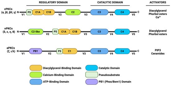
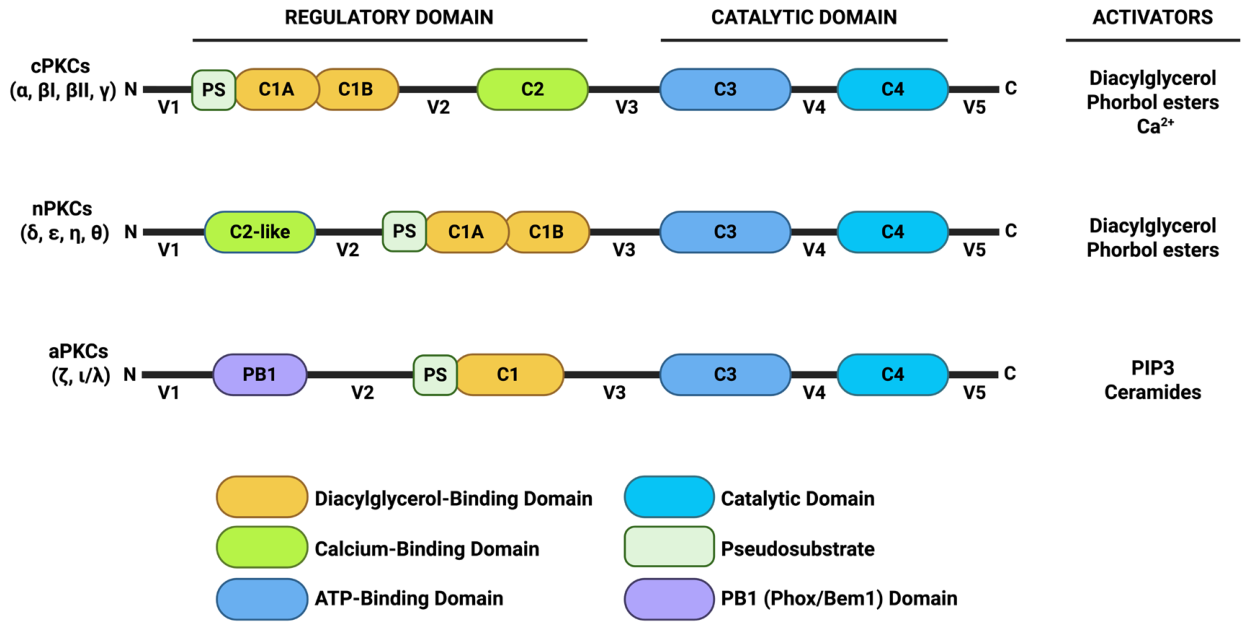
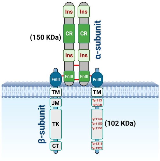
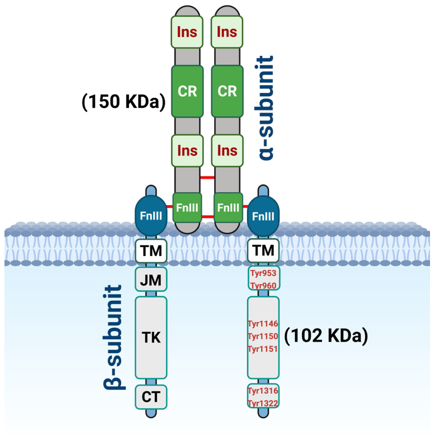
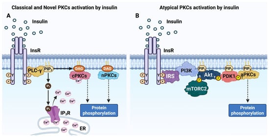
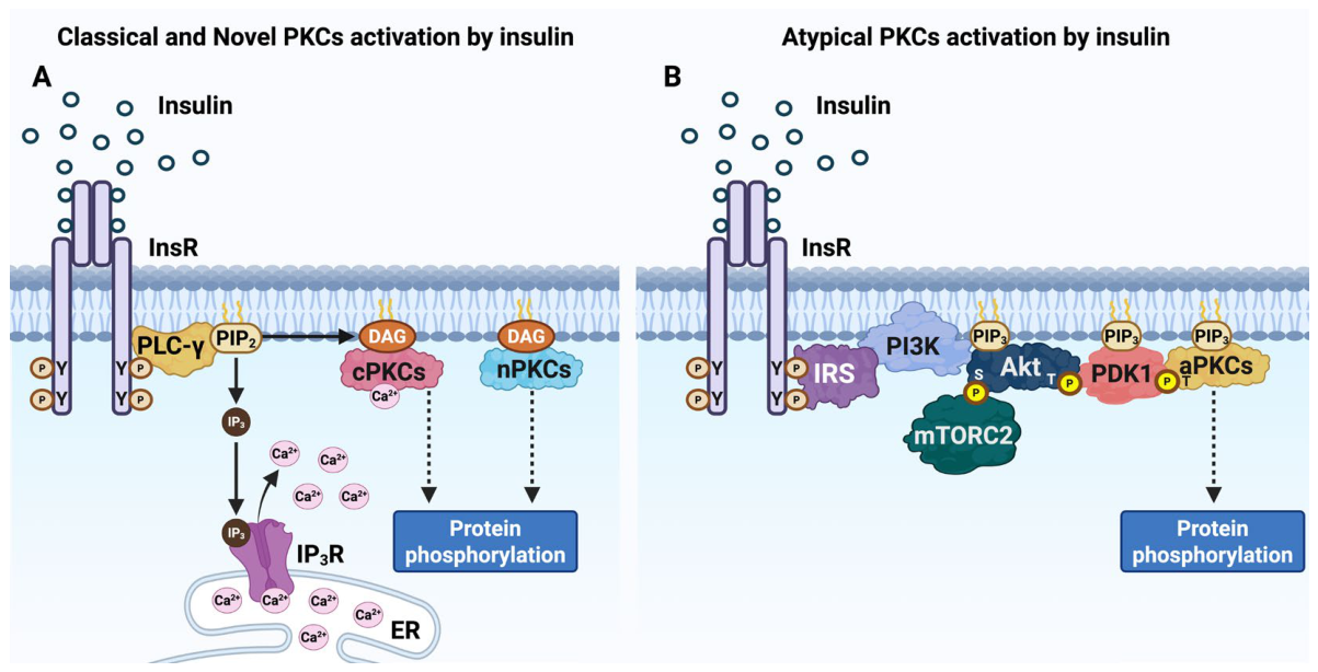
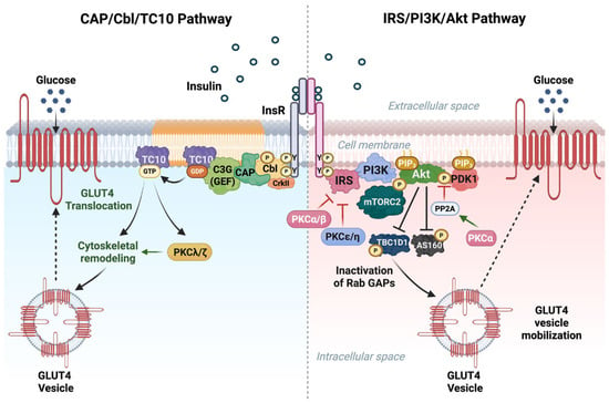
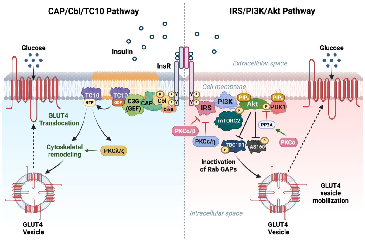
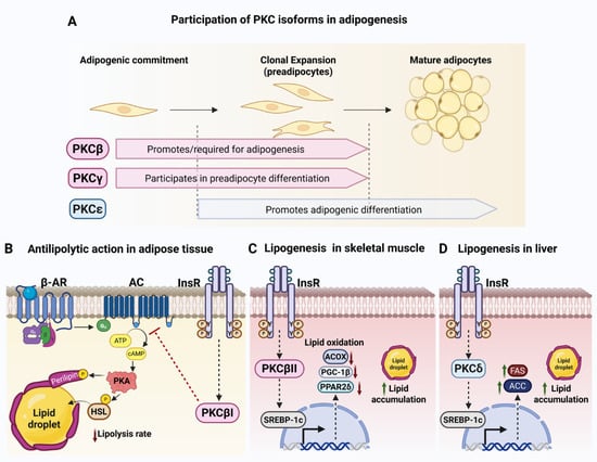
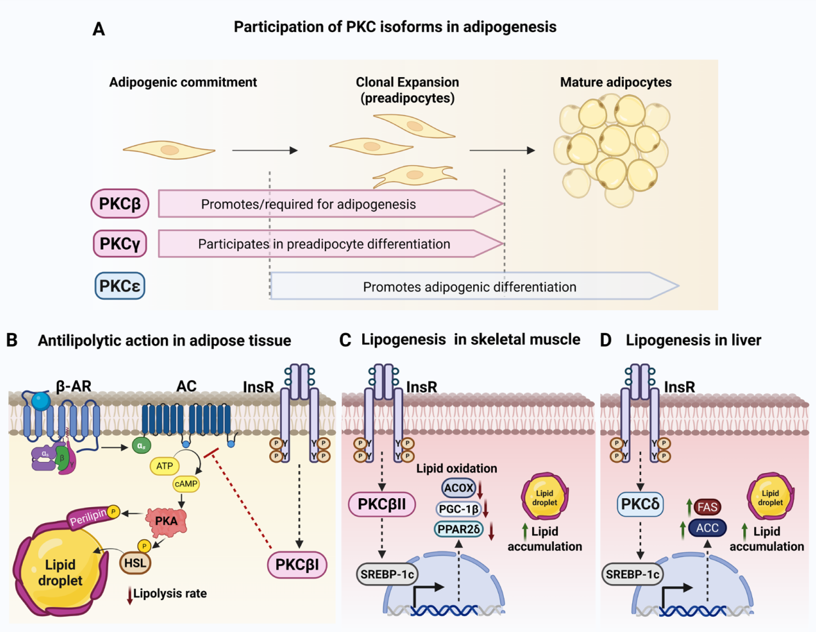
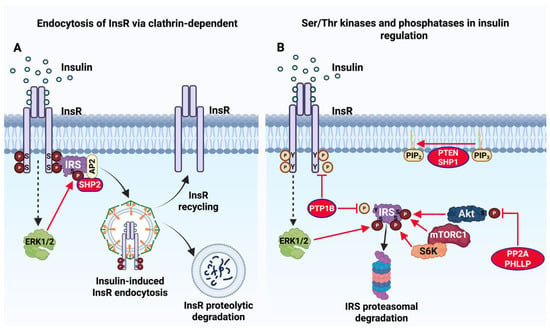
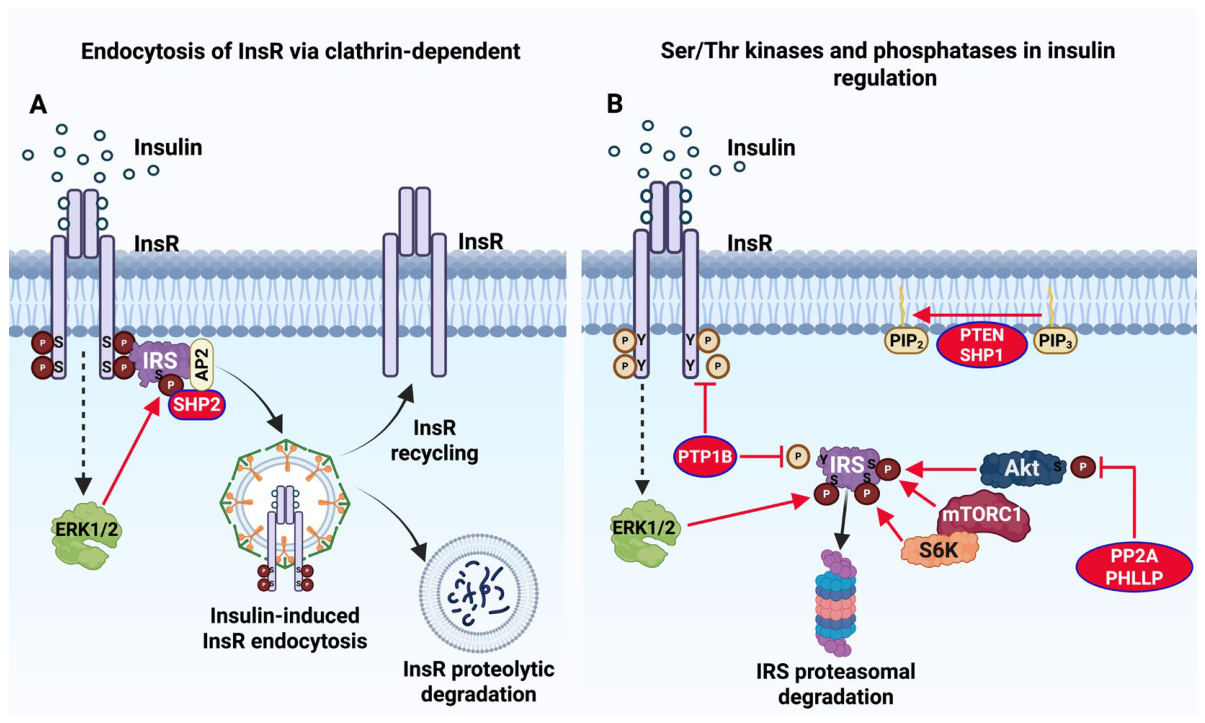
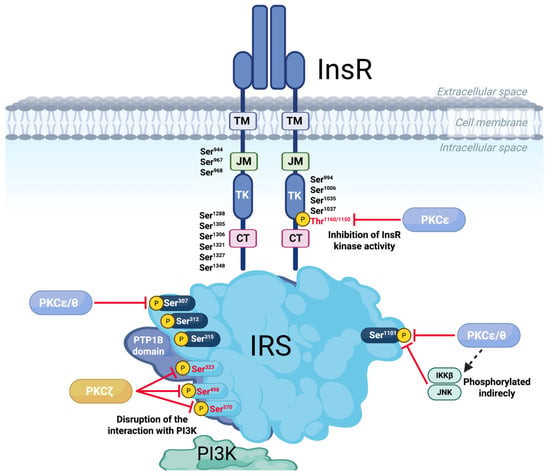
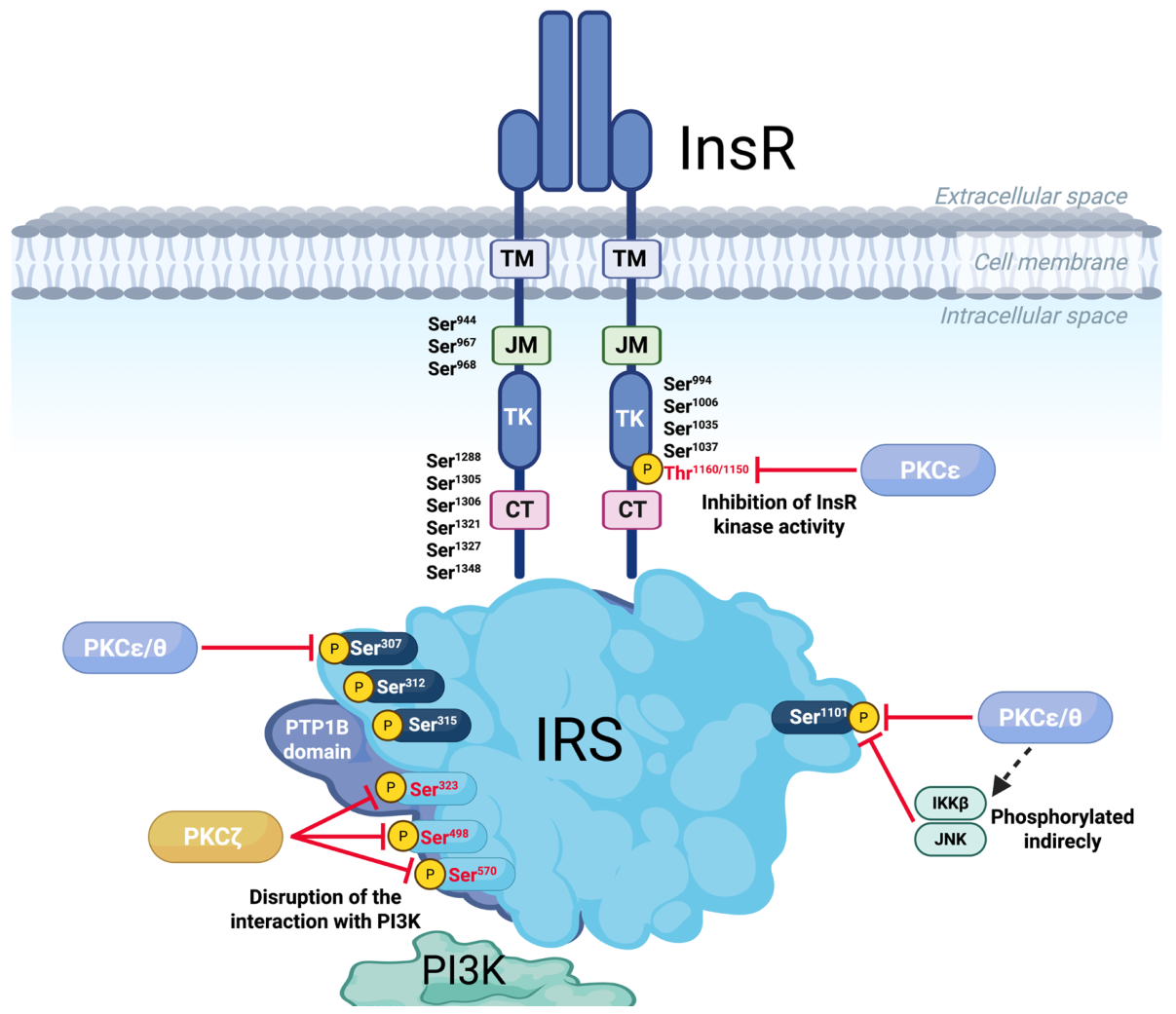
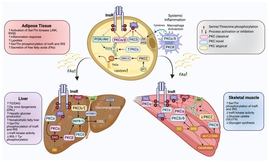
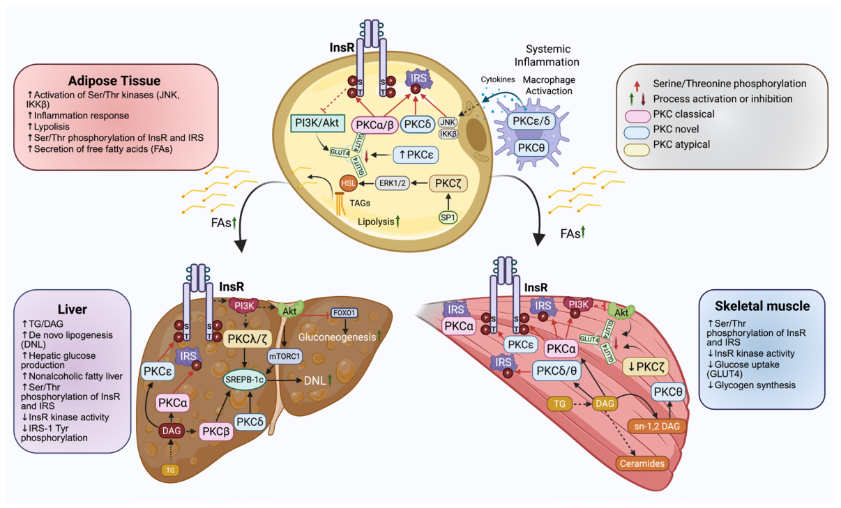

Disclaimer/Publisher’s Note: The statements, opinions and data contained in all publications are solely those of the individual author(s) and contributor(s) and not of MDPI and/or the editor(s). MDPI and/or the editor(s) disclaim responsibility for any injury to people or property resulting from any ideas, methods, instructions or products referred to in the content. |
© 2026 by the authors. Licensee MDPI, Basel, Switzerland. This article is an open access article distributed under the terms and conditions of the Creative Commons Attribution (CC BY) license.