


Abstract
Extracting high-molecular-weight (HMW) DNA from cactus tissues remains technically challenging due to the abundance of mucilage, pectins, polyphenols, and other metabolites that compromise DNA purity, increase viscosity, and reduce integrity, thereby limiting its suitability for long-read sequencing. This constraint is particularly relevant in Selenicereus megalanthus, a crop of increasing agronomic and genomic importance for which optimized protocols for third-generation sequencing remain limited. Here, we compared four CTAB-based DNA extraction protocols using dehydrated cladode tissue and evaluated DNA quality using NanoDrop spectrophotometry, Qubit fluorometry, agarose gel electrophoresis, and functional validation via sequencing on the Oxford Nanopore PromethION 2 Solo platform. Among the tested methods, our proposed optimized mucilage-adapted CTAB (MuCTAB) protocol, comprising 4% CTAB, 4% PVP-40, 0.5% β-mercaptoethanol, and proteinase K, showed the best overall performance. MuCTAB yielded the highest dsDNA concentration (239.63 ± 34.37 ng/µL), optimal purity ratios (A260/A280 = 1.96 ± 0.05; A260/A230 = 2.01 ± 0.01), and superior DNA integrity. Nanopore validation confirmed its effectiveness, with MuCTAB producing the highest sequencing yield (84.2 Gbp), read count, mean read quality score (Q18.4), read N50 (40.3 kbp), and maximum read length (1.9 Mbp). Overall, MuCTAB represents a low-cost, reproducible, and efficient method for HMW DNA extraction from mucilage-rich pitahaya tissues and shows promising potential for adaptation to other recalcitrant plant species.
1. Introduction
Pitahaya, or dragon fruit, comprises a group of climbing and epiphytic cacti important for production in tropical regions and is currently classified within the genus Selenicereus, previously known as Hylocereus [1,2]. Among the cultivated species, S. undatus, S. monacanthus, and S. megalanthus are the most prominent, differing in peel and pulp color, ploidy level, reproductive system, and agronomic traits [3,4,5,6]. In addition to their increasing commercial value, these fruits have attracted considerable interest due to their nutritional composition, bioactive compound content, and potential as an alternative crop for diversification in tropical and subtropical agroecosystems [5,7,8]. In Peru, cultivation has expanded to several regions, including Amazonas and San Martin, where recent studies have revealed morphological variability among cultivated accessions, suggesting the existence of valuable genetic resources for conservation, breeding and selection [9,10].
Despite its agronomic importance, genomic resources to support long-read sequencing in pitahaya remain limited across species. S. undatus already has a chromosome-scale nuclear genome, enabling deeper insight into genome evolution, chromosomal organization, and the regulation of betalain biosynthesis [11]. Likewise, studies based on chloroplast genomes and comparative plastomics have begun to provide valuable information for resolving phylogenetic relationships, patterns of structural variation, and taxonomic delimitation within the genus [12,13,14]. However, genomic development in yellow pitahaya (S. megalanthus) remains comparatively less advanced than in other cultivated Selenicereus species, despite its agronomic relevance and polyploid condition. This gap partly reflects the persistent technical difficulty of obtaining high-quality genomic DNA from mucilage-rich tissues, which has limited the generation of robust molecular and genomic resources for this species.[15].
The consolidation of long-read sequencing platforms, particularly those developed by Oxford Nanopore Technologies and PacBio, has reshaped modern plant genomics [16,17]. Unlike short-read sequencing, these technologies enable the recovery of repetitive regions, the detection of structural variants at higher resolution, reduced assembly fragmentation, and the reconstruction of complex organellar and nuclear genomes, which is especially valuable in non-model, heterozygous, or polyploid species [18,19,20,21,22]. However, their success depends critically on input DNA quality. The generation of stable libraries and informative long reads requires high-molecular-weight (HMW) DNA with high integrity, adequate purity, and minimal contamination from polysaccharides, phenolic compounds, proteins, and salts; in plant tissues, these factors often represent a major methodological bottleneck [23,24].
In cacti, particularly in pitahaya species, this challenge arises from the biochemical composition of the tissues. DNA extraction in this group is often compromised by the presence of mucilage and pectins, two polysaccharide fractions that promote the formation of viscous or gelatinous extracts, hinder phase separation, and facilitate the coprecipitation of contaminants with nucleic acids [1,25]. In addition, polyphenols and other secondary metabolites can oxidize, bind to DNA, or interfere with key enzymes in downstream applications. It has long been recognized that plant samples rich in polysaccharides and polyphenols require specific adjustments to extraction protocols, including modifications to the CTAB method, increased ionic strength, and the use of sequestering agents such as PVP [26]. Although important advances have been made in Cactaceae, most of these methods have been validated for PCR or conventional sequencing rather than under the more stringent requirements of long-read platforms [27,28].
Therefore, optimizing DNA extraction in pitahaya is not a minor adjustment but a prerequisite for advancing genomic, phylogenetic, diversity, and breeding studies in this crop. A truly effective protocol must reduce extract viscosity, minimize oxidation, decrease the coprecipitation of polysaccharides and proteins, and simultaneously preserve long, intact DNA fragments. Based on this need, the present study developed and evaluated an optimized protocol for extracting high-molecular-weight DNA from mucilage-rich pitahaya tissues. The suitability of the selected protocol for long-read sequencing applications and downstream genomic analyses was also assessed, with the aim of providing a reproducible methodological framework for pitahaya and, potentially, other mucilaginous cacti.
2. Materials and Methods
2.1. Plant Material
Tissue samples were collected from apical buds and subepidermal parenchyma of cladodes from adult S. megalanthus plants maintained in the germplasm collection of the Amazonas Agricultural Experimental Station of the Instituto Nacional de Innovación Agraria (INIA), Chachapoyas, Peru (6°13′ S, 77°52′ W; 2335 m a.s.l.).
2.2. Plant Tissue Preparation
Given the high-water content (>90%) and the characteristic mucilage of the succulent tissues of S. megalanthus, a dehydration step was implemented prior to processing. Fresh plant material (approximately 800 mg per sample) was cut into 2–3 cm fragments and placed on paper towels with silica gel beads (tissue:silica weight ratio of 1:10). Samples were incubated in an oven at 50 °C for 3–5 days, with the silica gel replaced every 12 h to maintain an active absorption gradient and prevent tissue rehydration, thereby reducing moisture content to below 5% [3,29]. The dehydrated tissue was ground in a sterile porcelain mortar to a fine homogeneous powder. Using this material (n = 5), four CTAB-based extraction protocols were evaluated: the conventional method [30], a CTAB method supplemented with PVP [26,31], the modified low-salt CTAB protocol (MoLSC) [32], and the optimized mucilage-adapted CTAB (MuCTAB) protocol for high-molecular-weight DNA developed in this study.
2.3. CTAB Protocol
Genomic DNA extraction using conventional CTAB was performed following the protocol of Doyle and Doyle [30], with minor modifications. Briefly, 100 mg of dehydrated powdered tissue was placed in 2 mL tubes and mixed with 800 µL of CTAB buffer (100 mM Tris-HCl pH 8.0, 20 mM EDTA pH 8.0, 1.4 M NaCl, 2% CTAB), 2 µL of β-mercaptoethanol, and 10 µL of proteinase K (20 mg/mL). The mixture was incubated at 65 °C for 1 h in a thermoblock with agitation at 600 rpm to promote cell lysis and nucleic acid solubilization. After incubation, 1 mL of chloroform:isoamyl alcohol (24:1) was added, the mixture was gently inverted to mix, followed by centrifugation at 10,000× g to separate the phases. The aqueous phase was transferred to a clean 1.5 mL tube and treated with RNase A according to the manufacturer’s recommendations. Subsequently, 750 µL of isopropanol was added to precipitate the DNA. The resulting pellet was washed twice with 750 µL of 70% ethanol, air-dried at room temperature for 30 min, and finally resuspended in 30 µL of sterile, ultrapure water.
2.4. CTAB + 1% PVP Protocol
The second protocol corresponded to the modified CTAB method previously proposed for cacti with high mucilage content [26]. Briefly, 100 mg of dehydrated powdered tissue was placed in 2 mL tubes and incubated with 700 µL of extraction buffer composed of 2% CTAB, 100 mM Tris-HCl (pH 8.0), 20 mM EDTA, 1.4 M NaCl, 1% PVP, and 1.4 µL of β-mercaptoethanol. The mixture was incubated at 65 °C for 20 min. Purification then followed the general workflow of the conventional CTAB method, including two extractions with chloroform:isoamyl alcohol (24:1). The recovered aqueous phase was treated with RNase A prior to DNA precipitation. DNA was subsequently precipitated with isopropanol, washed with 70% ethanol, and resuspended in 30 µL of ultrapure water.
2.5. MoLSC Protocol
The third protocol evaluated corresponded to the MoLSC method [32]. For this procedure, 100 mg of dehydrated powdered tissue was resuspended in 500 µL of CTAB lysis buffer composed of 100 mM Tris-HCl (pH 8.0), 20 mM EDTA, 2% CTAB, and 1.2 M NaCl, to which 30 µL of proteinase K (20 mg/mL) was added. Samples were incubated at 65 °C for 3 h in an incubator with agitation at 150 rpm. After cooling to room temperature, 500 µL of chloroform:isoamyl alcohol (24:1) was added, and the mixture was gently inverted to mix, followed by centrifugation for 15 min at 3000× g. The aqueous phase was transferred to a clean tube and supplemented with two volumes of diluted CTAB buffer (100 mM Tris-HCl, 20 mM EDTA, and 2% CTAB, without NaCl), followed by a second incubation at 60 °C for 30 min to promote formation of the CTAB-DNA complex. Samples were then centrifuged at 16,000× g for 3 min, the supernatant was discarded, and the pellet was washed with 75% ethanol. DNA was subsequently resuspended in 100 µL of high-salt TE buffer and treated with RNase A to a final concentration of 50 µg/mL for 15 min at 60 °C. Finally, a second wash with 75% ethanol was performed, the pellet was dried, and the DNA was resuspended in 30 µL of sterile, ultrapure water.
2.6. MuCTAB Protocol
The fourth protocol, developed and optimized in this study, was termed mucilage-adapted CTAB protocol for high-molecular-weight DNA (MuCTAB). This method was specifically designed for pitahaya tissues with high mucilage content and was based on the use of an enhanced lysis buffer composed of 4% CTAB, 100 mM Tris-HCl (pH 8.0), 20 mM EDTA, 2 M NaCl, and 4% PVP. For extraction, 100 mg of sample was placed in 2 mL tubes and mixed with 1500 µL of MuCTAB buffer, 5 µL of β-mercaptoethanol, and 12 µL of proteinase K. The mixture was gently homogenized by inversion for 1 min and incubated at 65 °C for 1 h, with gentle inversion every 5 min to avoid mechanical DNA fragmentation. After incubation, samples were centrifuged at 16,000× g for 10 min at room temperature, and the supernatant was transferred to a clean tube. Next, 100 µL of 5 M NaCl and 1000 µL of chloroform:isoamyl alcohol (24:1) were added, followed by gentle mixing by inversion for 1 min and centrifugation at 16,000× g for 10 min. The recovered aqueous phase was transferred to a new 1.5 mL tube and treated with RNase A according to the manufacturer’s instructions prior to DNA precipitation. Then, 50 µL of 3 M sodium acetate and 800 µL of cold isopropanol were added, followed by gentle mixing by inversion. The mixture was incubated at −20 °C for 2 h and then centrifuged at 16,000× g for 10 min at 4 °C. The pellet was washed with 800 µL of cold 70% ethanol, centrifuged again at 16,000× g for 15 min at 4 °C, air-dried at room temperature for 20 min, and finally resuspended in 30 µL of sterile, ultrapure water.
2.7. Assessment of DNA Concentration, Purity and Integrity
DNA concentration was determined by dual quantification methods: spectrophotometry (NanoDrop One, Thermo Fisher Scientific, Waltham, MA, USA) and dsDNA-specific fluorometry (Qubit 4 Fluorometer, Thermo Fisher Scientific). Purity was assessed using the A260/A280 ratio (optimal range 1.8–2.0) and A260/A230 ratio (optimal range: 2.0–2.2). The NanoDrop/Qubit ratio was calculated as an indirect indicator of spectrophotometric overestimation relative to fluorometric dsDNA quantification.
2.8. DNA Integrity
Genomic DNA integrity was evaluated by electrophoresis on 1% (w/v) agarose gels in 1X TAE buffer (40 mM Tris-acetate, 1 mM EDTA, pH 8.0), stained with Diamond nucleic acid dye. For each sample, 2 µL of DNA was loaded together with 1 µL of 6X loading buffer. A 1 kb GeneRuler Plus DNA ladder (Thermo Fisher Scientific) was used as a molecular weight reference. Electrophoresis was performed at 100 V for 30 min. Bands were visualized under ultraviolet light in a gel documentation system. The presence of a distinct high-molecular-weight band (>23 kbp) without significant degradation was considered indicative of DNA integrity suitable for long-read sequencing.
2.9. Validation by Long-Read Sequencing
DNA extracted using each protocol was functionally evaluated on the PromethION 2 Solo platform (Oxford Nanopore Technologies, Oxford, UK) using R10.4.1 flow cells and the SQK-LSK114 library preparation kit. Base calling was performed with Dorado v0.7 in super-accuracy mode. Sequencing quality metrics, including yield (Gbp), number of reads, N50, mean and median read lengths, threshold-based read distributions (≥10, 25, 50, 100, 200, and 500 kbp), and maximum read length, were obtained from the report generated by the sequencing platform.
2.10. Statistical Analysis
All analyses were performed in R v4.5.2 [33] and the RStudio graphical interface v2026.1.1.403 [34]. Normality and homoscedasticity assumptions were assessed using the Shapiro–Wilk and Levene tests, respectively. The effects of extraction protocol on concentration and purity variables were evaluated by one-way ANOVA, followed by Tukey’s HSD post hoc test (α = 0.05). Effect size was estimated using partial eta squared (η2p). In cases of heteroscedasticity, the Kruskal–Wallis test was applied. Principal component analysis (PCA) was performed on standardized data using FactoMineR [35] and biplots with 95% confidence ellipses were generated with factoextra [36]. The Pearson correlation matrix was calculated with Hmisc [37]. Data analysis was conducted using packages from the tidyverse ecosystem [38]; visualizations were generated with ggplot2 [39], radar charts with fmsb [40] and multipanel composition with gridExtra [41].
3. Results
3.1. Yield and Purity of Genomic DNA
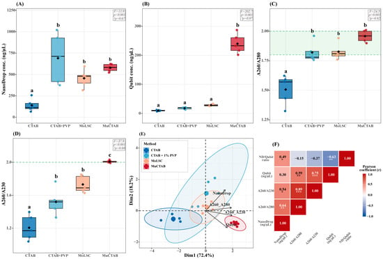
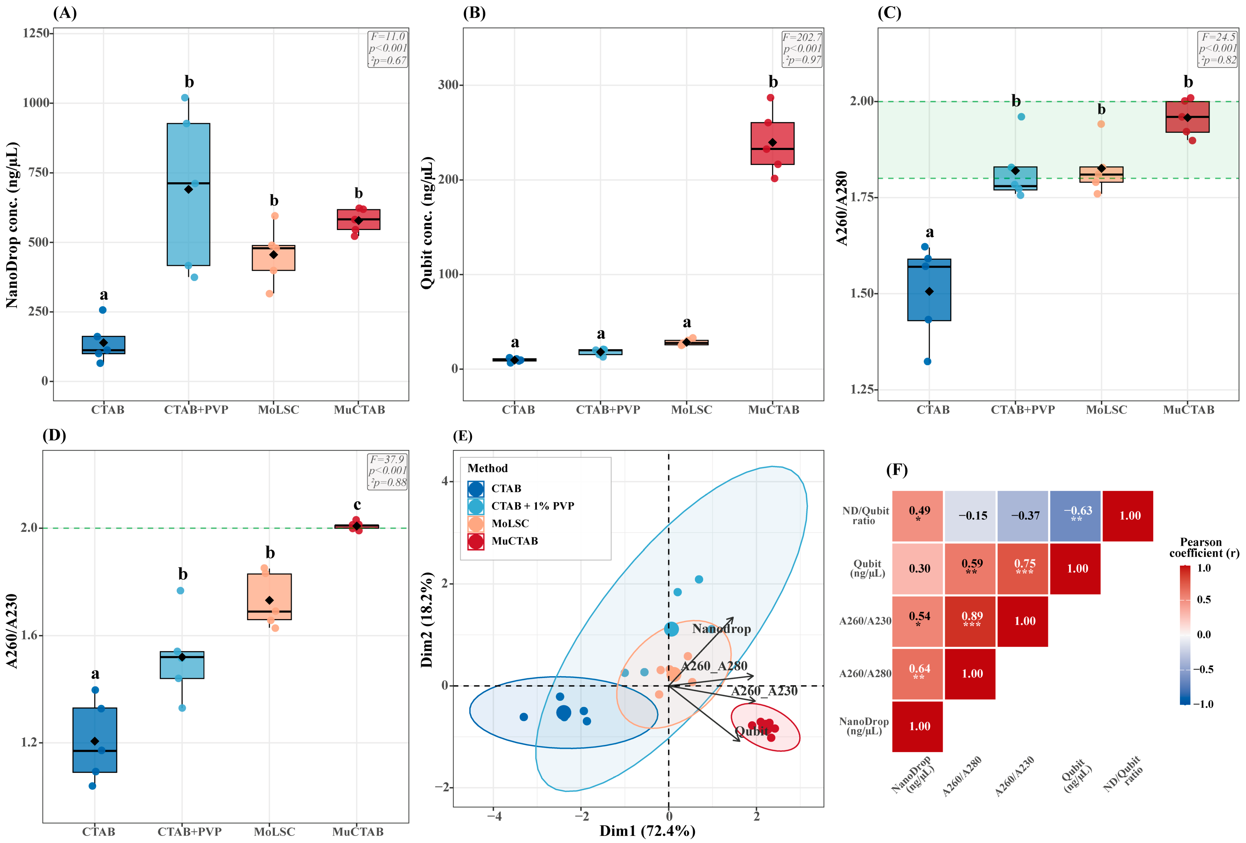
All four extraction protocols yielded detectable amounts of genomic DNA from dried S. megalanthus cladode tissue (n = 5), with highly significant differences among methods for all variables evaluated (Supplementary Table S1). Spectrophotometric quantification (NanoDrop) showed that the CTAB + 1% PVP (690.40 ± 291.20 ng/µL), MuCTAB (578.40 ± 43.17 ng/µL), and MoLSC (455.76 ± 104.06 ng/µL) protocols produced significantly higher concentrations than the conventional CTAB protocol (139.38 ± 73.83 ng/µL) (F(3, 16) = 11.0; p < 0.001; η2p = 0.67; Figure 1A). However, fluorometric quantification (Qubit), which specifically detects double-stranded DNA, revealed a different pattern: MuCTAB reached 239.63 ± 34.37 ng/µL, significantly higher than MoLSC (28.44 ± 3.37 ng/µL), CTAB + 1% PVP (18.09 ± 3.56 ng/µL), and the conventional CTAB protocol (9.56 ± 2.13 ng/µL) (F(3, 16) = 202.7; p < 0.001; η2p = 0.97; Figure 1B).
Figure 1.
Comparative evaluation of genomic DNA quality among four extraction protocols for S. megalanthus cladode tissue (n = 5). (A) DNA concentration measured by NanoDrop. (B) dsDNA concentration measured by Qubit. (C) A260/A280 ratio; green shaded area indicates the: optimal range (1.8–2.0). (D) A260/A230 ratio; dashed line indicates the optimal threshold (≥2.0). In panels A–D, different letters above the boxes indicate statistically significant differences according to Tukey’s HSD post hoc test (α = 0.05). (E) PCA biplot with 95% confidence ellipses. (F) Pearson correlation matrix of DNA quality variables; color intensity represents the Pearson correlation coefficient, ranging from negative correlations in blue to positive correlations in red. Asterisks indicate the level of statistical significance: * p < 0.05, ** p < 0.01, and *** p < 0.001.
Nucleic acid purity (A260/A280) differed significantly among protocols (F(3, 16) = 24.5; p < 0.001; η2p = 0.82; Figure 1C). MuCTAB reached 1.96 ± 0.05, consistently within the optimal range of 1.8–2.0, whereas the conventional CTAB protocol (1.51 ± 0.13) showed substantial residual protein contamination. Purity relative to other contaminants (A260/A230) provided the greatest discrimination among protocols (F(3, 16) = 37.9; p < 0.001; η2p = 0.88; Figure 1D). MuCTAB was the only protocol to reach the optimal threshold of ≥2.0 (2.01 ± 0.01; CV = 0.7%), with a clear progressive separation among methods: conventional CTAB < CTAB + 1% PVP ≈ MoLSC < MuCTAB. In addition, MuCTAB exhibited the lowest variability among biological replicates (Qubit CV = 14.3%; A260/A230 CV = 0.7%), indicating superior robustness and reproducibility.
Principal component analysis (PCA) based on the four DNA quality variables revealed that PC1 and PC2 explained 90.6% of the total variance (72.4% and 18.2%, respectively; Figure 1E). PC1 was positively associated with purity variables (A260/A280 and A260/A230) and Qubit concentration, representing an overall DNA quality gradient. PC2 was driven mainly by NanoDrop concentration, which was separated from Qubit in the two-dimensional space, highlighting the differential contribution of co-extracted contaminants to spectrophotometric readings. MuCTAB samples formed a compact and clearly differentiated cluster, whereas the conventional CTAB protocol was located at the opposite end of the quality gradient. The CTAB + 1% PVP and MoLSC protocols showed intermediate positions with greater dispersion.
A marked discrepancy was observed between concentrations estimated by NanoDrop and Qubit. The NanoDrop/Qubit ratio varied significantly among extraction methods (F(3, 16) = 27.1; p < 0.001), indicating marked differences in the agreement between spectrophotometric and fluorometric DNA quantification. CTAB + 1% PVP protocol showed the greatest spectrophotometric overestimation (ratio = 36.91 ± 9.89), followed by MoLSC (16.37 ± 4.95) and conventional CTAB protocol (14.33 ± 5.33), whereas MuCTAB showed a ratio of 2.43 ± 0.17, the closest to the ideal value of 1.0. The overall correlation between the two methods was weak and not statistically significant (r = 0.30; R2 = 0.09; p = 0.196; Figure 1F), likely attributable to heterogeneity in contamination profiles among protocols. The correlation matrix further showed that purity variables (A260/A280 and A260/A230) were strongly correlated with each other (r = 0.89; p < 0.001), and that Qubit concentration was positively associated with A260/A230 (r = 0.75; p < 0.001) and A260/A280 (r = 0.59; p < 0.01), whereas the NanoDrop/Qubit ratio was negatively correlated with Qubit (r = −0.63; p < 0.01; Figure 1F).
3.2. Integrity of Extracted DNA
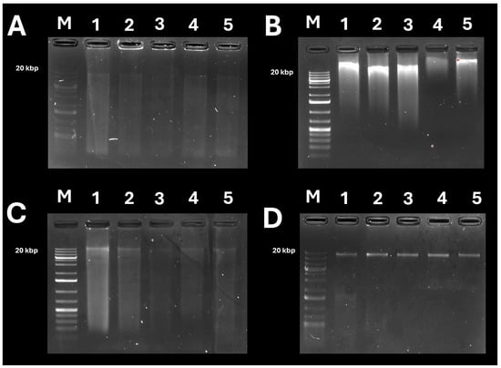
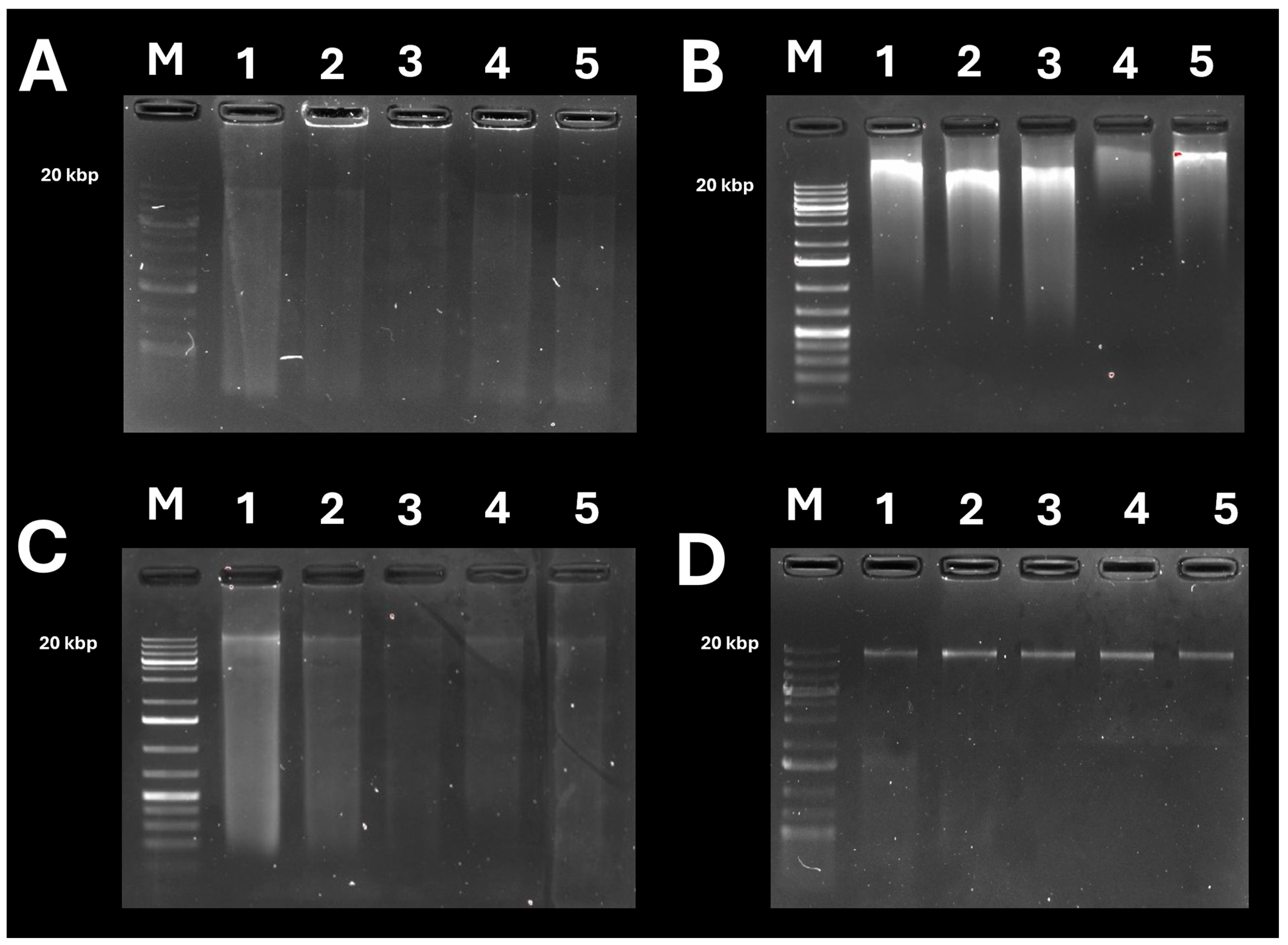
Agarose gel electrophoresis confirmed clear differences in the integrity of genomic DNA extracted from S. megalanthus cladodes among the evaluated protocols (Figure 2). The conventional CTAB protocol (Figure 2A) showed low signal intensity, consistent with its low DNA yield. The CTAB + 1% PVP and MoLSC methods (Figure 2B,C) exhibited more diffuse signals distributed along the lanes, suggesting increased degradation, smearing, or the co-extraction of interfering compounds. In contrast, the MuCTAB method (Figure 2D) produced more consistent high-molecular-weight DNA pattern, characterized by intense, compact bands near to the top gel with minimal smearing, indicating superior DNA integrity. These results are consistent with the spectrophotometric and fluorometric data, as MuCTAB yielded the highest Qubit concentration together with optimal A260/A280 and A260/A230 ratios, confirming that not only more abundant but also purer and more intact.
Figure 2.
Agarose gel electrophoresis of genomic DNA extracted from S. megalanthus cladode tissue using four extraction protocols. (A) CTAB, (B) CTAB + 1% PVP, (C) MoLSC, and (D) MuCTAB. M = 1 kb GeneRuler Plus DNA ladder (Thermo Fisher Scientific).
3.3. Functional Validation by Oxford Nanopore Sequencing
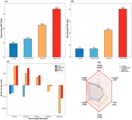
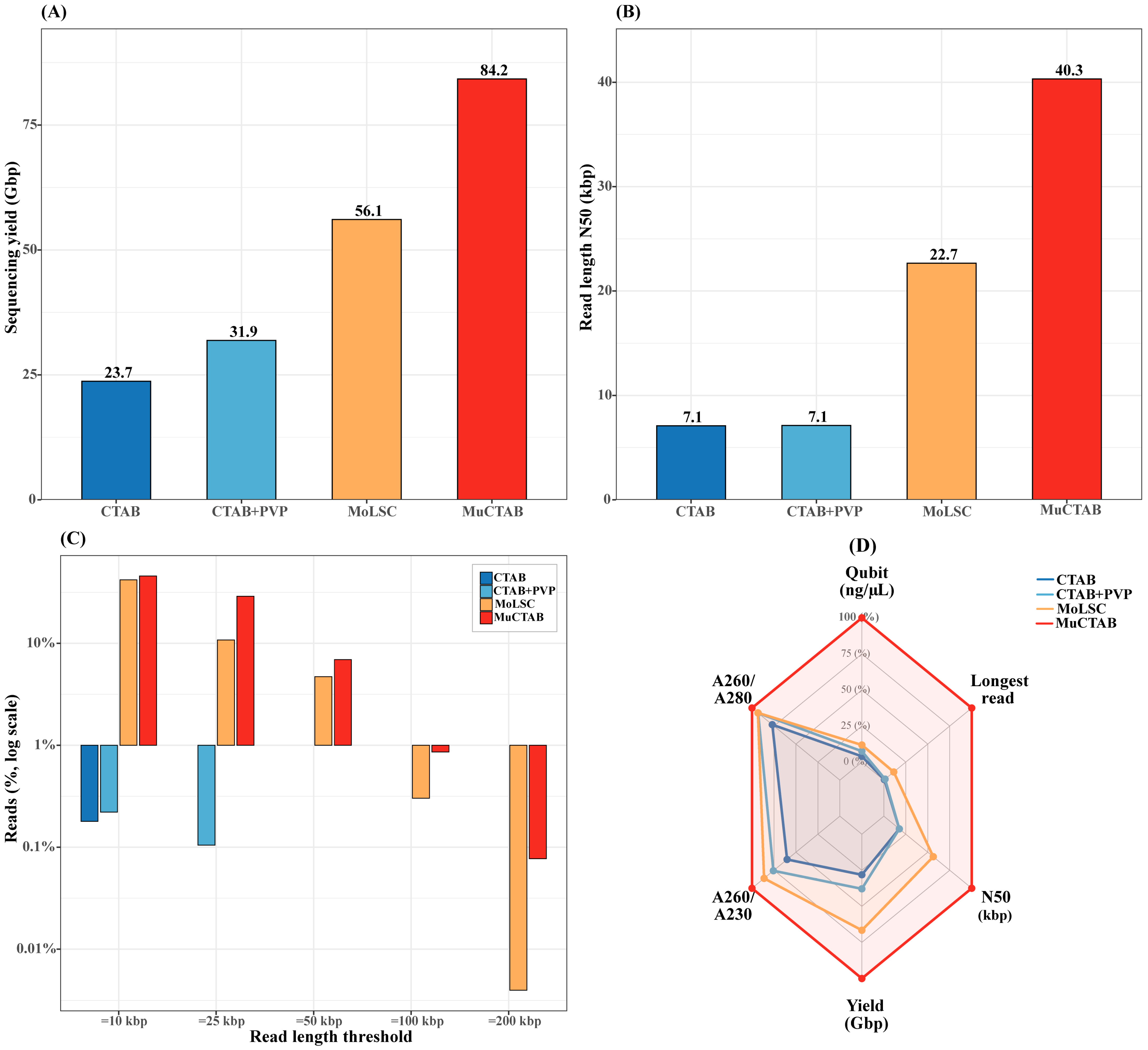
To evaluate the functional suitability of the extracted DNA for third-generation sequencing applications, one representative sample from each protocol was sequenced on the PromethION 2 Solo platform (Oxford Nanopore Technologies) using R10.4.1 flow cells (Table 1; Figure 3). MuCTAB method generated the highest yield (84.2 Gbp) and total number of reads (10.6 million), outperforming MoLSC (56.1 Gbp; 4.1 million), CTAB + 1% PVP (31.9 Gbp; 1.2 million), and the conventional CTAB protocol (23.7 Gbp; 0.6 million), representing a 3.6-fold increase relative to the conventional method (Figure 3A). Importantly, MuCTAB also produced the highest mean read quality score (Q18.4), followed by MoLSC (Q14.5), CTAB + 1% PVP (Q11.3), and conventional CTAB (Q11.1), indicating that the optimized protocol improved not only sequencing output and read length but also basecalling quality.
Table 1.
Oxford Nanopore PromethION 2 Solo sequencing metrics for genomic DNA.
Figure 3.
Functional validation of extracted DNA by Oxford Nanopore sequencing. (A) Total sequencing yield (Gbp). (B) Read N50 (kbp). (C) Cumulative read-length distribution (logarithmic scale). (D) Radar plot of normalized quality and sequencing performance metrics across the four protocols.
Read length differences were even more pronounced among protocols. The MuCTAB method achieved an N50 of 40.3 kbp, representing a 5.7-fold increase relative to both the conventional CTAB and CTAB + 1% PVP protocols (each approximately 7.1 kbp) and a 1.8-fold increase relative to MoLSC (22.7 kbp; Figure 3B). Only MuCTAB and MoLSC generated ultra-long reads (Figure 3C): MuCTAB produced 732,515 reads ≥50 kbp, 91,046 ≥ 100 kbp, and 278 ≥ 500 kbp, with a maximum read length of 1.9 Mbp. In contrast, the conventional CTAB and CTAB + 1% PVP protocols did not generate reads ≥50 kbp.
Integration of DNA quality parameters and sequencing performance using a normalized radar plot (Figure 3D) clearly identified MuCTAB as the optimal formulation for extracting genomic DNA from S. megalanthus compatible with long-read sequencing. This protocol consistently outperformed the others across the six evaluated metrics: dsDNA concentration (Qubit), nucleic acid purity (A260/A280 and A260/A230), sequencing yield (Gbp), read N50, and maximum read length.
4. Discussion
The extraction of high-quality genomic DNA from succulent tissues remains a significant technical challenge in Cactaceae, whose tissues contain high concentrations of mucilaginous polysaccharides, phenolic compounds, and secondary metabolites that coprecipitate with nucleic acids and inhibit downstream enzymatic reactions [26,28,42]. S. megalanthus presents similar challenges due to the mucilage-rich parenchyma of its cladodes [27,30]. In this study, we systematically evaluated four CTAB-based buffer formulations and validated the optimal protocol through long-read sequencing on the Oxford Nanopore PromethION platform, providing the first comprehensive assessment of DNA extraction quality for this species in the context of third-generation sequencing technologies.
Our results demonstrate that the MuCTAB protocol, which incorporates 4% CTAB, 4% PVP-40, 0.5% β-mercaptoethanol, and proteinase K (20 mg/mL), consistently outperformed the three alternative formulations across all evaluated quality parameters. The fluorometric yield obtained with MuCTAB (Qubit: 239.6 ± 34.4 ng/µL) was significantly higher than that of the conventional CTAB (9.56 ± 2.13 ng/µL), CTAB + PVP (18.09 ± 3.56 ng/µL), and MoLSC (28.44 ± 3.37 ng/µL) (p < 0.05, Tukey HSD). This 8.4 to 25.1-fold improvement reflects the synergistic effect of combining PVP-40 for polyphenol sequestration [43], elevated β-mercaptoethanol for disulfide bond reduction and oxidation prevention [44], and proteinase K for enzymatic protein degradation [45]. This multi-target approach extends the strategy used by Martínez-González et al. [28] for Opuntia, although our protocol was specifically optimized for compatibility with long-read sequencing.
The spectrophotometric purity ratios obtained with MuCTAB (A260/A280: 1.96 ± 0.05; A260/A230: 2.01 ± 0.01) fall within the optimal ranges for high-quality genomic DNA [46]. The NanoDrop/Qubit concentration ratio, used as an indirect indicator of contamination, was lowest for MuCTAB (2.43 ± 0.17) compared with MoLSC (16.37 ± 4.95), confirming minimal overestimation by spectrophotometric methods. This dual-quantification approach provides a more robust assessment of DNA quality than either method alone and should be considered standard practice for long-read sequencing applications [47,48].
Functional validation by PromethION 2 Solo sequencing revealed notable differences among protocols that were not fully captured by spectrophotometric assessment. The MuCTAB method achieved the highest sequencing yield (84.2 Gbp), number of reads (10.6 × 10⁶), and N50 (40.3 kbp), substantially exceeding values reported for other cactus species extracted with standard CTAB methods [49,50]. The progressive decline in sequencing performance from MuCTAB to MoLSC correlated with the purity metrics, suggesting that residual contaminants interfere with nanopore translocation kinetics and library preparation efficiency. Polysaccharide contamination increases solution viscosity and obstructs nanopore channels, thereby reducing read length and promoting premature pore inactivation [51].
An important practical contribution of this study is the demonstration that silica gel drying at 50 °C replaces expensive lyophilization equipment, achieving moisture levels below 5% within 3–5 days. This approach follows the methodologies previously reported for field-collected plant samples preserved in saturated NaCl/CTAB solution [29], and is particularly relevant for field stations in Andean regions where S. megalanthus is cultivated. Principal component analysis further confirmed that MuCTAB produces a qualitatively distinct DNA profile: the clear separation along PC1 (72.4% of the variance) was driven by Qubit concentration, the A260/A230 ratio, and sequencing N50.
Table 2 compares our protocol with previously published methods for Cactaceae and recalcitrant plant species. Several critical observations emerge. First, most existing protocols have validated DNA quality exclusively through PCR-based applications (RAPD, SSR, ITS, and matK), which require substantially lower DNA quality and integrity than long-read sequencing platforms [52]. Only one previous study [42] validated its CTAB protocol using next-generation sequencing (Illumina HiSeq), but with short-read technology that is less sensitive to DNA integrity than Oxford Nanopore platforms. To our knowledge, this is the first study to validate a CTAB-based extraction protocol for Cactaceae using long-read sequencing. Second, our A260/A230 ratio (2.01) exceeds most reported values and falls within the range previously reported for Opuntia [28], whereas many earlier studies did not report this critical metric. Third, the estimated cost (~$1.13 USD) is substantially lower than combined CTAB/kit approaches [53] (~$8–12) and commercial kit-based methods [54], while maintaining superior DNA quality for demanding applications.
Table 2.
Comparison of DNA extraction protocols for Cactaceae species and recalcitrant plants.
The MuCTAB protocol optimized in this study was specifically developed to address the biochemical challenges inherent to S. megalanthus cladode tissue, whose high mucilage and polyphenol contents have historically limited genomic studies in this species. This protocol provides a cost-effective, reproducible, and accessible solution compatible with the most demanding third-generation sequencing platforms, and it is expected to facilitate genome assembly, molecular marker development, and population genetics studies in this economically important tropical crop species [54].
Oxford Nanopore validation provided strong functional evidence of compatibility with long-read sequencing; however, the performance metrics appear to derive from representative libraries per treatment rather than from biologically replicated sequencing runs. Therefore, variables such as total yield, N50, and maximum read length should be interpreted as robust proof-of-concept evidence rather than as statistically exhaustive estimate of sequencing performance for each protocol [18,20,42].
Although the present study focused on S. megalanthus, the biochemical rationale of the MuCTAB workflow suggests that this approach may be adaptable to other mucilage-rich or otherwise recalcitrant plant tissues. In our laboratory, the protocol has been explored in additional plant materials, including cacao, strawberry, woody species of forestry interest, herbaceous crops and other Cactaceae, have shown encouraging preliminary performance. However, these evaluations were not included in the present manuscript because they fall outside its specific experimental scope and were not designed as formal cross-species validation assays. Therefore, broader validation should be addressed in future studies through comparative analyses including extraction yield, purity, DNA integrity, and long-read sequencing performance. From this perspective, MuCTAB provides a useful methodological framework not only for strengthening genomics research in S. megalanthus, but also for guiding future optimization in Cactaceae and other complex plant species constrained by the difficulty of recovering high-integrity genomic DNA for third-generation sequencing applications [22].
5. Conclusions
The MuCTAB protocol developed in this study represents a robust and economically accessible solution for extracting high-molecular-weight genomic DNA from mucilage-rich Selenicereus megalanthus cladodes. The combination of enhanced CTAB and PVP concentrations together with β-mercaptoethanol, proteinase K, controlled tissue dehydration, and gentle sample handling enabled the recovery of DNA with higher fluorometric yield (239.6 ± 34.4 ng/µL), better purity (A260/A280: 1.96; A260/A230: 2.01), and greater structural integrity than the three alternative protocols evaluated. More importantly, these advantages were not limited to conventional quality metrics but translated directly into superior Oxford Nanopore sequencing performance, reflected in higher total yield, longer reads, a markedly higher N50 (40.3 kbp), and the recovery of ultra-long reads. These results demonstrate that MuCTAB is not merely an incremental modification of the CTAB method but rather a workflow specifically suited for long-read sequencing applications in mucilage-rich and biochemically complex plant tissues. In addition to its technical effectiveness, the protocol offers practical value due to its low cost, reproducibility, and the possibility of implementation without lyophilization. The MuCTAB protocol thus provides a solid methodological framework for future genome assembly, comparative genomics, molecular marker development, and genetic conservation studies in pitahaya and potentially in other plant species with high mucilage content.
Supplementary Materials
The following supporting information can be downloaded at https://www.mdpi.com/article/10.3390/applbiosci5030054/s1. Table S1: Statistical summary of DNA yield and purity parameters across four extraction protocols from dried Selenicereus megalanthus cladode tissue. Standard Eta Squared (η2p).
Author Contributions
A.D.H.-A. designed the DNA extraction protocol, standardized four buffer formulations, performed DNA extractions, conducted gel electrophoresis and sequencing on the PromethION platform. J.C.S.-P. contributed to DNA extraction, performed statistical and bioinformatic analyses, prepared figures and tables, and drafted the original manuscript. J.Y.-D.l.C. contributed to DNA extraction and gel electrophoresis assays. J.A.C.-A. reviewed and edited the manuscript. J.C.G.-A. conceptualized the study, supervised the research, reviewed and edited the manuscript. All authors have read and agreed to the published version of the manuscript.
Funding
The authors declare that financial support was received for the research and/or publication of this article. This work was supported by the Consejo Nacional de Ciencia, Tecnología e Innovación Tecnológica (CONCYTEC) and the Programa Nacional de Investigación Científica y Estudios Avanzados (PROCIENCIA), project “OMICS in Andean crops under abiotic stress conditions in the context of climate change in the Amazonas region” (Contract No. PE501086005-2023-BM-PROCIENCIA), the project Creación de los Servicios de Investigación, Innovación y Desarrollo de Tecnología para el Sector Agrario y Agroindustrial de la UNTRM sede Chachapoyas, Departamento de Amazonas”, C.U.I. N° 2313205 and the Vicerrectorado de Investigación of the Universidad Nacional Toribio Rodríguez de Mendoza de Amazonas (UNTRM).
Institutional Review Board Statement
Not applicable.
Informed Consent Statement
Not applicable.
Data Availability Statement
The raw data supporting the conclusions of this article will be made available by the authors on request.
Acknowledgments
The authors acknowledge the support, equipment and facilities of the Plant Molecular Biology Laboratory of the Instituto de Investigación, Innovación y Desarrollo para el Sector Agrario y Agroindustrial (IIDAA), Faculty of Engineering and Agricultural Sciences, UNTRM.
Conflicts of Interest
The authors declare no conflicts of interest.
References
- Korotkova, N.; Borsch, T.; Arias, S. A Phylogenetic Framework for the Hylocereeae (Cactaceae) and Implications for the Circumscription of the Genera. Phytotaxa 2017, 327, 1–46. [Google Scholar] [CrossRef] [Scilit]
- Abirami, K.; Swain, S.; Baskaran, V.; Venkatesan, K.; Sakthivel, K.; Bommayasamy, N. Distinguishing Three Dragon Fruit (Hylocereus spp.) Species Grown in Andaman and Nicobar Islands of India Using Morphological, Biochemical and Molecular Traits. Sci. Rep. 2021, 11, 2894. [Google Scholar] [CrossRef] [Scilit] [PubMed]
- Tel-Zur, N.; Abbo, S.; Bar-Zvi, D.; Mizrahi, Y. Genetic Relationships among Hylocereus and Selenicereus Vine Cacti (Cactaceae): Evidence from Hybridization and Cytological Studies. Ann. Bot. 2004, 94, 527–534. [Google Scholar] [CrossRef] [Scilit] [PubMed]
- Morillo-Coronado, A.C.; Manjarres Hernández, E.H.; Forero-Mancipe, L. Phenotypic Diversity of Morphological Characteristics of Pitahaya (Selenicereus megalanthus Haw.) Germplasm in Colombia. Plants 2021, 10, 2255. [Google Scholar] [CrossRef] [Scilit] [PubMed]
- Shah, K.; Chen, J.; Chen, J.; Qin, Y. Pitaya Nutrition, Biology, and Biotechnology: A Review. Int. J. Mol. Sci. 2023, 24, 13986. [Google Scholar] [CrossRef] [Scilit] [PubMed]
- Valero, D.; Erazo-Lara, A.; García-Pastor, M.E.; Padilla-González, P.A.; Agulló, V.; El-Hiali, F.B.; Serrano, M. Yellow Pitahaya (Selenicereus megalanthus Haw.): The Less Known of the Pitahayas. Foods 2025, 14, 202. [Google Scholar] [CrossRef] [Scilit] [PubMed]
- Pérez-Orozco, A.F.; Sosa, V. Comparación del contenido de betalaínas y azúcares en frutos de cinco especies de Selenicereus (Cactaceae): Betalains and sugars in pitahayas. Acta Bot. Mex. 2022, 129. [Google Scholar] [CrossRef] [Scilit]
- Yang, Z.-Y.; Zheng, X.-W.; Jiang, W.-H.; Chen, G.-Z.; Liang, Q.-Z.; Xu, G.-Z.; Yi, R.-H. Selenicereus undatus (Dragon Fruit) Phytochemicals for Managing Three Human Pathogenic Bacteria: An In Vitro and In Silico Approach. Metabolites 2024, 14, 577. [Google Scholar] [CrossRef] [Scilit] [PubMed]
- Santos-Pelaez, J.C.; Saravia-Navarro, D.; Cruz-Delgado, J.H.I.; del Carpio-Salas, M.A.; Barboza, E.; Casanova Nuñez Melgar, D.P. Phenotypic Diversity of Morphological Traits of Pitahaya (Hylocereus spp.) and Its Agronomic Potential in the Amazonas Region, Peru. Agriculture 2024, 14, 1968. [Google Scholar] [CrossRef] [Scilit]
- Heredia-Montalvo, J.G.; Vallejos-Tapullima, A.; Carballar-Hernández, S.; Carvajal-Vallejos, F.M.; Pereira, C.F.; Oehl, F.; Guerrero-Abad, J.C.; Corazon-Guivin, M.A. Symbiotic Relationships between Arbuscular Mycorrhizal Fungi and Wild Accessions of Selenicereus undatus in the Peruvian Amazon. J. Appl. Bot. Food Qual. 2025, 98, 66–72. [Google Scholar] [CrossRef]
- Chen, J.-Y.; Xie, F.-F.; Cui, Y.-Z.; Chen, C.-B.; Lu, W.-J.; Hu, X.-D.; Hua, Q.-Z.; Zhao, J.; Wu, Z.-J.; Gao, D.; et al. A Chromosome-Scale Genome Sequence of Pitaya (Hylocereus undatus) Provides Novel Insights into the Genome Evolution and Regulation of Betalain Biosynthesis. Hortic. Res. 2021, 8, 164. [Google Scholar] [CrossRef] [Scilit] [PubMed]
- Liu, J.; Liu, Z.-Y.; Zheng, C.; Niu, Y.-F. Complete Chloroplast Genome Sequence and Phylogenetic Analysis of Dragon Fruit (Selenicereus undatus (Haw.) D.R.Hunt). Mitochondrial DNA Part B 2021, 6, 1154–1156. [Google Scholar] [CrossRef] [Scilit] [PubMed]
- Qin, Q.; Li, J.; Zeng, S.; Xu, Y.; Han, F.; Yu, J. The Complete Plastomes of Red Fleshed Pitaya (Selenicereus monacanthus) and Three Related Selenicereus Species: Insights into Gene Losses, Inverted Repeat Expansions and Phylogenomic Implications. Physiol. Mol. Biol. Plants 2022, 28, 123–137. [Google Scholar] [CrossRef] [Scilit] [PubMed]
- Zheng, E.; Yisilam, G.; Li, C.; Jiao, F.; Ling, Y.; Lu, S.; Wang, Q.; Tian, X. Comparative Analysis of Chloroplast Genomes and Phylogenetic Relationships of Different Pitaya Cultivars. BMC Genom. 2025, 26, 463. [Google Scholar] [CrossRef] [Scilit] [PubMed]
- Zaman, Q.U.; Raza, A.; Hui, L.; Nazir, M.F.; Garg, V.; Ikram, M.; Wang, G.; Lv, W.; Khan, D.; Khokhar, A.A.; et al. Chromosome-Level Genome Assembly of the Autotetraploid Yellow Pitaya Provides Novel Insights into Evolution of Trait Patterning in Pitaya Species with Different Ploidy. Genome Biol. 2025, 26, 234. [Google Scholar] [CrossRef] [Scilit] [PubMed]
- Koo, H.; Lee, G.-W.; Ko, S.-R.; Go, S.; Kwon, S.-Y.; Kim, Y.-M.; Shin, A.-Y. Two Long Read-Based Genome Assembly and Annotation of Polyploidy Woody Plants, Hibiscus syriacus L. Using PacBio and Nanopore Platforms. Sci. Data 2023, 10, 713. [Google Scholar] [CrossRef] [Scilit] [PubMed]
- van Rengs, W.M.J.; Schmidt, M.H.-W.; Effgen, S.; Le, D.B.; Wang, Y.; Zaidan, M.W.A.M.; Huettel, B.; Schouten, H.J.; Usadel, B.; Underwood, C.J. A Chromosome Scale Tomato Genome Built from Complementary PacBio and Nanopore Sequences Alone Reveals Extensive Linkage Drag during Breeding. Plant J. 2022, 110, 572–588. [Google Scholar] [CrossRef] [Scilit] [PubMed]
- Dumschott, K.; Schmidt, M.H.-W.; Chawla, H.S.; Snowdon, R.; Usadel, B. Oxford Nanopore Sequencing: New Opportunities for Plant Genomics? J. Exp. Bot. 2020, 71, 5313–5322. [Google Scholar] [CrossRef] [Scilit] [PubMed]
- Sebastiani, L.; Sarfraz, I.; Francini, A.; Celii, M.; Wing, R.A.; Zuccolo, A. PacBio Genome Assembly of Olea europaea L. subsp. europaea Cultivars Frantoio and Leccino. Sci. Data 2025, 12, 1095. [Google Scholar] [CrossRef] [Scilit] [PubMed]
- Kang, M.; Chanderbali, A.; Lee, S.; Soltis, D.E.; Soltis, P.S.; Kim, S. High-Molecular-Weight DNA Extraction for Long-Read Sequencing of Plant Genomes: An Optimization of Standard Methods. Appl. Plant Sci. 2023, 11, e11528. [Google Scholar] [CrossRef] [Scilit] [PubMed]
- Zhu, Q.; Wang, Y.; Yao, N.; Ni, X.; Wang, C.; Wang, M.; Zhang, L.; Liang, W. Chromosome-Level Genome Assembly of an Endangered Plant Prunus Mongolica Using PacBio and Hi-C Technologies. DNA Res. 2023, 30, dsad012. [Google Scholar] [CrossRef] [Scilit] [PubMed]
- Lu, D.; Liu, C.; Ji, W.; Xia, R.; Li, S.; Liu, Y.; Liu, N.; Liu, Y.; Deng, X.W.; Li, B. Nanopore Ultra-Long Sequencing and Adaptive Sampling Spur Plant Complete Telomere-to-Telomere Genome Assembly. Mol. Plant 2024, 17, 1773–1786. [Google Scholar] [CrossRef] [Scilit] [PubMed]
- Russo, A.; Mayjonade, B.; Frei, D.; Potente, G.; Kellenberger, R.T.; Frachon, L.; Copetti, D.; Studer, B.; Frey, J.E.; Grossniklaus, U.; et al. Low-Input High-Molecular-Weight DNA Extraction for Long-Read Sequencing from Plants of Diverse Families. Front. Plant Sci. 2022, 13, 883897. [Google Scholar] [CrossRef] [Scilit] [PubMed]
- Nishii, K.; Möller, M.; Foster, R.G.; Forrest, L.L.; Kelso, N.; Barber, S.; Howard, C.; Hart, M.L. A High Quality, High Molecular Weight DNA Extraction Method for PacBio HiFi Genome Sequencing of Recalcitrant Plants. Plant Methods 2023, 19, 41. [Google Scholar] [CrossRef] [Scilit] [PubMed]
- Otálora, M.C.; Wilches-Torres, A.; Gómez Castaño, J.A. Mucilage from Yellow Pitahaya (Selenicereus megalanthus) Fruit Peel: Extraction, Proximal Analysis, and Molecular Characterization. Molecules 2023, 28, 786. [Google Scholar] [CrossRef] [Scilit] [PubMed]
- Porebski, S.; Bailey, L.G.; Baum, B.R. Modification of a CTAB DNA Extraction Protocol for Plants Containing High Polysaccharide and Polyphenol Components. Plant Mol. Biol. Rep. 1997, 15, 8–15. [Google Scholar] [CrossRef] [Scilit]
- Tel-zur, N.; Abbo, S.; Myslabodski, D.; Mizrahi, Y. Modified CTAB Procedure for DNA Isolation from Epiphytic Cacti of the Genera Hylocereus and Selenicereus (Cactaceae). Plant Mol. Biol. Report. 1999, 17, 249–254. [Google Scholar] [CrossRef] [Scilit]
- Martínez-González, C.R.; Ramírez-Mendoza, R.; Jiménez-Ramírez, J.; Gallegos-Vázquez, C.; Luna-Vega, I. Improved Method for Genomic DNA Extraction for Opuntia Mill. (Cactaceae). Plant Methods 2017, 13, 82. [Google Scholar] [CrossRef] [Scilit] [PubMed]
- Štorchová, H.; Hrdličková, R.; Chrtek, J., Jr.; Tetera, M.; Fitze, D.; Fehrer, J. An Improved Method of DNA Isolation from Plants Collected in the Field and Conserved in Saturated NaCl/CTAB Solution. Taxon 2000, 49, 79–84. [Google Scholar] [CrossRef] [Scilit]
- Doyle, J.J.; Doyle, J.L. A Rapid DNA Isolation Procedure for Small Quantities of Fresh Leaf Tissue. Phytochem. Bull. 1987, 19, 11–15. [Google Scholar]
- Jobes, D.V.; Hurley, D.L.; Thien, L.B. Plant DNA Isolation: A Method to Efficiently Remove Polyphenolics, Polysaccharides, and RNA. Taxon 1995, 44, 379–386. [Google Scholar] [CrossRef] [Scilit]
- Arseneau, J.-R.; Steeves, R.; Laflamme, M. Modified Low-Salt CTAB Extraction of High-Quality DNA from Contaminant-Rich Tissues. Mol. Ecol. Resour. 2017, 17, 686–693. [Google Scholar] [CrossRef] [Scilit] [PubMed]
- R Core Team. R: The R Project for Statistical Computing; R Foundation for Statistical Computing: Vienna, Austria, 2025. [Google Scholar]
- Posit Team. RStudio: Integrated Development Environment for R; R Foundation for Statistical Computing: Vienna, Austria, 2024. [Google Scholar]
- Lê, S.; Josse, J.; Husson, F. FactoMineR: An R Package for Multivariate Analysis. J. Stat. Softw. 2008, 25, 1–18. [Google Scholar] [CrossRef] [Scilit]
- Kassambara, A.; Mundt, F. Factoextra: Extract and Visualize the Results of Multivariate Data Analyses. CRAN Contributed Packages. 2020. Available online: https://cran.r-project.org/web/packages/factoextra/index.html (accessed on 21 April 2026).
- Harrell, F.E., Jr.; Beck, C.; Dupont, C. Hmisc: Harrell Miscellaneous. 2026. Available online: https://cran.r-project.org/package=Hmisc (accessed on 27 January 2026).
- Wickham, H.; Averick, M.; Bryan, J.; Chang, W.; McGowan, L.D.A.; François, R.; Grolemund, G.; Hayes, A.; Henry, L.; Hester, J.; et al. Welcome to the tidyverse. J. Open Source Softw. 2019, 4, 1686. [Google Scholar] [CrossRef] [Scilit]
- Wickham, H. Ggplot2: Elegant Graphics for Data Analysis; Springer International Publishing: Cham, Switzerland, 2016. [Google Scholar]
- Nakazawa, M. Fmsb: Functions for Medical Statistics Book with Some Demographic Data. 2024. Available online: https://cran.r-project.org/package=fmsb (accessed on 12 February 2026).
- Baptiste, A. gridExtra: Miscellaneous Functions for “Grid” Graphics. Available online: https://cran.r-project.org/package=gridExtra (accessed on 20 February 2026).
- Healey, A.; Furtado, A.; Cooper, T.; Henry, R.J. Protocol: A Simple Method for Extracting next-Generation Sequencing Quality Genomic DNA from Recalcitrant Plant Species. Plant Methods 2014, 10, 21. [Google Scholar] [CrossRef] [Scilit] [PubMed]
- Loomis, W.D. Overcoming Problems of Phenolics and Quinones in the Isolation of Plant Enzymes and Organelles. In Methods in Enzymology; Academic Press: Cambridge, MA, USA, 1974; Volume 31, pp. 528–544. [Google Scholar]
- Doyle, J. DNA Protocols for Plants. In Molecular Techniques in Taxonomy; Hewitt, G.M., Johnston, A.W.B., Young, J.P.W., Eds.; Springer: Berlin/Heidelberg, Germany, 1991; pp. 283–293. [Google Scholar]
- Varma, A.; Padh, H.; Shrivastava, N. Plant Genomic DNA Isolation: An Art or a Science. Biotechnol. J. 2007, 2, 386–392. [Google Scholar] [CrossRef] [Scilit] [PubMed]
- Wilfinger, W.W.; Mackey, K.; Chomczynski, P. Effect of pH and Ionic Strength on the Spectrophotometric Assessment of Nucleic Acid Purity. BioTechniques 1997, 22, 474–481. [Google Scholar] [CrossRef] [Scilit] [PubMed]
- Simbolo, M.; Gottardi, M.; Corbo, V.; Fassan, M.; Mafficini, A.; Malpeli, G.; Lawlor, R.T.; Scarpa, A. DNA Qualification Workflow for Next Generation Sequencing of Histopathological Samples. PLoS ONE 2013, 8, e62692. [Google Scholar] [CrossRef] [Scilit] [PubMed]
- Robin, J.D.; Ludlow, A.T.; LaRanger, R.; Wright, W.E.; Shay, J.W. Comparison of DNA Quantification Methods for Next Generation Sequencing. Sci. Rep. 2016, 6, 24067. [Google Scholar] [CrossRef] [Scilit] [PubMed]
- Zheng, J.; Meinhardt, L.W.; Goenaga, R.; Zhang, D.; Yin, Y. The Chromosome-Level Genome of Dragon Fruit Reveals Whole-Genome Duplication and Chromosomal Co-Localization of Betacyanin Biosynthetic Genes. Hortic. Res. 2021, 8, 63. [Google Scholar] [CrossRef] [Scilit] [PubMed]
- De Coster, W.; D’Hert, S.; Schultz, D.T.; Cruts, M.; Van Broeckhoven, C. NanoPack: Visualizing and Processing Long-Read Sequencing Data. Bioinformatics 2018, 34, 2666–2669. [Google Scholar] [CrossRef] [Scilit] [PubMed]
- Mayjonade, B.; Gouzy, J.; Donnadieu, C.; Pouilly, N.; Marande, W.; Callot, C.; Langlade, N.; Muños, S. Extraction of High-Molecular-Weight Genomic DNA for Long-Read Sequencing of Single Molecules. BioTechniques 2016, 61, 203–205. [Google Scholar] [CrossRef] [Scilit] [PubMed]
- Jain, M.; Koren, S.; Miga, K.H.; Quick, J.; Rand, A.C.; Sasani, T.A.; Tyson, J.R.; Beggs, A.D.; Dilthey, A.T.; Fiddes, I.T.; et al. Nanopore Sequencing and Assembly of a Human Genome with Ultra-Long Reads. Nat. Biotechnol. 2018, 36, 338–345. [Google Scholar] [CrossRef] [Scilit] [PubMed]
- Raimundo, J.; Reis, C.M.G.; Ribeiro, M.M. Rapid, Simple and Potentially Universal Method for DNA Extraction from Opuntia spp. Fresh Cladode Tissues Suitable for PCR Amplification. Mol. Biol. Rep. 2018, 45, 1405–1412. [Google Scholar] [CrossRef] [Scilit] [PubMed]
- Fehlberg, S.D.; Allen, J.M.; Church, K. A Novel Method of Genomic DNA Extraction for Cactaceae. Appl. Plant Sci. 2013, 1, 1200013. [Google Scholar] [CrossRef] [Scilit] [PubMed]
- de la Cruz, M.; Ramirez, F.; Hernandez, H. DNA Isolation and Amplification from Cacti. Plant Mol. Biol. Report. 1997, 15, 319–325. [Google Scholar] [CrossRef] [Scilit]
- Mondragon-Jacobo, C.; Doudareva, N.; Bordelon, B.P. DNA Extraction from Several Cacti. HortScience 2000, 35, 1124–1126. [Google Scholar] [CrossRef] [Scilit]
- Griffith, M.P.; Porter, J.M. Back to the Basics: A Simple Method of DNA Extraction for Mucilaginous Cacti. Bradleya 2003, 2003, 126–128. [Google Scholar] [CrossRef] [Scilit]
- Cota-Sánchez, J.H.; Remarchuk, K.; Ubayasena, K. Ready-to-Use DNA Extracted with a CTAB Method Adapted for Herbarium Specimens and Mucilaginous Plant Tissue. Plant Mol. Biol. Rep. 2006, 24, 161–167. [Google Scholar] [CrossRef] [Scilit]
- Sánchez-Hernández, C.; Gaytán-Oyarzún, J.C. Two mini-preparation protocols to DNA extraction from plants with high polysaccharide and secondary metabolites. Afr. J. Biotechnol. 2006, 5, 1864–1867. [Google Scholar]

Disclaimer/Publisher’s Note: The statements, opinions and data contained in all publications are solely those of the individual author(s) and contributor(s) and not of MDPI and/or the editor(s). MDPI and/or the editor(s) disclaim responsibility for any injury to people or property resulting from any ideas, methods, instructions or products referred to in the content. |
© 2026 by the authors. Licensee MDPI, Basel, Switzerland. This article is an open access article distributed under the terms and conditions of the Creative Commons Attribution (CC BY) license.