


Abstract
Periodontitis is a chronic inflammatory disease initiated by dysbiotic biofilms and sustained by an altered host response, resulting in progressive destruction of the tooth-supporting tissues. Alongside immune-mediated damage, epithelial–mesenchymal transition has been proposed as a contributing mechanism in gingival barrier disruption and periodontal tissue remodeling. This narrative review examines the roles of the EMT-related transcription factors Snail1 and Twist1 in periodontitis, with particular focus on their involvement in epithelial junction loss, mesenchymal activation, and disease-associated tissue change. Relevant literature was identified through targeted searches of PubMed/MEDLINE and Google Scholar using terms related to periodontitis, gingival epithelium, EMT, Snail, and Twist, while selected non-periodontal studies were used only for mechanistic context where direct evidence was limited. Current findings suggest that periodontal disease is commonly associated with reduced epithelial adhesion markers and increased mesenchymal-associated markers, with Snail1 appearing more closely linked to epithelial repression and Twist1 to motility and matrix remodeling. However, the evidence remains largely preclinical or associative. Overall, Snail1 and Twist1 may participate in EMT-like remodeling in periodontitis, but periodontal-specific mechanistic and functional studies are still needed to clarify causality and clinical relevance.
1. Introduction
Periodontitis is a highly prevalent chronic inflammatory disease that causes irreversible destruction of the tooth-supporting apparatus, including gingiva, periodontal ligament, and alveolar bone [1,2,3,4]. The overall etiology of periodontitis is multifactorial, with its origin based on complex interactions between a dysbiotic subgingival microbiome and an aberrant host immune response [5,6,7]. Direct effects of bacterial virulence factors paired with the host inflammatory cascade have been widely described in terms of clinical and microbiome shifts; however, the cellular and molecular processes that drive profound tissue remodeling and breakdown are less well understood [8,9]. More recently, a cellular plasticity, or EMT, that involves a series of cellular morphologic, behavioral, and functional changes has been increasingly discussed as a potential contributor to periodontal epithelial remodeling and barrier disruption [10,11,12].
EMT is a complex biology whereby polarized epithelial cells undergo a series of morphological and molecular events to adopt a mesenchymal phenotype characterized by increased motility, invasiveness, and anti-apoptotic behavior [11,13]. Hallmarks of the transition include downregulation of epithelial markers such as E-cadherin, which is necessary to maintain adherens junctions, and upregulation of the mesenchymal markers N-cadherin, Vimentin, and fibronectin [11]. This transformation relies on transcription factors that are considered the master regulators of the process [10]. Among these proteins, the zinc-finger protein Snail1 (Snail) and basic helix–loop–helix protein Twist1 are considered master regulators of EMT during development and pathological cases [12,14].
First identified in embryology, EMT has been established as an important process in wound healing, tissue fibrosis, and cancer metastasis; research is increasingly investigating its role in chronic inflammatory diseases, with evidence for its involvement in several conditions, from kidney fibrosis to inflammatory bowel disease [15]. With regard to oral health, EMT has been linked to the development of oral squamous cell carcinoma and, more recently, inflammation surrounding dental implants and peri-implantitis [16,17]. This progress provides a strong rationale to further explore EMT’s role in the parallel pathology of periodontitis, a chronic inflammatory disease characterized by tissue destruction. Preclinical rat models, as well as human gingival tissue samples from periodontitis patients, have identified classical EMT hallmarks, which provide evidence of EMT not only as an idealized process, but as an active phenomenon in infected periodontal tissues [5,11,13].
EMT is likely to be activated in the periodontal micro-environment due to the chronic stimulation of inflammation; inflammatory cytokine and growth factor stimuli (and signaling from periodontal pathogens) will activate intracellular signaling pathways leading to the convergence on EMT-inducing transcription factors [5,12]. The induction of these transcriptional programs will have an impact on the homeostatic balance of periodontal tissues. Loss of E-cadherin due to Snail1 reduces the cohesion of the junctional and sulcular epithelia, and therefore diminishes the protective barrier function of this epithelium against deeper bacterial invasion into the subjacent connective tissue, promoting inflammation [12]. Activation of the mesenchymal phenotype through the action of factors such as Twist1 leads to an increase in cell motility and invasiveness, contributing to the apical migration of the epithelial attachment and to the disruption of the organization of the subjacent connective tissue [10,12].
Several reviews have discussed EMT-like changes in periodontitis in the broader context of inflammation-driven epithelial remodeling. However, the specific contribution of EMT transcriptional regulators to periodontal barrier dysfunction and tissue remodeling is often discussed briefly or extrapolated from non-periodontal disease models. In this focused narrative review, we concentrate on two EMT transcription factors, Snail1 and Twist1, because they represent complementary regulatory nodes: Snail1 is strongly linked to epithelial junction repression, while Twist1 is associated with activation of mesenchymal gene programs relevant to motility and matrix remodeling.
The novelty of this review lies in reframing periodontal EMT around two complementary regulators: Snail1, mainly linked to epithelial junction loss, and Twist1, mainly linked to mesenchymal activation. It also integrates these factors with oxidative stress, immune signaling, regulated cell death, and microbial challenge. This approach helps distinguish periodontal-specific evidence from mechanisms extrapolated from cancer or fibrosis models. Practically, Snail1 and Twist1 are presented as mechanistic biomarkers and experimental targets, not immediate clinical therapeutic targets.
2. Methodology
This article is a focused narrative review. A targeted search was performed in PubMed/Medline and Google Scholar to identify studies evaluating EMT or EMT-related markers in periodontitis and periodontal-relevant models, with emphasis on Snail1 and Twist1. Search terms included combinations of: (“periodontitis” OR “gingivitis” OR “gingival epithelium”) AND (“epithelial–mesenchymal transition” OR “EMT”) AND (“Snail” OR “SNAI1” OR “Twist” OR “TWIST1”). Studies were selected according to their relevance to the review focus. Studies that evaluated EMT or EMT-related markers in human periodontal tissues, animal models of periodontitis, or non-malignant oral/gingival epithelial models exposed to periodontal pathogens or inflammatory mediators were included. Studies were excluded as direct evidence if they focused exclusively on malignancies, non-oral tissues, or unrelated inflammatory diseases. However, selected non-periodontal mechanistic studies were used cautiously when clarifying pathways relevant to Snail1, Twist1, inflammatory signaling, epithelial barrier disruption, or EMT regulation. Because this was a narrative review, no formal risk-of-bias assessment or meta-analysis was performed; instead, the strengths and limitations of the evidence were discussed qualitatively.
3. Epithelial–Mesenchymal Transition (EMT) in the Pathogenesis of Periodontitis
Periodontitis is a chronic inflammatory disease caused by a dysbiotic microbial biofilm that results in the gradual destruction of the tooth-supporting apparatus, including the gingiva, periodontal ligament (PDL), and alveolar bone [5,18,19]. While the host inflammatory response to pathogenic bacteria remains important in tissue damage, an emerging body of literature defines a cellular program, termed epithelial–mesenchymal transition (EMT), which contributes to tissue destruction possibly in a manner not previously considered [20]. EMT is defined as a complicated biological process in which epithelial cells lose their epithelial polarity and cell–cell adhesion, resulting in remodeling of the cytoskeleton to adopt a mesenchymal phenotype with increased migration and invasion capacity [13]. EMT was initially described as a process that occurred during embryonic development, and tissue repair or regeneration, but dysregulation of EMT has increasingly been exploited in pathologic processes including fibrosis and cancer progression [21]. In periodontitis, EMT-like epithelial changes shift the view of the gingival epithelium from a passive barrier to an active participant in inflammation-driven tissue remodeling [22]. This review will provide an overview of the molecular hallmarks of EMT, the effects of the inflammatory microenvironment on EMT, evidence of the occurrence of EMT in periodontal tissues, and discuss the implications of the changes to epithelial barrier function and disease progression.
3.1. Defining EMT: Molecular Hallmarks and Phenotypic Changes
Epithelial–mesenchymal transition is not a switch from epithelium to mesenchyme, but a dynamic and reversible continuum of epithelial and mesenchymal states [15]. EMT and MET are crucial for cellular plasticity [23]. During embryogenesis, EMT enables epithelial cells to migrate and form mesoderm and neural crest, while MET allows mesenchymal cells to re-establish epithelial structures. In tissue repair, partial EMT-like states aid epithelial movement and wound closure, but persistent EMT can cause fibrosis. In cancer, EMT leads to loss of adhesion, increased invasion, apoptosis resistance, and metastasis; MET helps tumor cells regain epithelial traits and form secondary lesions. This EMT/MET concept is relevant in periodontitis as gingival epithelial cells may partially transition under chronic inflammation [24].
The transition is mediated by signaling pathways targeting EMT-inducing transcription factors (EMT-TFs), including Snail, members of the Twist family, and ZEB family [25]. These master regulators coordinate transcriptional reprogramming that defines the molecular hallmarks of EMT [25]. At the molecular level, the key event is downregulation of epithelial markers, primarily E-cadherin [26]. E-cadherin is a transmembrane protein required for adherens junctions that connects epithelial cells to maintain tissue integrity [26]. Losing E-cadherin is the first significant event initiating EMT, resulting from transcriptional repression by EMT-TFs, causing dissolution of cell–cell junctions [27]. The downregulation of epithelial markers occurs with upregulation of mesenchymal markers [28]. N-cadherin replaces E-cadherin, providing transient cell–cell interactions favoring migration and invasion [28]. Vimentin replaces the cytokeratin network, providing cell flexibility and motility [29]. Mesenchymal markers like fibronectin and matrix metalloproteinases (MMPs) facilitate cell migration and ECM degradation [30].
From these molecular changes arise drastic phenotypic changes. Epithelial cells, which normally exist in tightly packed sheets with apical-basal polarity, transition to elongated, spindle shaped cells resembling fibroblasts [5]. A functional change accompanies these morphologic changes [5]. Cells from EMT gain increased migration and invasion capacity, lose contact inhibition, and show higher apoptosis resistance [31]. The transition from cohesive epithelial cells to migratory mesenchymal cells defines EMT’s functional aspects [12]. This cellular change affects tissue homeostasis and disease progression in the periodontium [10].
3.2. The Role of the Inflammatory Microenvironment in Inducing EMT
The periodontal pocket environment exhibits chronic inflammation [6,19,32]. The host response to the dysbiotic, subgingival microbiome triggers pro-inflammatory cytokines, chemokines and growth factors from resident and traffic immune cells [5,33]. The inflammatory environment drives the Epithelial–Mesenchymal Transition (EMT) process. Key cytokines in periodontitis, including Transforming Growth Factor-beta (TGF-β), Tumor Necrosis Factor-alpha (TNF-α), Interleukin-1 beta (IL-1β) and Interleukin-6 (IL-6), act as EMT drivers in other diseases [34]. TGF-β is one of the strongest EMT drivers [35]. TGF-β engages canonical Smad signaling by binding to its receptor, activating EMT transcription factors (EMT-TFs) like Snail1 and Twist1 [36]. Additionally, TGF-β and inflammatory mediators activate non-canonical pathways like MAPK and PI3K/Akt that enhance the EMT program [37].
Oxidative stress is also a central signaling component of the periodontal inflammatory microenvironment [38]. In periodontitis, excessive reactive oxygen species (ROS) arise from activated neutrophils, macrophages, mitochondrial dysfunction, and bacterial-host inflammatory interactions [39]. Among ROS, hydrogen peroxide (H2O2) is especially important as a diffusible signaling molecule because it can cross membranes and reversibly oxidize redox-sensitive proteins. Superoxide anion (O2•−) is also important as an upstream ROS generated by mitochondrial and NADPH oxidase activity, while hydroxyl radical (•OH) is highly reactive and mainly contributes to irreversible macromolecular injury rather than controlled signaling [40]. ROS can promote EMT by activating TGF-β/Smad and non-Smad pathways, MAPK signaling including ERK, p38, and JNK, PI3K/Akt signaling through oxidative inhibition of PTEN, NF-κB activation, and Wnt/β-catenin-related signaling [41]. These pathways increase Snail1 and Twist1 expression or stability and subsequently reduce E-cadherin by transcriptional repression of CDH1 and disruption of adherens junctions. Therefore, oxidative stress may act upstream of Snail1/Twist1-driven epithelial barrier weakening in periodontitis, although the exact ROS species and pathway dominance in gingival tissues require further direct investigation [42].
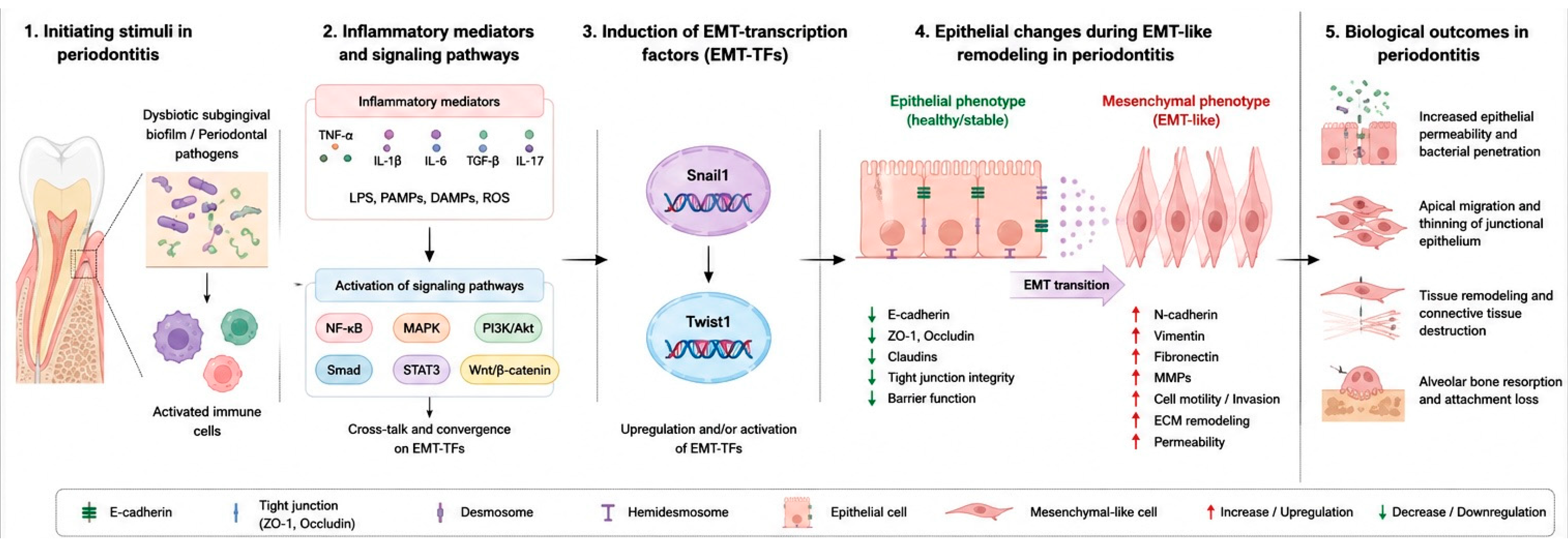
Periodontal pathogens and their components (e.g., lipopolysaccharide (LPS)) can directly induce EMT-associated genes [10,20]. Porphyromonas gingivalis and Fusobacterium nucleatum alter host cell signaling to generate EMT-like phenotypes in oral epithelial cells [10,43,44]. These pathogens activate NF-κB and MAPK pathways, inducing inflammation and intersecting with EMT pathways. F. nucleatum drives EMT in oral and colorectal cancers, demonstrating microbes’ capacity to prime such transitions [45,46]. This creates a destructive feedback loop where microbial challenge causes inflammation and EMT in gingival epithelium [10,20,47]. The mesenchymal-like cells produce pro-inflammatory mediators, perpetuating inflammation and tissue destruction [48]. Additionally, tobacco extracts can elicit EMT in gingival epithelial cells, augmenting the inflammatory damage [49] (Figure 1).
Figure 1.
Schematic overview of epithelial–mesenchymal transition in periodontitis. Dysbiotic subgingival biofilms and periodontal pathogens stimulate epithelial cells and immune cells, leading to release of inflammatory mediators such as TNF-α, IL-1β, IL-6, and TGF-β. These signals activate NF-κB, MAPK, PI3K/Akt, and Smad-related pathways, which converge on EMT-inducing transcription factors including Snail1 and Twist1. The resulting epithelial changes include reduced E-cadherin and junctional integrity, increased mesenchymal markers such as vimentin and N-cadherin, increased epithelial permeability, apical migration, and tissue remodeling.
p53 may link inflammation, epithelial integrity, and EMT-like remodeling, though direct periodontal evidence is limited [50]. In non-periodontal EMT models, wild-type p53 suppresses EMT by regulating microRNA networks like miR-34/Snail and miR-200/ZEB, maintaining epithelial differentiation and E-cadherin expression [50,51]. Loss or dysfunction of p53 can favor EMT-TF activity and repress epithelial adhesion programs. In periodontitis, recent evidence suggests p53 may reduce disease progression by decreasing M1-type macrophage differentiation and inflammatory cytokine expression. Thus, p53 might influence periodontal EMT indirectly by modulating the inflammatory microenvironment rather than being a direct EMT regulator in gingival epithelium [52]. Further periodontal-specific studies are needed to see if p53 signaling affects Snail1/Twist1 expression, E-cadherin loss, and epithelial barrier disruption in diseased periodontal tissues.
Besides activating EMT-related transcriptional programs, the inflammatory periodontal microenvironment may compromise the epithelial barrier through regulated cell death. Recent oral mucosal injury studies indicate epithelial depletion and barrier breakdown can be driven by ferroptosis, an iron-dependent regulated cell death form characterized by lipid peroxidation and impaired antioxidant defense [53]. Although evidence mainly comes from mucosal injury models, it is relevant as the periodontal pocket also experiences oxidative stress, microbial challenge, and inflammation. Periodontal studies support this, showing ferroptosis-associated proteins in gingival crevicular fluid during periodontitis progression [54]. Thus, epithelial barrier disruption in periodontitis likely reflects overlapping mechanisms, including EMT-like plasticity, oxidative stress, ferroptosis, apoptosis, and other programmed cell deaths [55].
3.3. Evidence of EMT in Preclinical and Clinical Models of Periodontitis
The conceptual link between inflammation and the process of EMT has triggered an investigation into the presence of EMT in periodontitis. Evidence from animal models and the literature using human samples all support the presence of EMT-like changes in periodontal disease [5,11,12]. For example, evidence of EMT in an animal model of ligature-induced periodontitis in rats produced traditional evidence of EMT in the gingival epithelium, loss of E-cadherin, and upregulation of mesenchymal markers such as vimentin, which all provide direct evidence in vivo that the disease process induces this cellular program [5]. Animal models provide the opportunity to investigate the temporal and spatial dynamics related to the initiation and progression of periodontitis, which is an important component of discovery [5].
Human translational studies agreed with preclinical studies. A key study comparing gingival tissues of patients with chronic periodontitis to healthy controls showed significantly lower E-cadherin expression with higher N-cadherin and EMT-TF Snail1 expression [11]. The expression levels of these markers correlated with clinical severity, suggesting EMT may track periodontal destruction progression [11]. Another study of periodontitis gingival tissues showed clear EMT phenotype evidence, supporting EMT’s role in active periodontitis [12]. Additional studies have linked EMT to peri-implant disease, suggesting it as a common pathway in inflammatory diseases of natural teeth and dental implants [16]. The consistent presence of EMT markers in diseased periodontal tissues supports its relevance in humans and highlights the need for periodontal-specific mechanistic and interventional studies.
To help synthesize the available evidence, the main periodontal and periodontal-relevant studies reporting EMT-associated changes are summarized in Table 1.
Table 1.
Summary of key studies reporting EMT-related changes relevant to periodontitis, Snail1, and Twist1.
3.4. Implications of EMT: Compromised Epithelial Barrier and Tissue Destruction
EMT processes lead to a significant detrimental influence on the integrity and function of periodontal tissue [56]. The gingival epithelium serves as a barrier between connective tissue and the oral cavity environment of oro bacteria [57]. And downregulation of E-cadherin impairs the AJs and epithelial barrier function [58]. Increased epithelial permeability permits further penetration of pathogenic bacteria, virulence factors such as LPS and increased microbial encounter [20]. The inflamed epithelium is unable to elicit an adequate response for continued exposure to microorganisms [59]. This triggers increased immune cell influx and release of tissue-damaging enzymes [60], amplifying collagen fiber breakdown in connective tissue and PDL [60]. EMT-related phenotypic changes cause tissue remodeling and destruction. Epithelial cells gain migratory capabilities, allowing junctional epithelium to move apically, leading to periodontal pockets and attachment loss [5,61]. Mesenchymal-like cells from EMT contribute to fibrotic changes by producing excessive disorganized ECM [62], limiting tissue function and regeneration [62]. The association between EMT in periodontitis and more severe disease states, such as oral squamous cell carcinoma has also been proposed, where the cellular plasticity induced by chronic inflammation and periodontal pathogens were suggested to establish a microenvironment that may lead to cancerous transformation [31,63]. At the same time, the destructive periodontal microenvironment cannot be explained by epithelial EMT alone. Immune-driven connective tissue destruction and bone resorption remain central features of periodontitis, particularly in metabolically compromised conditions such as diabetic periodontitis. Recent experimental work using bioactive Zn–V–Si–Ca glass nanoparticle hydrogel microneedles in diabetic periodontitis showed that modulation of the local microenvironment was associated with downregulation of JAK-STAT and NF-κB inflammatory signaling pathways and improved alveolar bone regeneration [64]. These findings support the view that persistent inflammatory signaling can directly drive connective tissue breakdown and osteoclastic bone loss, while EMT-like epithelial remodeling may mainly contribute to barrier weakening, epithelial migration, and altered tissue architecture. Thus, EMT should be interpreted as part of a wider pathological network that includes immune dysregulation, oxidative stress, programmed cell death, and inflammatory bone destruction.
4. Snail1 Functions as a Transcriptional Regulator of Periodontal EMT
The initiation and progression of epithelial–mesenchymal transition (EMT) rely on a core set of transcription factors that drive the extensive reprogramming of gene expression needed for a cell to lose its epithelial identity and acquire mesenchymal characteristics. One of the most significant core regulators is Snail1 (also referred to as Snail), a member of the Snail family of zinc-finger transcription factors. Snail1 is a strong transcriptional repressor because it represses genes that define the epithelial phenotype (e.g., genes involved in cell adhesion) [65]. The transcriptional effects of Snail1 are regulated by numerous signaling pathways, many of which are active in the inflamed periodontal environment, which will be further discussed in this chapter. In this chapter, we will outline the role of Snail1 in driving periodontal EMT by discussing its structure and function, its primary mechanism of action through E-cadherin repression, the literature tying Snail1 activation to periodontal pathogens, and its correlation with disease severity [65].
4.1. Snail1 Structure, Function, and Upstream Signaling Pathways
Snail1 is a member of the superfamily of Snail transcription factors and contains a highly conserved C-terminal region that possesses 4 to 6 C2H2-type zinc finger motifs. The zinc finger domain is required for function because it binds to specific DNA sequences named E-boxes (5′-CANNTG-3′) in target gene promoter regions [66]. The N-terminal region of the Snail1 protein has the SNAG domain (Snail/Gfi-1) that acts as a transcription repressor domain by recruiting a number of co-repressor complexes, including the histone deacetylase complex (HDAC) and several components of Polycomb repressive complex 2 (PRC2), which help condense and silence target gene expression in the epigenetic context, giving Snail1 a powerful and stable repressive ability [67].
Snail1 primarily serves as a master switch to drive EMT [67]. Snail1 expression is normally low or absent in healthy differentiated epithelia, but can be rapidly induced by specific developmental cues, tissue injury, or pathological stimuli [68]. Following activation, Snail1 initiates a signaling cascade that leads to loss of epithelial polarity, acquisition of a migratory and invasive phenotype, and resistance to apoptosis [68]. While much is known based on developmental biology and cancer metastasis literature, the proposed function as a pathogenic mediator in chronic inflammatory diseases such as periodontitis is still relatively unexplored [13].
Like many transcription factors, expression and activity of Snail1 are regulated by a complex network of upstream signaling pathways, many of which are highly relevant to the periodontal inflammatory environment [69]. Key inducers include:
- Transforming Growth Factor-β (TGF-β) Signaling: The TGF-β/Smad pathway is perhaps the most potent and well-characterized inducer of Snail1 [70]. As a result of ligand binding, TGF-β receptors phosphorylate Smad2 and Smad3, which then heterodimerize with Smad4 [70]. This complex translocates to the nucleus and directly activates transcription of the SNAI1 gene. With the abundance of TGF-β measured in inflamed and fibrotic tissues of the periodontium, the TGF-β pathway is likely a primary mechanism of Snail1 activation in periodontitis [70].
- Wnt/β-catenin Signaling: The canonical Wnt pathway can also induce Snail1 expression as it leads to stabilization and nuclear accumulation of β-catenin [71]. In the nucleus, β-catenin forms a complex with TCF/LEF transcription factors to activate target genes, including SNAl1. Although largely documented crosstalk has been shown between Wnt signaling and inflammation, there is likely another pathway for Snail1 induction in periodontal pockets [72].
- Receptor Tyrosine Kinase (RTK) Pathways: Growth factors such as epidermal growth factor (EGF), fibroblast growth factor (FGF), and hepatocyte growth factor (HGF) can activate RTKs, leading to downstream activation of the Ras/MAPK and PI3K/Akt signaling cascades. Both pathways can increase Snail1 expression and stability, typically by phosphorylating and inhibiting Glycogen Synthase Kinase 3β (GSK3β), a kinase that specifically targets Snail1 for proteasomal degradation [73].
- Inflammatory Signaling: Pro-inflammatory cytokines such as tumor necrosis factor-α (TNF-α) and Interleukin-6 (IL-6) can increase Snail1 expression through signaling pathways such as NF-κB and STAT3. These findings provide a potential direct link between the chronic inflammatory state of periodontitis and active core EMT machinery [74].
The convergence of these multiple signaling cascades onto the SNA1 gene serves to emphasize its central position as a hub for pro-EMT signals. In periodontitis, the sustained release of bacterial products along with pro-inflammatory cytokines and growth factors establishes a microenvironment that would tend to maintain Snail1 constantly activated leading to an indefinite maladaptive EMT program (Table 2).
Table 2.
Upstream factors and signaling pathways associated with Snail1 and Twist1 activation in periodontal or periodontal-relevant EMT.
4.2. Snail1-Mediated E-Cadherin Repression and Disruption of Cell Adhesion
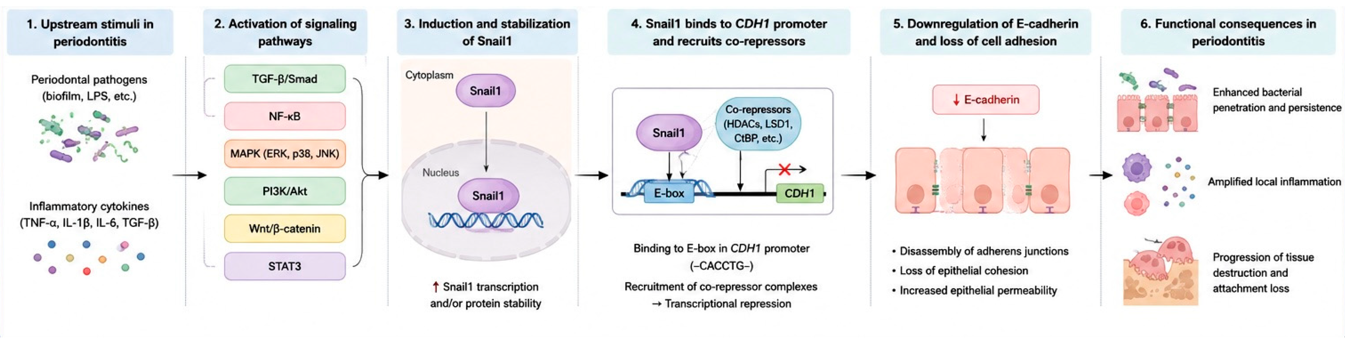
Snail1 is particularly important in periodontal EMT because it directly represses epithelial adhesion programs. Its best-characterized target is CDH1, the gene encoding E-cadherin, which is essential for adherens junction integrity and epithelial cohesion [20]. By binding to E-box sequences within the CDH1 promoter and recruiting co-repressor complexes, Snail1 epigenetically suppresses E-cadherin transcription and weakens epithelial architecture [75].
The implications of downregulating E-cadherin are deep and far-reaching. The first immediate implication is that dissolving strong intercellular adhesions frees cells from the epithelial sheet and promotes cell motility [20]. The second implication is that loss of E-cadherin removes sequestration of β-catenin at the cell membrane. Free of adherens junction complex, β-catenin can translocate to the nucleus and acts as a transcriptional co-activator for genes responsible for cell proliferation, and mesenchymal differentiation, including Snail1, resulting in a positive feedback loop that supports the EMT state [12].
Snail1 represses E-cadherin and has broad repression mechanisms of epithelial genes. Snail1 represses junction proteins, including claudins and occludins (tight junction components), and desmoplakin and desmoglein (desmosomal components) [76]. This attack on cell adhesion complexes dismantles epithelial barrier function [12]. Within the gingival epithelium, disruption of epithelial seal by Snail1 has pathological significance. Compromised barrier function allows bacterial toxins and antigens from dental plaque biofilm to penetrate connective tissues [20]. The influx of microbial products aggravates local inflammation, creating a cycle where inflammation increases Snail1 expression—leading to greater barrier dysfunction and severe tissue destruction [20]. Therefore, Snail1 repression of E-cadherin is not just a cellular event but a critical step in periodontitis pathogenesis (Figure 2).
Figure 2.
Mechanistic role of Snail1 in periodontal epithelial–mesenchymal transition. Periodontal pathogens and inflammatory mediators activate upstream pathways, including TGF-β/Smad, NF-κB, MAPK, PI3K/Akt, Wnt/β-catenin, and STAT3 signaling. These pathways increase Snail1 expression and/or stability. Activated Snail1 translocates to the nucleus, binds E-box sequences within the CDH1 promoter, and recruits transcriptional co-repressor complexes, resulting in E-cadherin suppression. Loss of E-cadherin weakens adherens junctions, increases epithelial permeability, facilitates bacterial product penetration, and may amplify local inflammation and periodontal tissue destruction.
Stress-response chaperones may also influence E-cadherin stability and EMT-related epithelial cohesion. HSP70 appears to have context-dependent effects, as it may preserve epithelial features in some models but promote EMT-like changes through p38/MAPK signaling in others [77]. HSP70-2/HSPA2 has also been linked to EMT regulation, with its suppression associated with increased E-cadherin and reduced Snail/Twist-related mesenchymal markers [78]. Although these findings are not periodontitis-specific, they suggest that chaperone responses may modify epithelial adhesion under chronic inflammatory stress.
4.3. Links Between Periodontal Pathogens and Snail1 Activation
The chronic microbial stimulus from a dysbiotic subgingival biofilm is the major etiologic agent of periodontitis. Increasing evidence now indicates that certain periodontal pathogens are also directly involved in activation of the EMT program, with Snail1 often being proposed as a central player [20]. These pathogens have developed complex strategies to control host cell signaling networks, often involving those that drive EMT.
Research has shown that keystone periodontal pathogens can alter the expression of transcription factors related to the irreversible epithelial–mesenchymal transition (EMT) program [31]. For example, Porphyromonas gingivalis, a recognized bacterial pathogen of chronic periodontitis, demonstrated the ability to modulate host cell signaling in ways that affected Snail1 expression [20]. In some situations, such as in models of esophageal cancer, the research reported that P. gingivalis stimulated signaling pathways that inhibited Snail protein; however, as is often the case with bacteria, the role of P. gingivalis in the host periodontal inflammatory environment is likely context-dependent [79]. The usage of in vitro models has demonstrated that stimulation of oral epithelial cells with components of periodontal pathogens, such as lipopolysaccharide (LPS), have led to the development of an EMT-like phenotype, where E-cadherin was decreased, while mesenchymal markers were increased. This process is often marked by a dramatic upregulation of Snail1, suggesting the presence of bacterial products were able to stimulate the transcriptional machinery of EMT in epithelial cells, and that these products had directly engaged the infection and resulted in the activation of the core genes involved in EMT [5].
These pathways through which periodontal pathogens can activate Snail1 are complex in nature. They can directly stimulate host cell receptors, such as the toll-like receptor (TLR) family, which detects pathogen-associated molecular patterns (PAMPs) such as LPS, which ultimately trigger downstream inflammatory signaling cascades such as NF-κB and MAPK. As we mentioned previously, these signaling pathways activate Snail1 expression [80]. Additionally, pathogens can induce a local inflammatory microenvironment, rich in cytokines, such as TGF-β and TNF-α, that also act as strong activators of Snail1 [80]. Thus, periodontal pathogens can activate Snail1 directly, by engaging epithelial cells, and indirectly by orchestrating a pro-EMT inflammatory microenvironment [20]. The positive link between microbial challenge and activation of a master regulator of EMT is an important piece to connect the root cause of periodontitis (bacterial dysbiosis) with a fundamental cellular mechanism of destruction (EMT) [12].
4.4. Snail1 Expression and Correlation with Clinical Severity of Periodontitis
Immunohistochemical studies of gingival tissue biopsies from patients with periodontitis show evidence of upregulated Snail1 in diseased tissues compared to cells from healthy controls [11]. These studies showed that Snail1 expression was localized to the basal and suprabasal layers of the pocket epithelium, which is the anatomical point of the greatest interaction between the microbial biofilm and host tissues [11].
A very important confirmation of the Snail1 role in periodontal disease relates to the observation that it is elevated in tissues with deeper periodontal pockets and greater clinical attachment loss, and greater degrees of inflammation [11]. In particular, the Snail1 protein staining intensity was significantly stronger in biopsies of diseased periodontal tissue with higher clinical severity of periodontitis compared to those from patients with healthy periodontium [11]. This dose-dependency indicates that Snail1 is not a passive molecule but rather, has an active and functional effect in facilitating pathological processes within the gingival tissues in terms of promoting disease progression. Moreover, in human tissues, increased levels of Snail1 occurred concomitantly with decreased amounts of E-cadherin and therefore served as an in vivo evidence for repressive role other than impairing the epithelial barrier during periodontitis patients [11]. The upregulation of Snail1 by inflammation and disease in periodontal lesions has several implications. First, it validates in vitro and pre-clinical model system data showing that Snail1-induced EMT is active in human periodontitis. Second, it indicates a potential role of Snail1 as a biomarker for disease activity and severity. It seems possible that the monitoring of Snail1 in gingival biopsies (or perhaps even in GCF), given its ability to predict, could indicate a patient at risk for continuous tissue loss. From a clinical point of view, the increased Snail1 expression levels could be indicative of a more inflamed and remodeled epithelium and may be worth investigating as potential severity marker [11,20]. However, functional validation is needed to determine whether Snail1 has a direct role in mediating periodontal destruction or acts as an inflammation-driven phenomenon [12,81].
5. The Role of Twist1 in Controlling the Mesenchymal Shift
Twist-related protein 1 (Twist1) is a crucial transcription factor. In contrast to repressor Snail1, Twist1 acts as a master regulator by directly stimulating the expression of genes inclined to define mesenchymal phenotypes related to motility, invasion, and matrix remodeling rather than bestowing repression or suppression [82]. Twist1, however, had been demonstrated to be also dysregulated in pathological conditions such as chronic inflammatory diseases, including periodontitis [11]. Thus, activation of Twist1in the periodontal microenvironment is another important event that can lead to transition from compromised epithelial barrier function to a more aggressive tissue-damaging phenotype and a causative factor in the pathogenesis of periodontitis [11].
5.1. Twist1 as a Master Regulator of Mesenchymal Gene Expression
Unlike Snail1, which is mainly associated with repression of epithelial junctional genes, Twist1 is more closely linked to the activation of mesenchymal programs [83]. Through its basic helix–loop–helix structure and E-box binding activity, Twist1 promotes genes involved in motility, cytoskeletal remodeling, extracellular matrix interaction, and invasion [84]. These include N-cadherin, vimentin, fibronectin, and matrix metalloproteinases.
In this way, Twist1 contributes to cellular reprogramming by reducing epithelial stability and promoting a more migratory phenotype. In oral diseases and other related pathologies, increased Twist1 expression has been suggested as a marker of EMT activation and tissue remodeling [83].
5.2. The Consequences of Twist1 Activation on Cell Motility, Invasion, and Fibrosis of Tissues in the Periodontium
Activation of Twist1 within periodontal epithelium plays a role in tissue homeostasis, and in periodontitis development by facilitating cellular motility, invasion and fibrotic activities. These phenotypes result in degradation of the gingival barrier and abnormal remodelling of connective tissue that leads to loss of the tooth-supporting apparatus [13]. Twist1 promotes cell motility and invasion through the upregulation of genes associated with cytoskeletal remodeling and ECM proteolysis. It upregulates front-rear polarity and cytoskeletal plasticity (required for motility) by inducing N-cadherin and vimentin genes [85]. Twist1 also induces MMP expression to degrade the basement membrane, which is essential for cell invasion through the lamina propria. In periodontitis, such invasion ruptures the integrity of junctional epithelium leading to pathogen entry and pro-inflammatory loops [85]. Furthermore, invasive mesenchymal cells derived from epithelia migrate together with activated fibroblast-myofibroblasts that drive tissue remodeling [12].
Twist1-regulated-mesenchymal cells deposit collagen ECM proteins that lead to dense non-oriented scar-like tissue [86]. This fibrotic remodeling creates dense periodontal ligament tissue that loses organized structure, resulting in dysfunction. Combined with sustained inflammation, this creates a stiffened microenvironment unable to support tooth regeneration [87].
6. Integrated Roles of Snail1 and Twist1 in Periodontal EMT
6.1. Synergistic and Independent Roles of Twist1 and Snail1
Although Snail1 and Twist1 are discussed separately, periodontal EMT-like remodeling is unlikely to be controlled by a single transcription factor [88]. A more realistic interpretation is that Snail1 and Twist1 act as complementary regulators within the same inflammatory epithelial plasticity program [88]. Snail1 appears more closely related to the early loss of epithelial cohesion through repression of E-cadherin and other junctional proteins, whereas Twist1 appears more closely related to mesenchymal activation, motility, extracellular matrix interaction, and invasive behavior [76] (Table 3).
Table 3.
Downstream molecules and phenotypic effects associated with Snail1 and Twist1 activation.
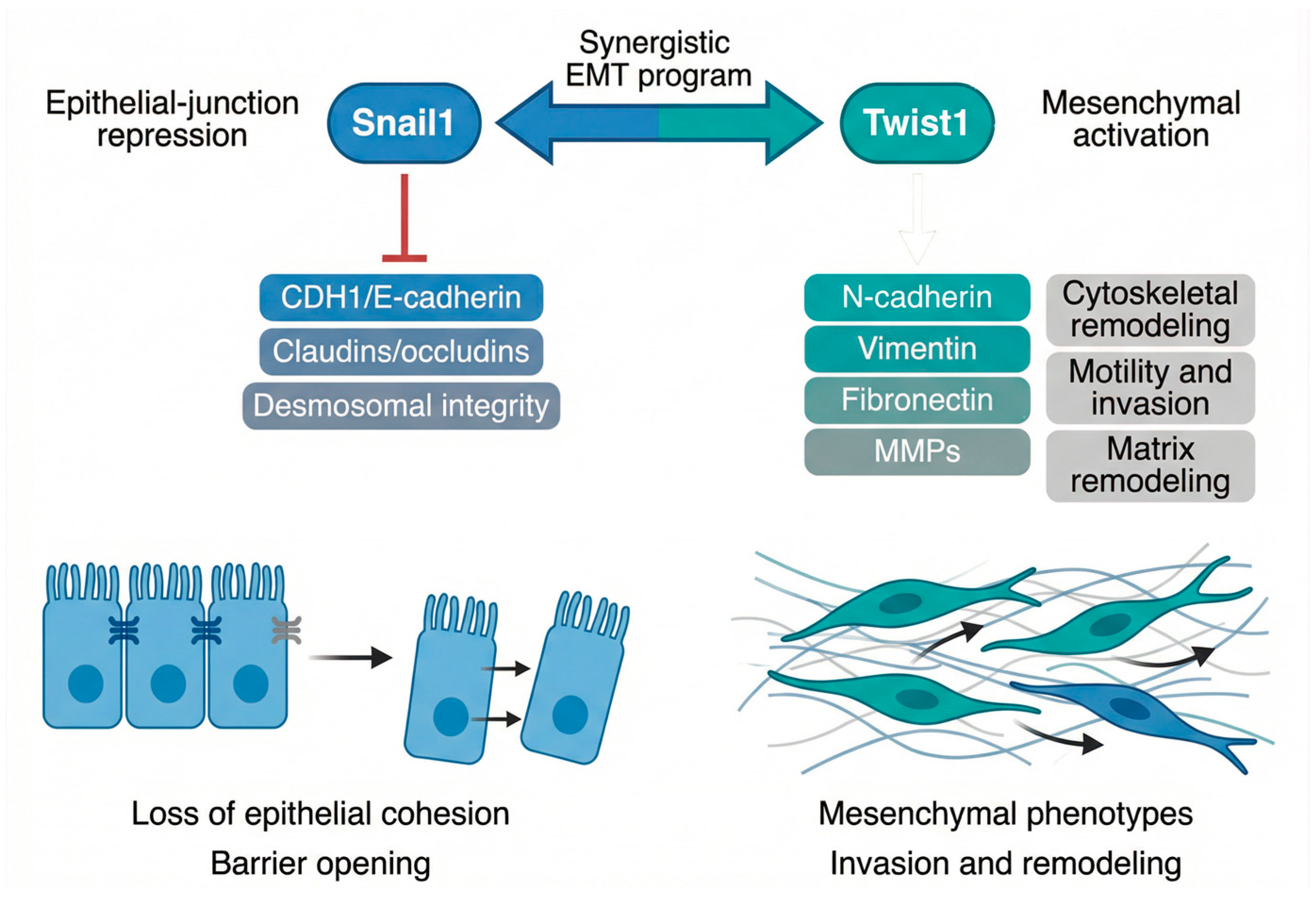
Snail1 and Twist1 collaborate to induce epithelial–mesenchymal transition, which also leads to disassembly of adherens junctions. Snail1 directly inhibits the transcription of E-cadherin promoter by binding to its E-boxes [89]. Twist1 promotes a “cadherin switch,” regulating E-cadherin and activating mesenchymal genes like N-cadherin [12], weakening epithelial adhesion and altering signaling cascades for cell motility [84]. Whereas Snail1 dissolves cell-cell adhesions, Twist1 promotes mesenchymal adhesion and the engagement of migration machinery [90]. They act both separately and interdependently, differentiating roles from their evolutionary predecessor [90]. Snail1, a zinc-finger transcription factor, represses transcription during de-epithelialization [76]. Twist1, a bHLH transcription factor regulates the mesenchymal genes leading to enhancing the invasiveness, resistance to apoptosis and modulation of matrix [91]. EMT involves loss of epithelial features and mesenchymal phenotype acquisition [92]. In that disease, Snail1 dismantles barriers to bacterial entry, while Twist1 initiates lymphocyte-independent chronic inflammation and fibrotic remodeling [13] (Figure 3).
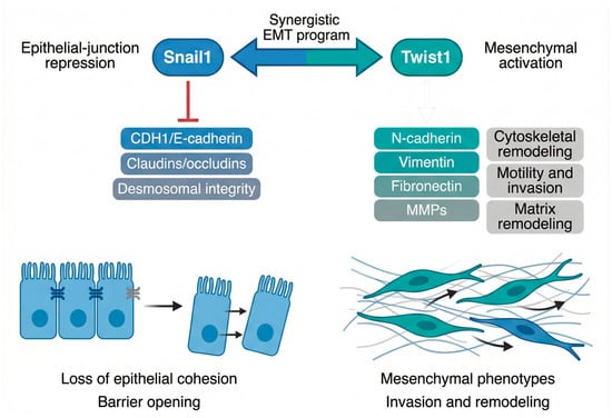
Figure 3.
Distinct but synergistic functions of Snail 1 and Twist 1: Snail1 primarily represses epithelial-junction programs, including CDH1/E-cadherin, claudins/occludins, and desmosomal integrity, thereby promoting loss of epithelial cohesion and barrier opening. Twist1 promotes mesenchymal activation, including N-cadherin, vimentin, fibronectin, MMPs, cytoskeletal remodeling, and invasion; together, both factors cooperate to drive EMT-like remodeling in periodontitis.
6.2. Unified Model of Snail1/Twist1-Mediated Epithelial Remodeling in Periodontitis
A unified model can be proposed in which dysbiotic subgingival biofilms initiate a persistent inflammatory and oxidative microenvironment. Bacterial products, cytokines, growth factors, and ROS activate redox-sensitive and inflammation-related pathways, including NF-κB, MAPK, PI3K/Akt, STAT3, Wnt/β-catenin, and TGF-β/Smad signaling [13,93]. These pathways converge on EMT-associated transcription factors, particularly Snail1 and Twist1. Snail1 mainly represses epithelial adhesion genes such as CDH1/E-cadherin and contributes to junctional weakening, while Twist1 supports mesenchymal activation, cytoskeletal remodeling, matrix interaction, and cell motility [5]. In parallel, immune activation, ferroptosis or other regulated cell death pathways, matrix metalloproteinase activity, osteoclast activation, and connective tissue breakdown contribute to the destructive periodontal phenotype [53,55]. Thus, EMT-like remodeling should be viewed as one component of a broader disease network, linking epithelial barrier disruption to inflammatory tissue destruction rather than replacing the established immune-microbial model of periodontitis.
7. Translational Considerations: Challenges and Future Directions
7.1. Challenges in Translating Snail1/Twist1 Biology into Periodontal Therapy
Although Snail1 and Twist1 are attractive mechanistic targets, their translation into periodontal therapy remains premature. The first challenge is causality. Most periodontal evidence is still based on marker expression in diseased tissues or experimental models, and increased Snail1 or Twist1 expression does not prove that these factors directly drive clinical attachment loss or bone destruction. The second challenge is biological specificity. EMT-related transcription factors are not exclusively pathological; they also participate in epithelial repair, wound healing, and tissue turnover. Broad inhibition may therefore interfere with healing after periodontal therapy. A third challenge is therapeutic delivery. Transcription factors are difficult to inhibit directly, and systemic modulation of upstream pathways such as TGF-β, NF-κB, STAT3, MAPK, or PI3K/Akt may carry unacceptable off-target effects. For periodontal application, local delivery into periodontal pockets would be more realistic, but this approach still requires careful control of drug retention, dose, safety, and timing.
Another important limitation is that EMT is only one part of the periodontal destructive network. Immune activation, oxidative stress, ferroptosis, bacterial dysbiosis, matrix metalloproteinase activity, osteoclast activation, and impaired repair all interact with epithelial plasticity. For this reason, Snail1 and Twist1 should not yet be presented as direct therapeutic targets in clinical periodontology. At present, they are better considered as mechanistic biomarkers or experimental targets that may help clarify how epithelial barrier dysfunction contributes to periodontal progression.
7.2. Future Directions
Future work should first establish whether Snail1 and Twist1 are causally involved in periodontal tissue destruction or mainly reflect the inflammatory state of diseased tissues. This will require periodontal-specific mechanistic studies using human gingival epithelial cells, organotypic models, animal periodontitis models, and spatial tissue analysis. Studies should combine EMT markers with epithelial barrier markers, regulated cell death markers, inflammatory mediators, and bone resorption markers rather than relying on single-marker interpretation.
A second priority is to improve the spatial and cellular resolution of EMT assessment in periodontal tissues. Immunohistochemistry remains useful, but future studies would benefit from multiplex staining, spatial transcriptomics, and single-cell approaches to determine whether EMT-like changes occur mainly in junctional epithelium, pocket epithelium, basal epithelial layers, or other epithelial compartments. This is particularly important because inflammatory stromal and immune cells can express markers that overlap with EMT-associated signatures.
A third direction is translational testing. Before any clinical use is considered, interventions that modulate Snail1/Twist1-related pathways should be tested in preclinical periodontal models using both molecular and clinical endpoints. These should include E-cadherin and tight junction preservation, epithelial permeability, inflammatory cytokine reduction, probing-depth-related outcomes, attachment preservation, and alveolar bone protection. Local delivery systems may be especially relevant because they could target the periodontal pocket while reducing systemic exposure. However, these approaches need to be evaluated cautiously because excessive suppression of epithelial plasticity could impair repair.
Several plant-derived, animal-derived, and synthetic agents have been reported to modulate EMT-related pathways in cancer, fibrosis, or oral disease models; however, their periodontal use remains experimental and should not be interpreted as current clinical recommendation (Table 4).
Table 4.
Examples of pharmacological agents reported to modulate EMT-related pathways.
8. Conclusions
EMT-like remodeling seems to be part of periodontal inflammation. Across studies, diseased tissues and experimental models tend to show lower epithelial junction markers and higher mesenchymal markers. Snail1 and Twist1 are often highlighted because they occupy different points in this pathway, with Snail1 linked to loss of epithelial adhesion and Twist1 linked to the mesenchymal activation and matrix changes. Still, most of the evidence is preclinical or only associative in human samples, so questions about which cells are driving it, whether EMT is truly causal, and whether it can be safely targeted remain open. Future studies will likely need improved marker panels and more cell- and spatial-level methods, plus periodontal-specific functional work, to determine whether modulating these programs can actually improve barrier function and clinical outcomes.
Author Contributions
Conceptualization, H.M.A. and S.S.S.; methodology, H.M.A.; software, H.M.A.; validation, H.M.A. and S.S.S.; formal analysis, H.M.A.; investigation, H.M.A.; resources, H.M.A.; data curation, H.M.A.; writing—original draft preparation, H.M.A.; writing—review and editing, H.M.A. and S.S.S.; visualization, H.M.A.; supervision, S.S.S.; project administration, H.M.A.; funding acquisition, not applicable. All authors have read and agreed to the published version of the manuscript.
Funding
This research received no external funding.
Institutional Review Board Statement
Not applicable.
Informed Consent Statement
Not applicable.
Data Availability Statement
No new experimental data were generated. The illustrative figures created for this review are available from the corresponding author upon reasonable request.
Acknowledgments
During the preparation of this manuscript, the authors used Nano Banana 2 (Google DeepMind) to enhance and refine the visual quality of figures based on author-created sketches and figure designs. The tool was not used for study design, data generation, data analysis, interpretation of results, or the creation of scientific content. All figures were reviewed and edited by the authors, who take full responsibility for the content of this publication.
Conflicts of Interest
The authors declare no conflicts of interest.
Abbreviations
The following abbreviations are used in this manuscript:
| Akt | Protein Kinase B |
| bHLH | Basic Helix–Loop–Helix |
| CDH1 | The gene that encodes E-cadherin |
| ECM | Extracellular Matrix |
| EGF | Epidermal Growth Factor |
| EMT | Epithelial–Mesenchymal Transition |
| EMT-TFs | EMT-Inducing Transcription Factors |
| FGF | Fibroblast Growth Factor |
| GSK3β | Glycogen Synthase Kinase 3 beta |
| HDAC | Histone Deacetylase |
| HGF | Hepatocyte Growth Factor |
| IL-1β | Interleukin-1 |
| IL-6 | Interleukin-6 |
| lncRNAs | Long Non-Coding RNAs |
| LPS | Lipopolysaccharide |
| MAPK | Mitogen-Activated Protein Kinase |
| miRNAs | MicroRNAs |
| MMPs | Matrix Metalloproteinases |
| NF-κB | Nuclear Factor kappa-light-chain-enhancer of activated B cells |
| PAMPs | Pathogen-Associated Molecular Patterns |
| PDL | Periodontal Ligament |
| PI3K | Phosphatidylinositol 3-Kinase |
| PRC2 | Polycomb Repressive Complex 2 |
| RTK | Receptor Tyrosine Kinase |
| siRNA | Small Interfering RNA |
| Smad | Mothers Against Decapentaplegic Homolog |
| SNAI1 | The gene that encodes Snail1 |
| SNAG domain | Snail/Gfi-1 domain |
| Snail1 (Snail) | Snail Family Transcriptional Repressor 1 |
| STAT3 | Signal Transducer and Activator of Transcription 3 |
| TGF-β | Transforming Growth Factor-beta |
| TLR | Toll-Like Receptor |
| TNF-α | Tumor Necrosis Factor-alpha |
| Twist1 | Twist-related protein 1 |
| Wnt | Wingless-related integration site |
| ZEB | Zinc Finger E-Box Binding Homeobox |
References
- Caetano, A.J.; Yianni, V.; Volponi, A.; Booth, V.; D’Agostino, E.M.; Sharpe, P. Defining human mesenchymal and epithelial heterogeneity in response to oral inflammatory disease. eLife 2021, 10, e62810. [Google Scholar] [CrossRef] [Scilit] [PubMed]
- Sczepanik, F.S.C.; Grossi, M.L.; Casati, M.; Goldberg, M.; Glogauer, M.; Fine, N.; Tenenbaum, H.C. Periodontitis is an inflammatory disease of oxidative stress: We should treat it that way. Periodontology 2020, 84, 45–68. [Google Scholar] [CrossRef] [Scilit] [PubMed]
- Alvarez, C.; Abdalla, H.; Suliman, S.; Rojas, P.; Wu, Y.-C.; Almarhoumi, R.; Huang, R.-Y.; Galindo, M.; Vernal, R.; Kantarci, A. RvE1 impacts the gingival inflammatory infiltrate by inhibiting the T cell response in experimental periodontitis. Front. Immunol. 2021, 12, 664756. [Google Scholar] [CrossRef] [Scilit] [PubMed]
- Imran, N.K.; Abdulbaqi, H.R.; Milward, M. The prevalence of periodontitis in an Iraqi population using the 2017 classification. J. Baghdad Coll. Dent. 2024, 36, 1–10. [Google Scholar] [CrossRef] [Scilit]
- Alanbari, B.F.; Al-Taweel, F.B.; Cooper, P.R.; Milward, M.R. Induction of Epithelial–Mesenchymal Transition in Periodontitis Rat Model. Eur. J. Dent. 2025, 19, 428–437. [Google Scholar] [CrossRef] [Scilit] [PubMed]
- Mohammed, A.; Fadhil, R.; Mahmood, M.S.; Al-Waeli, H.A. Diagnostic biomarkers for periodontitis (observational case-control study). J. Baghdad Coll. Dent. 2024, 36, 11–19. [Google Scholar] [CrossRef] [Scilit]
- Al-Rihaymee, S.; Mahmood, M.S. Platelet-rich fibrin potential role in periodontal regeneration: A review study. Med. J. Babylon 2023, 20, 1–6. [Google Scholar] [CrossRef] [Scilit] [PubMed]
- Sharaf, S.; Hijazi, K. Modulatory mechanisms of pathogenicity in porphyromonas gingivalis and other periodontal pathobionts. Microorganisms 2022, 11, 15. [Google Scholar] [CrossRef] [Scilit] [PubMed]
- Dahlen, G.; Basic, A.; Bylund, J. Importance of virulence factors for the persistence of oral bacteria in the inflamed gingival crevice and in the pathogenesis of periodontal disease. J. Clin. Med. 2019, 8, 1339. [Google Scholar] [CrossRef] [Scilit] [PubMed]
- Abdulkareem, A.; Shelton, R.; Landini, G.; Cooper, P.; Milward, M. Periodontal pathogens promote epithelial-mesenchymal transition in oral squamous carcinoma cells in vitro. Cell Adhes. Migr. 2018, 12, 127–137. [Google Scholar] [CrossRef] [Scilit] [PubMed]
- Saliem, S.S.; Bede, S.Y.; Abdulkareem, A.A.; Abdullah, B.H.; Milward, M.R.; Cooper, P.R. Gingival tissue samples from periodontitis patients demonstrate epithelial–mesenchymal transition phenotype. J. Periodontal Res. 2023, 58, 247–255. [Google Scholar] [CrossRef] [Scilit] [PubMed]
- Abdulkareem, A.A. Potential Involvement of Epithelial-Mesenchymal Transition in the Pathogenesis of Periodontitis; University of Birmingham: Birmingham, UK, 2017; Available online: https://etheses.bham.ac.uk/7340/1/Abdulkareem17PhD.pdf (accessed on 11 May 2026).
- Saliem, S.S.; Bede, S.Y.; Cooper, P.R.; Abdulkareem, A.A.; Milward, M.R.; Abdullah, B.H. Pathogenesis of periodontitis–A potential role for epithelial-mesenchymal transition. Jpn. Dent. Sci. Rev. 2022, 58, 268–278. [Google Scholar] [CrossRef] [Scilit] [PubMed]
- Dieterle, M.P.; Husari, A.; Steinberg, T.; Wang, X.; Ramminger, I.; Tomakidi, P. Role of mechanotransduction in periodontal homeostasis and disease. J. Dent. Res. 2021, 100, 1210–1219. [Google Scholar] [CrossRef] [Scilit] [PubMed]
- Acloque, H.; Yang, J.; Theveneau, E. Epithelial-to-mesenchymal plasticity from development to disease: An introduction to the special issue. Genesis 2023, 62, e23581. [Google Scholar] [CrossRef] [Scilit] [PubMed]
- Alnajaty, T.A.K.; Abdulbaqi, H.R.; Soliman, O. Epithelial-mesenchymal transition: A novel target for preventing and treating peri-implant diseases. J. Med. Pharm. Chem. Res. 2025, 7, 63–1144. [Google Scholar] [CrossRef]
- Li, S.; Liu, X.; Zhou, Y.; Acharya, A.; Savkovic, V.; Xu, C.; Wu, N.; Deng, Y.; Hu, X.; Li, H.; et al. Shared genetic and epigenetic mechanisms between chronic periodontitis and oral squamous cell carcinoma. Oral Oncol. 2018, 86, 216–224. [Google Scholar] [CrossRef] [Scilit] [PubMed]
- Đapić, I.; Aurer, A.; Žučko, J.; Mravak-Stipetić, M.; Baričević, M.B.; Pavelić, K.; Ozer, F.; Pavelić, S.K. Enhancing Professional Periodontal Therapy with a Novel PMA-Zeolite Application: A Clinical Study on Periodontal Outcomes and Microbiological Changes. J. Funct. Biomater. 2025, 16, 270. [Google Scholar] [CrossRef] [Scilit] [PubMed]
- Bhuyan, R.; Bhuyan, S.K.; Mohanty, J.N.; Das, S.; Juliana, N.; Abu, I.F. Periodontitis and its inflammatory changes linked to various systemic diseases: A review of its underlying mechanisms. Biomedicines 2022, 10, 2659. [Google Scholar] [CrossRef] [Scilit] [PubMed]
- Abdulkareem, A.A.; Shelton, R.; Landini, G.; Cooper, P.; Milward, M.R. Potential role of periodontal pathogens in compromising epithelial barrier function by inducing epithelial-mesenchymal transition. J. Periodontal Res. 2018, 53, 565–574. [Google Scholar] [CrossRef] [Scilit] [PubMed]
- Marconi, G.D.; Fonticoli, L.; Rajan, T.S.; Pierdomenico, S.D.; Trubiani, O.; Pizzicannella, J.; Diomede, F. Epithelial-mesenchymal transition (EMT): The type-2 EMT in wound healing, tissue regeneration and organ fibrosis. Cells 2021, 10, 1587. [Google Scholar] [CrossRef] [Scilit] [PubMed]
- Debnath, D.; Tatuskar, P.; Attar, S.M.F. From bacterial origins to multifactorial complexity: Tracing the evolution of periodontitis pathogenesis from early era to 2017: A review article. Int. J. Oral Health Sci. 2024, 14, 55–66. [Google Scholar] [CrossRef] [Scilit] [PubMed]
- Kalluri, R.; Weinberg, R.A. The basics of epithelial-mesenchymal transition. J. Clin. Investig. 2009, 119, 1420–1428. [Google Scholar] [CrossRef] [Scilit] [PubMed]
- Kim, D.H.; Xing, T.; Yang, Z.; Dudek, R.; Lu, Q.; Chen, Y.-H. Epithelial mesenchymal transition in embryonic development, tissue repair and cancer: A comprehensive overview. J. Clin. Med. 2017, 7, 1. [Google Scholar] [CrossRef] [Scilit] [PubMed]
- Saitoh, M. (Ed.) Transcriptional regulation of EMT transcription factors in cancer. In Seminars in Cancer Biology; Elsevier: Amsterdam, The Netherlands, 2023. [Google Scholar] [CrossRef] [Scilit] [PubMed]
- Aban, C.E.; Lombardi, A.; Neiman, G.; Biani, M.C.; La Greca, A.; Waisman, A.; Moro, L.N.; Sevlever, G.; Miriuka, S.; Luzzani, C. Downregulation of E-cadherin in pluripotent stem cells triggers partial EMT. Sci. Rep. 2021, 11, 2048. [Google Scholar] [CrossRef] [Scilit] [PubMed]
- Fang, C.; Kang, Y. E-cadherin: Context-dependent functions of a quintessential epithelial marker in metastasis. Cancer Res. 2021, 81, 5800–5802. [Google Scholar] [CrossRef] [Scilit] [PubMed]
- Shash, L.S.; Ibrahim, R.A.; Elgohary, S.A. E-cadherin and N-cadherin immunohistochemical expression in proliferating urothelial lesions: Potential novel cancer predictive EMT profiles. Appl. Immunohistochem. Mol. Morphol. 2021, 29, 657–666. [Google Scholar] [CrossRef] [Scilit] [PubMed]
- Usman, S.; Waseem, N.H.; Nguyen, T.K.N.; Mohsin, S.; Jamal, A.; Teh, M.-T.; Waseem, A. Vimentin is at the heart of epithelial mesenchymal transition (EMT) mediated metastasis. Cancers 2021, 13, 4985. [Google Scholar] [CrossRef] [Scilit] [PubMed]
- Debnath, P.; Huirem, R.S.; Bhowmick, A.; Ghosh, A.; Ghosh, D.; Dutta, P.; Maity, D.; Palchaudhuri, S. Epithelial mesenchymal transition induced nuclear localization of the extracellular matrix protein Fibronectin. Biochimie 2024, 219, 142–145. [Google Scholar] [CrossRef] [Scilit] [PubMed]
- Ma, Y.; Yu, Y.; Yin, Y.; Wang, L.; Yang, H.; Luo, S.; Zheng, Q.; Pan, Y.; Zhang, D. Potential role of epithelial–mesenchymal transition induced by periodontal pathogens in oral cancer. J. Cell. Mol. Med. 2024, 28, e18064. [Google Scholar] [CrossRef] [Scilit] [PubMed]
- Albuslutan, A.A.; Saliem, S.S.; Shibli, J. Impact of oral hygiene instructions on cytokines in smokers and vapers with gingivitis. J. Baghdad Coll. Dent. 2025, 37, 78–89. [Google Scholar] [CrossRef] [Scilit]
- Ali, A.M.; Mohammed, A.N.; Al-Waeli, H.A.; Al-Ani, H.A. Association of salivary Interleukin-6 levels in smokers with periodontitis. J. Baghdad Coll. Dent. 2024, 36, 39–49. [Google Scholar] [CrossRef] [Scilit]
- Sisto, M.; Lisi, S.; Ribatti, D. The role of the epithelial-to-mesenchymal transition (EMT) in diseases of the salivary glands. Histochem. Cell Biol. 2018, 150, 133–147. [Google Scholar] [CrossRef] [Scilit] [PubMed]
- Foroutan, M.; Cursons, J.; Hediyeh-Zadeh, S.; Thompson, E.W.; Davis, M.J. A transcriptional program for detecting TGFβ-induced EMT in cancer. Mol. Cancer Res. 2017, 15, 619–631. [Google Scholar] [CrossRef] [Scilit] [PubMed]
- Saitoh, M. Epithelial–mesenchymal transition by synergy between transforming growth factor-β and growth factors in cancer progression. Diagnostics 2022, 12, 2127. [Google Scholar] [CrossRef] [Scilit] [PubMed]
- Giarratana, A.O.; Prendergast, C.M.; Salvatore, M.M.; Capaccione, K.M. TGF-β signaling: Critical nexus of fibrogenesis and cancer. J. Transl. Med. 2024, 22, 594. [Google Scholar] [CrossRef] [Scilit] [PubMed]
- Menaka, S. Redox Status in Periodontal and Systemic Inflammatory Conditions Including Associated Neoplasias: Antioxidants as Adjunctive Therapy? Infect. Disord. Drug Targets 2009, 9, 415–427. [Google Scholar] [CrossRef] [Scilit] [PubMed]
- Shang, J.; Liu, H.; Zheng, Y.; Zhang, Z. Role of oxidative stress in the relationship between periodontitis and systemic diseases. Front. Physiol. 2023, 14, 1210449. [Google Scholar] [CrossRef] [Scilit] [PubMed]
- Akter, S.; Madhuvilakku, R.; Kar, A.K.; Nila, I.S.; Liu, P.; Inuzuka, H.; Wei, W.; Hong, Y. Reactive oxygen species (ROS) in cancer: From mechanism to therapeutic implications. Signal Transduct. Target. Ther. 2026, 11, 111. [Google Scholar] [CrossRef] [Scilit] [PubMed]
- Wang, Z.; Li, Y.; Sarkar, F.H. Signaling mechanism (s) of reactive oxygen species in epithelial-mesenchymal transition reminiscent of cancer stem cells in tumor progression. Curr. Stem Cell Res. Ther. 2010, 5, 74–80. [Google Scholar] [CrossRef] [Scilit] [PubMed]
- Jiang, J.; Wang, K.; Chen, Y.; Chen, H.; Nice, E.C.; Huang, C. Redox regulation in tumor cell epithelial–mesenchymal transition: Molecular basis and therapeutic strategy. Signal Transduct. Target. Ther. 2017, 2, 17036. [Google Scholar] [CrossRef] [Scilit] [PubMed]
- Shao, W.; Fujiwara, N.; Mouri, Y.; Kisoda, S.; Yoshida, K.; Yoshida, K.; Yumoto, H.; Ozaki, K.; Ishimaru, N.; Kudo, Y. Conversion from epithelial to partial-EMT phenotype by Fusobacterium nucleatum infection promotes invasion of oral cancer cells. Sci. Rep. 2021, 11, 14943. [Google Scholar] [CrossRef] [Scilit] [PubMed]
- Alameer, M.I.; Abdulbaqi, H.R.; Martin, D. Antibacterial effect of green tea against Porphyromonas gingivalis: A literature review. J. Baghdad Coll. Dent. 2024, 36, 70–78. [Google Scholar] [CrossRef] [Scilit]
- Chiscuzzu, F.; Crescio, C.; Varrucciu, S.; Rizzo, D.; Sali, M.; Delogu, G.; Bussu, F. Current Evidence on the Relation Between Microbiota and Oral Cancer—The Role of Fusobacterium nucleatum—A Narrative Review. Cancers 2025, 17, 171. [Google Scholar] [CrossRef] [Scilit] [PubMed]
- Yu, M.R.; Kim, H.J.; Park, H.R. Fusobacterium nucleatum accelerates the progression of colitis-associated colorectal cancer by promoting EMT. Cancers 2020, 12, 2728. [Google Scholar] [CrossRef] [Scilit] [PubMed]
- Akram, H.M.; Saliem, S.S. From Dysbiosis to Tissue Destruction: Periodontal Pathogens as Inducers of Gingival Epithelial–Mesenchymal Transition (A Narrative Review). J. Mol. Pathol. 2026, 7, 11. [Google Scholar] [CrossRef] [Scilit]
- Wang, X. Human umbilical cord mesenchymal stem cells and their extracellular vesicles modulate pro-and anti-inflammatory cytokines in ligature-induced periodontitis. Iran. J. Immunol. 2023, 20, 446–455. [Google Scholar] [CrossRef] [Scilit] [PubMed]
- Yamaga, S.; Tanigaki, K.; Nakamura, E.; Sasaki, N.; Kato, Y.; Kuboniwa, M.; Matsusaki, M.; Amano, A.; Takeuchi, H. Cigarette smoke extract impairs gingival epithelial barrier function. Sci. Rep. 2023, 13, 9228. [Google Scholar] [CrossRef] [Scilit] [PubMed]
- Kim, N.H.; Kim, H.S.; Li, X.-Y.; Lee, I.; Choi, H.-S.; Kang, S.E.; Cha, S.Y.; Ryu, J.K.; Yoon, D.; Fearon, E.R.; et al. A p53/miRNA-34 axis regulates Snail1-dependent cancer cell epithelial–mesenchymal transition. J. Cell Biol. 2011, 195, 417–433. [Google Scholar] [CrossRef] [Scilit] [PubMed]
- Kim, T.; Veronese, A.; Pichiorri, F.; Lee, T.J.; Jeon, Y.-J.; Volinia, S.; Pineau, P.; Marchio, A.; Palatini, J.; Suh, S.-S.; et al. p53 regulates epithelial–mesenchymal transition through microRNAs targeting ZEB1 and ZEB2. J. Exp. Med. 2011, 208, 875–883. [Google Scholar] [CrossRef] [Scilit] [PubMed]
- Liu, T.; Chen, D.; Tang, S.; Zou, Z.; Yang, F.; Zhang, Y.; Wang, D.; Lu, H.; Liao, G.; Liu, X. P53 alleviates the progression of periodontitis by reducing M1-type macrophage differentiation. Inflammation 2024, 47, 1170–1184. [Google Scholar] [CrossRef] [Scilit] [PubMed]
- Yu, W.; Jiao, K.; Wang, K.; Li, X.; Wan, Q.; Qin, W.; Hao, X.; Wan, M.; Ma, Y.; Wu, J.; et al. Metabolic support protects mucosa from ferroptosis in radiation-induced mucositis. Nat. Commun. 2025, 17, 520. [Google Scholar] [CrossRef] [Scilit] [PubMed]
- Torres, A.; Michea, M.A.; Végvári, Á.; Arce, M.; Pérez, V.; Alcota, M.; Morales, A.; Vernal, R.; Budini, M.; Zubarev, R.A.; et al. A multi-platform analysis of human gingival crevicular fluid reveals ferroptosis as a relevant regulated cell death mechanism during the clinical progression of periodontitis. Int. J. Oral Sci. 2024, 16, 43. [Google Scholar] [CrossRef] [Scilit] [PubMed]
- Guan, P.; Ruan, Q.; Li, J.; Xi, M.; Qi, W.; Ko, K.I.; Ni, J. Ferroptosis in periodontitis: Mechanisms, impacts, and systemic connections. Cell Death Discov. 2025, 11, 283. [Google Scholar] [CrossRef] [Scilit] [PubMed]
- Panda, A.; Mishra, P.; Mohanty, A.; Sundaragiri, K.S.; Singh, A.; Jha, K. Is epithelial-mesenchymal transition a new roadway in the pathogenesis of oral submucous fibrosis: A comprehensive review. Cureus 2022, 14, e29636. [Google Scholar] [CrossRef] [Scilit] [PubMed]
- Ko, Y.K.; Hong, S.; Kim, H.M.; Liu, M.; Moon, E.; Kim, P.; Choi, Y. Characterization of junctional structures in the gingival epithelium as barriers against bacterial invasion. J. Periodontal Res. 2022, 57, 799–810. [Google Scholar] [CrossRef] [Scilit] [PubMed]
- Zheng, X.; Heijink, I.; Nawijn, M. Implications of reduced E-cadherin for abnormal airway epithelial damage and repair responses in COPD. Eur. Respir. Soc. 2023, 62, PA1267. [Google Scholar] [CrossRef] [Scilit]
- Vitkov, L.; Singh, J.; Schauer, C.; Minnich, B.; Krunić, J.; Oberthaler, H.; Gamsjaeger, S.; Herrmann, M.; Knopf, J.; Hannig, M. Breaking the gingival barrier in periodontitis. Int. J. Mol. Sci. 2023, 24, 4544. [Google Scholar] [CrossRef] [Scilit] [PubMed]
- Neurath, N.; Kesting, M. Cytokines in gingivitis and periodontitis: From pathogenesis to therapeutic targets. Front. Immunol. 2024, 15, 1435054. [Google Scholar] [CrossRef] [Scilit] [PubMed]
- Fernandez-Calero, T.; Lopez, I.; Davyt, M.; Chalar, C.; Ehrlich, R.; Marin, M. Ready to migrate? Reading cellular signs of migration in an epithelial to mesenchymal transition model. Biocell 2022, 46, 2353–2356. [Google Scholar] [CrossRef] [Scilit]
- Meng, Z.; Yang, T.; Liu, D. Type-2 epithelial-mesenchymal transition in oral mucosal nonneoplastic diseases. Front. Immunol. 2022, 13, 1020768. [Google Scholar] [CrossRef] [Scilit] [PubMed]
- Pigossi, S.C.; Oliveira, J.A.; de Medeiros, M.C.; Soares, L.F.; D’Silva, N.J. Demystifying the link between periodontitis and oral cancer: A systematic review integrating clinical, pre-clinical, and in vitro data. Cancer Metastasis Rev. 2025, 44, 67. [Google Scholar] [CrossRef] [Scilit] [PubMed]
- Li, L.; Qin, W.; Ye, T.; Wang, C.; Qin, Z.; Ma, Y.; Mu, Z.; Jiao, K.; Tay, F.R.; Niu, W.; et al. Bioactive Zn–V–Si–Ca glass nanoparticle hydrogel microneedles with antimicrobial and antioxidant properties for bone regeneration in diabetic periodontitis. ACS Nano 2025, 19, 7981–7995. [Google Scholar] [CrossRef] [Scilit] [PubMed]
- de Herreros, A.G. Dual role of Snail1 as transcriptional repressor and activator. Biochim. Biophys. Acta (BBA)-Rev. Cancer 2024, 1879, 189037. [Google Scholar] [CrossRef] [Scilit] [PubMed]
- Chen, P.-H.; Chuang, Y.-T.; Huang, C.-F.; Lu, H.-K. Expression of epithelial–mesenchymal transition-associated proteins and proliferating cell nuclear antigen in dihydropyridine-induced gingival overgrowth fibroblasts: A preliminary study. J. Dent. Sci. 2023, 18, 551–559. [Google Scholar] [CrossRef] [Scilit] [PubMed]
- Christofori, G. Snail1 links transcriptional control with epigenetic regulation. EMBO J. 2010, 29, 1787–1789. [Google Scholar] [CrossRef] [Scilit] [PubMed]
- Loubat-Casanovas, J.; Peña, R.; Gonzàlez, N.; Alba-Castellón, L.; Rosell, S.; Francí, C.; Navarro, P.; de Herreros, A.G. Snail1 is required for the maintenance of the pancreatic acinar phenotype. Oncotarget 2015, 7, 4468. [Google Scholar] [CrossRef] [Scilit] [PubMed]
- Baulida, J.; Díaz, V.M.; García de Herreros, A. Snail1: A transcriptional factor controlled at multiple levels. J. Clin. Med. 2019, 8, 757. [Google Scholar] [CrossRef] [Scilit] [PubMed]
- Atfi, A.; Bourgeade, M.-F. Regulators of Smad2/3 transcription and phosphorylation. In Transforming Growth Factor-β in Cancer Therapy, Volume I: Basic and Clinical Biology; Springer: Berlin/Heidelberg, Germany, 2008; pp. 77–90. [Google Scholar] [CrossRef] [Scilit]
- Katoh, M.; Katoh, M. WNT signaling and cancer stemness. Essays Biochem. 2022, 66, 319–331. [Google Scholar] [CrossRef] [Scilit] [PubMed]
- Zhou, Y.; Sztukowska, M.; Wang, Q.; Inaba, H.; Potempa, J.; Scott, D.A.; Wang, H.; Lamont, R.J. Noncanonical activation of β-catenin by Porphyromonas gingivalis. Infect. Immun. 2015, 83, 3195–3203. [Google Scholar] [CrossRef] [Scilit] [PubMed]
- Gaborit, N.; Yarden, Y. Growth factors and associated signalling pathways in tumour progression and in cancer treatment. In Oxford Textbook of Cancer Biology; Oxford Academic: Oxford, UK, 2019; pp. 105–122. [Google Scholar] [CrossRef] [Scilit]
- Nakase, H.; Shimomori, Y. Association of Chronic Inflammation-Associated Cancer with Cytokines. Cureus 2025, 17, e92239. [Google Scholar] [CrossRef] [Scilit] [PubMed]
- Tan, E.-J.; Kahata, K.; Idås, O.; Thuault, S.; Heldin, C.-H.; Moustakas, A. The high mobility group A2 protein epigenetically silences the Cdh1 gene during epithelial-to-mesenchymal transition. Nucleic Acids Res. 2015, 43, 162–178. [Google Scholar] [CrossRef] [Scilit] [PubMed]
- Kaufhold, S.; Bonavida, B. Central role of Snail1 in the regulation of EMT and resistance in cancer: A target for therapeutic intervention. J. Exp. Clin. Cancer Res. 2014, 33, 62. [Google Scholar] [CrossRef] [PubMed]
- Shi, F.; Ma, M.; Zhai, R.; Ren, Y.; Li, K.; Wang, H.; Xu, C.; Huang, X.; Wang, N.; Zhou, F.; et al. Overexpression of heat shock protein 70 inhibits epithelial-mesenchymal transition and cell migration induced by transforming growth factor-β in A549 cells. Cell Stress Chaperones 2021, 26, 505–513. [Google Scholar] [CrossRef] [Scilit] [PubMed]
- Kasioumi, P.; Vrazeli, P.; Vezyraki, P.; Zerikiotis, S.; Katsouras, C.; Damalas, A.; Angelidis, C. Hsp70 (HSP70A1A) downregulation enhances the metastatic ability of cancer cells. Int. J. Oncol. 2019, 54, 821–832. [Google Scholar] [CrossRef] [Scilit] [PubMed]
- Olsen, I.; Yilmaz, Ö. Possible role of Porphyromonas gingivalis in orodigestive cancers. J. Oral Microbiol. 2019, 11, 1563410. [Google Scholar] [CrossRef] [Scilit] [PubMed]
- Ferrara, E.; Mastrocola, F. Pattern Recognition Receptors in Periodontal Disease: Molecular Mechanisms, Signaling Pathways, and Therapeutic Implications. J. Mol. Pathol. 2024, 5, 497–511. [Google Scholar] [CrossRef] [Scilit]
- Nam, M.-W.; Kim, C.-W.; Choi, K.-C. Epithelial-mesenchymal transition-inducing factors involved in the progression of lung cancers. Biomol. Ther. 2022, 30, 213. [Google Scholar] [CrossRef] [Scilit] [PubMed]
- Wang, T.; Rho, O.; Eguiarte-Solomon, F.; DiGiovanni, J. Twist1 as a target for prevention of cutaneous squamous cell carcinoma. Mol. Carcinog. 2023, 62, 62–76. [Google Scholar] [CrossRef] [Scilit] [PubMed]
- Pomella, S.; Melaiu, O.; Dri, M.; Martelli, M.; Gargari, M.; Barillari, G. Effects of angiogenic factors on the epithelial-to-mesenchymal transition and their impact on the onset and progression of oral squamous cell carcinoma: An overview. Cells 2024, 13, 1294. [Google Scholar] [CrossRef] [Scilit] [PubMed]
- Kazem, N.M.; Abdulkareem, A.A.; Cooper, P.R.; Milward, M.R. E-cadherin Single Nucleotide Variants Are Associated with Increasing Susceptibility to Periodontitis. Eur. J. Dent. 2025, 19, 650–659. [Google Scholar] [CrossRef] [Scilit] [PubMed]
- Mikheeva, S.A.; Mikheev, A.M.; Petit, A.; Beyer, R.; Oxford, R.G.; Khorasani, L.; Maxwell, J.-P.; Glackin, C.A.; Wakimoto, H.; González-Herrero, I.; et al. TWIST1 promotes invasion through mesenchymal change in human glioblastoma. Mol. Cancer 2010, 9, 194. [Google Scholar] [CrossRef] [Scilit] [PubMed]
- García-Palmero, I.; Torres, S.; Bartolome, R.A.; Peláez-García, A.; Larriba, M.J.; López-Lucendo, M.; Peña, C.; Escudero-Paniagua, B.; Munoz, A.; Casal, J.I. Twist1-induced activation of human fibroblasts promotes matrix stiffness by upregulating palladin and collagen α1 (VI). Oncogene 2016, 35, 5224–5236. [Google Scholar] [CrossRef] [Scilit] [PubMed]
- Lorencini, M.; Silva, J.; Almeida, C.; Bruni-Cardoso, A.; Carvalho, H.; Stach-Machado, D. A new paradigm in the periodontal disease progression: Gingival connective tissue remodeling with simultaneous collagen degradation and fibers thickening. Tissue Cell 2009, 41, 43–50. [Google Scholar] [CrossRef] [Scilit] [PubMed]
- Bastos, V.A.F.; Souza, A.G.D.; Guedes, V.C.S.; Cunha, T.M. Shared and Context-Specific Mechanisms of EMT and Cellular Plasticity in Cancer and Fibrotic Diseases. Int. J. Mol. Sci. 2025, 26, 9476. [Google Scholar] [CrossRef] [Scilit] [PubMed]
- Papiewska-Pająk, I.; Kowalska, M.A.; Boncela, J. Expression and activity of SNAIL transcription factor during Epithelial to Mesenchymal Transition (EMT) in cancer progression. History 2016, 70, 968–980. [Google Scholar] [CrossRef] [Scilit] [PubMed]
- Huang, Z.; Zhang, Z.; Zhou, C.; Liu, L.; Huang, C. Epithelial–mesenchymal transition: The history, regulatory mechanism, and cancer therapeutic opportunities. MedComm 2022, 3, e144. [Google Scholar] [CrossRef] [Scilit] [PubMed]
- Khales, S.A.; Mozaffari-Jovin, S.; Geerts, D.; Abbaszadegan, M.R. TWIST1 activates cancer stem cell marker genes to promote epithelial-mesenchymal transition and tumorigenesis in esophageal squamous cell carcinoma. BMC Cancer 2022, 22, 1272. [Google Scholar] [CrossRef] [Scilit] [PubMed]
- Ramos, F.S.; Wons, L.; Cavalli, I.J.; Ribeiro, E. Epithelial-mesenchymal transition in cancer: An overview. Integr. Cancer Sci. Ther. 2017, 4, 1–5. [Google Scholar] [CrossRef] [Scilit]
- Kirkwood, K.L. Targeting MAPK/MKP signaling as a therapeutic axis in periodontal disease. In Emerging Therapies in Periodontics; Springer: Berlin/Heidelberg, Germany, 2020; pp. 55–71. [Google Scholar] [CrossRef] [Scilit]
- Avila-Carrasco, L.; Majano, P.; Sánchez-Toméro, J.A.; Selgas, R.; López-Cabrera, M.; Aguilera, A.; Mateo, G.G. Natural plants compounds as modulators of epithelial-to-mesenchymal transition. Front. Pharmacol. 2019, 10, 715. [Google Scholar] [CrossRef] [Scilit] [PubMed]
- Lee, C.H. Reversal of epithelial–mesenchymal transition by natural anti-inflammatory and pro-resolving lipids. Cancers 2019, 11, 1841. [Google Scholar] [CrossRef] [Scilit] [PubMed]
- Chea, C.; Miyauchi, M.; Inubushi, T.; Okamoto, K.; Haing, S.; Nguyen, P.T.; Imanaka, H.; Takata, T. Bovine lactoferrin reverses programming of epithelial-to-mesenchymal transition to mesenchymal-to-epithelial transition in oral squamous cell carcinoma. Biochem. Biophys. Res. Commun. 2018, 507, 142–147. [Google Scholar] [CrossRef] [Scilit] [PubMed]
- Pandey, P.; Khan, F.; Khan, M.A.; Kumar, R.; Upadhyay, T.K. An updated review summarizing the anticancer efficacy of melittin from bee venom in several models of human cancers. Nutrients 2023, 15, 3111. [Google Scholar] [CrossRef] [Scilit] [PubMed]
- Das, S.; Becker, B.N.; Hoffmann, F.M.; Mertz, J.E. Complete reversal of epithelial to mesenchymal transition requires inhibition of both ZEB expression and the Rho pathway. BMC Cell Biol. 2009, 10, 94. [Google Scholar] [CrossRef] [Scilit] [PubMed]

Disclaimer/Publisher’s Note: The statements, opinions and data contained in all publications are solely those of the individual author(s) and contributor(s) and not of MDPI and/or the editor(s). MDPI and/or the editor(s) disclaim responsibility for any injury to people or property resulting from any ideas, methods, instructions or products referred to in the content. |
© 2026 by the authors. Licensee MDPI, Basel, Switzerland. This article is an open access article distributed under the terms and conditions of the Creative Commons Attribution (CC BY) license.