


Abstract
Colorectal cancer remains a significant global health challenge, highlighting the need for targeted therapeutic strategies. Plant-derived bioactive compounds are of interest due to their antiproliferative and pro-apoptotic properties. This study investigated the total phenolic and flavonoid contents, antioxidant activity and anticancer potential of Origanum majorana, Salvia officinalis, and Ribes nigrum extracts using normal human intestinal epithelial cells (HIEC-6) and human colorectal carcinoma cells (HCT-116). Cell viability was assessed using the MTT assay, while apoptosis and necrosis were evaluated by Annexin V-FITC/propidium iodide staining followed by flow cytometry. Ribes nigrum exhibited the highest phenolic content and antioxidant activity, whereas Origanum majorana contained the highest flavonoid concentration. All tested extracts reduced HCT-116 cell viability in a concentration-dependent manner while maintaining high viability in HIEC-6 cells (>93%). At the highest tested concentration, R. nigrum and S. officinalis reduced HCT-116 viability to approximately 13–14% and 16–18%, respectively, whereas O. majorana reduced viability to 18–19%. Annexin V/PI analysis demonstrated marked increases in late apoptotic and necrotic populations in HCT-116 cells, confirming selective pro-apoptotic activity. Overall, these extracts demonstrated selective antiproliferative and pro-apoptotic activities while exerting minimal toxicity toward normal intestinal epithelial cells. These findings support the potential of extracts as complementary sources of bioactive compounds for colorectal cancer prevention and therapy.
1. Introduction
Colorectal cancer (CRC) remains a leading cause of cancer-related morbidity and mortality worldwide. While hereditary forms are relatively uncommon, CRC development is strongly associated with modifiable factors, including diet, chronic inflammation, and alterations in the gut microbiota [1]. Despite advances in surgery, chemotherapy, immunotherapy and targeted therapies, treatment efficacy is constrained by chemoresistance, systemic toxicity, tumour heterogeneity and disease recurrence. These limitations have increased interest in naturally occurring bioactive compounds capable of selectively targeting cancer cells while minimising damage to normal tissues [2].
As a result, research has increasingly focused on natural compounds that modulate oxidative stress, inflammation and early tumorigenic processes [1].
Among these compounds, polyphenols and flavonoids have emerged as promising candidates due to their antioxidant, anti-inflammatory, antiproliferative, and pro-apoptotic properties. Polyphenols regulate molecular pathways involved in tumour growth and progression, whereas flavonoids influence apoptosis, cell proliferation and inflammatory signalling. In addition, flavonoids interact closely with the gut microbiota, contributing to the detoxification of carcinogens and potentially reducing CRC risk [1,3,4]. Epidemiological evidence further suggests an inverse association between dietary flavonoid intake and colorectal cancer incidence [4].
Beyond their well-established antioxidant properties, polyphenols regulate multiple intracellular signalling pathways involved in carcinogenesis, including oxidative stress responses, inflammation, cell-cycle progression, mitochondrial dysfunction and apoptosis. Their ability to simultaneously modulate several molecular targets has attracted considerable attention as a complementary strategy for colorectal cancer prevention and treatment [5].
Origanum majorana (marjoram), Salvia officinalis (sage) and Ribes nigrum (blackcurrant) represent valuable botanical sources of phenolic phytochemicals with documented biological activities. O. majorana is particularly rich in rosmarinic acid and flavonoids, compounds associated with antioxidant, anti-inflammatory and antiproliferative effects. S. officinalis contains high concentrations of rosmarinic acid together with phenolic diterpenes such as carnosic acid and carnosol, which modulate cell-cycle progression and apoptosis in several cancer models. In contrast, R. nigrum is rich in anthocyanins and other polyphenols with strong antioxidant capacity and promising chemopreventive potential. Despite these promising phytochemical characteristics, comparative investigations evaluating their selective effects on normal intestinal epithelial cells and colorectal cancer cells remain scarce [6,7,8].
The three selected botanical species were not chosen solely for their high phenolic content, but because they represent complementary phytochemical profiles associated with distinct biological activities [9]. Origanum majorana is particularly enriched in flavonoids and rosmarinic acid; Salvia officinalis contains both rosmarinic acid and phenolic diterpenes such as carnosic acid and carnosol; whereas Ribes nigrum is distinguished by its high anthocyanin content. These bioactive compounds have been reported to modulate different molecular pathways involved in oxidative stress, cell-cycle regulation, mitochondrial dysfunction, and apoptosis. Therefore, comparing these three species under identical experimental conditions provides a suitable experimental model. This approach enables assessment of whether differences in phytochemical composition translate into differences in antioxidant capacity, antiproliferative activity, and induction of apoptosis. The biological activity of plant extracts is largely determined by their phytochemical profile, particularly their content of phenolic compounds and flavonoids [10]. Comparative analyses have shown that Salvia officinalis (sage), Origanum majorana (marjoram) and Ribes nigrum (blackcurrant) are valuable sources of these bioactive compounds, with Salvia officinalis exhibiting the highest polyphenol content, followed by Origanum majorana and Ribes nigrum [9].
Despite growing interest in their anticancer potential, limited data are available regarding the comparative effects of these extracts on normal intestinal and colorectal cancer cells. Unlike previous studies that have primarily investigated these plant species individually, the present work provides a comparative assessment of extracts from Origanum majorana, Salvia officinalis, and Ribes nigrum under identical experimental conditions. Furthermore, by simultaneously evaluating their effects on both normal intestinal epithelial cells and colorectal cancer cells and relating these biological responses to their phytochemical and antioxidant profiles, this study provides new insight into their selective anticancer potential.
The qualitative phytochemical composition of the investigated extracts has been previously characterised by HPLC–MS [9]. Therefore, rather than performing additional phytochemical profiling, the present study focused on evaluating the biological activity of these extracts and correlating it with the spectrophotometrically determined total phenolic and flavonoid contents.
2. Materials and Methods
2.1. Chemicals and Reagents
Ethanol (96%; Sigma-Aldrich, St. Louis, MO, USA) was used for extract preparation. The Plant Flavonoids Colorimetric Assay Kit was purchased from Elabscience Biotechnology Inc. (Houston, TX, USA). Cell culture media and supplements were obtained from Gibco Life Technologies (Paisley, UK), whereas enzymatic dissociation reagents were purchased from Sigma-Aldrich (St. Louis, MO, USA). The Annexin V-FITC/propidium iodide (PI) apoptosis detection kit was obtained from Thermo Fisher Scientific (Waltham, MA, USA). The remaining reagents used were acquired from Sigma-Aldrich (Darmstadt, Germany) and Merck (Darmstadt, Germany).
2.2. Determination of Total Phenolic and Total Flavonoid Contents
2.2.1. Harvesting Plants and Fruit Samples
Dried aerial parts of Origanum majorana and Salvia officinalis were purchased in commercially dried and ground form from a certified local phytotherapeutic supplier. According to the supplier, the plants were cultivated under natural conditions without chemical treatments and harvested at the stage considered optimal for maximising the content of bioactive compounds. However, the supplier did not provide detailed information on the exact phenological stage, drying temperature, or particle size after grinding.
Freeze-dried whole fruits of Ribes nigrum were obtained from the same supplier. This preservation method involves rapid freezing followed by vacuum dehydration, allowing water removal by sublimation. Freeze-drying was selected because it minimises the degradation of thermolabile compounds, particularly anthocyanins, thereby preserving the phytochemical profile of the fruits [9]. Before extraction, all samples were stored in sealed, light-protected containers at room temperature under dry conditions.
2.2.2. Preparation of Extracts
Dried powdered plant material (1 g) was extracted successively with 96% ethanol under magnetic stirring (300 rpm) at 22 ± 2 °C for 60 min. For Origanum majorana and Salvia officinalis, the initial extraction was performed with 60 mL of ethanol (plant-to-solvent ratio 1:60, g/mL), followed by two additional extractions with 30 mL each. For Ribes nigrum, the initial extraction was performed with 30 mL of ethanol, followed by two additional extractions with 20 mL each. These extraction volumes were selected to ensure complete wetting and suspension of the plant material while maintaining sufficient solvent volume during stirring and filtration. Ethanol (96%) was selected as the extraction solvent because of its high efficiency in extracting a broad range of phenolic compounds. The combined filtrates were concentrated under reduced pressure at 40–45 °C using a Concentrator Plus evaporator (Eppendorf, Hamburg, Germany) until complete removal of the solvent. The resulting dried residues were stored at −20 °C in airtight containers protected from light until use. For the cell-based assays, each dried extract was reconstituted in 2 mL dimethyl sulfoxide (DMSO) to prepare stock solutions, which were subsequently diluted with culture medium to the desired working concentrations. The final DMSO concentration was identical in all experimental groups, including the vehicle control, and did not affect cell viability.
2.2.3. Total Phenolic Content Assay
The Total Phenolic Content (TPC) was assessed using the Folin–Ciocalteu method. Aliquots of 25 µL of the sample were mixed with 1.8 mL of distilled water in a 24-well microplate. Each extract was mixed with 125 µL of Folin–Ciocalteu reagent (0.2 N) and allowed to sit at room temperature for 5 min. Subsequently, 340 µL of a 7.5% (w/v) Na2CO3 solution was added to the mixture to create primary conditions (pH ~10) for the redox reaction between phenolic compounds and Folin–Ciocalteu reagent. The resulting solution was then incubated in the dark at 25 °C for 2 h. A methanol blank was used for baseline correction. Absorbances were measured at 760 nm with a microplate reader (BioTek Instruments, Winooski, VT, USA). Gallic acid (0.01–1.00 mg/mL) was used to create the standard curve and the TPC in the samples was expressed as gallic acid equivalents (GAE) in mg/g DW. The results were calculated using the formula: y = 2.4368x + 0.0482; R2 = 0.9969. The analysis was conducted in triplicate.
2.2.4. Total Flavonoid Content Assay
The total flavonoid content (TFC) of the extracts was determined using the aluminum chloride (AlCl3) colorimetric method, as described by Zhishen et al. (1999) [11], with minor modifications. Briefly, the extracts were diluted with 720 µL of distilled water and 90 µL of 5% NaNO2 was added. After a 5-min incubation, the mixture was treated with 90 µL of 10% AlCl3, followed by an additional 5-min incubation. Then, 600 µL of 1 N NaOH was added to the mixture. The absorbance was measured at 510 nm, using quercetin as the reference standard. Each determination was performed in triplicate.
The equation describing the calibration curve: y = 15.19x − 0.0016; R2 = 0.9881.
The total flavonoid content was expressed as milligrams of quercetin equivalent (mg QE/g DW).
2.3. Antioxidant Capacity
2.3.1. Radical Scavenging Activity Assay (DPPH)
The antioxidant activity of the extracts was evaluated using the 2,2-diphenyl-1-picrylhydrazyl (DPPH) free radical scavenging assay, according to the method described by Brand-Williams et al. (1995) [12], with minor modifications. Briefly, 35 µL of each extract was mixed with 250 µL of methanolic DPPH solution in a 96-well microplate. The reaction mixtures were incubated in the dark at room temperature (22 ± 2 °C) for 30 min, after which the absorbance was measured at 515 nm using a multimode microplate reader (BioTek, Winooski, VT, USA). All measurements were performed in triplicate. Antioxidant activity was calculated using the Trolox calibration curve (y = −0.0017x + 0.5642, R2 = 0.9915) and expressed as milligrams of Trolox equivalents per gram of dry weight (mg TE/g DW).
2.3.2. ABTS Radical Scavenging Activity Assay (ABTS)
The antioxidant activity of the extracts was also evaluated using the 2,2′-azino-bis (3-ethylbenzothiazoline-6-sulfonic acid) (ABTS) radical cation decolorization assay, according to the method described by Arnao et al. (2001) [13], with minor modifications adapted to a 96-well microplate format. Briefly, 20 µL of Trolox standard or sample extract was mixed with 170 µL of ABTS•+ working solution. After incubation for 6 min in the dark at room temperature, the absorbance was measured at 734 nm using a multimode microplate reader (BioTek, Winooski, VT, USA). All measurements were performed in triplicate. Antioxidant activity was calculated using the Trolox calibration curve (y = 0.0013x + 0.0016, R2 = 0.9975) and expressed as milligrams of Trolox equivalents per gram of dry weight (mg TE/g DW).
2.4. Cell Cultures
The human colorectal carcinoma cell line HCT-116 (ATCC® CCL-247™) and the normal human intestinal epithelial cell line HIEC-6 (ATCC® CRL-3266™) were used in this study. Both cell lines were originally obtained from the American Type Culture Collection (ATCC, Manassas, VA, USA). They were supplied by the Oncology Institute “Prof. Dr. Ion Chiricuță”, Cluj-Napoca, Romania, and the MedFuture Research Center for Advanced Medicine, “Iuliu Hațieganu” University of Medicine and Pharmacy, Cluj-Napoca, Romania.
2.4.1. Intestinal Epithelial Cell Cultures
Cytotoxicity of the tested compounds was evaluated using the human intestinal epithelial cell line HIEC-6 and the colorectal adenocarcinoma cell line HCT-116. Cells were maintained in standard propagation medium under normal culture conditions (37 °C, 5% CO2, humidified atmosphere). For the assay, cells were seeded in 96-well plates at a density of 1 × 10⁴ cells per well and allowed to adhere for 24 h. Subsequently, the medium was replaced and cells were treated with four concentrations of each of three hydroalcoholic plant extracts (10, 20, 30 and 40 μL). The dried residues obtained from the ethanolic extracts were dissolved in DMSO to prepare stock solutions, which were added to the culture medium immediately before treatment. A positive control consisting of 1 μM Doxorubicin was included, while untreated cells maintained in standard culture medium served as the negative control. All experimental conditions were performed in triplicate. After 24 h of treatment, cell viability was assessed using a colorimetric/metabolic assay and results were expressed as percentage viability relative to control cells maintained under identical conditions. The experiment was conducted in three independent assays to ensure reproducibility.
2.4.2. Cell Viability Assay
Cell viability and cytotoxic potential were assessed using the MTT (3-(4,5-dimethylthiazol-2-yl)-2,5-diphenyltetrazolium bromide) colorimetric assay, which relies on the reduction of the yellow tetrazolium salt to purple formazan crystals by mitochondrial enzymes of metabolically active cells. The resulting formazan was solubilized in isopropanol. Its absorbance was measured spectrophotometrically at 450 nm, the signal being directly proportional to the number of viable cells. The assay was performed in 96-well plates, at a seeding density of 1 × 10⁴ cells/well (200 μL cell suspension), followed by a 24 h incubation under standard culture conditions.
Subsequently, the cells were treated with different volumes of the tested extracts (10, 20, 30 and 40 μL), corresponding to four concentration levels for each sample: C (1–4) for Ribes nigrum, M (1–4) for Origanum majorana, and S (1–4) for Salvia officinalis. The concentrations were established based on the total polyphenolic content of each extract. The final concentrations of total phenolics are presented in Table 1. Untreated cells served as the negative control, while doxorubicin-treated cells served as the positive control. The Selectivity Index (SI) was calculated for each extract and concentration as the ratio between the percentage viability of HIEC-6 cells and the percentage viability of HCT-116 cells (SI = HIEC-6 viability (%)/HCT-116 viability (%)). An SI value greater than 1 indicates preferential cytotoxicity toward colorectal cancer cells relative to normal intestinal epithelial cells, with higher SI values reflecting greater selectivity.
Table 1.
Final total polyphenol concentrations in the cell culture medium corresponding to the tested extract volumes (expressed as µg GAE).
After 24 h of treatment, the culture medium was removed and replaced with 100 μL of MTT solution (1 mg/mL), followed by incubation for 2–3 h at 37 °C in the dark. The MTT solution was then discarded and the formed formazan crystals were dissolved in 150 μL DMSO. Absorbance was recorded using a BioTek Synergy 2 spectrophotometer ((BioTek Instruments, Winooski, VT, USA).
Cell viability was calculated as follows: Viability (%) = AT/AC × 100, where AT is the absorbance of treated cells and AC that of control cells. All measurements were performed in triplicate and the protocol was adapted from previously reported methods with minor modifications.
2.4.3. Cell Apoptotic Assay
Apoptosis was evaluated using an Annexin V-FITC/propidium iodide (PI) apoptosis detection kit (Thermo Fisher Scientific, Waltham, MA, USA) according to the manufacturer’s instructions, with minor modifications. Following treatment with the extracts, cells were harvested, centrifuged (300× g, 5 min), and washed twice with phosphate-buffered saline (PBS). The cell pellet was resuspended in 500 µL of Annexin V binding buffer, stained with 5 µL Annexin V-FITC and 5 µL PI, and incubated for 15 min at room temperature in the dark. Samples were analyzed immediately by flow cytometry. Cell populations were classified as viable, early apoptotic, late apoptotic and necrotic. Cells were treated with the same extract volumes as those used in the cell viability assay, and the corresponding total phenolic concentrations are summarized in Table 1.
2.5. Statistical Analysis
Results are presented as the mean ± standard deviation (SD) of three independent experiments performed in triplicate. Statistical analyses were performed using GraphPad Prism 8 (GraphPad Software, San Diego, CA, USA). Differences among multiple groups were evaluated using one-way analysis of variance (ANOVA), while comparisons between individual treatment groups and the corresponding control were performed using Student’s t-test. Differences were considered statistically significant at p < 0.05. No formal normality testing or post hoc multiple comparison analysis was performed.
3. Results
3.1. Determination of Total Phenolic and Total Flavonoid Contents
The spectrophotometric determination of total phenolic and total flavonoid contents (Table 2) revealed species-dependent differences among the investigated extracts. Ribes nigrum exhibited the highest total phenolic content, suggesting a greater contribution to the overall antioxidant potential among the investigated samples. In contrast, Salvia officinalis and Origanum majorana exhibited slightly lower but comparable levels.
Table 2.
Total phenolic and total flavonoid concentration.
In contrast, total flavonoid content followed a different distribution pattern, with Origanum majorana showing the most pronounced flavonoid enrichment. Intermediate values were observed for Salvia officinalis, while Ribes nigrum presented the lowest flavonoid concentration.
Taken together, these results demonstrate species-dependent differences in the total phenolic and total flavonoid contents of the investigated extracts. Ribes nigrum exhibited the highest total phenolic content, whereas Origanum majorana showed the highest total flavonoid content. These differences in spectrophotometrically determined phytochemical composition may contribute to the variations observed in antioxidant and antiproliferative activities.
3.2. Antioxidant Capacity
Table 3 presents the antioxidant capacity of the examined extracts as determined by the DPPH and ABTS assays. Species-dependent differences in radical scavenging activity were observed, reflecting differences in their ability to neutralize free radicals depending on the assay.
Table 3.
Antioxidant activity of the extracts.
In the DPPH test, Ribes nigrum showed the highest antioxidant activity, closely followed by Salvia officinalis, with Origanum majorana displaying lower values. The ABTS assay revealed a similar but more distinct pattern, with Ribes nigrum exhibiting significantly higher activity, indicating a stronger overall electron-donating ability. Consistent with its higher phenolic content, Ribes nigrum exhibited the greatest radical scavenging activity in both DPPH and ABTS assays, whereas Origanum majorana showed comparatively lower antioxidant capacity despite its higher flavonoid concentration, suggesting that overall antioxidant activity reflects the combined contribution of multiple phenolic constituents.
3.3. Antiproliferative and Pro-Apoptotic Effects of Origanum majorana, Salvia officinalis and Ribes nigrum Extracts on Normal Human Intestinal Epithelial Cells (HIEC-6) and Human Colorectal Carcinoma Cells (HCT-116)
3.3.1. Evaluation of Cell Viability by MTT Assay
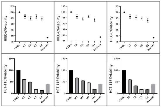
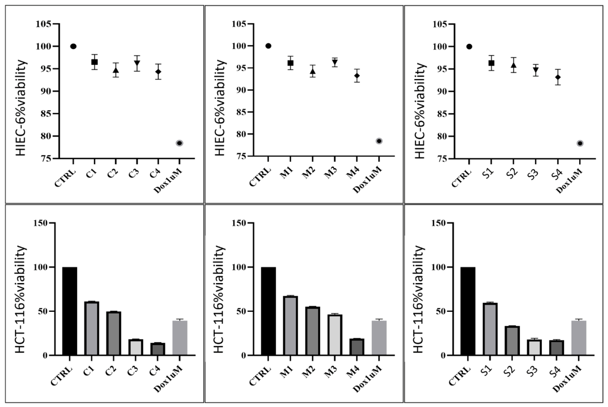
The antiproliferative activity of Origanum majorana, Salvia officinalis, and Ribes nigrum extracts was evaluated in normal human intestinal epithelial (HIEC-6) and colorectal carcinoma (HCT-116) cells following 24 h of treatment using the MTT assay. As shown in Figure 1, all three extracts exhibited concentration-dependent effects, although the magnitude of the response differed markedly between the two cell lines. HCT-116 cells were considerably more susceptible to treatment than HIEC-6 cells, indicating selective cytotoxic activity against colorectal cancer cells.
Figure 1.
Percentage viability of non-cancerous HIEC-6 intestinal epithelial cells and HCT-116 colorectal cancer cells following treatment with Ribes nigrum (C), Origanum majorana (M), and Salvia officinalis (S) extracts at different concentrations. Treatments included 10 µL (C1, M1, S1), 20 µL (C2, M2, S2), 30 µL (C3, M3, S3) and 40 µL (C4, M4, S4). Doxorubicin at 1 µM (Dox) was used as the positive control. Cell viability values are expressed relative to the untreated control group.
All three plant extracts exhibited minimal cytotoxicity toward normal HIEC-6 intestinal epithelial cells at the tested concentrations. Ribes nigrum maintained cell viability between 97.5% and 95.2%, values that remained close to the untreated control (Figure 1). Origanum majorana preserved HIEC-6 cell viability between 94.6% and 96.8% across all tested concentrations. Salvia officinalis also demonstrated low toxicity, with viability values ranging from 93.1% to 97.4%. A slight decrease in viability was observed for the latter at higher concentrations. Nevertheless, none of the extracts reduced viability more strongly than the positive control. Doxorubicin (1 µM) decreased HIEC-6 viability to approximately 78%, indicating that the plant extracts were considerably less toxic to normal intestinal epithelial cells.
In parallel, the colorectal cancer cell line HCT-116 exhibited a pronounced concentration-dependent decrease in viability following treatment with the extracts. Among the three tested extracts, Ribes nigrum demonstrated the strongest effect. At 8.24 µg/µL, HCT-116 cell viability decreased to approximately 60–61%. At 15.77 µg/µL, viability further declined to 49–50%. At 22.61 µg/µL, viability was reduced to 17–18%, while at 28.88 µg/µL, it reached only 13–14% (Figure 1). HCT-116 cell viability decreased from approximately 61% at 8.24 µg to 13–14% at 28.88 µg following treatment with Ribes nigrum extract.
Origanum majorana also induced a clear dose-dependent reduction in HCT-116 cell viability, although the effect was less pronounced than that observed for Ribes nigrum. At 6.86 µg/µL, viability was approximately 67–68%. At 13.14 µg/µL, viability decreased to around 54–55%. At 18.87 µg/µL, viability was 45–47%, whereas at 24.13 µg/µL, it declined to 18–19% (Figure 1). Origanum majorana reduced HCT-116 cell viability from approximately 68% at 6.86 µg to 18–19% at 24.13 µg.
Similarly, Salvia officinalis demonstrated strong cytotoxicity against HCT-116 cells, particularly at higher concentrations. At 9.47 µg/µL, cell viability decreased to approximately 58–60%. At 18.94 µg/µL, viability was reduced to approximately 33%. At 28.42 µg/µL, viability ranged between 16 and 19%, while at 37.89 µg/µL, it remained between 16 and 18% (Figure 1). Treatment with Salvia officinalis reduced HCT-116 viability to approximately 16–18% at 37.89 µg, comparable to that of Ribes nigrum.
At the highest tested concentrations, Ribes nigrum and Salvia officinalis reduced cell viability to a greater extent than doxorubicin, highlighting their strong anticancer potential. Doxorubicin (1 µM), used as the positive control, reduced HCT-116 viability to approximately 37–41% (Figure 1).
Overall, all three extracts reduced HCT-116 cell viability in a concentration-dependent manner, whereas HIEC-6 cell viability remained above 93% under all treatment conditions (Table 4).
Table 4.
Summary of antiproliferative activity (MTT assay).
All extracts showed Selectivity Index (SI) values greater than 1 across the tested concentrations, indicating preferential cytotoxic effects toward HCT-116 colorectal cancer cells compared with normal HIEC-6 intestinal epithelial cells. The highest SI values were observed at the highest concentrations tested, particularly for Ribes nigrum and Origanum majorana, confirming their superior selectivity toward colorectal cancer cells (Table 5). The observed effects were concentration-dependent, with Ribes nigrum and Salvia officinalis exhibiting the most pronounced cytotoxic and antiproliferative activities. Origanum majorana demonstrated a moderate but still significant anticancer effect.
Table 5.
Selectivity Index (SI) values of the investigated extracts.
3.3.2. Evaluation of Cell Apoptotic Rate by Annexin-V FITC Assay
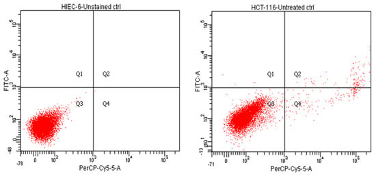
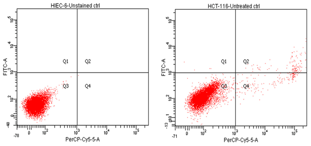
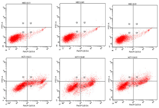
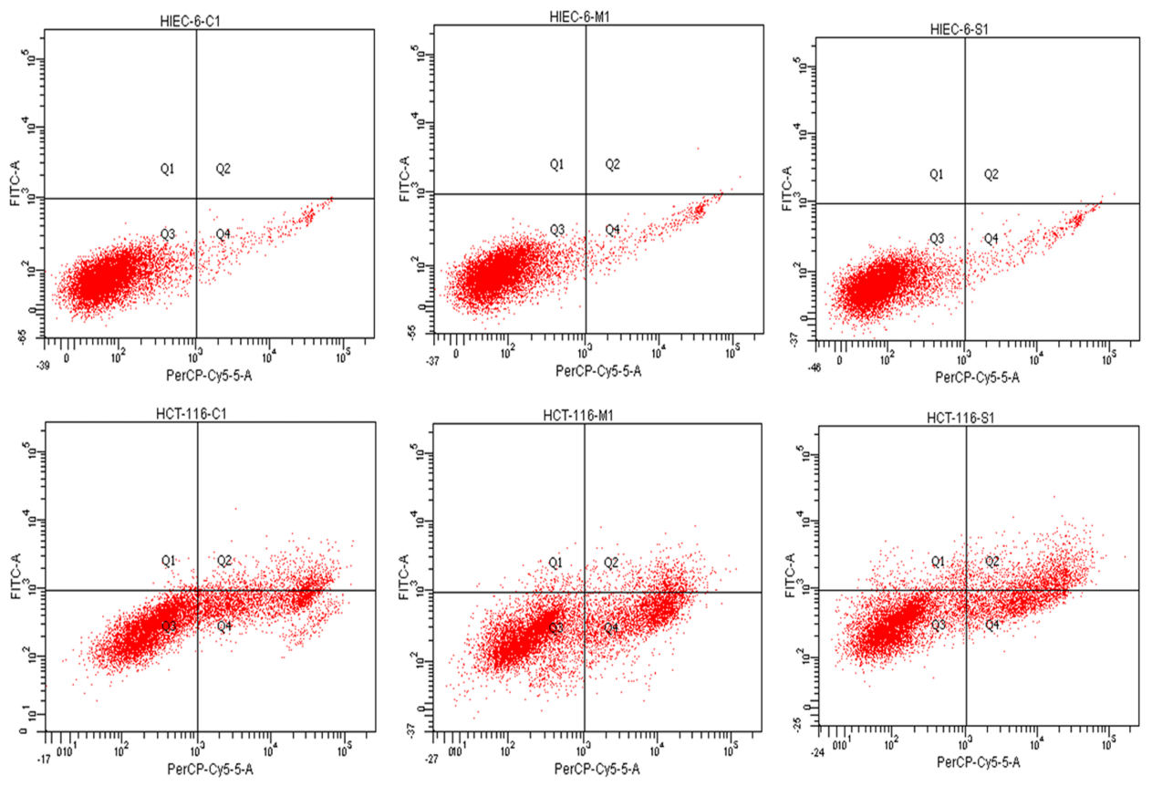
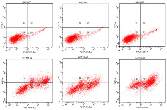
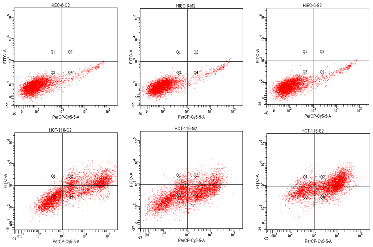
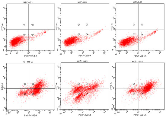
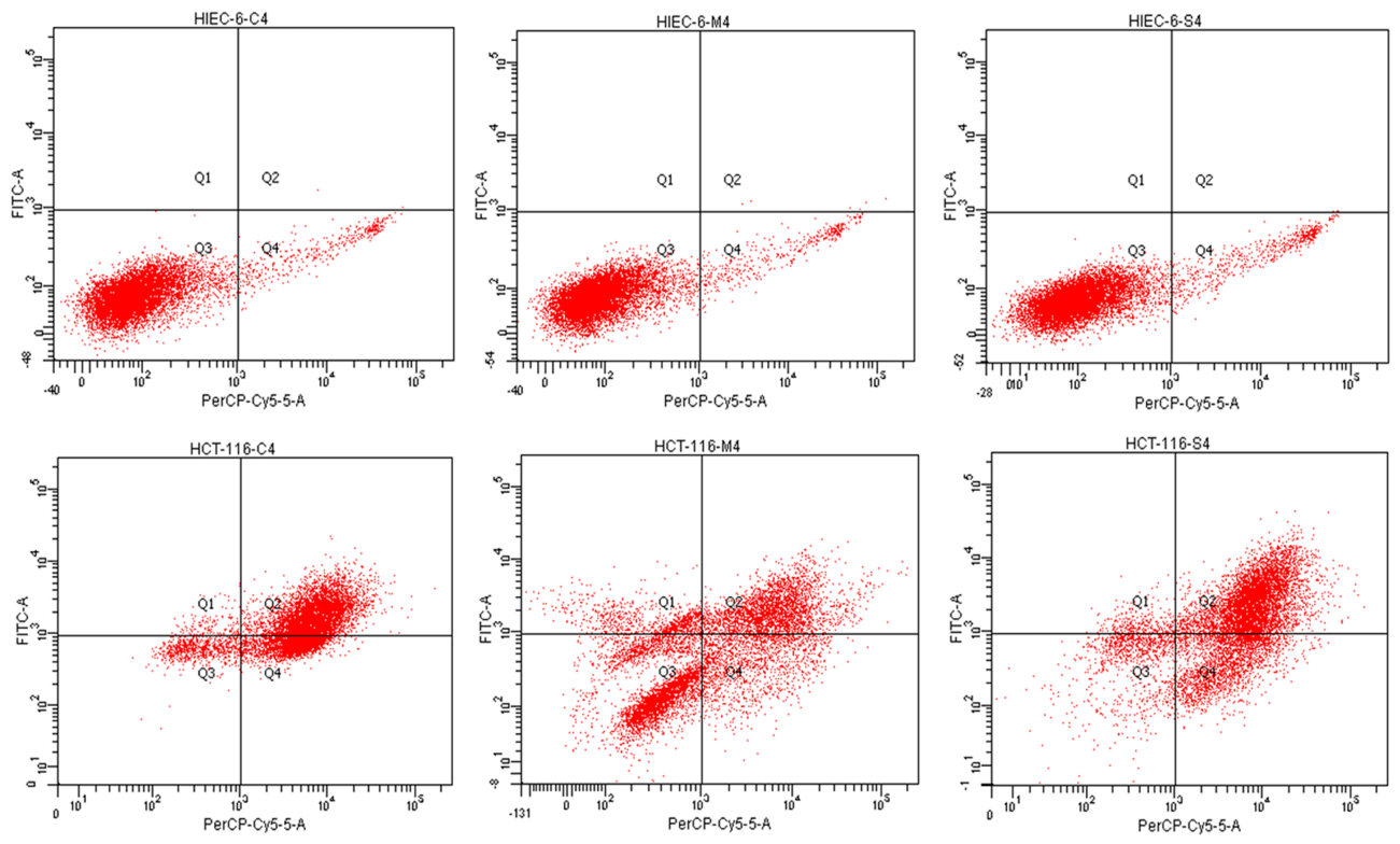
To determine whether the reduction in cell viability observed in the MTT assay was associated with apoptosis induction, HIEC-6 and HCT-116 cells were subjected to Annexin V-FITC/propidium iodide (PI) staining after 24 h of treatment with the same concentrations of Ribes nigrum, Origanum majorana, and Salvia officinalis extracts used in the cell viability assay (Table 1). Representative flow cytometry dot plots are shown in Figure 2, Figure 3, Figure 4, Figure 5 and Figure 6. The quantitative distribution of viable, early apoptotic, late apoptotic, and necrotic cell populations, along with the statistical analysis of these data, is presented in Figure 7 and Figure 8.
Figure 2.
Flow cytometric evaluation of cell viability in untreated non-cancerous HIEC-6 and cancerous HCT-116 intestinal epithelial cell lines. Cell populations were discriminated into early apoptotic (Q1), late apoptotic (Q2), viable (Q3) and necrotic (Q4) fractions based on Annexin V-FITC/PI staining profiles. Data are presented as mean ± standard deviation (SD) of three independent experiments.
Figure 3.
Flow cytometric assessment of cell viability in non-cancerous HIEC-6 and cancerous HCT-116 intestinal epithelial cell lines following treatment with 10 µL of selected plant extracts (Ribes nigrum-C1, Origanum majorana-M1, Salvia officinalis-S1). Based on Annexin V-FITC/PI staining, cell populations were classified as early apoptotic (Q1), late apoptotic (Q2), viable (Q3), and necrotic (Q4). Data are presented as mean ± standard deviation (SD) from three independent experiments.
Figure 4.
Flow cytometric assessment of cell viability in non-cancerous HIEC-6 and cancerous HCT-116 intestinal epithelial cell lines following treatment with 20 µL of selected plant extracts (Ribes nigrum-C2, Origanum majorana-M2, Salvia officinalis-S2). Based on Annexin V-FITC/PI staining, cell populations were classified as early apoptotic (Q1), late apoptotic (Q2), viable (Q3) and necrotic (Q4). Data are presented as mean ± standard deviation (SD) from three independent experiments.
Figure 5.
Flow cytometric evaluation of cell viability in non-cancerous HIEC-6 and cancerous HCT-116 intestinal epithelial cell lines after exposure to 30 µL of selected plant extracts (Ribes nigrum-C3, Origanum majorana-M3, Salvia officinalis-S3). Cell populations were identified by Annexin V-FITC/PI staining as early apoptotic (Q1), late apoptotic (Q2), viable (Q3), and necrotic (Q4). Results are presented as mean ± standard deviation (SD) from three independent experiments.
Figure 6.
Flow cytometric analysis of cell viability in non-cancerous HIEC-6 and cancerous HCT-116 intestinal epithelial cell lines following treatment with 40 µL of selected plant extracts (Ribes nigrum-C4, Origanum majorana-M4, Salvia officinalis-S4). Apoptotic and necrotic cell populations were determined using Annexin V-FITC/PI staining and classified as early apoptotic (Q1), late apoptotic (Q2), viable (Q3) and necrotic (Q4). Data are expressed as mean ± standard deviation (SD) of three independent experiments.
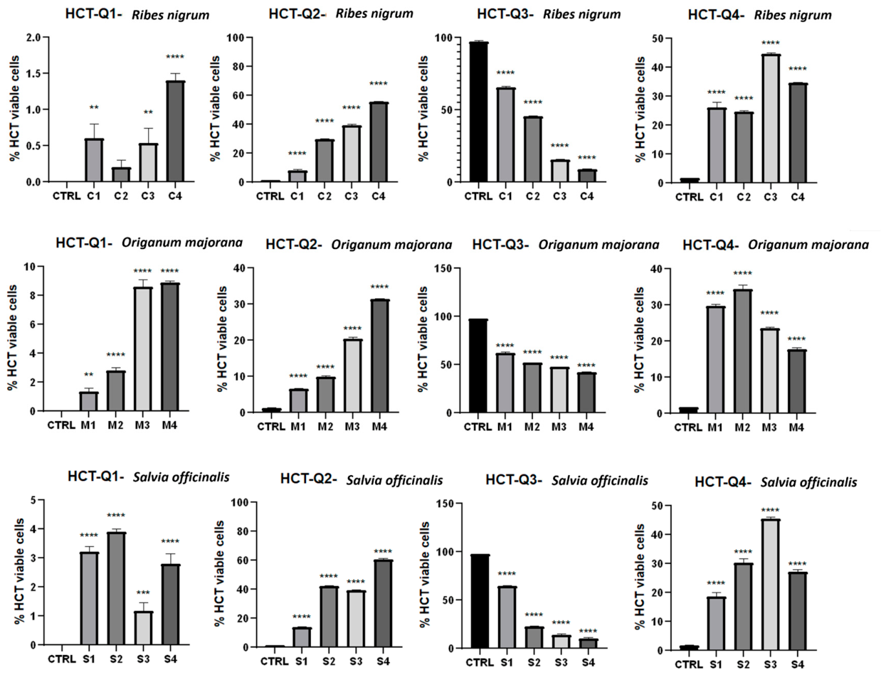
Figure 7.
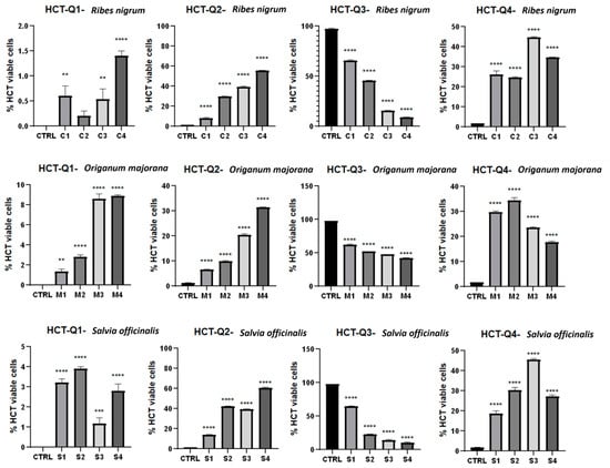
Flow cytometric analysis of apoptosis and necrosis in HCT-116 colorectal cancer cells following treatment with Ribes nigrum (C1–C4), Origanum majorana (M1–M4) and Salvia officinalis (S1–S4) extracts. Cell populations were identified by Annexin V-FITC/PI staining and classified as early apoptotic (Q1), late apoptotic (Q2), viable (Q3) and necrotic (Q4) cells. All extracts induced significant changes in cell population distribution, characterized by decreased viability and increased apoptotic and necrotic fractions compared with untreated control cells. Data are presented as mean ± standard deviation (SD) of three independent experiments performed in triplicate. Statistical analysis was performed using one-way ANOVA and paired/unpaired Student’s t-tests. Statistical significance was defined as follows: p < 0.05 versus control (*), p < 0.01 (**), p < 0.001 (***), and p < 0.0001 (****).
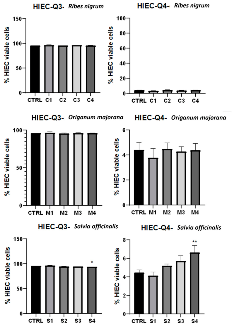
Figure 8.
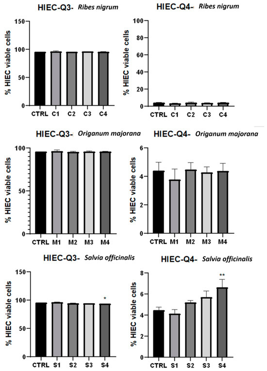
Flow cytometric analysis of apoptosis and necrosis in HIEC-6 cells following treatment with Ribes nigrum (C1–C4), Origanum majorana (M1–M4) and Salvia officinalis (S1–S4) extracts. Cell populations were identified by Annexin V-FITC/PI staining and classified as early apoptotic (Q1), late apoptotic (Q2), viable (Q3) and necrotic (Q4) cells. All extracts induced significant changes in cell population distribution, characterized by decreased viability and increased apoptotic and necrotic fractions compared with untreated control cells. Data are presented as mean ± standard deviation (SD) of three independent experiments performed in triplicate. Statistical analysis was performed using one-way ANOVA and paired/unpaired Student’s t-tests. Statistical significance was defined as follows: p < 0.05 versus control (*), p < 0.01 (**), p < 0.001 (***), and p < 0.0001 (****).
Untreated control samples from both cell lines exhibited predominantly viable cell populations, confirming the suitability of the experimental model. HIEC-6 cells showed 95.6% viability, with only 4.4% necrotic cells and virtually no apoptotic population. Similarly, untreated HCT-116 cells showed approximately 97.1% viability, whereas late apoptotic and necrotic populations accounted for only 1.2% and 1.6%, respectively (Figure 2, Figure 7 and Figure 8).
Treatment of HIEC-6 cells with all three extracts produced only minor alterations in cell population distribution. Regardless of extract type or concentration, cell viability consistently exceeded 93%, while early and late apoptotic populations remained negligible throughout the experiment. A slight concentration-dependent increase in necrotic cells was observed, particularly after treatment with Salvia officinalis, with necrosis reaching approximately 6% at the highest concentration. However, these modest changes did not substantially affect overall cell viability, indicating that the extracts exerted minimal cytotoxic effects on normal intestinal epithelial cells (Figure 8).
In contrast, HCT-116 colorectal carcinoma cells exhibited a pronounced concentration-dependent transition from viable cells toward apoptotic and necrotic populations following treatment with all three extracts. Even at the lowest concentration (R. nigrum 8.24 μg/μL; O. majorana 6.86 μg/μL; S. officinalis 9.47 μg/μL), all extracts significantly reduced the proportion of viable cells compared with untreated controls and induced measurable increases in late apoptosis and necrosis (Figure 3). These effects became progressively more pronounced as extract concentration increased, demonstrating a clear dose-dependent response (Figure 7).
Treatment with Ribes nigrum produced the most pronounced concentration-dependent cytotoxic response. Viable HCT-116 cells decreased progressively from approximately 65% at 8.24 μg/μL to less than 10% at 28.88 μg/μL, accompanied by a marked increase in late apoptotic (>55%) and necrotic (>30%) cell populations (Figure 3, Figure 4, Figure 5 and Figure 6). Early apoptosis remained comparatively low, indicating rapid progression toward late apoptotic stages (Figure 7).
Salvia officinalis exhibited a comparable but distinct apoptotic profile. Cell viability declined progressively from approximately 64% at the lowest concentration to approximately 10–14% at the two highest concentrations, while late apoptosis increased to approximately 60%, representing the highest apoptotic response among the investigated extracts (Figure 3, Figure 4, Figure 5 and Figure 6). Necrosis also increased with concentration but remained lower than after Ribes nigrum treatment, suggesting apoptosis as the predominant mechanism of cell death (Figure 7).
Treatment with Origanum majorana also induced concentration-dependent apoptosis, although its cytotoxic activity was less pronounced than that of the other extracts. Cell viability declined gradually from approximately 63% to approximately 42%, accompanied by progressive increases in both apoptotic and necrotic populations (Figure 3, Figure 4, Figure 5 and Figure 6). Compared with the other extracts, O. majorana produced a more balanced apoptotic-necrotic profile, suggesting a slower progression of programmed cell death (Figure 7).
Comparison of the three extracts revealed distinct cytotoxic profiles. Ribes nigrum resulted in the greatest reduction in viable cells and extensive late apoptosis, accompanied by pronounced necrosis. Salvia officinalis resulted in the highest proportion of late apoptotic cells, indicating apoptosis as the predominant mechanism of cell death. These differences indicate that variations in phytochemical composition may influence both the extent and the mode of cell death induced by each extract. In contrast, Origanum majorana produced a more moderate yet significant response, characterized by concurrent increases in apoptotic and necrotic cell populations (Figure 7).
Flow cytometric analysis yielded results consistent with those of the MTT assay. The progressive decline in metabolic activity observed in HCT-116 cells was accompanied by an increase in apoptotic and necrotic cell populations, confirming that the antiproliferative effects of the investigated extracts were primarily associated with induction of programmed cell death rather than temporary metabolic inhibition. In contrast, the sustained high viability of HIEC-6 cells and the minimal apoptotic changes observed support the selective cytotoxic activity of these extracts toward colorectal cancer cells.
Together, these findings demonstrate that the reduction in HCT-116 cell viability observed in the MTT assay was primarily associated with apoptosis and increased necrosis at higher concentrations, rather than nonspecific metabolic inhibition. In contrast, normal intestinal epithelial cells remained largely unaffected by treatment.
4. Discussion
4.1. Total Phenolic and Flavonoid Composition and Antioxidant Capacity
It should be noted that the present study did not aim to provide a full phytochemical profile. Instead, it quantified total phenolic and total flavonoid contents using spectrophotometric assays, while the qualitative HPLC-MS characterization of these extracts has been reported previously [9]. The biological assays were performed using DMSO-reconstituted dried residues obtained from the ethanolic extracts rather than the crude ethanolic extracts themselves. Therefore, the observed biological activities reflect the phytochemical fraction extracted and remaining after solvent removal rather than the extraction solvent itself.
Analysis of total phenolic and flavonoid contents revealed clear differences among the investigated extracts, indicating distinct phytochemical profiles for each plant species. Although Ribes nigrum exhibited the highest total phenolic content (TPC), Origanum majorana had the highest concentration of total flavonoid content (TFC), demonstrating that these parameters do not necessarily correlate. Flavonoids represent only one subclass of phenolic compounds, whereas total phenolic content reflects a much broader group of secondary metabolites. Therefore, the biological activity of plant extracts depends not only on the overall abundance of phenolic compounds but also on their qualitative composition and the interactions among individual phytochemicals [9].
The antioxidant assays further supported the relationship between phytochemical composition and biological activity. Ribes nigrum displayed the highest radical-scavenging capacity in both DPPH and ABTS assays, followed by S. officinalis and O. majorana. The superior antioxidant capacity of Ribes nigrum observed in both the DPPH and ABTS assays is consistent with its elevated total phenolic content and agrees with previous reports describing blackcurrant as an exceptionally rich source of anthocyanins and other phenolic compounds with strong radical-scavenging activity [9,14]. Anthocyanins, together with hydroxycinnamic acids and other phenolic constituents, are recognized as major contributors to the antioxidant properties of Ribes nigrum, and their synergistic interactions probably account for the high antioxidant activity observed in the present study.
On the other hand, Origanum majorana exhibited the highest flavonoid concentration but showed comparatively lower antioxidant activity than Ribes nigrum. This finding suggests that antioxidant capacity depends not only on the quantity of flavonoids but also on their chemical structure, redox properties and interactions with other phenolic constituents. Similar observations have been reported previously, indicating that the biological activity of medicinal plants is influenced by the qualitative composition of phenolic compounds rather than by the concentration of a single phytochemical class [9,15].
Salvia officinalis also showed notable antioxidant activity. Despite its lower total phenolic content compared with Ribes nigrum, the extract exhibited considerable radical-scavenging activity, supporting previous reports identifying sage as an important source of rosmarinic acid, caffeic acid derivatives, carnosic acid and carnosol [16,17]. Rosmarinic acid has been identified as one of the principal contributors to the antioxidant properties of Salvia species, whereas carnosic acid and carnosol provide additional antioxidant and anti-inflammatory activities that may further enhance the extract’s biological potential [7,17].
Another important observation is that the ranking of antioxidant activity closely paralleled total phenolic content but not total flavonoid concentration. This finding reinforces the concept that antioxidant potential is determined by the diversity and synergistic interactions of phenolic constituents rather than by the abundance of flavonoids alone. Similar conclusions have been reported for numerous medicinal plants, in which the combined activity of phenolic acids, flavonoids and anthocyanins results in greater antioxidant capacity than would be expected from the concentration of any individual compound [9,15].
These compounds possess strong electron-donating capacity and efficiently scavenge reactive oxygen species, thereby contributing to the high antioxidant potential of blackcurrant extracts [14,18]. Conversely, although Origanum majorana contained the highest flavonoid concentration, it displayed lower antioxidant activity than Ribes nigrum, suggesting that antioxidant efficacy depends more on the structural characteristics and redox properties of individual phenolic compounds than on flavonoid concentration alone [9].
Differences between the DPPH and ABTS assays should also be considered. While both methods evaluate radical-scavenging activity, they differ in their sensitivity toward hydrophilic and lipophilic antioxidants and involve distinct reaction mechanisms. Consequently, slight variations between the assays most likely reflect differences in phytochemical composition rather than inconsistencies in antioxidant potential [15]. Nevertheless, both assays consistently identified Ribes nigrum as the most active extract, supporting the robustness of the present findings.
Overall, the phytochemical and antioxidant analyses indicate that the biological potential of Origanum majorana, Salvia officinalis and Ribes nigrum cannot be explained solely by quantitative measurements of total phenolics or flavonoids. Instead, the results suggest that the antioxidant properties of these extracts are determined by the diversity, structural characteristics and synergistic interactions of their phenolic constituents. These differences in phytochemicals likely help explain the unique antiproliferative and pro-apoptotic effects observed in later cell experiments and provide a biochemical basis for the selective anticancer effects discussed in the following sections.
4.2. Relationship Between Antioxidant Properties and Antiproliferative Activity
The biological relevance of the phytochemical differences observed among the investigated extracts became evident during the evaluation of antiproliferative activity. All three extracts reduced the viability of HCT-116 colorectal carcinoma cells in a concentration-dependent manner while exerting only minimal cytotoxic effects on normal HIEC-6 intestinal epithelial cells. This selective response represents one of the principal findings of the present study and suggests that the investigated extracts preferentially target malignant rather than normal intestinal cells.
Selective cytotoxicity is considered a desirable characteristic of potential anticancer agents because malignant cells generally exhibit higher basal oxidative stress, accelerated proliferation, altered mitochondrial metabolism and dysregulated intracellular signalling pathways than their normal counterparts. Consequently, colorectal cancer cells are more susceptible to additional oxidative and metabolic disturbances induced by polyphenolic compounds, whereas normal intestinal epithelial cells possess more efficient antioxidant defence mechanisms and greater capacity to maintain cellular homeostasis [19,20]. The preservation of HIEC-6 viability observed in the present study therefore suggests that the investigated extracts selectively interfere with biological processes associated with malignant transformation rather than inducing nonspecific cytotoxicity.
Among the three investigated species, Ribes nigrum exhibited the most pronounced antiproliferative activity, producing the greatest reduction in HCT-116 cell viability. This finding is consistent with its high total phenolic content and superior antioxidant capacity and agrees with previous studies demonstrating that blackcurrant extracts inhibit colorectal cancer cell proliferation through the combined activity of anthocyanins, flavonols and phenolic acids [19,21]. Similarly, Ginovyan et al. (2022) [22] reported that phenolic-rich extracts from medicinal plants exert selective cytotoxic effects against colorectal carcinoma cells while exhibiting limited toxicity toward normal cells, emphasising the importance of phytochemical composition in determining biological activity.
Although Salvia officinalis contained lower total phenolic levels than Ribes nigrum, its antiproliferative activity was comparable at higher concentrations. This observation supports previous reports identifying rosmarinic acid, carnosic acid and carnosol as major contributors to the anticancer activity of Salvia species [16,17]. These compounds have been shown to inhibit cell proliferation through modulation of oxidative stress, suppression of pro-survival signalling pathways and induction of programmed cell death, thereby contributing to the selective inhibition of colorectal cancer cells [23].
In contrast, Origanum majorana exhibited a more moderate but still significant concentration-dependent antiproliferative effect. Despite presenting the highest total flavonoid content, its cytotoxic activity was lower than that of Ribes nigrum and Salvia officinalis. This finding reinforces the concept that biological activity depends not only on the quantity of flavonoids but also on their structural diversity, relative abundance and synergistic interactions with other phenolic constituents [9,24]. Similar observations have been reported for other medicinal plants, in which differences in phytochemical composition led to marked variations in antiproliferative efficacy despite comparable total phenolic or flavonoid content.
Comparison with doxorubicin further supports the biological potential of the investigated extracts. Although direct comparisons between crude plant extracts and an established chemotherapeutic agent should be interpreted cautiously, the highest concentrations of Ribes nigrum and Salvia officinalis produced greater reductions in HCT-116 cell viability than 1 μM doxorubicin under identical experimental conditions, while causing only minimal cytotoxic effects in HIEC-6 cells. These findings indicate a favourable selectivity profile and support the growing evidence that polyphenol-rich plant extracts may represent valuable complementary agents capable of enhancing anticancer efficacy while reducing adverse effects on healthy tissues [20,23].
Overall, the antiproliferative activity observed in the present study appears to be closely associated with the total phenolic and flavonoid contents of the investigated extracts. The superior activity of Ribes nigrum and Salvia officinalis, together with the selective preservation of normal intestinal epithelial cells, suggests that the combined action of multiple phenolic constituents rather than individual compounds is responsible for the observed biological effects. These findings provide a rationale for further studies to elucidate the molecular mechanisms responsible for the selective anticancer activity of these medicinal plants.
The selective response observed between HCT-116 and HIEC-6 cells represents one of the most important findings of the present study. Cancer cells generally exhibit increased basal oxidative stress, accelerated proliferation, altered mitochondrial metabolism and constitutive activation of survival pathways such as PI3K/Akt and NF-κB. Consequently, they are considerably more susceptible than normal epithelial cells to additional oxidative and metabolic disturbances induced by polyphenolic compounds. Conversely, normal intestinal epithelial cells maintain more efficient antioxidant defence systems and tighter regulation of mitochondrial homeostasis, which may explain their greater resistance to the investigated extracts. Therefore, the preservation of HIEC-6 viability together with the marked inhibition of HCT-116 proliferation suggests that these phytochemical-rich extracts preferentially target biological processes associated with malignant transformation rather than cause nonspecific cytotoxicity [5,25].
4.3. Apoptosis as the Primary Mechanism of Cytotoxicity
To further elucidate the mechanisms underlying the observed reduction in cell viability, flow cytometry analysis was performed. The results provided important evidence that the investigated extracts not only suppress cellular metabolism but also actively promote programmed cell death pathways in colorectal cancer cells.
The flow cytometry analysis confirmed that the reduction in HCT-116 cell viability observed in the MTT assay was primarily associated with induction of apoptosis, rather than nonspecific cytotoxicity. Following treatment with all three extracts, HCT-116 cells exhibited a concentration-dependent increase in apoptotic populations, whereas HIEC-6 cells remained predominantly viable throughout the experimental period. The close agreement between the MTT and Annexin V-FITC/PI assays therefore demonstrates that the observed decrease in metabolic activity reflected genuine programmed cell death rather than transient inhibition of cellular metabolism. Although Annexin V/PI staining demonstrated increased apoptotic cell populations following treatment, the present study did not investigate the molecular pathways responsible for apoptosis induction. Therefore, it cannot be determined whether the observed effects involve the intrinsic mitochondrial pathway, the extrinsic death receptor pathway, or other signalling mechanisms. Future studies should evaluate key apoptosis-related markers, including Bax, Bcl-2, caspase-3, caspase-9, PARP cleavage, and p53, together with mitochondrial membrane potential and reactive oxygen species production, to further elucidate the molecular basis of the observed antiproliferative effects.
The predominance of late apoptotic cells after 24 h indicates that apoptosis had progressed substantially during the incubation period. Polyphenolic compounds present in Origanum majorana, Salvia officinalis and Ribes nigrum, including rosmarinic acid, caffeic acid derivatives, anthocyanins, carnosic acid and carnosol, have previously been reported to activate the intrinsic mitochondrial apoptotic pathway through disruption of mitochondrial membrane potential, modulation of the Bax/Bcl-2 ratio, release of cytochrome c and subsequent activation of caspase-9 and caspase-3 [7,17,26]. Although these molecular events were not examined in the present study, the marked increase in Annexin V-positive/PI-positive cells is consistent with mechanisms previously reported for these phytochemicals.
The three investigated extracts, however, exhibited distinct apoptotic profiles. Ribes nigrum produced the greatest reduction in HCT-116 cell viability and was associated with substantial increases in both late apoptotic and necrotic cell populations. These findings are in agreement with previous reports demonstrating that anthocyanin-rich blackcurrant extracts effectively suppress colorectal cancer cell growth by promoting apoptosis and impairing mitochondrial function [19,21]. The strong biological activity of Ribes nigrum is therefore likely attributable to the combined effects of anthocyanins, phenolic acids and other polyphenolic constituents acting synergistically.
Although Salvia officinalis induced a comparable reduction in cell viability, its biological response was characterized by the highest proportion of late apoptotic cells and comparatively lower levels of necrosis. This observation suggests that apoptosis represented the predominant mechanism of cell death induced by the extract and agrees with previous studies reporting that rosmarinic acid, carnosic acid and carnosol promote apoptosis through regulation of mitochondrial signalling pathways and activation of caspase-dependent mechanisms [16,17,23]. The pronounced apoptotic response observed in the present study further supports the contribution of these bioactive compounds to the anticancer potential of Salvia officinalis.
In contrast, Origanum majorana exhibited a more moderate but significant pro-apoptotic effect. Despite containing the highest flavonoid concentration, its antiproliferative activity was comparatively weaker than that of Ribes nigrum and Salvia officinalis. Similar observations have been reported previously, indicating that the biological activity of medicinal plant extracts depends not only on the total concentration of flavonoids but also on their qualitative composition and synergistic interactions with other phenolic constituents [9,24]. Consequently, differences in phytochemical composition likely explain the distinct apoptotic responses observed among the extracts investigated.
In addition to apoptosis, a moderate increase in necrotic cells was observed at higher extract concentrations, particularly following treatment with Ribes nigrum. This finding most likely reflects the progression of apoptotic cells toward secondary necrosis after prolonged incubation rather than direct induction of primary necrotic cell death. Similar patterns have been described for polyphenol-rich extracts, in which extensive apoptosis during prolonged exposure is accompanied by increased membrane permeability, as detected by propidium iodide staining [23,27].
The flow cytometry findings indicate that apoptosis represents the principal mechanism underlying the antiproliferative activity of the investigated extracts. The close relationship among phytochemical composition, reduced cell viability and apoptosis induction further supports the hypothesis that multiple phenolic constituents act synergistically to selectively eliminate colorectal cancer cells while exerting only minimal effects on normal intestinal epithelial cells. These observations provide a mechanistic basis for the selective cytotoxic activity demonstrated in the present study and reinforce the therapeutic potential of these medicinal plants as complementary candidates for colorectal cancer prevention and treatment.
4.4. Biological Significance, Limitations and Future Perspectives
The present study provides further evidence that polyphenol-rich plant extracts possess considerable potential as complementary agents for colorectal cancer prevention and therapy. Although antioxidant activity is often considered one of the principal biological properties of medicinal plants, the present findings demonstrate that the investigated extracts also exert selective antiproliferative and pro-apoptotic effects against colorectal carcinoma cells while largely preserving the viability of normal intestinal epithelial cells. This selective response is particularly important because one of the major limitations of conventional chemotherapy is its toxicity toward rapidly proliferating healthy tissues, especially the gastrointestinal epithelium [20,23].
The relationship observed between phytochemical composition, antioxidant capacity and anticancer activity suggests that the biological effects of Origanum majorana, Salvia officinalis and Ribes nigrum cannot be attributed to a single compound. Instead, the combined activity of anthocyanins, phenolic acids, flavonoids and other secondary metabolites likely contributes to the selective inhibition of tumour cell proliferation and apoptosis induction. Similar conclusions have been reported in previous studies, emphasising that synergistic interactions among phytochemicals frequently produce greater biological activity than isolated constituents [9,17]. The superior biological activity of Ribes nigrum and Salvia officinalis observed in the present study further supports this concept and highlights the importance of evaluating the qualitative composition of plant extracts rather than only their total phenolic or flavonoid content.
The selective preservation of HIEC-6 cell viability observed throughout the study deserves particular attention. In contrast to HCT-116 cells, normal intestinal epithelial cells exhibited only minimal reductions in viability and negligible induction of apoptosis following treatment with the investigated extracts. This observation suggests that normal cells are less susceptible to phytochemical-induced stress and are better able to maintain cellular homeostasis under the experimental conditions employed. Although the molecular basis of this selectivity remains unclear, previous studies suggest that differences in oxidative metabolism, antioxidant defences, and intracellular signalling contribute to the greater sensitivity of tumour cells to polyphenol-rich extracts [19,20].
Several limitations of the present study should nevertheless be acknowledged. First, the biological activity of the extracts was evaluated exclusively in vitro using a single colorectal carcinoma cell line and one normal intestinal epithelial cell line. Consequently, the results cannot be directly extrapolated to the complex physiological environment encountered in vivo. Second, only total phenolic and total flavonoid contents were determined using spectrophotometric assays. However, the qualitative phytochemical composition of these extracts has been previously characterized by HPLC–MS [9]. No chromatographic analyses were performed within the present study. Consequently, it was not possible to attribute the observed biological activities to individual phytochemicals or specific classes of compounds. Furthermore, the MTT assay evaluates mitochondrial metabolic activity as an indirect indicator of cell viability and does not distinguish between reduced proliferation, metabolic inhibition, and cell death. To strengthen the biological interpretation, the MTT results were complemented by Annexin V/PI flow cytometry, which confirmed apoptosis and necrosis in treated HCT-116 cells. Nevertheless, the present study did not investigate the molecular mechanisms underlying these effects. Therefore, additional studies evaluating cell-cycle distribution, caspase activation, mitochondrial membrane potential, reactive oxygen species (ROS) production, and the expression of apoptosis-related proteins are warranted to further elucidate the mechanisms responsible for the observed antiproliferative activity. Finally, although apoptosis induction was clearly demonstrated by Annexin V-FITC/PI staining, the molecular pathways responsible for these effects, including oxidative stress modulation, mitochondrial dysfunction and activation of apoptotic signalling cascades, were not investigated directly.
Future research should focus on identifying the principal bioactive constituents responsible for the observed antiproliferative activity and elucidating their molecular mechanisms of action. In particular, future investigations should combine chromatographic characterization with bioactivity-guided fractionation and mechanistic studies to identify the compounds responsible for the observed biological effects. Particular attention should be directed toward evaluating reactive oxygen species generation, mitochondrial membrane potential, cell-cycle regulation, caspase activation and the expression of apoptosis-related proteins. Additionally, investigations using additional colorectal cancer models, three-dimensional cell culture systems and in vivo experimental models will be essential for confirming the biological activity, bioavailability and safety of these extracts. Evaluating potential synergistic interactions with conventional chemotherapeutic agents also represents an important area for future research, as such combinations may improve therapeutic efficacy while reducing adverse effects associated with current anticancer treatments [17,23].
Overall, the present findings demonstrate that the biological activity of Origanum majorana, Salvia officinalis and Ribes nigrum extends beyond their antioxidant properties and includes selective inhibition of colorectal cancer cell proliferation accompanied by apoptosis induction. The close relationship between phytochemical composition and biological activity observed in the present study further supports the therapeutic potential of these medicinal plants as valuable sources of bioactive compounds for complementary strategies aimed at colorectal cancer prevention and treatment.
5. Conclusions
This study demonstrated that the investigated plant extracts exhibited distinct antioxidant capacities and selective biological activity against HCT-116 colorectal cancer cells while showing minimal cytotoxicity toward normal HIEC-6 intestinal epithelial cells. Among the tested extracts, Ribes nigrum and Salvia officinalis produced the strongest antiproliferative and pro-apoptotic effects, whereas Origanum majorana showed moderate activity. These findings suggest that differences in phytochemical composition, reflected by the spectrophotometrically determined total phenolic and flavonoid contents, may contribute to the observed biological responses. Overall, the results support the potential of these plant extracts as promising sources of bioactive compounds for complementary research on colorectal cancer.
Although the present findings are promising, they should be interpreted within the limitations of an in vitro study. Only one colorectal cancer cell line and one normal intestinal epithelial cell line were evaluated, and the investigated extracts represent complex mixtures of bioactive compounds whose individual contributions remain to be determined. Therefore, further studies are required to identify the principal active constituents, elucidate the molecular pathways responsible for their selective anticancer activity, evaluate potential synergistic interactions with conventional chemotherapeutic agents, and validate these observations in in vivo models of colorectal cancer.
Author Contributions
Conceptualization, O.-R.H., E.P. and S.A.; methodology, O.-R.H., E.P., M.C. and K.S.; software, D.-A.D.; validation, O.-R.H., E.P., M.C., K.S. and S.A.; formal analysis, O.-R.H. and D.-A.D.; investigation, O.-R.H., E.P., M.C. and K.S.; resources, E.P., M.C., K.S. and S.A.; data curation, O.-R.H. and D.-A.D.; writing—original draft preparation, O.-R.H.; writing—review and editing, E.P., M.C., K.S. and S.A.; visualization, O.-R.H.; supervision, S.A. and E.P. All authors have read and agreed to the published version of the manuscript.
Funding
This research received no external funding.
Institutional Review Board Statement
Not applicable.
Informed Consent Statement
Not applicable.
Data Availability Statement
The data presented in this study are available within the article.
Conflicts of Interest
The authors declare no conflicts of interest.
Abbreviations
The following abbreviations are used in this manuscript:
| ABTS | 2,2′-Azino-bis (3-ethylbenzothiazoline-6-sulfonic acid) |
| ANOVA | Analysis of Variance |
| CRC | Colorectal Cancer |
| DPPH | 2,2-Diphenyl-1-picrylhydrazyl |
| DMSO | Dimethyl Sulfoxide |
| FITC | Fluorescein Isothiocyanate |
| GAE | Gallic Acid Equivalents |
| HCT-116 | Human Colorectal Carcinoma Cell Line |
| HIEC-6 | Human Intestinal Epithelial Cell Line |
| MTT | 3-(4,5-Dimethylthiazol-2-yl)-2,5-diphenyltetrazolium Bromide |
| PBS | Phosphate-Buffered Saline |
| PI | Propidium Iodide |
| QE | Quercetin Equivalents |
| SD | Standard Deviation |
| TFC | Total Flavonoid Content |
| TPC | Total Phenolic Content |
References
- López-Gómez, L.; Uranga, J.A. Polyphenols in the Prevention and Treatment of Colorectal Cancer: A Systematic Review of Clinical Evidence. Nutrients 2024, 16, 2735. [Google Scholar] [CrossRef] [Scilit] [PubMed]
- Siegel, R.L.; Kratzer, T.B.; Giaquinto, A.N.; Sung, H.; Jemal, A. Cancer Statistics, 2025. CA A Cancer J. Clin. 2025, 75, 10–45. [Google Scholar] [CrossRef] [Scilit] [PubMed]
- Li, Y.; Zhang, T.; Chen, G.Y. Flavonoids and Colorectal Cancer Prevention. Antioxidants 2018, 7, 187. [Google Scholar] [CrossRef] [Scilit] [PubMed]
- Okpoghono, J.; Isoje, E.F.; Igbuku, U.A.; Ekayoda, O.; Omoike, G.O.; Adonor, T.O.; Igue, U.B.; Okom, S.U.; Ovowa, F.O.; Stephen-Onojedje, Q.O.; et al. Natural Polyphenols: A Protective Approach to Reduce Colorectal Cancer. Heliyon 2024, 10, e32390. [Google Scholar] [CrossRef] [Scilit] [PubMed]
- Esmeeta, A.; Adhikary, S.; Dharshnaa, V.; Swarnamughi, P.; Ummul Maqsummiya, Z.; Banerjee, A.; Pathak, S.; Duttaroy, A.K. Plant-Derived Bioactive Compounds in Colon Cancer Treatment: An Updated Review. Biomed. Pharmacother. 2022, 153, 113384. [Google Scholar] [CrossRef] [Scilit] [PubMed]
- Liang, J.; Liang, J.; Hao, H.; Lin, H.; Wang, P.; Wu, Y.; Jiang, X.; Fu, C.; Li, Q.; Ding, P.; et al. The Extracts of Morinda Officinalis and Its Hairy Roots Attenuate Dextran Sodium Sulfate-Induced Chronic Ulcerative Colitis in Mice by Regulating Inflammation and Lymphocyte Apoptosis. Front. Immunol. 2017, 8, 905. [Google Scholar] [CrossRef] [Scilit] [PubMed]
- Czerwińska, K.; Radziejewska, I. Rosmarinic Acid: A Potential Therapeutic Agent in Gastrointestinal Cancer Management—A Review. Int. J. Mol. Sci. 2024, 25, 11704. [Google Scholar] [CrossRef] [Scilit] [PubMed]
- Medic, N.; Tramer, F.; Passamonti, S. Anthocyanins in Colorectal Cancer Prevention. A Systematic Review of the Literature in Search of Molecular Oncotargets. Front. Pharmacol. 2019, 10, 675. [Google Scholar] [CrossRef] [Scilit] [PubMed]
- Haralambie, O.-R.; Novac, C.-Ș.; Hodor, D.; Ranga, F.; Andrei, S. Antibacterial Activity of Ethanolic Extracts of Origanum Majorana, Salvia Officinalis, and Ribes Nigrum Against Digestive Pathogens: Polyphenolic Composition and In Vitro Assessment. Molecules 2025, 30, 3341. [Google Scholar] [CrossRef] [Scilit] [PubMed]
- Navarro García, N.E.; Trujillo, J.; Ramírez, V. Polyphenols and Health Benefits: 2nd Edition. Foods 2025, 14, 4340. [Google Scholar] [CrossRef] [Scilit] [PubMed]
- Zhishen, J.; Mengcheng, T.; Jianming, W. The Determination of Flavonoid Contents in Mulberry and Their Scavenging Effects on Superoxide Radicals. Food Chem. 1999, 64, 555–559. [Google Scholar] [CrossRef] [Scilit]
- Brand-Williams, W.; Cuvelier, M.E.; Berset, C. Use of a Free Radical Method to Evaluate Antioxidant Activity. LWT-Food Sci. Technol. 1995, 28, 25–30. [Google Scholar] [CrossRef] [Scilit]
- Arnao, M.B.; Cano, A.; Acosta, M. The Hydrophilic and Lipophilic Contribution to Total Antioxidant Activity. Food Chem. 2001, 73, 239–244. [Google Scholar] [CrossRef] [Scilit]
- Ejaz, A.; Waliat, S.; Afzaal, M.; Saeed, F.; Ahmad, A.; Din, A.; Ateeq, H.; Asghar, A.; Shah, Y.A.; Rafi, A.; et al. Biological Activities, Therapeutic Potential, and Pharmacological Aspects of Blackcurrants (Ribes nigrum L.): A Comprehensive Review. Food Sci. Nutr. 2023, 11, 5799–5817. [Google Scholar] [CrossRef] [Scilit] [PubMed]
- Lisjak, M.; Špoljarević, M.; Ravlić, J.; Lončarić, Z.; Galić, L. Phenolic Compounds and Antioxidant Activity: Analytical Methods and Current Knowledge—A Review. Methods Protoc. 2026, 9, 60. [Google Scholar] [CrossRef] [Scilit] [PubMed]
- Bilginoğlu, E.; Kızıl, H.E.; Öğütcü, H.; Ağar, G.; Bağcı, Y. Pharmacological Potential and Bioactive Components of Wild Anatolian Sage (Salvia aethiopis L.). Food Sci. Nutr. 2025, 13, e70118. [Google Scholar] [CrossRef] [Scilit] [PubMed]
- Molonia, M.S.; Salamone, F.L.; Cimino, F.; D’Arrigo, M.; Cristani, M.; Pulvirenti, L.; Saija, A.; Speciale, A.; Napoli, E. Phenolic Profile and In Vitro Anti-Inflammatory Activities of Salvia officinalis L. Hydrodistillation Wastewater. Chem. Biodivers. 2026, 23, e02271. [Google Scholar] [CrossRef] [Scilit] [PubMed]
- Cortez, R.E.; Gonzalez De Mejia, E. Blackcurrants (Ribes nigrum): A Review on Chemistry, Processing, and Health Benefits. J. Food Sci. 2019, 84, 2387–2401. [Google Scholar] [CrossRef] [Scilit] [PubMed]
- Mazewski, C.; Liang, K.; Gonzalez De Mejia, E. Comparison of the Effect of Chemical Composition of Anthocyanin-Rich Plant Extracts on Colon Cancer Cell Proliferation and Their Potential Mechanism of Action Using in Vitro, in Silico, and Biochemical Assays. Food Chem. 2018, 242, 378–388. [Google Scholar] [CrossRef] [PubMed]
- Habchi, C.; Badran, A.; Srour, M.; Daou, A.; Baydoun, E.; Hamade, K.; Hijazi, A. Determination of the Antioxidant and Antiproliferative Properties of Pomegranate Peel Extract Obtained by Ultrasound on HCT-116 Colorectal Cancer Cell Line. Processes 2023, 11, 1111. [Google Scholar] [CrossRef] [Scilit]
- Lyashenko, S.; López-Ruiz, R.; García-Cervantes, A.M.; Rodríguez-García, I.; Yunusova, S.; Guil-Guerrero, J.L. Phenolic Profiles and Antitumor Activity against Colorectal Cancer Cells of Seeds from Selected Ribes Taxa. Appl. Sci. 2024, 14, 2428. [Google Scholar] [CrossRef] [Scilit]
- Ginovyan, M.; Bartoszek, A.; Koss-Mikołajczyk, I.; Kusznierewicz, B.; Andreoletti, P.; Cherkaoui-Malki, M.; Sahakyan, N. Growth Inhibition of Cultured Cancer Cells by Ribes nigrum Leaf Extract. AIMS Biophys. 2022, 9, 282–293. [Google Scholar] [CrossRef] [Scilit]
- Luca, S.V.; Skalicka-Woźniak, K.; Mihai, C.-T.; Gradinaru, A.C.; Mandici, A.; Ciocarlan, N.; Miron, A.; Aprotosoaie, A.C. Chemical Profile and Bioactivity Evaluation of Salvia Species from Eastern Europe. Antioxidants 2023, 12, 1514. [Google Scholar] [CrossRef] [Scilit] [PubMed]
- Benhalilou, N.; Alsamri, H.; Alneyadi, A.; Athamneh, K.; Alrashedi, A.; Altamimi, N.; Al Dhaheri, Y.; Eid, A.H.; Iratni, R. Origanum Majorana Ethanolic Extract Promotes Colorectal Cancer Cell Death by Triggering Abortive Autophagy and Activation of the Extrinsic Apoptotic Pathway. Front. Oncol. 2019, 9, 795. [Google Scholar] [CrossRef] [Scilit] [PubMed]
- Dekker, E.; Tanis, P.J.; Vleugels, J.L.A.; Kasi, P.M.; Wallace, M.B. Colorectal Cancer. Lancet 2019, 394, 1467–1480. [Google Scholar] [CrossRef] [Scilit] [PubMed]
- Jiang, M.; Zhang, X.; Jin, X.; Shi, B.; Xu, Y.; Wang, Z. Relieving Effect of Artemisia Ordosica Krasch Extract on DSS-Induced Colitis by Regulating Immunity, Antioxidant Function, Gut Microbiota, and Bile Acid Metabolism in Mice. Antioxidants 2025, 14, 45. [Google Scholar] [CrossRef] [Scilit] [PubMed]
- Rupasinghe, V.; Jiang, Y.; Zhang, L. The Anticancer Properties of Phytochemical Extracts from Salvia Plants. Bot. Targets Ther. 2016, 6, 25–44. [Google Scholar] [CrossRef] [Scilit][Green Version]

Disclaimer/Publisher’s Note: The statements, opinions and data contained in all publications are solely those of the individual author(s) and contributor(s) and not of MDPI and/or the editor(s). MDPI and/or the editor(s) disclaim responsibility for any injury to people or property resulting from any ideas, methods, instructions or products referred to in the content. |
© 2026 by the authors. Licensee MDPI, Basel, Switzerland. This article is an open access article distributed under the terms and conditions of the Creative Commons Attribution (CC BY) license.