


Abstract
Oncofetal reprogramming has recently emerged as a critical concept in translational cancer research, particularly for its role in driving therapeutic resistance across a variety of malignancies. This biological process refers to a pattern of gene expression that is restricted to embryogenesis, but becomes expressed again in a subpopulation of cancer cells. These genes are typically suppressed after embryogenesis, and their aberrant re-expression in tumors endows cancer cells with stem-like properties and enhanced adaptability. The goal of this review is the following: (i) comprehensively examine the multifaceted nature of oncofetal reprogramming; (ii) elucidate its underlying molecular mechanisms, including its regulators and effectors; and (iii) evaluate its consequences for the therapeutic response in different cancer types. We comprehensively integrate the latest findings from colorectal, breast, lung, liver, and other cancers to provide a detailed understanding of how oncofetal programs interfere with tumor response to treatment. Among the candidates, YAP1 and AP-1 have emerged as central transcriptional drivers of this reprogramming process, especially in colorectal and breast cancers. We also explore the distinct expression patterns of oncofetal genes across different tumor types and how these patterns correlate with treatment outcomes and patient survival. Lastly, we propose a dual-targeting therapeutic strategy that simultaneously targets both cancer stem cells and oncofetal-reprogrammed populations as a more effective approach to overcome resistance and limit recurrence.
1. Introduction
Oncofetal reprogramming, the reactivation of embryonic and fetal gene expression programs in cancer cells, has emerged as a key concept in translational oncology with profound implications for clinical treatment regimens [1]. Under physiological conditions, the set of oncofetal genes is exclusively expressed during embryogenesis, where they play specialized roles in cell fate specification, tissue morphogenesis, and pluripotency [2,3,4]. Following development, these genes are robustly suppressed, ensuring the maintenance of lineage identity and tissue homeostasis throughout adult life [2,3,4]. However, accumulating evidence has revealed that malignant transformations in several cancers, including colorectal, breast, lung, and liver, can hijack these early developmental programs, resulting in tumor cells with enhanced plasticity and the capacity to evade therapeutic interventions [5,6,7,8,9].
Reactivation of oncofetal genes is not a universal phenomenon across all tumor cells; rather, it marks a distinct subpopulation within a tumor mass, one that exhibits heightened adaptability and resistance to pharmacological and immune-mediated therapies [6,9]. These cells often exploit oncofetal transcriptional regulators to modulate differentiation trajectories, remodel the tumor microenvironment, and orchestrate evasive responses that hinder the efficacy of standard-of-care treatments [6]. It is critical to distinguish between oncofetal reprogramming and other similar but distinct phenomena, such as cancer stem cells (CSCs) or injury-induced regenerative/fetal-like repair responses. On one hand, CSCs are a functionally defined tumor cellular subpopulation that possesses self-renewal and tumorigenic properties, whereas oncofetal reprogramming drives a transcriptional and epigenetic phenotype that involves the reactivation of embryonic gene expression profiles in both CSCs and non-CSCs in cancers [1,2]. Likewise, injury-induced regenerative or fetal-like repair responses are transient and tightly regulated activations of developmental gene expression in non-neoplastic cells in response to injury, whereas oncofetal reprogramming in cancers engages a distinct subset of embryonic genes, and is uncoupled from the normal regulatory constraints that govern developmental gene expression in healthy cells [2,4]. Recent multi-omic and single-cell profiling studies have mapped the landscape of oncofetal gene reactivation across cancer types, revealing both shared and context-dependent drivers in different cancers to pioneer more effective therapeutic interventions [9,10,11]. Among the key molecular drivers, the transcriptional regulators YAP1 and AP-1 (c-Jun/Fos family) have emerged as central players in oncofetal reprogramming [9,12,13,14,15,16]. YAP1, a principal effector of the Hippo pathway, operates as an initiator that drives the aberrant expression of embryonic gene modules in malignancies, such as colorectal cancer (CRC) and breast cancer (Figure 1) [17,18]. YAP1’s overactivation is frequently associated with stemness, epithelial-to-mesenchymal transition (EMT), and resistance to chemotherapy or targeted modalities [17,18]. AP-1 family members further reinforce this transcriptional state, cooperating with YAP1 and other cofactors to establish oncogenic chromatin landscapes that are permissive of oncofetal gene expression [19,20,21]. Moreover, tumor recurrence and resistance frequently correlate with the presence of YAP1- and AP-1-driven oncofetal cell populations, which exhibit both intrinsic insensitivity to conventional therapies and the capacity to repopulate a tumor following treatment-induced damage [9,22,23]. In some cancers, such as lung cancer, patients with high expression of YAP1 will have lower overall survival (Table 1). Notably, recent work has demonstrated that oncofetal program activation can modulate the tumor microenvironment, promote immune evasion, and suppress antitumor immune responses, thus complicating the efficacy of immunotherapy in cancers such as lung and liver [6,24,25]. Understanding oncofetal reprogramming will allow clinicians to shift toward dual-targeting strategies that simultaneously attack conventional CSCs and oncofetal-reprogrammed populations, aiming to more durably overcome resistance and reduce relapse rates.
Figure 1.
YAP1 expression in pan-cancer. Median expression ratio of YAP1 in tumor tissue compared to adjacent normal tissue across multiple cancer types (pan-cancer analysis) using The Cancer Genome Atlas (TCGA) data. Cancer type abbreviations (TCGA): CHOL (cholangiocarcinoma), GBM (glioblastoma multiforme), BRCA (breast invasive carcinoma), COAD (colon adenocarcinoma), LIHC (liver hepatocellular carcinoma), STAD (stomach adenocarcinoma), THYM (thymoma), READ (rectum adenocarcinoma), SARC (sarcoma), PAAD (pancreatic adenocarcinoma), SKCM (skin cutaneous melanoma), THCA (thyroid carcinoma), PRAD (prostate adenocarcinoma), HNSC (head and neck squamous cell carcinoma), LUAD (lung adenocarcinoma), LUSC (lung squamous cell carcinoma), KIRC (kidney renal clear cell carcinoma), PRAD (prostate adenocarcinoma), KICH (kidney chromophobe), KIRP (kidney renal papillary cell carcinoma), CESC (cervical squamous cell carcinoma and endocervical adenocarcinoma), ESCA (esophageal carcinoma), UCEC (uterine corpus endometrial carcinoma), PCPG (pheochromocytoma and paraganglioma).
Table 1.
Human YAP1 expression and survival in cancers.
Although YAP1 expression and survival associations vary across cancer types, this review does not prioritize tumor entities solely based on their statistical prognostic significance (Table 1). Instead, CRC, breast, lung, and liver cancers are selected because they represent the most extensively studied and mechanistically validated models of oncofetal reprogramming. Conceptually, this review follows a unified framework. We first describe how developmental programs normally restricted to embryogenesis are reactivated under injury and oncogenic stress (Section 1 and Section 2). We then define the core transcriptional and epigenetic machinery, centered on YAP/TAZ and AP-1, that stabilizes fetal-like, drug-tolerant states (Section 3). Next, we compare how this regulatory engine is activated across tissues to generate distinct oncofetal identities (Section 4). Finally, we discuss how these insights can be translated clinically to stratify patients, guide treatment strategies, and intercept resistance before relapse (Section 5).
2. Methods—Data Synthesis and Analysis
2.1. Pan-Cancer Analysis Using TCGA
RNA-seq transcript per million (TPM) values for YAP1 were retrieved from The Cancer Genome Atlas (TCGA) (https://portal.gdc.cancer.gov/) (accessed on 1 December 2025) for all tumor types that contained both primary tumor and matched normal tissue samples. For each cancer type, samples were stratified into two groups: (i) normal tissue samples and (ii) primary tumor samples. Median YAP1 expression values were calculated independently for each group. Tumor-to-normal expression ratios were computed as follows: Tumor/Normal ratio = Median (TPM_tumor)/Median (TPM_normal). A ratio > 1 indicates upregulation of YAP1 in tumors, whereas a ratio < 1 indicates downregulation relative to normal tissue.
2.2. Survival Analysis
Overall survival analyses were performed using Kaplan–Meier (KM) plots generated from Human Protein Atlas database (proteinatlas.org) (accessed on 1 December 2025). Patients were stratified into high- and low-YAP1 expression groups based on median expression cutoff within each cancer cohort. Survival differences were assessed using log-rank tests. Significance was set at *** p < 0.05.
2.3. Analysis of Oncofetal Gene Signatures in Selected Cancers
To examine relationship between YAP1 activation and oncofetal reprogramming, four tumor types were selected for in-depth analysis: colorectal adenocarcinoma (TCGA-COAD), breast invasive carcinoma (TCGA-BRCA), lung adenocarcinoma (TCGA-LUAD), and liver hepatocellular carcinoma (TCGA-LIHC). For each dataset, TPM values of curated oncofetal signature genes were extracted and compared between normal and tumor samples. Statistical differences were evaluated using unpaired Student’s t-test. Significance was defined as *** p < 0.0001.
3. Mechanistic Principles of Oncofetal Reprogramming
This section defines the mechanistic core of oncofetal reprogramming. Using CRC as the most completely mapped model, we outline how injury, mechanical cues, and oncogenic signaling converge for YAP/TAZ-AP-1-driven enhancer reactivation. The subsequent subsections on breast, lung, and liver cancers highlight how this same transcriptional logic is redeployed in tissue-specific ways.
3.1. CRC
In CRC, oncofetal reprogramming represents a coordinated form of developmental plasticity, in which injury and oncogenic signaling converge to drive fetal-like, stem cell states on adult epithelium. The idea started with damaged tissue: In DSS colitis and 3D organoids, Yui et al. showed that severe injury drives the colonic epithelium into a transient fetal-like state. Adult stem and differentiation cell markers are suppressed, fetal genes are induced, and collagen I deposited during extracellular matrix remodeling activates integrin α2β1, FAK/Src, and nuclear YAP/TAZ [26]. When crypt-derived organoids are grown in collagen I with WNT, this YAP/TAZ-dependent state is maintained and imprints a transcriptional program marked by CLU, LY6A/SCA-1, and TACSTD2. Building on this, Ayyaz et al. identified CLU-high LY6A/SCA-1+ revival stem cells (revSCs) that arise after radiation or LGR5+ ablation, which depend on YAP1 and regenerate the LGR5+ compartment [27]. These observations established a YAP-high fetal-like reference state that normal tissue can enter during repair and that CRC later co-opts.
Tumors begin capturing these regenerative circuits early. Roulis et al. discovered PTGS2+ peri-cryptal fibroblasts that constitutively produce PGE2. A stromal PGE2 signal occurs via epithelial PTGER4 to expand a Sca-1+ YAP-dependent reserve-like stem cell compartment, and loss of PTGS2 in fibroblasts or YAP1 in the epithelium sharply reduces tumor initiation [28]. A second route operates entirely within the epithelium. Jacquemin et al. found that APC-mutant tumoroids secrete THBS1, which induces nuclear YAP and expression of a regenerative program in neighboring wild-type organoids. This confers WNT-independent hyperproliferative growth and mirrors the fetal-like YAP states observed in colitis and collagen cultures [29]. Together, the PGE2-PTGER4-YAP and THBS1-YAP loops show how both stromal prostaglandins and epithelial matricellular ligands ignite fetal-like programs during the earliest stages of neoplasia. Once activated, these transient regenerative states can be stabilized by oncogenic and TGF-β signaling. Han et al. demonstrated that KRASG12D or BRAFV600E combined with TP53 and SMAD4 loss, followed by a brief TGF-β pulse, drives a YAP/TAZ-dependent “embryonic intestinal” identity that persists independently of canonical WNT. In this state, YAP/TAZ effectively replaces β-catenin/TCF as the main growth module [30]. In parallel, Cheung et al. showed that disrupting Hippo signaling through LATS1/2 or MST1/2 loss, or YAP overexpression, reprograms LGR5+ stem cells into a regenerative-like, low-WNT state with altered differentiation and metastatic potential [31]. These findings position the YAP/Hippo pathway as a switch between adult LGR5+ identity and an oncofetal-like state. Importantly, this state is not only regenerative but provides tumor cells with the plasticity and lineage flexibility required for adaptation and metastasis.
As tumors develop, these fetal-like programs no longer appear binary, but instead form a continuum of CSC states. Vazquez et al. mapped a spectrum from LGR5+ CBC-like CSCs to LGR5- regenerative CSCs (RSC-CSCs) enriched for CLU, ANXA1, LY6A/SCA-1, and TACSTD2/TACSTD2. They also showed that tumors with high plasticity shift toward this fetal-like RSC compartment under neoadjuvant FOLFOX treatment and respond poorly to FOxTROT therapy [32]. Qin et al. clarified how upstream pathways steer cells along this spectrum. Using a multiplexed single-cell perturbation atlas, they found that fibroblast-derived WNT3A and TGF-β, combined with low MAPK and PI3K activity, induce a CLU+ revival CSC (revCSC) state that depends strictly on the epithelial YAP. When APC loss is coupled with KRASG12D and high MAPK/PI3K activity, however, cells are driven instead into an LRIG1+/BIRC5+ proCSC attractor state that is hyperproliferative and comparatively stromal independent [33]. In vivo, Paneth cell models have revealed a similar logic: differentiated Paneth cells can dedifferentiate through a CLU+ YAP1-high revival intermediate under inflammation and APC/KRAS/TP53 mutations, placing this YAP-driven oncofetal-like state at the center of colitis-associated CRC initiation [34].
Chromatin-level work has shown how these states gain molecular stability. Kobayashi et al. profiled “collagen spheres”—organoids grown on collagen I to mimic stiff, inflamed ECM. Collagen I engagement of integrin α2β1 and FAK/Src induced nuclear YAP/TAZ; upregulated WWTR1 (TAZ) and TEAD4; and activated a fetal-like inflammatory program including LY6A, CLU, TACSTD2, KRT80, FN1, IL33, and PTGS2. ATAC-seq revealed an extensive enhancer opening enriched in TEAD and AP-1 motifs, coinciding with strong induction of FOSL1 (FRA-1) and RUNX2 [35]. Enhancer-regulated transcription by AP/TAZ, TEAD, AP-1, and RUNX provides a molecular scaffold for maintaining oncofetal identity. Metastatic studies have directly demonstrated how this regenerative plasticity becomes a prerequisite for successful colonization. Heinz et al. combined intravital liver imaging with patient-derived CRC organoids and single-cell transcriptomics. Early micro-metastases, initially depleted of LGR5+ markers, require a transient burst of YAP activity to survive and begin expansion. If YAP remains too high, lesions fail to grow; if YAP cannot be induced, colonization collapses [36]. Moorman et al. extended this view by analyzing matched normal colon, primary CRC, and metastases. The primary tumors are dominated by LGR5+ intestinal stem-like states, while the metastases pass through a conserved fetal-like, progenitor state that later branches into squamous and neuroendocrine-like lineages. Chemotherapy expands this fetal progenitor pool and increases the non-canonical differentiation states, both correlating with poor survival [37].
The depth of mechanistic studies increased even further with Mzoughi et al., who described a primitive oncofetal stem state (OnF) that emerges immediately after APC loss. Using APC-mutant mouse models, human CRC organoids, lineage tracing, and single-cell multi-omics, they showed that loss of RXRα activity in mature LGR5+ cells initiates an epigenetic circuit that YAP and AP-1 can then activate. Together, these factors remodel enhancers; activate a fetal-like OnF program; and generate highly plastic, lineage-infidel cells [9]. OnF and LGR5+ CSCs coexist, but differ sharply in their therapy responses. LGR5+ cells remain FOLFIRI sensitive, whereas OnF cells are intrinsically drug tolerant. Chemotherapy can even drive LGR5+ cells into the OnF state, and AP-1 hyperactivation pushes cells beyond OnF into even more dysregulated configurations. Loss of RXRα control leaves a lasting “OnF memory” in the chromatin structure [9].
Across these systems, a coherent set of oncofetal signatures—revSC, revCSC, RSC-CSC, collagen-induced fetal programs, conserved fetal progenitors in metastases, and RXRα-gated OnF states—stratifies tumors by their plasticity and differentiation state. They track consistently with chemotherapy resistance and metastatic potential. Moreover, they point to various therapeutic opportunities, which include YAP/TAZ-TEAD or AP-1 complex inhibition, RXR ligand use to erase OnF memory, COX-2/PGE2/PTGER4 or THBS1-YAP loop blocking, targeting integrin–FAK/Src mechanotransduction to impede collagen-induced inflammation–fetal programs, and timing of YAP-directed interventions at micro-metastasis. The details are provided in [9,27,28,29,32,35,36,37]. A schematic of oncofetal reprogramming mechanisms and profound oncofetal signatures in CRC are illustrated in Figure 2 and Figure 3a, respectively.
Figure 2.
Oncofetal reprogramming in CRC. Injury and oncogenic signaling remodel the ECM, promoting collagen I deposition and activation of epithelial integrin α2β1–FAK/Src signaling. In parallel, stromal PGE2 produced by Ptgs2+ fibroblasts signals via epithelial PTGER4, while epithelial THBS1 functions in an autocrine/paracrine manner. In addition, reduced expression of RXRα provides another route of pathway dysregulation. These signals converge on nuclear activation of YAP/TAZ, which induces a fetal-like regenerative transcriptional program marked by CLU, Ly6a/Sca-1, and TACSTD2 to promote high cellular plasticity and metastatic CSC potential. Created in BioRender. Nguyen, A. (2026) https://BioRender.com/izpnrzv.
Figure 3.
Oncofetal gene signatures in selected TCGA cancers. Heatmaps illustrating the upregulated expression of selected oncofetal genes in tumors compared to the adjacent normal tissues in (a) colorectal cancer (TCGA-COAD); (b) breast cancer (TCGA-BRCA); (c) lung cancer (TCGA-LUAD); (d) liver cancer (TCGA_LIHC). The expression values are calculated as log2 (TPM + 1) (transcripts per million), followed by mean normalization to highlight the differences. The color intensity corresponds to the log2 (TPM + 1) expression level, as indicated by the scale bars adjacent to each heatmap. Statistical significance: Differentially expressed genes are identified using a Student’s t-test. ***: p < 0.0001.
3.2. Breast Cancer
In breast cancer, evidence of oncofetal reprogramming is more scattered than in CRC, but the different strands of evidence converge on a similar idea. Aggressive tumors often revive fetal or embryonic mammary programs at the transcriptomic, regulatory, and microenvironmental levels [38,39,40]. Basal-like, triple-negative, and some HER2-enriched subtypes show a propensity for occupation of transcriptional states that are reminiscent of fetal mammary epithelium [41,42]. These tumors re-express oncofetal regulators and reshape their niche to harbor extracellular matrix and glycan features normally seen only during development.
Comparative transcriptomics gives one of the clearest windows into these developmental trajectories. Spike et al. purified fetal mammary stem cells from late mouse embryogenesis and showed they represent a self-renewing, multipotent population that co-expresses luminal and basal markers. Their fetal stem cells and surrounding stroma are closely similar to human basal-like and HER2-enriched tumors and enriched in ErbB and FGF signaling [43]. Zvelebil et al. extended this view by profiling mid-gestation mammary bud epithelium. They defined an embryonic mammary signature marked by ER-negative and PR-negative status with co-expression of KRT5, KRT14, and p63. This resembles basal-like and triple-negative disease, and elements such as BCL11A and SOX11 are activated in BRCA1-null mouse tumors and human basal-like and HER2+ cancers. High SOX11 also predicts poor survival [44]. The clinical relevance of these developmental signatures was also demonstrated. Pfefferle et al. integrated mammary cell datasets to generate luminal progenitor, basal stem cell, and fetal MaSC signatures. When applied to large neoadjuvant chemotherapy cohorts, fetal and progenitor-like signatures strongly predicted a pathological complete response to anthracyclines and taxanes across the intrinsic subtypes [45]. Additional work identified a 323-gene adult mammary stem cell signature enriched for motility and morphogenesis functions. This signature separates triple-negative tumors into high- and low-risk groups, with high activity marking rapidly metastasizing cancers [46]. Single-cell sequencing further revealed a gestational stem cell population normally restricted to pregnancy and early development. Its markers appear at high levels in basal-like and triple-negative tumors, aligning with cancer stem-like compartments [47].
Our understanding becomes deeper when examining specific regulators. The long non-coding RNA H19 stands out as a prototypical oncofetal factor [48]. It is abundant during fetal life, silenced in most adult tissues, and re-expressed in many carcinomas, often in the tumor stroma. Gain and loss experiments showed that H19 promotes proliferation, while its repression reduces growth [48]. Matouk et al. demonstrated that TGF-beta and hypoxia induce H19 and miR-675 along with Slug. This creates a PI3K- and AKT-dependent loop in which Slug depends on H19, Slug boosts H19 promoter activity and H19, and miR675 suppress E-cadherin. The result is a reinforcing EMT-type circuit that promotes invasion, metastasis, and multidrug resistance [48]. Regulation also occurs post-transcriptionally. IGF2BP1, also known as IMP1, is widely expressed during embryogenesis, but silenced in most adult epithelia. It is re-expressed in aggressive breast cancers and binds to E2F transcription factor mRNAs, stabilizing them and amplifying E2F-driven cell-cycle programs. Pharmacologic inhibition of IGF2BP1 RNA binding reduces E2F signatures, lengthens the G1 phase, and suppresses tumor growth in vivo [49,50]. A broader catalog of oncofetal regulators includes OCT4, SOX2, NANOG, KLF4, MYC, SALL4, and FOXM1, and pathways such as Wnt and beta-catenin, Hedgehog, Notch, TGF beta, and Hippo. These factors normally act in embryonic development but re-emerge in cancer stem-like subpopulations [51]. In breast cancer, many of these regulators, along with CRIPTO and selected HOX proteins, appear in small but highly tumorigenic compartments capable of extensive plasticity [52,53,54].
This developmental reawakening is mirrored in the tumor microenvironment. Early work on fibronectin identified oncofetal isoforms recognized by FDC-6 and BC-1. These isoforms are found in fetal tissues, placentas, and tumors, but are absent from adult tissues and plasma fibronectin [55,56]. In a study of 171 breast specimens, Kaczmarek et al. showed that while all normal and hyperplastic tissues expressed fibronectin, none expressed the oncofetal isoforms detected by FDC-6 or BC-1. In contrast, 93 percent of invasive ductal carcinomas and 99 percent of invasive lobular carcinomas stained positive, often with strong stromal and vascular localization [55,57]. This indicates that invasive breast cancers reconstruct a fibronectin environment similar to that found in fetal tissues, which likely influences integrin engagement, tissue stiffness, growth factor presentation, and mechanotransduction.
Glycosylation adds yet another developmental layer. The Thomsen–Friedenreich antigen is a classic oncofetal carbohydrate epitope that is usually masked in adult tissues. In breast cancer, hypo-glycosylation of MUC1 exposes the TF core. Experiments show that TF-high MCF 7 cells, but not TF-low T 47D cells, undergo strong growth inhibition and apoptosis when exposed to galectin 1 [58]. Several oncofetal or trophoblast-like antigens are now being explored as therapeutic targets. The trophoblast glycoprotein 5T4 is minimally expressed in normal adult tissues but strongly expressed in many carcinomas. An antibody drug conjugate targeting 5T4, called A1mcMMAF, produces durable tumor regressions and complete responses in xenograft models driven by 5T4-positive tumor-initiating cells [59]. Another candidate, MIG 7, is expressed in fetal cytotrophoblasts and various carcinomas but absent from normal adult tissues. A monoclonal antibody against an N-terminal MIG 7 peptide selectively recognizes MIG 7 on cancer cells and inhibits MCF 7 growth in vitro [60]. Additional targets include OFA iLRP and HOX transcription factors, such as HOXB3, HOXB4, and HOXC6, which are rarely expressed in adult tissues but strongly reactivated in breast carcinomas [61,62].
Taken together, these various lines of evidence define a multilayer oncofetal program in breast cancer (Figure 4). Tumors often adopt fetal or embryonic mammary expression states with clear prognostic and predictive significance [43,44,45,46,47]. They re-express fetal regulators, such as H19, IGF2BP1, and CRIPTO, and selected HOX factors promoting EMT, proliferation, stemness, and stress tolerance [48,51,63]. At the same time, they rebuild their niche by means of oncofetal ECM, TF glycans, and trophoblast-like antigens [55,57,58,59,60]. Some oncofetal signatures in breast cancer are illustrated in Figure 3b.
Figure 4.
Multilayer oncofetal reprogramming in breast cancer. (1) Fetal regulators (lncRNA H19, miR675, IGF2BP1, HOX factors, and CRIPTO) drive EMT, proliferation, stemness, and multidrug resistance via PI3K/AKT signaling, E-cadherin suppression, and E2F stabilization. (2) Tumor niche adopts fetal-like state through oncofetal fibronectin isoforms (FDC-6, BC-1), MUC1 hypoglycosylation with Thomsen–Friedenreich (TF) antigen exposure, and trophoblast-like antigens (5T4, MIG7), enhancing invasiveness. (3) Reactivation of Wnt/β-catenin, TGF-β, Notch, and Hippo pathways sustains fetal/stem-like gene programs that drive tumor plasticity and aggressiveness. Schematic created using BioRender.com. Nguyen, A. (2026) https://BioRender.com/b8bv5bc.
3.3. Lung Cancer (NSCLC)
Oncofetal reprogramming in non-small-cell lung cancer (NSCLC) refers to the re-emergence of embryonic developmental pathways and antigens that are typically expressed during fetal development in the lungs but are often silenced in adult tissues. In lung cancer, very similar to CRC, oncofetal reprogramming represents a coordinated form of developmental plasticity in which injury, oncogenic signaling, and stromal cues converge to impose fetal-like stem cell states on an adult epithelium. This idea stems from the data retrieved from tissue damage and regeneration. Comparative transcriptomics gives one of the clearest windows into these molecular changes.
Laughney et al. utilized a single-cell analysis of 40,505 cells from adjacent non-tumor-involved lung tissue, primary lung adenocarcinoma (LUAD), and three LUAD metastases from three separate sites. These cells were used to create a global cell atlas of twenty different cell types. Within their analysis of primary tumor cells, they identified two progenitor cell types implicated in the regeneration of severely injured lungs. Interestingly, they found that metastatic cells revert to a more embryonic-like state with enrichment of SOX2 and SOX9, which are responsible for the re-emergence of these developmental pathways [64]. Early work by Hassan et al. found a similar result using a microarray gene set analysis to examine the gene expression data for 443 lung adenocarcinomas and 130 squamous cell lung cancers for expression changes in the embryonic stem cell profile genes. The embryonic gene set was previously identified by Ben-Porath et al. and includes embryonic stem cell genes, Polycomb targets; NANOG, OCT4, and SOX2 targets; and MYC targets [65]. In the lung adenocarcinomas, they found that enrichment of these gene sets correlated with poorly differentiated tumors and worse overall survival. This finding did not apply to the squamous cell lung cancers, highlighting the heterogeneity of lung cancer types [66]. Recent work on early-stage NSCLC by Wang et al. included a comprehensive profiling of paired tumors (n = 122) from stage I NSCLC patients; they found that PRAME, a well-known cancer testis antigen that is an established oncofetal driver, was the most significantly upregulated in recurrent LUAD and was hypomethylated. They proposed this antigen as a possible immunotherapy target [67].
Our understanding becomes deeper when examining specific regulators. Specific drivers of oncofetal reprogramming have also been examined in NSCLC. As noted above, YAP is one of the pivotal drivers of oncofetal reprogramming. Similar to other cancers, YAP1 overexpression is correlated with worse a prognosis in NSCLC patients and is found to drive proliferation and invasion in NSCLC [68]. A well-established oncofetal antigen, 5T4, is typically expressed only during embryonic development. Damelin et al. showed that 5T4 is associated with worse clinical outcomes and expressed in a tumor-initiating, subpopulation of cells [69,70]. Similarly, an oncofetal chondrotin sulfate modification on cell surface proteoglycans was highly expressed in NSCLC, but absent in adult normal tissue. Across four cohorts of early-stage NSCLC patients, high oncofetal chondrotin sulfate was linked to worse overall survival [8]. Non-coding RNAs have been implicated in oncofetal reprogramming, including lncRNA H19, which is highly expressed during embryonic development and is downregulated in adults. Several studies have shown that lncRNA H19 is increased in both serum and expression levels, and is associated with a worse prognosis in NSCLC patients [71]. Small RNA-sequencing of 25 human fetal lungs, adult non-malignant lungs, and LUAD identified 13 miRNAs with oncofetal expression, and 3 of the 13 were linked to shorter overall survival [72]. Interestingly, the oncofetal RBP IGF2BP3 was found by Fujiwara et al. to directly promote Drosha cleavage and selectively regulate the production of miRNA isoforms. Subsequently, miRNA isoforms alter the seed sequence and targeting of the miRNome, leading to a cancer-specific regulatory network that is linked to recurrence risk [73]. Another class of small non-coding regulatory RNA that has been implicated in oncofetal reprogramming is PIWI-interacting RNAs (piRNAs). Michelle et al. analyzed the piRNA profiles in fetal lungs, adjacent normal LUAD, and LUSC. They identified 37 oncofetal piRNAs in LUAD and 46 in LUSC. They identified an 8-piRNA signature that could stratify the signatures that predict poor outcomes [74].
This developmental reawakening is mirrored in the tumor microenvironment. The tumor microenvironment has been implicated in oncofetal reprogramming in NSCLC. Early work by Schor et al. found that in NSCLC, a truncated isoform of migration-stimulating factor (MSF), normally only found in fetal tissues, is re-expressed in cancers and secreted by tumor-associated stromal cells and promotes tumor progression [75]. A recent single-cell analysis was performed on 900,000 cells from 25 treatment-naive patients with lung cancer [76]. Their analysis showed that the tumor-associated macrophages had a transcriptional signature similar to fetal lung development in STAB1+ TAMs, and showed an increase in the gene expression that promotes iron release into the environment [76].
Together, these findings position NSCLC as a non-canonical example of oncofetal reprogramming, in which tumor cells, non-coding regulatory networks, and the microenvironment converge to reinstate fetal-like programs that drive plasticity, immune modulation, and therapeutic resistance. These molecular mechanisms are depicted schematically in Figure 5, while the specific OnF signatures in NSCLC are illustrated in Figure 3c.
Figure 5.
Molecular mechanisms of oncofetal reprogramming in NSCLC. NSCLC cells reactivate embryonic transcriptional programs marked by SOX2, SOX9, OCT4, NANOG, and MYC, activating developmental targets, such as BCL11A, SOX11, CRIPTO, and SALL. This is accompanied by induction of oncofetal antigens (PRAME, 5T4, oncofetal chondroitin sulfate (FDC-6 epitope), and truncated migration-stimulating factor (MSF)). Non-coding RNAs, including lncRNA H19, oncofetal miRNAs, piRNAs, and RNA-binding protein IGF2BP3, modulate miRNA processing and targeting. Integration of these signals through YAP1 activation and Polycomb target genes establishes fetal-like regulatory networks. Created in BioRender. Nguyen, A. (2026) https://BioRender.com/5e0ikeu.
3.4. Liver Cancer (HCC)
Since the early 1960s, there has been a growing body of research in HCC implicating oncofetal reprogramming as a driver of tumorigenesis. One of the earliest discoveries was the fetal antigen α-Fetoprotein (AFP) that is expressed by the liver and yolk sac during fetal development and re-expressed during HCC [77]. Serum AFP remains one of the most commonly used diagnostic and screening methods in HCC [77], and a comprehensive review of AFP is provided elsewhere [78]. Notably, a high expression of AFP has been associated with an increased expression of YAP1, the well-known developmental master regulator discussed above [79]. In the liver, YAP1 has been shown to reactivate embryonic enhancers and drive dedifferentiation and proliferation [80]. Biagioni et al. identified a YAP target gene signature and used it to classify liver cancer, finding that it correlated with poor prognosis [80]. Consistently, multiple clinical investigations have reported that YAP1 overexpression is common in HCC and associated with poor outcomes [77,79]. YAP1 expression also correlates with other oncofetal and stemness markers, including NANOG and OCT3/4 and CD133 in HCC [81]. Also, its overexpression has been implicated in increased proliferation, enhanced EMT, organ size, and tumor progression [77,82,83]. In a murine model of acute liver failure, Hyun et al. found reactivation of YAP1 and other fetal markers, and an increase in fetal-like hepatocytes [84]. Highlighting its relevance, Fitamant et al. found that inhibiting YAP restores hepatocyte differentiation ability and induces tumor regression [85]. Recent detailed reviews of YAP1 in HCC can be found in [77,86].
A key YAP1 target gene is Glypican-3 (GPC3), and its expression is positively correlated with YAP1. GPC3 is normally detected in fetal livers and is re-expressed in HCC, making it an attractive potential biomarker candidate. Several studies have shown that GPC3 expression is associated with worse overall survival, larger tumor size, and increased metastasis. A recent review of GPC3 in HCC can be found in [87].
Another well-known oncofetal marker, LIN-28B, is an RNA-binding protein (RBP) whose overexpression increases the expression of oncofetal stemness markers OCT4, SOX2, and NANOG [88]. More recently, LIN28B was shown to participate and drive the formation of a oncofetal regulatory network with 15 other RBPs that collectively drive HCC tumor initiation [14]. In patients with HCC, elevated serum LIN28B levels are associated with a higher tumor grade, larger tumor size, and early recurrence [88]. Recent single-cell RNA sequencing (scRNA-seq) and bulk RNA sequencing of fetal livers, normal livers, and liver tumors have further delineated oncofetal transcriptional programs in liver cancer. These findings underscore the central role RBPs in oncofetal reprogramming. In particular, this work identified a high presence of RBPs, such as TRIM71, which the authors proposed acted as a driver of oncofetal reprogramming [5].
Other studies have focused on the transcription factor Splat-like protein 4 (SALL4) that is involved in embryonic development and is particularly important for stem cell function [1]. Unlike GPC3 and AF, SALL4 is not highly expressed in regenerating liver tissue, but is elevated in patients with HCC, making it a potentially more sensitive biomarker candidate [78]. SALL4 is associated with poor prognosis and is considered to be a promising therapeutic target currently under clinical development [78,89,90,91].
Additionally, non-coding RNAs have been implicated in oncofetal reprogramming, including lncRNA H19, which is highly expressed during embryonic development. In particular, lncRNA H19 is upregulated in the fetal liver and is downregulated in adults [92,93]. Although increased in HCC, lncRNA H19’s oncogenic role is controversial (reviewed in ref. [92]). Another long non-coding RNA implicated in oncofetal reprogramming in HCC is lncRNA PVT1, whose expression is associated with AFP and poor prognosis [94].
Oncofetal reprogramming has also been implicated in changes in the tumor microenvironment, including the re-emergence of fetal-associated endothelial cells (PLVAP/VEGFR2) and fetal-like (FOLR2) tumor-associated macrophages [95]. The re-emergence of these cellular populations is believed to contribute to the immunosuppressive features of a fetal liver. The information on these findings is reviewed in refs. [1,7]. For comparison, a schematic diagram of these processes is depicted in Figure 6, and the common OnF signatures in liver cancer are illustrated in Figure 3d.
Figure 6.
Oncofetal network in HCC. HCC reactivates fetal gene programs that converge on YAP1-driven stemness and dedifferentiation. AFP is upregulated and correlates with YAP1 activity, while YAP1 induces GPC3 and is associated with OCT3/4, SOX2, NANOG, and CD133. LIN28B and SALL4 establish an oncofetal regulatory network linked to stemness and poor prognosis, with lncRNAs H19 and PVT1 further reinforcing this state. Together, these molecular alterations drive hepatocyte dedifferentiation, proliferation, EMT, tumor progression, and poor clinical outcomes. Red arrow = upregulation/increased activity. Created in BioRender. Nguyen, A. (2026) https://BioRender.com/zc2o9kj.
5. Clinical Translation: Implications of Oncofetal YAP/TEAD-AP-1 Programs for Patient Stratification and Treatment Sequencing
The mechanistic findings outlined above have important and increasingly supported implications for clinical oncology. The main opportunity lies in using OnF/YAP/AP-1 activity as both a risk assessment tool and a predictive biomarker to guide the timing of treatment, rather than treating these programs as fixed descriptions of tumor biology [107,108,109,110,111,112,113,114,115].
A unifying model emerging from various studies across tumor types suggests a two-phase adaptive path. Initial treatment causes the activation of stress- and inflammation-related transcriptional programs, often involving AP-1 activity, which help cells survive [116,117,118]. These early adaptive states then develop into more stable YAP/AP-1-driven fetal-like programs that support persistent drug tolerance, metastatic ability, and relapse [95,116,119,120,121]. This process has been seen across various clinical and translational situations, including KRASG12C and EGFR-treated CRC [122,123,124,125], chemotherapy-treated pancreatic ductal adenocarcinoma (PDAC) [126,127,128,129], drug-tolerant persister paths in TNBC, and resistance to targeted therapy in lung cancer [130,131,132]. This sequence explains why initial tumor responses often fail to provide lasting clinical benefits. This section translates the mechanistic and comparative insights above into clinically actionable frameworks. We discuss when oncofetal programs emerge during therapy, how they can be measured for patient stratification, and how treatment timing may be optimized to intercept YAP/TAZ–AP-1-driven resistance states. A summary of current approaches targeting OnF drivers is presented in Table 3.
Table 3.
OnF driver-targeted approaches in clinical trials (data obtained from clinicaltrials.gov).
5.1. Clinical Contexts Where OnF Programs Become Useful
CRC serves as a leading example of OnF reprogramming in translation. Here, OnF states are defined as YAP- and AP-1-driven transcriptional programs influenced by altered RXR signaling, and are inherently more drug-tolerant than typical LGR5+ intestinal stem-like states [133,134,135]. Notably, fetal-like programs grow during metastatic progression and increase further after chemotherapy, directly linking to poorer survival [136,137,138]. These findings suggest that while a standard cytotoxic treatment can reduce the size of differentiated tumor populations, it also selects for OnF-reprogrammed cells that can evade complete elimination. Clinically, this indicates that YAP/AP-1/OnF-high CRC is a relapse-prone type of cancer that likely needs combination or sequential strategies beyond standard chemotherapy, especially in metastatic and post-treatment situations [133,135,136,138,139]. In CRC treated with both KRASG12C and EGFR inhibition, resistant tumors showed higher levels of mesenchymal, YAP-active, and fetal-like gene signatures [127,128,135,140]. Longitudinal studies have shown that inflammatory and stress-response programs appear before stabilized fetal-like resistance states, creating an important window where early intervention against adaptive signaling could stop later consolidation into YAP-driven resistance [141]. TBK1 has been suggested as an upstream target to block this early adaptive phase [126,127,128,135]. In RAS/RAF wild-type CRC, phosphorylation of YAP1 by AURKA has been connected to primary resistance to cetuximab, and blocking AURKA restores cetuximab sensitivity in translational models [138]. More broadly, in KRAS-mutant solid tumors, including CRC, lung cancer, and PDAC, YAP/TEAD activation surfaces as a common resistance mechanism that allows cells to grow and survive regardless of KRAS-MAPK signaling [128,133,138,140]. Preclinical studies have shown that inhibiting TEAD can resensitize KRAS-mutant tumors as inhibitors of KRASG12C and MAPK pathways, supporting the hypothesis that YAP/TEAD activity provides protection to cancer cells under targeted therapy pressure [128,133,138,140].
In HCC, OnF programs help distinguish prognosis and treatment responses using integrated multi-omic signatures and molecular targets [129,142,143,144,145,146,147]. These include DUSP9-driven lipid metabolism changes related to sorafenib resistance, the HLF-c-Jun/AP-1 axis boosting tumor-initiating ability, TRIM71-associated metabolic dependencies, and upstream kinases like Yes that regulate YAP/TAZ activity [129,143,145,146]. Unlike CRC, where OnF reprogramming is mainly seen as a feature of tumor cell adaptability, HCC emphasizes multi-compartment OnF systems that include endothelial and stromal cells, creating immunosuppressive and tumor-favorable environments [144,148]. Translationally, these findings suggest that OnF-high HCC is a high-risk group that needs precision therapy and logical therapeutic combinations that address both tumor-specific programs and microenvironmental support.
In breast cancer, bulk transcriptomic analyses have shown that YAP target gene signatures relate to poor survival, particularly in HER2-positive and TNBC subtypes, while neoadjuvant cohorts have connected YAP protein expression to complete pathological responses and relapse events [130,131,133]. Mechanistically, TNBC drug-tolerant persister states are controlled by AP-1, particularly FOSL1, and reflect stress and inflammatory transcription characteristics, with the persister populations reverting to bulk-like states upon relapse [130]. In lung cancer, YAP-centered resistance programs are linked to failures in EGFR tyrosine kinase inhibitors and drug resistance related to EMT [130,132,149,150,151,152]. Together, these findings create various clinically relevant scenarios for using OnF/YAP/AP-1 biomarkers, including predicting risk in neoadjuvant settings, stratifying post-therapy minimal residual disease (MRD), and targeting resistance states after treatment failures.
5.2. Clinical Trial Landscape and Translation of OnF
Several first-in-human studies of TEAD or YAP-TEAD inhibitors are currently underway for advanced solid tumors, especially for cases with alterations in the Hippo pathway, like NF2 or LATS loss (Table 3) [137,139,153,154,155,156]. These developments could fundamentally change the landscape by linking OnF biology with specific therapeutic agents instead of remaining theoretical. VT3989, a TEAD palmitoylation inhibitor, is being tested in an open-label phase I/II trial for metastatic or refractory solid tumors, focusing on mesothelioma [157]. IAG933, a direct YAP-TEAD inhibitor, is in phase I trials for advanced mesothelioma and other solid tumors with specific Hippo pathway characteristics [158]. IK-930, an oral TEAD inhibitor, is also undergoing first-in-human phase I trials for advanced solid tumors [159]. While these studies do not specifically target OnF transcriptional signatures, they show the feasibility of focusing on the foundational transcriptional machinery driving OnF programs. These agents could allow for the development of trial strategies that consider timing and biomarkers. Moreover, TEAD inhibition represents a sensible way to counter resistance to KRASG12C and broader MAPK/ERK pathway inhibitors in tumors enriched for YAP/TEAD or OnF-like signatures [127,128,140,160,161,162]. In chemotherapy, the YAP1-COX2 circuit induced by treatment in PDAC shows a specific vulnerability, where targeting both the tumor-intrinsic YAP1 and stromal COX2 increases sensitivity to gemcitabine and extends survival in models [126]. These findings support strategies for chemo-sensitization and relapse prevention that incorporates therapy-induced OnF reprogramming.
The most compelling, though least clinically developed, opportunity exists in post-therapy and MRD situations. Treatment selection seems to enhance and partially standardize OnF states, boosting their relative presence within residual disease [145,146,163]. This opens up a therapeutic window where TEAD inhibitors, RXR modulators, FAK/Src/Yes inhibitors, metabolic treatments targeting lipid or serine/glycine pathways, and possibly therapies targeting oncofetal antigens might have the greatest effect [143,146,153]. Conceptually, this matches an MRD-focused precision oncology model: identify the cell state primarily responsible for relapse, measure it over time, and intervene at the most optimal therapeutic window.
5.3. Existing Gaps and Opportunities
Despite the increasing alignment of mechanistic, translational, and early clinical evidence, some gaps persist. Large-scale prospective studies that clearly define OnF signature cutoffs and hazard ratios are limited, especially in breast, lung, and PDAC groups. Real MRD samples, like circulating tumor cells or micro-metastatic deposits, with complete OnF profiling are rare. The interaction of OnF biology with immunotherapy outcomes has not been well studied, even though there is clear evidence of immunosuppressive OnF ecosystems in HCC [144]. Finally, ongoing trials of YAP/TEAD inhibitors do not yet include OnF signatures as criteria for inclusion or stratification, emphasizing the need for studies designed around biomarkers [137,139,153,154,155,156].
Overall, the evidence supports a cohesive framework where conventional treatments eliminate differentiated tumor populations while selecting for adaptable reprogrammed OnF states that drive resistance and relapse. OnF programs therefore represent an important area of tumor adaptability that is currently overlooked by standard treatment approaches. By actively incorporating OnF biomarkers and treatment timing, future clinical strategies could more effectively disrupt relapse-prone populations and enhance the durability of treatment responses in solid tumors.
6. Conclusions
OnF reprogramming is a key aspect of tumor plasticity with important clinical effects. The main takeaway is that these cellular programs are not just side effects of aggressive disease. Instead, they create a cell state that reacts to therapy and reduces the effectiveness of standard treatments. Importantly, OnF reprogramming does not create a fixed tumor type. It represents a flexible, dynamic, and reversible state that cancer cells can access when under therapeutic stress. This flexibility helps explain why tumors that are genetically similar can have vastly different clinical outcomes. It also shows why resistance often appears even without the obvious mutations that promote it. The research has consistently found that the populations enriched in OnF are more likely to survive treatment. This pattern, across colorectal, pancreatic, breast, lung, and liver cancers, suggests that OnF reprogramming represents a shared adaptive strategy rather than a tumor-specific phenomenon.
Overall, these insights indicate that achieving lasting therapeutic benefits will require approaches that go beyond just targeting tumor size or traditional cancer stem cell populations. To effectively counteract the oncofetal mechanisms behind therapy resistance and high relapse rates, we suggest a dual-targeting strategy. This approach would aim at both standard CSCs and OnF-reprogrammed cells. By combining the biomarkers that reflect developmental states with targeted therapies based on timing, this strategy offers a sensible way to disrupt tumor plasticity, eliminate cancer dormancy, and ultimately improve long-term clinical outcomes.
Author Contributions
Conceptualization, A.N. and M.L.; Methodology, A.N. and M.L.; Software, A.N.; Validation, A.N., M.L., and B.M.B.; Formal Analysis, A.N., M.L., and B.M.B.; Investigation, A.N., M.L., and B.M.B.; Resources, A.N. and M.L.; Data Curation, A.N. and M.L.; Writing—Original Draft Preparation, A.N. and M.L.; Writing—Review and Editing, A.N., M.L., and B.M.B.; Visualization, A.N. and M.L.; Supervision, B.M.B.; Project Administration, B.M.B. All authors have read and agreed to the published version of the manuscript.
Funding
This study was funded by generous support from The Lisa Dean Moseley Foundation (2024; BMB), The Cawley Center for Translational Cancer Research Fund (2024, BMB, ML, AN), The Carpenter Foundation (2024, BMB), University of Delaware Department of Biological Sciences (2025; ML, AN), and INBRE NIH/NIGMS GM103446 (BMB).
Data Availability Statement
No new data were created or analyzed in this study. The results published here are partly based upon data generated by The Cancer Genome Atlas (TCGA) Research Network (https://www.cancer.gov/tcga) (accessed on 1 December 2025) and the Human Protein Atlas database (proteinatlas.org) (accessed on 1 December 2025).
Acknowledgments
We thank Nicholas Petrelli and Lynn Opdenaker at the Helen F. Graham Cancer Center and Research Institute for their support.
Conflicts of Interest
The authors do not have any conflicts of interest.
Abbreviations
The following abbreviations are used in this manuscript:
| YAP1 | Yes-Associated Protein 1 |
| AP-1 | Activator Protein 1 |
| CRC | Colorectal Cancer |
| EMT | Epithelial-to-Mesenchymal Transition |
| revSCs | Revival Stem Cells |
| NSCLC | Non-Small-Cell Lung Cancer |
| HCC | Hepatocellular Carcinoma |
| AFP | α-Fetoprotein |
| GPC3 | Glypican-3 |
| LUAD | Lung Adenocarcinoma |
References
- Sharma, A.; Blériot, C.; Currenti, J.; Ginhoux, F. Oncofetal reprogramming in tumour development and progression. Nat. Rev. Cancer 2022, 22, 593–602. [Google Scholar] [CrossRef] [Scilit]
- Zaidi, S.K.; Frietze, S.E.; Gordon, J.A.; Heath, J.L.; Messier, T.; Hong, D.; Boyd, J.R.; Kang, M.; Imbalzano, A.N.; Lian, J.B.; et al. Bivalent Epigenetic Control of Oncofetal Gene Expression in Cancer. Mol. Cell. Biol. 2017, 37, e00352-17. [Google Scholar] [CrossRef] [Scilit]
- Huang, J.-F.; Jiang, H.-Y.; Cai, H.; Liu, Y.; Zhu, Y.-Q.; Lin, S.-S.; Hu, T.-T.; Wang, T.-T.; Yang, W.-J.; Xiao, B.; et al. Genome-wide screening identifies oncofetal lncRNA Ptn-dt promoting the proliferation of hepatocellular carcinoma cells by regulating the Ptn receptor. Oncogene 2019, 38, 3428–3445. [Google Scholar] [CrossRef] [Scilit]
- West, R.C.; Bouma, G.J.; Winger, Q.A. Shifting perspectives from “oncogenic” to oncofetal proteins; how these factors drive placental development. Reprod. Biol. Endocrinol. 2018, 16, 101. [Google Scholar] [CrossRef] [Scilit] [PubMed]
- Su, Y.; Li, Z.; Li, Q.; Guo, X.; Zhang, H.; Li, Y.; Meng, Z.; Huang, S.; Hu, Z. Oncofetal TRIM71 drives liver cancer carcinogenesis through remodeling CEBPA-mediated serine/glycine metabolism. Theranostics 2024, 14, 4948–4966. [Google Scholar] [CrossRef] [Scilit]
- Cao, J.; Zhang, Z.; Zhou, L.; Luo, M.; Li, L.; Li, B.; Nice, E.C.; He, W.; Zheng, S.; Huang, C. Oncofetal reprogramming in tumor development and progression: Novel insights into cancer therapy. Medcomm 2023, 4, e427. [Google Scholar] [CrossRef] [Scilit]
- Currenti, J.; Mishra, A.; Wallace, M.; George, J.; Sharma, A. Immunosuppressive mechanisms of oncofetal reprogramming in the tumor microenvironment: Implications in immunotherapy response. Biochem. Soc. Trans. 2023, 51, 597–612. [Google Scholar] [CrossRef] [Scilit]
- Oo, H.Z.; Lohinai, Z.; Khazamipour, N.; Lo, J.; Kumar, G.; Pihl, J.; Adomat, H.; Nabavi, N.; Behmanesh, H.; Zhai, B.; et al. Oncofetal chondroitin sulfate is a highly expressed therapeutic target in non-small cell lung cancer. Cancers 2021, 13, 4489. [Google Scholar] [CrossRef] [Scilit] [PubMed]
- Mzoughi, S.; Schwarz, M.; Wang, X.; Demircioglu, D.; Ulukaya, G.; Mohammed, K.; Zorgati, H.; Torre, D.; Tomalin, L.E.; Di Tullio, F.; et al. Oncofetal reprogramming drives phenotypic plasticity in WNT-dependent colorectal cancer. Nat. Genet. 2025, 57, 402–412. [Google Scholar] [CrossRef] [Scilit]
- Liang, A.; Kong, Y.; Chen, Z.; Qiu, Y.; Wu, Y.; Zhu, X.; Li, Z. Advancements and applications of single-cell multi-omics techniques in cancer research: Unveiling heterogeneity and paving the way for precision therapeutics. Biochem. Biophys. Rep. 2024, 37, 101589. [Google Scholar] [CrossRef] [Scilit] [PubMed]
- Zou, X.; Wang, Y.; Luan, M.; Zhang, Y. Multi-omics and single-cell approaches reveal molecular subtypes and key cell interactions in hepatocellular carcinoma. Front. Pharmacol. 2025, 16, 1605162. [Google Scholar] [CrossRef] [Scilit]
- Kim, M.; Park, Y.; Covitz, R.; Kwon, J.; Liu, J.-J.; Liu, S.; Ko, S. SALL4 Is Required for YAP1-Dependent Malignant and Regenerative Hepatocyte-to-Cholangiocyte Reprogramming. Cancer Res. Commun. 2025, 5, 1714–1727. [Google Scholar] [CrossRef] [Scilit]
- Hu, S.; Molina, L.; Tao, J.; Liu, S.; Hassan, M.; Singh, S.; Poddar, M.; Bell, A.; Sia, D.; Oertel, M.; et al. NOTCH-YAP1/TEAD-DNMT1 Axis Drives Hepatocyte Reprogramming Into Intrahepatic Cholangiocarcinoma. Gastroenterology 2022, 163, 449–465. [Google Scholar] [CrossRef] [Scilit]
- Hsieh, M.-H.; Wei, Y.; Li, L.; Nguyen, L.H.; Lin, Y.-H.; Yong, J.M.; Sun, X.; Wang, X.; Luo, X.; Knutson, S.K.; et al. Liver cancer initiation requires translational activation by an oncofetal regulon involving LIN28 proteins. J. Clin. Investig. 2024, 134, e165734. [Google Scholar] [CrossRef] [Scilit]
- Faraji, F.; Ramirez, S.I.; Clubb, L.M.; Sato, K.; Burghi, V.; Hoang, T.S.; Officer, A.; Quiroz, P.Y.A.; Galloway, W.M.G.; Mikulski, Z.; et al. YAP-driven malignant reprogramming of oral epithelial stem cells at single cell resolution. Nat. Commun. 2025, 16, 498. [Google Scholar] [CrossRef] [Scilit]
- Schott, A.; Simon, T.; Müller, S.; Rausch, A.; Busch, B.; Glaß, M.; Misiak, D.; Dipto, M.; Elrewany, H.; Peters, L.M.; et al. The IGF2BP1 oncogene is a druggable m6A-dependent enhancer of YAP1-driven gene expression in ovarian cancer. NAR Cancer 2025, 7, zcaf006. [Google Scholar] [CrossRef] [Scilit] [PubMed]
- Bala, R.; Madaan, R.; Bedi, O.; Singh, A.; Taneja, A.; Dwivedi, R.; Figueroa-González, G.; Reyes-Hernández, O.D.; Quintas-Granados, L.I.; Cortés, H.; et al. Targeting the Hippo/YAP Pathway: A Promising Approach for Cancer Therapy and Beyond. Medcomm 2025, 6, e70338. [Google Scholar] [CrossRef] [Scilit] [PubMed]
- Watt, K.I.; Turner, B.J.; Hagg, A.; Zhang, X.; Davey, J.R.; Qian, H.; Beyer, C.; Winbanks, C.E.; Harvey, K.F.; Gregorevic, P. The Hippo pathway effector YAP is a critical regulator of skeletal muscle fibre size. Nat. Commun. 2015, 6, 6048. [Google Scholar] [CrossRef] [Scilit]
- Wu, Z.; Nicoll, M.; Ingham, R.J. AP-1 family transcription factors: A diverse family of proteins that regulate varied cellular activities in classical hodgkin lymphoma and ALK+ ALCL. Exp. Hematol. Oncol. 2021, 10, 4. [Google Scholar] [CrossRef] [Scilit] [PubMed]
- He, L.; Pratt, H.; Gao, M.; Wei, F.; Weng, Z.; Struhl, K. YAP and TAZ are transcriptional co-activators of AP-1 proteins and STAT3 during breast cellular transformation. eLife 2021, 10, e67312. [Google Scholar] [CrossRef] [Scilit]
- Zhuo, B.; Zhang, Q.; Xie, T.; Wang, Y.; Chen, Z.; Zuo, D.; Guo, B. Integrative epigenetic analysis reveals AP-1 promotes activation of tumor-infiltrating regulatory T cells in HCC. Cell. Mol. Life Sci. 2023, 80, 103. [Google Scholar] [CrossRef] [Scilit]
- Szulzewsky, F.; Holland, E.C.; Vasioukhin, V. YAP1 and its fusion proteins in cancer initiation, progression and therapeutic resistance. Dev. Biol. 2021, 475, 205–221. [Google Scholar] [CrossRef] [Scilit]
- Frost, T.C.; Gartin, A.K.; Liu, M.; Cheng, J.; Dharaneeswaran, H.; Keskin, D.B.; Wu, C.J.; Giobbie-Hurder, A.; Thakuria, M.; DeCaprio, J.A. YAP1 and WWTR1 expression inversely correlates with neuroendocrine markers in Merkel cell carcinoma. J. Clin. Investig. 2023, 133, e157171. [Google Scholar] [CrossRef] [Scilit] [PubMed]
- Zhang, H.; Li, S.; Wang, D.; Liu, S.; Xiao, T.; Gu, W.; Yang, H.; Wang, H.; Yang, M.; Chen, P. Metabolic reprogramming and immune evasion: The interplay in the tumor microenvironment. Biomark. Res. 2024, 12, 96. [Google Scholar] [CrossRef] [Scilit] [PubMed]
- Li, Z.; Pai, R.; Gupta, S.; Currenti, J.; Guo, W.; Di Bartolomeo, A.; Feng, H.; Zhang, Z.; Li, Z.; Liu, L.; et al. Presence of onco-fetal neighborhoods in hepatocellular carcinoma is associated with relapse and response to immunotherapy. Nat. Cancer 2024, 5, 167–186. [Google Scholar] [CrossRef] [Scilit] [PubMed]
- Yui, S.; Azzolin, L.; Maimets, M.; Pedersen, M.T.; Fordham, R.P.; Hansen, S.L.; Larsen, H.L.; Guiu, J.; Alves, M.R.; Rundsten, C.F.; et al. YAP/TAZ-Dependent Reprogramming of Colonic Epithelium Links ECM Remodeling to Tissue Regeneration. Cell Stem Cell 2018, 22, 35–49.e7. [Google Scholar] [CrossRef] [Scilit]
- Ayyaz, A.; Kumar, S.; Sangiorgi, B.; Ghoshal, B.; Gosio, J.; Ouladan, S.; Fink, M.; Barutcu, S.; Trcka, D.; Shen, J.; et al. Single-cell transcriptomes of the regenerating intestine reveal a revival stem cell. Nature 2019, 569, 121–125. [Google Scholar] [CrossRef] [Scilit]
- Roulis, M.; Kaklamanos, A.; Schernthanner, M.; Bielecki, P.; Zhao, J.; Kaffe, E.; Frommelt, L.-S.; Qu, R.; Knapp, M.S.; Henriques, A.; et al. Paracrine orchestration of intestinal tumorigenesis by a mesenchymal niche. Nature 2020, 580, 524–529. [Google Scholar] [CrossRef] [Scilit]
- Jacquemin, G.; Wurmser, A.; Huyghe, M.; Sun, W.; Homayed, Z.; Merle, C.; Perkins, M.; Qasrawi, F.; Richon, S.; Dingli, F.; et al. Paracrine signalling between intestinal epithelial and tumour cells induces a regenerative programme. eLife 2022, 11, e76541. [Google Scholar] [CrossRef] [Scilit]
- Han, T.; Goswami, S.; Hu, Y.; Tang, F.; Zafra, M.P.; Murphy, C.; Cao, Z.; Poirier, J.T.; Khurana, E.; Elemento, O.; et al. Lineage reversion drives wnt independence in intestinal cancer. Cancer Discov. 2020, 10, 1590–1609. [Google Scholar] [CrossRef] [Scilit]
- Cheung, P.; Xiol, J.; Dill, M.T.; Yuan, W.-C.; Panero, R.; Roper, J.; Osorio, F.G.; Maglic, D.; Li, Q.; Gurung, B.; et al. Regenerative Reprogramming of the Intestinal Stem Cell State via Hippo Signaling Suppresses Metastatic Colorectal Cancer. Cell Stem Cell 2020, 27, 590–604.e9. [Google Scholar] [CrossRef] [Scilit]
- Gil Vazquez, E.; Nasreddin, N.; Valbuena, G.N.; Mulholland, E.J.; Belnoue-Davis, H.L.; Eggington, H.R.; Schenck, R.O.; Wouters, V.M.; Wirapati, P.; Gilroy, K.; et al. Dynamic and adaptive cancer stem cell population admixture in colorectal neoplasia. Cell Stem Cell 2022, 29, 1213–1228.e8, Erratum in Cell Stem Cell 2022, 29, 1612. [Google Scholar] [CrossRef] [Scilit]
- Qin, X.; Rodriguez, F.C.; Sufi, J.; Vlckova, P.; Claus, J.; Tape, C.J. An oncogenic phenoscape of colonic stem cell polarization. Cell 2023, 186, 5554–5568.e18. [Google Scholar] [CrossRef] [Scilit] [PubMed]
- Verhagen, M.P.; Joosten, R.; Schmitt, M.; Välimäki, N.; Sacchetti, A.; Rajamäki, K.; Choi, J.; Procopio, P.; Silva, S.; van der Steen, B.; et al. Non-stem cell lineages as an alternative origin of intestinal tumorigenesis in the context of inflammation. Nat. Genet. 2024, 56, 1456–1467. [Google Scholar] [CrossRef] [Scilit] [PubMed]
- Kobayashi, S.; Ogasawara, N.; Watanabe, S.; Yoneyama, Y.; Kirino, S.; Hiraguri, Y.; Inoue, M.; Nagata, S.; Okamoto-Uchida, Y.; Kofuji, S.; et al. Collagen type I-mediated mechanotransduction controls epithelial cell fate conversion during intestinal inflammation. Inflamm. Regen. 2022, 42, 49. [Google Scholar] [CrossRef] [Scilit]
- Heinz, M.C.; Peters, N.A.; Oost, K.C.; Lindeboom, R.G.; van Voorthuijsen, L.; Fumagalli, A.; van der Net, M.C.; de Medeiros, G.; Hageman, J.H.; Verlaan-Klink, I.; et al. Liver Colonization by Colorectal Cancer Metastases Requires YAP-Controlled Plasticity at the Micrometastatic Stage. Cancer Res. 2022, 82, 1953–1968. [Google Scholar] [CrossRef] [Scilit] [PubMed]
- Moorman, A.; Benitez, E.K.; Cambulli, F.; Jiang, Q.; Mahmoud, A.; Lumish, M.; Hartner, S.; Balkaran, S.; Bermeo, J.; Asawa, S.; et al. Progressive plasticity during colorectal cancer metastasis. Nature 2025, 637, 947–954, Correction in Nature 2025, 637, E28. [Google Scholar] [CrossRef] [Scilit]
- Bobbitt, J.R.; Seachrist, D.D.; Keri, R.A. Chromatin Organization and Transcriptional Programming of Breast Cancer Cell Identity. Endocrinology 2023, 164, bqad100. [Google Scholar] [CrossRef] [Scilit]
- Iglesia, M.D.; Jayasinghe, R.G.; Chen, S.; Terekhanova, N.V.; Herndon, J.M.; Storrs, E.; Karpova, A.; Zhou, D.C.; Al Deen, N.N.; Shinkle, A.T.; et al. Differential chromatin accessibility and transcriptional dynamics define breast cancer subtypes and their lineages. Nat. Cancer 2024, 5, 1713–1736. [Google Scholar] [CrossRef] [Scilit]
- Fredlund, E.; Staaf, J.; Rantala, J.K.; Kallioniemi, O.; Borg, Å.; Ringnér, M. The gene expression landscape of breast cancer is shaped by tumor protein p53 status and epithelial-mesenchymal transition. Breast Cancer Res. 2012, 14, R113. [Google Scholar] [CrossRef] [Scilit]
- Franco, H.L.; Nagari, A.; Malladi, V.S.; Li, W.; Xi, Y.; Richardson, D.; Allton, K.L.; Tanaka, K.; Li, J.; Murakami, S.; et al. Enhancer transcription reveals subtype-specific gene expression programs controlling breast cancer pathogenesis. Genome Res. 2017, 28, 159–170. [Google Scholar] [CrossRef] [Scilit] [PubMed]
- Shi, W.; Zhong, B.; Dong, J.; Hu, X.; Li, L. Super enhancer-driven core transcriptional regulatory circuitry crosstalk with cancer plasticity and patient mortality in triple-negative breast cancer. Front. Genet. 2023, 14, 1258862. [Google Scholar] [CrossRef] [Scilit] [PubMed]
- Spike, B.T.; Engle, D.D.; Lin, J.C.; Cheung, S.K.; La, J.; Wahl, G.M. A mammary stem cell population identified and characterized in late embryogenesis reveals similarities to human breast cancer. Cell Stem Cell 2012, 10, 183–197. [Google Scholar] [CrossRef] [Scilit]
- Zvelebil, M.; Oliemuller, E.; Gao, Q.; Wansbury, O.; Mackay, A.; Kendrick, H.; Smalley, M.J.; Reis-Filho, J.S.; A Howard, B. Embryonic mammary signature subsets are activated in Brca1 -/- and basal-like breast cancers. Breast Cancer Res. 2013, 15, R25. [Google Scholar] [CrossRef] [Scilit] [PubMed]
- Pfefferle, A.D.; Spike, B.T.; Wahl, G.M.; Perou, C.M. Luminal progenitor and fetal mammary stem cell expression features predict breast tumor response to neoadjuvant chemotherapy. Breast Cancer Res. Treat. 2015, 149, 425–437. [Google Scholar] [CrossRef] [Scilit]
- Soady, K.J.; Kendrick, H.; Gao, Q.; Tutt, A.; Zvelebil, M.; Ordonez, L.D.; Quist, J.; Tan, D.W.-M.; Isacke, C.M.; Grigoriadis, A.; et al. Mouse mammary stem cells express prognostic markers for triple-negative breast cancer. Breast Cancer Res. 2015, 17, 31. [Google Scholar] [CrossRef] [Scilit]
- McMullen, J.R.W.; Soto, U. Newly identified breast luminal progenitor and gestational stem cell populations likely give rise to HER2-overexpressing and basal-like breast cancers. Discov. Oncol. 2022, 13, 38. [Google Scholar] [CrossRef] [Scilit]
- Matouk, I.J.; Raveh, E.; Abu-Lail, R.; Mezan, S.; Gilon, M.; Gershtain, E.; Birman, T.; Gallula, J.; Schneider, T.; Barkali, M.; et al. Oncofetal H19 RNA promotes tumor metastasis. Biochim. Biophys. Acta (BBA)—Mol. Cell Res. 2014, 1843, 1414–1426. [Google Scholar] [CrossRef] [Scilit]
- Huang, X.; Zhang, H.; Guo, X.; Zhu, Z.; Cai, H.; Kong, X. Insulin-like growth factor 2 mRNA-binding protein 1 (IGF2BP1) in cancer. J. Hematol. Oncol. 2018, 11, 88. [Google Scholar] [CrossRef] [Scilit]
- Li, S.; Jiang, M. Elevated insulin-like growth factor 2 mRNA binding protein 1 levels predict a poor prognosis in patients with breast carcinoma using an integrated multi-omics data analysis. Front. Genet. 2022, 13, 994003. [Google Scholar] [CrossRef] [Scilit]
- Yan, Q.; Fang, X.; Li, C.; Lan, P.; Guan, X. Oncofetal proteins and cancer stem cells. Essays Biochem. 2022, 66, 423–433. [Google Scholar] [CrossRef] [Scilit]
- Paço, A.; de Bessa Garcia, S.A.; Castro, J.L.; Costa-Pinto, A.; Freitas, R. Roles of the HOX proteins in cancer invasion and metastasis. Cancers 2021, 13, 10. [Google Scholar] [CrossRef] [Scilit]
- Ishii, H.; Afify, S.M.; Hassan, G.; Salomon, D.S.; Seno, M. Cripto-1 as a potential target of cancer stem cells for immunotherapy. Cancers 2021, 13, 2491. [Google Scholar] [CrossRef] [Scilit]
- de Bessa Garcia, S.A.; Araujo, M.; Pereira, T.; Mouta, J.; Freitas, R. HOX genes function in Breast Cancer development. Biochim. Biophys. Acta (BBA)—Bioenerg. 2020, 1873, 188358. [Google Scholar] [CrossRef] [Scilit] [PubMed]
- Matsuura, H.; Hakomori, S.-I. The oncofetal domain of fibronectin defined by monoclonal antibody FDC-6: Its presence in fibronectins from fetal and tumor tissues and its absence in those from normal adult tissues and plasma (hepatoma/COOH-terminal region). Proc. Natl. Acad. Sci. USA 1985, 82, 6517–6521. [Google Scholar] [CrossRef] [Scilit] [PubMed]
- Guller, S.; Ma, Y.; Raju, U.; Kadner, S.; Thung, S.; Colasacco, L.; Malek, A.; Schneider, H. Release of oncofetal fibronectin from human placenta. Placenta 2003, 24, 843–850. [Google Scholar] [CrossRef] [Scilit]
- Kaczmarek, J.; Castellani, P.; Nicolo, G.; Spina, B.; Allemanni, G.; Zardi, L. Distribution of oncofetal fibronectin isoforms in normal, hyperplastic and neoplastic human breast tissues. Int. J. Cancer 1994, 59, 11–16. [Google Scholar] [CrossRef] [Scilit] [PubMed]
- Geiger, P.; Mayer, B.; Wiest, I.; Schulze, S.; Jeschke, U.; Weissenbacher, T. Binding of galectin-1 to breast cancer cells MCF7 induces apoptosis and inhibition of proliferation in vitro in a 2D- and 3D- cell culture model. BMC Cancer 2016, 16, 870. [Google Scholar] [CrossRef] [Scilit]
- Sapra, P.; Damelin, M.; DiJoseph, J.; Marquette, K.; Geles, K.G.; Golas, J.; Dougher, M.; Narayanan, B.; Giannakou, A.; Khandke, K.; et al. Long-term tumor regression induced by an antibody–drug conjugate that targets 5T4, an oncofetal antigen expressed on tumor-initiating cells. Mol. Cancer Ther. 2013, 12, 38–47. [Google Scholar] [CrossRef] [Scilit]
- Tapaneeyakorn, S.; Chantima, W.; Thepthai, C.; Dharakul, T. Production, characterization, and in vitro effects of a novel monoclonal antibody against Mig-7. Biochem. Biophys. Res. Commun. 2016, 475, 149–153. [Google Scholar] [CrossRef] [Scilit]
- An, Y.; Hu, Y.; Li, X.; Li, Z.; Duan, J.; Yang, X.-D. Selection of a novel DNA aptamer against OFA/iLRP for targeted delivery of doxorubicin to AML cells. Sci. Rep. 2019, 9, 7343. [Google Scholar] [CrossRef] [Scilit]
- Bhatlekar, S.; Fields, J.Z.; Boman, B.M. Role of HOX genes in stem cell differentiation and cancer. Stem Cells Int. 2018, 2018, 1–15. [Google Scholar] [CrossRef] [Scilit] [PubMed]
- Donlic, A.; Zafferani, M.; Padroni, G.; Puri, M.; Hargrove, A.E. Regulation of MALAT1 triple helix stability and in vitro degradation by diphenylfurans. Nucleic Acids Res. 2020, 48, 7653–7664. [Google Scholar] [CrossRef] [Scilit] [PubMed]
- Laughney, A.M.; Hu, J.; Campbell, N.R.; Bakhoum, S.F.; Setty, M.; Lavallée, V.-P.; Xie, Y.; Masilionis, I.; Carr, A.J.; Kottapalli, S.; et al. Regenerative lineages and immune-mediated pruning in lung cancer metastasis. Nat. Med. 2020, 26, 259–269. [Google Scholar] [CrossRef] [Scilit]
- Ben-Porath, I.; Thomson, M.W.; Carey, V.J.; Ge, R.; Bell, G.W.; Regev, A.; Weinberg, R.A. An embryonic stem cell–like gene expression signature in poorly differentiated aggressive human tumors. Nat. Genet. 2008, 40, 499–507. [Google Scholar] [CrossRef] [Scilit] [PubMed]
- Hassan, K.A.; Chen, G.; Kalemkerian, G.P.; Wicha, M.S.; Beer, D.G. An embryonic stem cell–like signature identifies poorly differentiated lung adenocarcinoma but not squamous cell carcinoma. Clin. Cancer Res. 2009, 15, 6386–6390. [Google Scholar] [CrossRef] [Scilit]
- Wang, C.; Li, J.; Chen, J.; Wang, Z.; Zhu, G.; Song, L.; Wu, J.; Li, C.; Qiu, R.; Chen, X.; et al. Multi-omics analyses reveal biological and clinical insights in recurrent stage I non-small cell lung cancer. Nat. Commun. 2025, 16, 1477. [Google Scholar] [CrossRef] [Scilit]
- Wang, Y.; Dong, Q.; Zhang, Q.; Li, Z.; Wang, E.; Qiu, X. Overexpression of yes-associated protein contributes to progression and poor prognosis of non-small-cell lung cancer. Cancer Sci. 2010, 101, 1279–1285. [Google Scholar] [CrossRef] [Scilit]
- Damelin, M.; Geles, K.G.; Follettie, M.T.; Yuan, P.; Baxter, M.; Golas, J.; DiJoseph, J.F.; Karnoub, M.; Huang, S.; Diesl, V.; et al. Delineation of a cellular hierarchy in lung cancer reveals an oncofetal antigen expressed on tumor-initiating cells. Cancer Res. 2011, 71, 4236–4246. [Google Scholar] [CrossRef] [Scilit]
- Stern, P.L.; Harrop, R. 5T4 oncofoetal antigen: An attractive target for immune intervention in cancer. Cancer Immunol. Immunother. 2016, 66, 415–426. [Google Scholar] [CrossRef] [Scilit]
- Ahmad, I.; Jasim, S.A.; Sergeevna, K.N.; Jyothi, S.R.; Kumar, A.; Dusanov, A.; Alubiady, M.H.S.; Sinha, A.; Al-Abdeen, S.H.Z.; Hjazi, A. Emerging roles of long noncoding RNA H19 in human lung cancer. Cell Biochem. Funct. 2024, 42, e4072. [Google Scholar] [CrossRef] [Scilit]
- Cohn, D.E.; Barros-Filho, M.C.; Minatel, B.C.; Pewarchuk, M.E.; Marshall, E.A.; Vucic, E.A.; Sage, A.P.; Telkar, N.; Stewart, G.L.; Jurisica, I.; et al. Reactivation of multiple fetal miRNAs in lung adenocarcinoma. Cancers 2021, 13, 2686. [Google Scholar] [CrossRef] [Scilit]
- Fujiwara, Y.; Takahashi, R.-U.; Saito, M.; Umakoshi, M.; Shimada, Y.; Koyama, K.; Yatabe, Y.; Watanabe, S.-I.; Koyota, S.; Minamiya, Y.; et al. Oncofetal IGF2BP3-mediated control of microRNA structural diversity in the malignancy of early-stage lung adenocarcinoma. Proc. Natl. Acad. Sci. USA 2024, 121, e2407016121. [Google Scholar] [CrossRef] [Scilit] [PubMed]
- Pewarchuk, M.E.; Souza, V.G.P.; Cohn, D.E.; Telkar, N.; Stewart, G.L.; Bénard, K.H.; Reis, P.P.; Martinez, V.D.; Robinson, W.P.; Lam, W.L. Identification of oncofetal PIWI-interacting RNAs as potential prognostic biomarkers in non-small cell lung cancer. Front. Genet. 2025, 16, 1611805. [Google Scholar] [CrossRef] [Scilit] [PubMed]
- Schor, S.L.; Ellis, I.R.; Jones, S.J.; Baillie, R.; Seneviratne, K.; Clausen, J.; Motegi, K.; Vojtesek, B.; Kankova, K.; Furrie, E.; et al. Migration-stimulating factor: A genetically truncated onco-fetal fibronectin isoform expressed by carcinoma and tumor-associated stromal cells. Cancer Res. 2003, 63, 8827–8836. [Google Scholar] [PubMed]
- De Zuani, M.; Xue, H.; Park, J.S.; Dentro, S.C.; Seferbekova, Z.; Tessier, J.; Curras-Alonso, S.; Hadjipanayis, A.; Athanasiadis, E.I.; Gerstung, M.; et al. Single-cell and spatial transcriptomics analysis of non-small cell lung cancer. Nat. Commun. 2024, 15, 4388. [Google Scholar] [CrossRef] [Scilit]
- Wu, H.; Liu, Y.; Liao, Z.; Mo, J.; Zhang, Q.; Zhang, B.; Zhang, L. The role of YAP1 in liver cancer stem cells: Proven and potential mechanisms. Biomark. Res. 2022, 10, 42. [Google Scholar] [CrossRef] [Scilit]
- Liu, J.; Park, K.; Shen, Z.; Lee, H.; Geetha, P.; Pakyari, M.; Chai, L. Immunotherapy, targeted therapy, and their cross talks in hepatocellular carcinoma. Front. Immunol. 2023, 14, 1285370. [Google Scholar] [CrossRef] [Scilit]
- Han, S.-X.; Bai, E.; Jin, G.-H.; He, C.-C.; Guo, X.-J.; Wang, L.-J.; Li, M.; Ying, X.; Zhu, Q. Expression and clinical significance of YAP, TAZ, and AREG in hepatocellular carcinoma. J. Immunol. Res. 2014, 2014, 1–10. [Google Scholar] [CrossRef] [Scilit]
- Biagioni, F.; Croci, O.; Sberna, S.; Donato, E.; Sabò, A.; Bisso, A.; Curti, L.; Chiesa, A.; Campaner, S. Decoding YAP dependent transcription in the liver. Nucleic Acids Res. 2022, 50, 7959–7971. [Google Scholar] [CrossRef] [Scilit]
- Simile, M.M.; Latte, G.; Demartis, M.I.; Brozzetti, S.; Calvisi, D.F.; Porcu, A.; Feo, C.F.; Seddaiu, M.A.; Daino, L.; Berasain, C.; et al. Post-translational deregulation of YAP1 is genetically controlled in rat liver cancer and determines the fate and stem-like behavior of the human disease. Oncotarget 2016, 7, 49194–49216. [Google Scholar] [CrossRef] [Scilit]
- Li, H.; Wolfe, A.; Septer, S.; Edwards, G.; Zhong, X.; Abdulkarim, A.B.; Ranganathan, S.; Apte, U. Deregulation of Hippo kinase signalling in Human hepatic malignancies. Liver Int. 2012, 32, 38–47. [Google Scholar] [CrossRef] [Scilit]
- Patel, S.H.; Camargo, F.D.; Yimlamai, D. Hippo Signaling in the Liver Regulates Organ Size, Cell Fate, and Carcinogenesis. Gastroenterology 2017, 152, 533–545. [Google Scholar] [CrossRef] [Scilit]
- Hyun, J.; Oh, S.-H.; Premont, R.T.; Guy, C.D.; Berg, C.L.; Diehl, A.M. Dysregulated activation of fetal liver programme in acute liver failure. Gut 2019, 68, 1076–1087. [Google Scholar] [CrossRef] [Scilit] [PubMed]
- Fitamant, J.; Kottakis, F.; Benhamouche, S.; Tian, H.S.; Chuvin, N.; Parachoniak, C.A.; Nagle, J.M.; Perera, R.M.; Lapouge, M.; Deshpande, V.; et al. YAP Inhibition Restores Hepatocyte Differentiation in Advanced HCC, Leading to Tumor Regression. Cell Rep. 2015, 10, 1692–1707. [Google Scholar] [CrossRef] [Scilit] [PubMed]
- Cubero, F.J.; Martinez-Chantar, M.L. Plasticity of adult hepatocytes and readjustment of cell fate: A novel dogma in liver disease. Gut 2019, 68, 954–956. [Google Scholar] [CrossRef] [Scilit]
- Devan, A.R.; Nair, B.; Pradeep, G.K.; Alexander, R.; Vinod, B.S.; Nath, L.R.; Calina, D.; Sharifi-Rad, J. The role of glypican-3 in hepatocellular carcinoma: Insights into diagnosis and therapeutic potential. Eur. J. Med. Res. 2024, 29, 490. [Google Scholar] [CrossRef] [Scilit] [PubMed]
- Cheng, S.-W.; Tsai, H.-W.; Lin, Y.-J.; Cheng, P.-N.; Chang, Y.-C.; Yen, C.-J.; Huang, H.-P.; Chuang, Y.-P.; Chang, T.-T.; Lee, C.-T.; et al. Lin28B Is an oncofetal circulating cancer stem cell-like marker associated with recurrence of hepatocellular carcinoma. PLoS ONE 2013, 8, e80053. [Google Scholar] [CrossRef] [Scilit]
- Yong, K.J.; Gao, C.; Lim, J.S.; Yan, B.; Yang, H.; Dimitrov, T.; Kawasaki, A.; Ong, C.W.; Wong, K.-F.; Lee, S.; et al. Oncofetal Gene SALL4 in Aggressive Hepatocellular Carcinoma. N. Engl. J. Med. 2013, 368, 2266–2276. [Google Scholar] [CrossRef] [Scilit]
- Yin, F.; Han, X.; Yao, S.-K.; Wang, X.-L.; Yang, H.-C. Importance of SALL4 in the development and prognosis of hepatocellular carcinoma. World J. Gastroenterol. 2016, 22, 2837–2843. [Google Scholar] [CrossRef] [Scilit]
- Han, S.-X.; Wang, J.-L.; Guo, X.-J.; He, C.-C.; Ying, X.; Ma, J.-L.; Zhang, Y.-Y.; Zhao, Q.; Zhu, Q. Serum SALL4 is a novel prognosis biomarker with tumor recurrence and poor survival of patients in hepatocellular carcinoma. J. Immunol. Res. 2014, 2014, 1–7. [Google Scholar] [CrossRef] [Scilit]
- Tietze, L.; Kessler, S.M. The Good, the Bad, the Question–H19 in Hepatocellular Carcinoma. Cancers 2020, 12, 1261. [Google Scholar] [CrossRef] [Scilit]
- Wang, S.; Wu, X.; Liu, Y.; Yuan, J.; Yang, F.; Huang, J.; Meng, Q.; Zhou, C.; Liu, F.; Ma, J.; et al. Long noncoding RNA H19 inhibits the proliferation of fetal liver cells and the Wnt signaling pathway. FEBS Lett. 2016, 590, 559–570. [Google Scholar] [CrossRef] [Scilit]
- Ding, C.; Haiyang, X.; Lv, Z.; Du, C.; Xiao, H.; Peng, C.; Cheng, S.; Xie, H.; Zhou, L.; Wu, J.; et al. Long non-coding RNA PVT1 is associated with tumor progression and predicts recurrence in hepatocellular carcinoma patients. Oncol. Lett. 2015, 9, 955–963. [Google Scholar] [CrossRef] [Scilit]
- Sharma, A.; Seow, J.J.W.; Dutertre, C.-A.; Pai, R.; Blériot, C.; Mishra, A.; Wong, R.M.M.; Singh, G.S.N.; Sudhagar, S.; Khalilnezhad, S.; et al. Onco-fetal Reprogramming of Endothelial Cells Drives Immunosuppressive Macrophages in Hepatocellular Carcinoma. Cell 2020, 183, 377–394.e21. [Google Scholar] [CrossRef] [Scilit] [PubMed]
- Nanki, N.; Fujita, J.; Yang, Y.; Hojo, S.; Bandoh, S.; Yamaji, Y.; Ishida, T. Expression of Oncofetal Fibronectin and Syndecan-1 mRNA in 18 Human Lung Cancer Cell Lines. Tumor Biol. 2001, 22, 390–396. [Google Scholar] [CrossRef] [Scilit] [PubMed]
- Samanta, S.; Sun, H.; Goel, H.L.; Pursell, B.; Chang, C.; Khan, A.; Greiner, D.L.; Cao, S.; Lim, E.; Shultz, L.D.; et al. IMP3 promotes stem-like properties in triple-negative breast cancer by regulating SLUG. Oncogene 2015, 35, 1111–1121. [Google Scholar] [CrossRef] [Scilit]
- dos Reis, J.S.; Santos, M.A.R.d.C.; da Costa, K.M.; Freire-De-Lima, C.G.; Morrot, A.; Previato, J.O.; Previato, L.M.; da Fonseca, L.M.; Freire-De-Lima, L. Increased expression of the pathological O-glycosylated form of oncofetal fibronectin in the multidrug resistance phenotype of cancer cells. Matrix Biol. 2023, 118, 47–68. [Google Scholar] [CrossRef] [Scilit]
- Yong, K.J.; Li, A.; Ou, W.-B.; Hong, C.K.Y.; Zhao, W.; Wang, F.; Tatetsu, H.; Yan, B.; Qi, L.; Fletcher, J.A.; et al. Targeting SALL4 by entinostat in lung cancer. Oncotarget 2016, 7, 75425–75440. [Google Scholar] [CrossRef] [Scilit]
- Glaß, M.; Hüttelmaier, S. IGF2BP1—An Oncofetal RNA-Binding Protein Fuels Tumor Virus Propagation. Viruses 2023, 15, 1431. [Google Scholar] [CrossRef] [Scilit] [PubMed]
- Xiang, D.-M.; Sun, W.; Zhou, T.; Zhang, C.; Cheng, Z.; Li, S.-C.; Jiang, W.; Wang, R.; Fu, G.; Cui, X.; et al. Oncofetal HLF transactivates c-Jun to promote hepatocellular carcinoma development and sorafenib resistance. Gut 2019, 68, 1858–1871. [Google Scholar] [CrossRef] [Scilit]
- Pandey, G.; Borcherding, N.; Kolb, R.; Kluz, P.; Li, W.; Sugg, S.; Zhang, J.; Lai, D.A.; Zhang, W. ROR1 potentiates FGFR signaling in basal-like breast cancer. Cancers 2019, 11, 718, Correction in Cancers 2022, 14, 4529.. [Google Scholar] [CrossRef] [Scilit]
- Nicolò, G.; Salvi, S.; Oliveri, G.; Borsi, L.; Castellani, P.; Zardi, L. Expression of tenascin and of the ED-B containing oncofetal fibronectin isoform in human cancer. Cell Differ. Dev. 1990, 32, 401–408. [Google Scholar] [CrossRef] [Scilit]
- Clausen, T.M.; Pereira, M.A.; Al Nakouzi, N.; Oo, H.Z.; Agerbæk, M.Ø.; Lee, S.; Ørum-Madsen, M.S.; Kristensen, A.R.; El-Naggar, A.; Grandgenett, P.M.; et al. Oncofetal chondroitin sulfate glycosaminoglycans are key players in integrin signaling and tumor cell motility. Mol. Cancer Res. 2016, 14, 1288–1299. [Google Scholar] [CrossRef] [Scilit]
- Pickholtz, I.; Saadyan, S.; Keshet, G.I.; Wang, V.S.; Cohen, R.; Bouwman, P.; Jonkers, J.; Byers, S.W.; Papa, M.Z.; Yarden, R.I. Cooperation between BRCA1 and vitamin D is critical for histone acetylation of the p21waf1 promoter and for growth inhibition of breast cancer cells and cancer stem-like cells. Oncotarget 2014, 5, 11827–11846. [Google Scholar] [CrossRef] [Scilit]
- Rokavec, M.; Li, H.; Jiang, L.; Hermeking, H. The p53/miR-34 axis in development and disease. J. Mol. Cell Biol. 2014, 6, 214–230. [Google Scholar] [CrossRef] [Scilit] [PubMed]
- Agarwal, A.; Bahadur, R.P. Modular architecture and functional annotation of human RNA-binding proteins containing RNA recognition motif. Biochimie 2023, 209, 116–130. [Google Scholar] [CrossRef] [Scilit]
- Ou, C.; Sun, Z.; Li, S.; Li, G.; Li, X.; Ma, J. Dual roles of yes-associated protein (YAP) in colorectal cancer. Oncotarget 2017, 8, 75727–75741. [Google Scholar] [CrossRef] [Scilit] [PubMed]
- Zhao, H.; Han, R.; Wang, Z.; Xian, J.; Bai, X. Colorectal Cancer stem cells and targeted agents. Pharmaceutics 2023, 15, 2763. [Google Scholar] [CrossRef] [Scilit] [PubMed]
- Peng, W.; Huang, H.; Zhao, Y.; Zhou, Q.; Cao, M.; Liu, L.; Liang, J.; Cui, H.; Chen, S.; Chen, W.; et al. Oncofetal dual-specificity phosphatase 9 drives stem-like properties through ERK1/2-PPARG-SCD axis-mediated lipid metabolism in hepatocellular carcinoma. Clin. Transl. Med. 2025, 15, e70550. [Google Scholar] [CrossRef] [Scilit]
- Saito, Y.; Xiao, Y.; Yao, J.; Li, Y.; Liu, W.; Yuzhalin, A.E.; Shyu, Y.-M.; Li, H.; Yuan, X.; Li, P.; et al. Targeting a chemo-induced adaptive signaling circuit confers therapeutic vulnerabilities in pancreatic cancer. Cell Discov. 2024, 10, 109. [Google Scholar] [CrossRef] [Scilit] [PubMed]
- Song, R.; Gu, D.; Zhang, L.; Zhang, X.; Yu, B.; Liu, B.; Xie, J. Functional significance of Hippo/YAP signaling for drug resistance in colorectal cancer. Mol. Carcinog. 2018, 57, 1608–1615. [Google Scholar] [CrossRef] [Scilit] [PubMed]
- Guégan, J.-P.; Lapouge, M.; Voisin, L.; Saba-El-Leil, M.K.; Tanguay, P.-L.; Lévesque, K.; Brégeon, J.; Mes-Masson, A.-M.; Lamarre, D.; Haibe-Kains, B.; et al. Signaling by the tyrosine kinase Yes promotes liver cancer development. Sci. Signal. 2022, 15, eabj4743. [Google Scholar] [CrossRef] [Scilit]
- Xu, Q.; Jin, Z.; Yuan, Z.; Yu, Z.; Gao, J.; Zhao, R.; Li, H.; Ren, H.; Cao, B.; Wei, B.; et al. YAP Promotes Chemoresistance to 5-FU in Colorectal Cancer Through mTOR/GLUT3 Axis. J. Cancer 2024, 15, 6784–6797. [Google Scholar] [CrossRef] [Scilit]
- Isaogullari, S.Y.; Topal, U.; Ozturk, F.; Gok, M.; Oz, B.; Akcan, A.C. The relationship of patients, giving or not giving a pathological full response, with YAP (yes associated protein) in breast cancer cases to which neo-adjuvant chemotherapy is applied. Ann. Ital. Di Chir. 2020, 93, 263–270. [Google Scholar] [CrossRef] [Scilit]
- Alonso, S.; Chu, K.; Parsons, M.J.; Granowsky, E.; Gunasinghe, H.; Shia, J.; Yaeger, R.; Dow, L.E. Concurrent genetic and non-genetic resistance mechanisms to KRAS inhibition in CRC. bioRxiv 2025. [Google Scholar] [CrossRef] [Scilit]
- Baudre, L.; Jouault, G.; Prompsy, P.; Saichi, M.; Gastineau, S.; Huret, C.; Sourd, L.; Dahmani, A.; Montaudon, E.; Dingli, F.; et al. Characterization of Drug-Tolerant Persister Cells in Triple-Negative Breast Cancer Identifies a Shared Persistence Program across Treatments and Patients. Cancer Res. 2025, OF1–OF16. [Google Scholar] [CrossRef] [Scilit] [PubMed]
- Leach, J.D.G.; Vlahov, N.; Tsantoulis, P.; Ridgway, R.A.; Flanagan, D.J.; Gilroy, K.; Sphyris, N.; Vázquez, E.G.; Vincent, D.F.; Faller, W.J.; et al. Oncogenic BRAF, unrestrained by TGFβ-receptor signalling, drives right-sided colonic tumorigenesis. Nat. Commun. 2021, 12, 3464. [Google Scholar] [CrossRef] [Scilit]
- Iqbal, S.; Andersson, S.; Nesta, E.; Pentinmikko, N.; Kumar, A.; Jha, S.K.; Borshagovski, D.; Webb, A.; Gebert, N.; Viitala, E.W.; et al. Fetal-like reversion in the regenerating intestine is regulated by mesenchymal asporin. Cell Stem Cell 2025, 32, 613–626.e8. [Google Scholar] [CrossRef] [Scilit]
- van der Net, M.C.; Vliem, M.J.; Kemp, L.J.; Perez-Gonzalez, C.; Haddad, T.S.; Strating, E.A.; Krotenberg-Garcia, A.; Houtekamer, R.M.; Pannekoek, W.-J.; Anker, K.B.v.D.; et al. Mechanosensitive calcium channels and integrins coordinate the reprogramming of colorectal cancer cells into a fetal-like state. Cell Rep. 2025, 44, 116308. [Google Scholar] [CrossRef] [Scilit]
- Liu, M.; Yan, Q.; Sun, Y.; Nam, Y.; Hu, L.; Loong, J.H.; Ouyang, Q.; Zhang, Y.; Li, H.-L.; Kong, F.-E.; et al. A hepatocyte differentiation model reveals two subtypes of liver cancer with different oncofetal properties and therapeutic targets. Proc. Natl. Acad. Sci. USA 2020, 117, 6103–6113. [Google Scholar] [CrossRef] [Scilit]
- Edwards, A.C.; Stalnecker, C.A.; Morales, A.J.; Taylor, K.E.; Klomp, J.E.; Klomp, J.A.; Waters, A.M.; Sudhakar, N.; Hallin, J.; Tang, T.T.; et al. TEAD Inhibition Overcomes YAP1/TAZ-Driven Primary and Acquired Resistance to KRASG12C Inhibitors. Cancer Res. 2023, 83, 4112–4129. [Google Scholar] [CrossRef] [Scilit]
- Edwards, A.C.; Stalnecker, C.A.; Morales, A.J.; Taylor, K.E.; Hallin, J.; Olson, P.; Tang, T.T.; Engstrom, L.; Post, L.; Christensen, J.G.; et al. Abstract B009: TEAD inhibition overcomes YAP1/TAZ-driven resistance to RAS inhibitors in KRASG12C-mutant cancers. Mol. Cancer Res. 2023, 21, B009. [Google Scholar] [CrossRef] [Scilit]
- Awad, M.; Liu, S.; Arbour, K.; Zhu, V.; Johnson, M.; Heist, R.; Patil, T.; Riely, G.; Jacobson, J.; Dilly, J.; et al. Abstract LB002: Mechanisms of acquired resistance to KRAS G12C inhibition in cancer. Cancer Res. 2021, 81, LB002. [Google Scholar] [CrossRef] [Scilit]
- Yaeger, R.; Mezzadra, R.; Sinopoli, J.; Bian, Y.; Marasco, M.; Kaplun, E.; Gao, Y.; Zhao, H.; Paula, A.D.C.; Zhu, Y.; et al. Molecular Characterization of Acquired Resistance to KRASG12C–EGFR Inhibition in Colorectal Cancer. Cancer Discov. 2023, 13, 41–55. [Google Scholar] [CrossRef] [Scilit] [PubMed]
- Kumar, R.; Toh, J.; Thian, J.; Mohideen, N.F.; Sun, J.; Chakraborty, S.; Gunaratne, J.; Hong, W. Abstract LB033: Identification of small molecule Pan-TEAD inhibitors disrupting YAP-TEAD protein-protein interaction and targeting gastric cancer cells. Cancer Res. 2024, 84, LB033. [Google Scholar] [CrossRef] [Scilit]
- Crawford, J.J.; Bronner, S.M.; Zbieg, J.R. Hippo pathway inhibition by blocking the YAP/TAZ–TEAD interface: A patent review. Expert Opin. Ther. Pat. 2018, 28, 867–873. [Google Scholar] [CrossRef] [Scilit]
- Lu, J.; Gao, M.; Du, W.; Fan, X.; Li, J.; Wang, M.; Feng, L.; Li, Y.; Yao, J.; Lu, J.; et al. Abstract 4585: Discovery of ETS-006, a highly potent YAP/TEADs PPI inhibitor with broad anti-tumor activity as a single agent. Cancer Res. 2024, 84, 4585. [Google Scholar] [CrossRef] [Scilit]
- Otsuki, H.; Uemori, T.; Inai, Y.; Suzuki, Y.; Araki, T.; Nan-Ya, K.-I.; Yoshinari, K. Reversible and monitorable nephrotoxicity in rats by the novel potent transcriptional enhanced associate domain (TEAD) inhibitor, K-975. J. Toxicol. Sci. 2024, 49, 175–191. [Google Scholar] [CrossRef] [Scilit]
- Fan, M.; Lu, W.; Che, J.; Kwiatkowski, N.P.; Gao, Y.; Seo, H.-S.; Ficarro, S.B.; Gokhale, P.C.; Liu, Y.; A Geffken, E.; et al. Covalent disruptor of YAP-TEAD association suppresses defective Hippo signaling. eLife 2022, 11, e78810. [Google Scholar] [CrossRef] [Scilit]
- Tang, T.T.; Post, L. Abstract 5364: The TEAD autopalmitoylation inhibitor VT3989 improves efficacy and increases durability of efficacy of osimertinib in preclinical EGFR mutant tumor models. Cancer Res. 2022, 82, 5364. [Google Scholar] [CrossRef] [Scilit]
- Lu, W.; Fan, M.; Ji, W.; Tse, J.; You, I.; Ficarro, S.B.; Tavares, I.; Che, J.; Kim, A.Y.; Zhu, X.; et al. Structure-based design of Y-Shaped covalent TEAD inhibitors. J. Med. Chem. 2023, 66, 4617–4632. [Google Scholar] [CrossRef] [Scilit]
- Schmelzle, T.; Chapeau, E.; Bauer, D.; Chene, P.; Faris, J.; Fernandez, C.; Furet, P.; Galli, G.; Gong, J.; Harlfinger, S.; et al. Abstract LB319: IAG933, a selective and orally efficacious YAP1/WWTR1(TAZ)-panTEAD protein-protein interaction inhibitor with pre-clinical activity in monotherapy and combinations. Cancer Res. 2023, 83, LB319. [Google Scholar] [CrossRef] [Scilit]
- Chapeau, E.A.; Sansregret, L.; Galli, G.G.; Chène, P.; Wartmann, M.; Mourikis, T.P.; Jaaks, P.; Baltschukat, S.; Barbosa, I.A.M.; Bauer, D.; et al. Direct and selective pharmacological disruption of the YAP–TEAD interface by IAG933 inhibits Hippo-dependent and RAS–MAPK-altered cancers. Nat. Cancer 2024, 5, 1102–1120. [Google Scholar] [CrossRef] [Scilit]
- Zhao, B.; Pobbati, A.V.; Rubin, B.P.; Stauffer, S. Leveraging Hot Spots of TEAD–Coregulator Interactions in the Design of Direct Small Molecule Protein-Protein Interaction Disruptors Targeting Hippo Pathway Signaling. Pharmaceuticals 2023, 16, 583. [Google Scholar] [CrossRef] [Scilit]
- Revollo, L.; Brenneman, J.; Paudyal, S.; Nguyen, T.; Lee, Y.-H.; Jin, T.J. Abstract C133: BBT-4437, a novel, brain-penetrable, reversible pan-TEAD inhibitor targeting the Hippo signaling pathway in solid tumors. Mol. Cancer Ther. 2023, 22, C133. [Google Scholar] [CrossRef] [Scilit]
- Byun, J.; Jung, S.H.; Moon, H.; Lim, S.; Park, S.; Baek, S.; Kim, Y.-Y.; Park, J.; Kim, J.; Lee, S.H.; et al. Abstract 7280: Potential activity of TEAD inhibitor in Hippo-altered cancer including NF2 mutant solid cancer and TAZ-CAMTA1 fusion-driven epithelioid hemangioendothelioma (EHE). Cancer Res. 2024, 84, 7280. [Google Scholar] [CrossRef] [Scilit]
- Wu, X. Abstract IA12: Targeting autopalmitoylation of TEAD transcription factors. Mol. Cancer Res. 2020, 18, IA12. [Google Scholar] [CrossRef] [Scilit]
- Sardo, F.L.; Strano, S.; Blandino, G. YAP and TAZ in Lung Cancer: Oncogenic Role and Clinical Targeting. Cancers 2018, 10, 137. [Google Scholar] [CrossRef] [Scilit] [PubMed]
- Li, Y.; Liu, S.; Ng, E.Y.; Li, R.; Poulsen, A.; Hill, J.; Pobbati, A.V.; Hung, A.W.; Hong, W.; Keller, T.H.; et al. Structural and ligand-binding analysis of the YAP-binding domain of transcription factor TEAD4. Biochem. J. 2018, 475, 2043–2055. [Google Scholar] [CrossRef] [Scilit]
- Szulzewsky, F.; Arora, S.; Arakaki, A.; Sievers, P.; Bonnin, D.A.; Paddison, P.; Sahm, F.; Cimino, P.; Gujral, T.; Holland, E. MODL-36. Expression of yap1-maml2 and constitutively active yap1 drive the formation of meningioma-like tumors in mice that resemble nf2-mutant meningiomas. Neuro-Oncology 2022, 24, vii298–vii299. [Google Scholar] [CrossRef] [Scilit]
- Tang, T.T.; Post, L. Abstract B088: VT3989, the first-in-class and first-in-human TEAD auto-palmitoylation inhibitor, enhances the efficacy and durability of multiple targeted therapies of the MAPK and P13K/AKT/mTOR pathways. Mol. Cancer Ther. 2023, 22, B088. [Google Scholar] [CrossRef] [Scilit]
- Chen, L.; de Marval, P.M.; Powell, K.; Johnson, M.; Falls, G.; Lawhorn, B.; Candi, A.; Kilonda, A.; Vanderhoydonck, B.; Marchand, A.; et al. Abstract 4964: SW-682: A novel TEAD inhibitor for the treatment of cancers bearing mutations in the Hippo signaling pathway. Cancer Res. 2023, 83, 4964. [Google Scholar] [CrossRef] [Scilit]
- Saha, S.; Jové, V.; Schirmer, A.; Celentano, L.; Chanelo, A.; Smith, C.; Ahn, S.; Smothers, J.; Shao, W.; Lin, T.-A.; et al. Abstract 5362: Molecular profiling of SW-682, a pan-TEAD inhibitor, as monotherapy and in combination with other treatments across tumor types. Cancer Res. 2025, 85, 5362. [Google Scholar] [CrossRef] [Scilit]
- Li, Q.; Wan, J.; Liu, J.; Shang, J.; Yu, J.; Zhu, W.; Chen, C.X.-J.; Qiao, J.; Wang, L.; Zhang, M.; et al. Abstract 1656: ISM6331, a novel and potent pan-TEAD inhibitor, exhibits strong anti-tumor activity in preclinical models of Hippo pathway-dysregulated cancers. Cancer Res. 2024, 84, 1656. [Google Scholar] [CrossRef] [Scilit]
- Tang, T.T.; Konradi, A.W.; Feng, Y.; Peng, X.; Ma, M.; Li, J.; Yu, F.-X.; Guan, K.-L.; Post, L. Small Molecule Inhibitors of TEAD Auto-palmitoylation Selectively Inhibit Proliferation and Tumor Growth of NF2-deficient Mesothelioma. Mol. Cancer Ther. 2021, 20, 986–998. [Google Scholar] [CrossRef] [Scilit]
- Pobbati, A.V.; Hong, W. A combat with the YAP/TAZ-TEAD oncoproteins for cancer therapy. Theranostics 2020, 10, 3622–3635. [Google Scholar] [CrossRef] [Scilit] [PubMed]
- Moure, C.J.; Vara, B.; Cheng, M.M.; Sondey, C.; Muise, E.; Park, E.; Ramirez, J.E.V.; Su, D.; D’sOuza, S.; Yan, Q.; et al. Activation of Hepatocyte Growth Factor/MET Signaling as a Mechanism of Acquired Resistance to a Novel YAP1/TEAD Small Molecule Inhibitor. Mol. Cancer Ther. 2024, 23, 1095–1108. [Google Scholar] [CrossRef] [Scilit] [PubMed]
- Kim, J.; Jin, H.; Kim, J.; Cho, S.Y.; Moon, S.; Wang, J.; Mao, J.; No, K.T. Leveraging the fragment molecular orbital and MM-GBSA methods in virtual screening for the discovery of novel non-covalent inhibitors targeting the TEAD lipid binding pocket. Int. J. Mol. Sci. 2024, 25, 5358. [Google Scholar] [CrossRef] [Scilit]
- Zhao, Y.; Sheldon, M.; Sun, Y.; Ma, L. New Insights into YAP/TAZ-TEAD-Mediated Gene Regulation and Biological Processes in Cancer. Cancers 2023, 15, 5497. [Google Scholar] [CrossRef] [Scilit] [PubMed]
- Heinrich, T.; Peterson, C.; Schneider, R.; Garg, S.; Schwarz, D.; Gunera, J.; Seshire, A.; Kötzner, L.; Schlesiger, S.; Musil, D.; et al. Optimization of TEAD P-Site Binding Fragment Hit into In Vivo active lead MSC-4106. J. Med. Chem. 2022, 65, 9206–9229. [Google Scholar] [CrossRef] [Scilit]
- Wang, Y.; Xu, X.; Maglic, D.; Dill, M.T.; Mojumdar, K.; Ng, P.K.-S.; Jeong, K.J.; Tsang, Y.H.; Moreno, D.; Bhavana, V.H.; et al. Comprehensive molecular characterization of the Hippo Signaling Pathway in Cancer. Cell Rep. 2018, 25, 1304–1317.e5. [Google Scholar] [CrossRef] [Scilit]
- Tang, T.T.; Konradi, A.W.; Feng, Y.; Peng, X.; Qiao, S.; Post, L. Abstract PR07: Targeting the Hippo-YAP pathway with small-molecule compounds. Mol. Cancer Res. 2020, 18, PR07. [Google Scholar] [CrossRef] [Scilit]
- Lauriola, A.; Uliassi, E.; Santucci, M.; Bolognesi, M.L.; Mor, M.; Scalvini, L.; Elisi, G.M.; Gozzi, G.; Tagliazucchi, L.; Marverti, G.; et al. Identification of a Quinone Derivative as a YAP/TEAD Activity Modulator from a Repurposing Library. Pharmaceutics 2022, 14, 391. [Google Scholar] [CrossRef] [Scilit]
- Gordon, J.A.; Dubauskaite, J.; DuPont, M.; Guan, N.; Holdgate, G.; Mlynarski, S.; Umbreit, N.; Sanchez, D.; Srivastava, A.; Suski, J.; et al. Abstract 6589: Discovery of potent and selective pan-TEAD autopalmitoylation inhibitors for the treatment of Hippo-pathway altered cancers. Cancer Res. 2024, 84, 6589. [Google Scholar] [CrossRef] [Scilit]
- Calvet, L.; Dos-Santos, O.; Spanakis, E.; Jean-Baptiste, V.; Le Bail, J.-C.; Buzy, A.; Paul, P.; Henry, C.; Valence, S.; Dib, C.; et al. YAP1 is essential for malignant mesothelioma tumor maintenance. BMC Cancer 2022, 22, 639. [Google Scholar] [CrossRef] [Scilit]
- Yap, T.A.; Kwiatkowski, D.J.; Dagogo-Jack, I.; Offin, M.; Zauderer, M.G.; Kratzke, R.; Desai, J.; Body, A.; Millward, M.; Tolcher, A.W.; et al. YAP/TEAD inhibitor VT3989 in solid tumors: A phase 1/2 trial. Nat. Med. 2025, 31, 4281–4290. [Google Scholar] [CrossRef] [Scilit]
- Li, H.; Ge, Z.; Lin, K.; He, W.; Chu, Q.; Zheng, M.; Zhang, S.; Xu, T. Design, Synthesis, and Bioevaluation of Transcriptional Enhanced Assocciated Domain (TEAD) PROTAC Degraders. ACS Med. Chem. Lett. 2024, 15, 631–639. [Google Scholar] [CrossRef] [Scilit] [PubMed]
- Tolcher, A.W.; Lakhani, N.J.; McKean, M.; Lingaraj, T.; Victor, L.; Sanchez-Martin, M.; Kacena, K.; Malek, K.S.; Santillana, S. A phase 1, first-in-human study of IK-930, an oral TEAD inhibitor targeting the Hippo pathway in subjects with advanced solid tumors. J. Clin. Oncol. 2022, 40, TPS3168. [Google Scholar] [CrossRef] [Scilit]
- Evsen, L.; Morris, P.J.; Thomas, C.J.; Ceribelli, M. Comparative assessment and high-throughput drug-combination profiling of TEAD-palmitoylation inhibitors in Hippo Pathway Deficient Mesothelioma. Pharmaceuticals 2023, 16, 1635. [Google Scholar] [CrossRef] [Scilit] [PubMed]
- Ortega, Á.; Vera, I.; Diaz, M.P.; Navarro, C.; Rojas, M.; Torres, W.; Parra, H.; Salazar, J.; De Sanctis, J.B.; Bermúdez, V. The YAP/TAZ Signaling Pathway in the Tumor Microenvironment and Carcinogenesis: Current Knowledge and Therapeutic Promises. Int. J. Mol. Sci. 2022, 23, 430. [Google Scholar] [CrossRef] [Scilit] [PubMed]
- Li, J.J.; Zhang, G.; Li, J. Abstract 3401: Discovery of GH658, a novel tead allosteric inhibitor as a cancer therapy. Cancer Res. 2023, 83, 3401. [Google Scholar] [CrossRef] [Scilit]
- Koroleva, O.A.; Kurkin, A.V.; Shtil, A.A. The Hippo pathway as an antitumor target: Time to focus on. Expert Opin. Investig. Drugs 2024, 33, 1177–1185. [Google Scholar] [CrossRef] [Scilit] [PubMed]
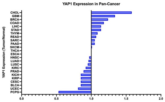
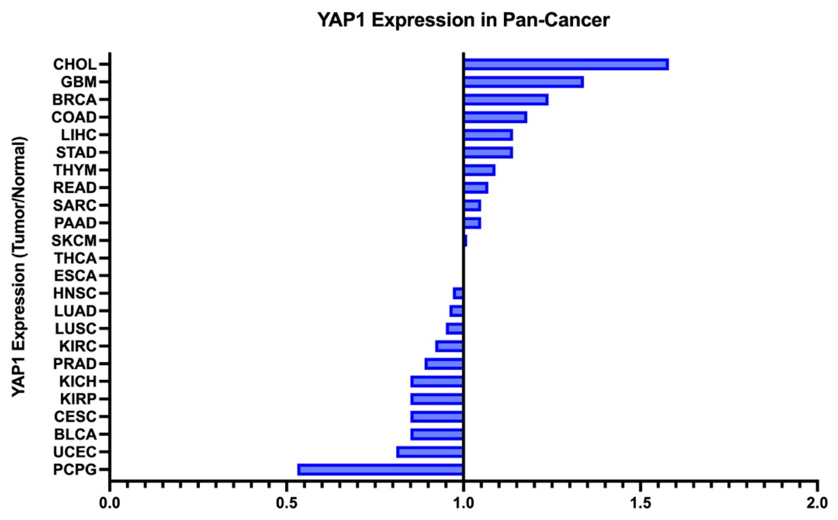
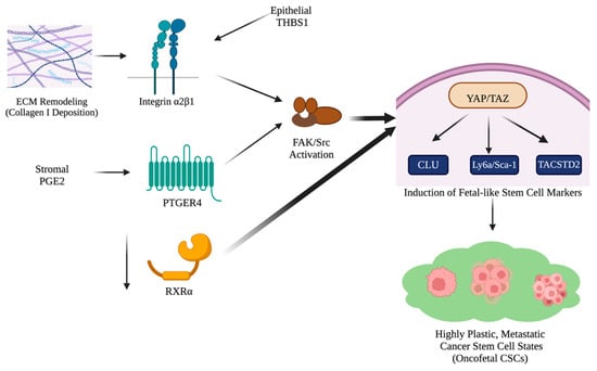
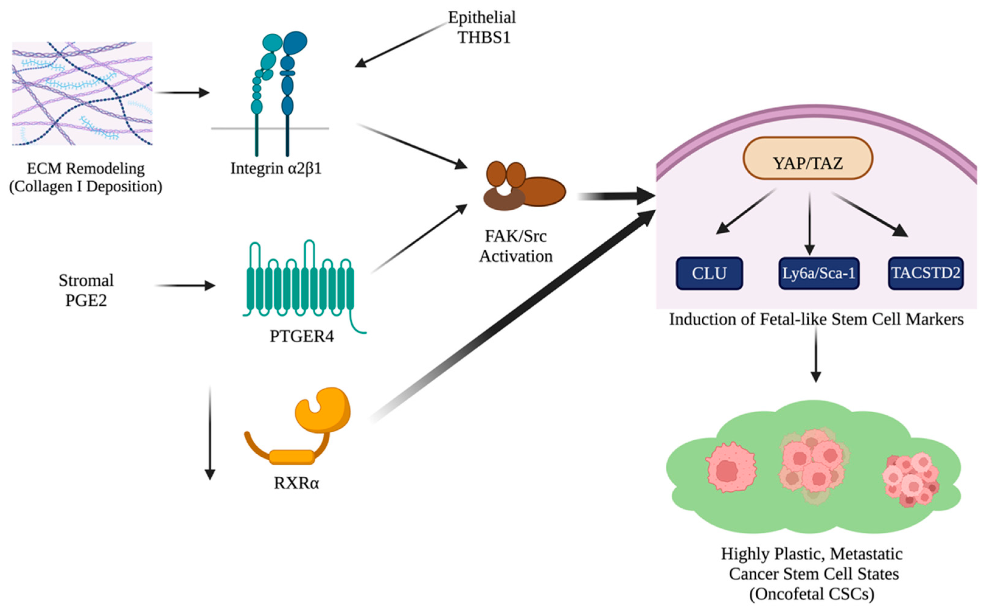
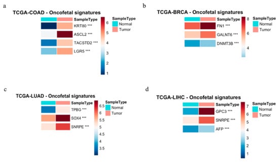
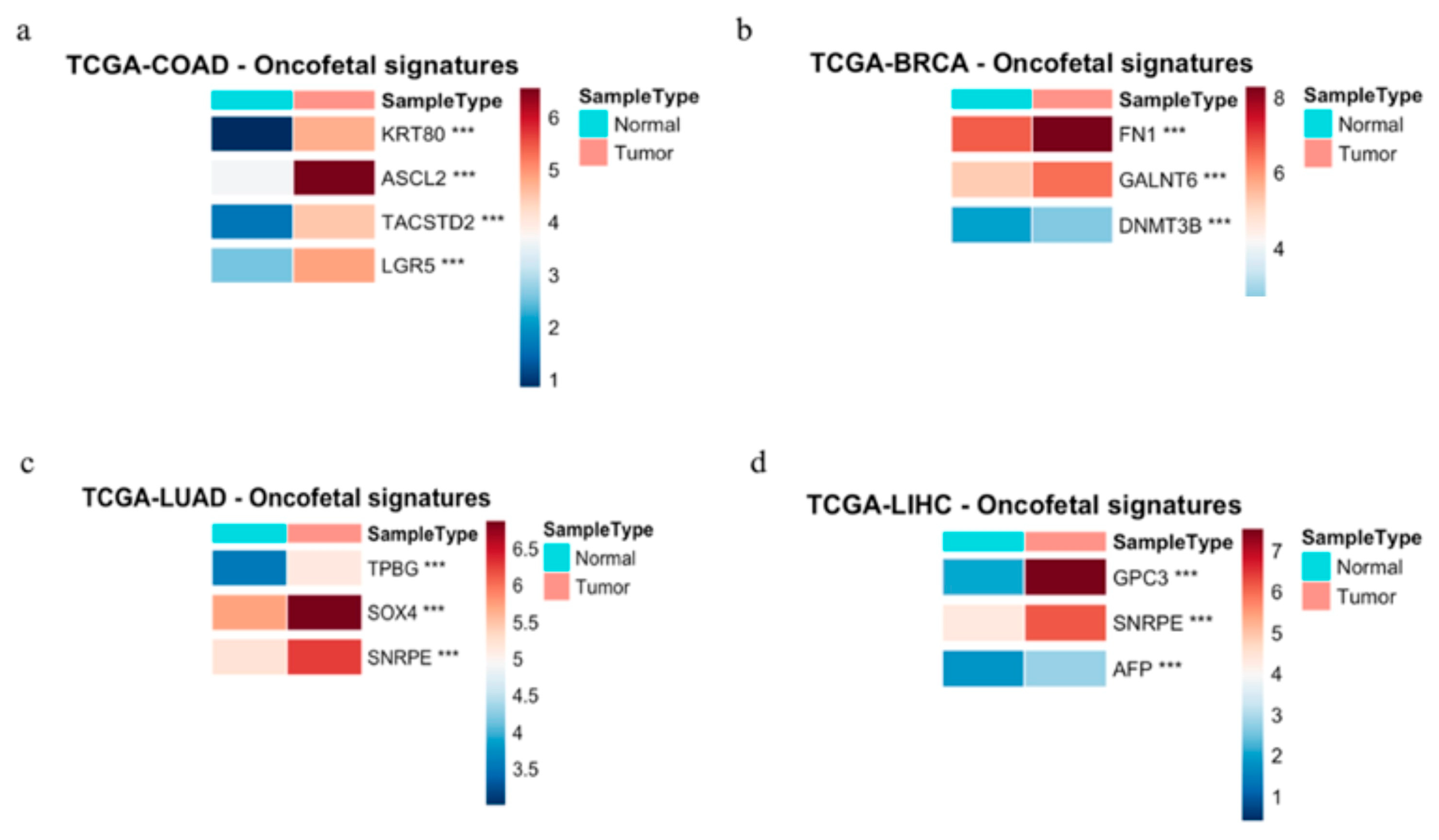
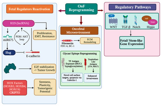
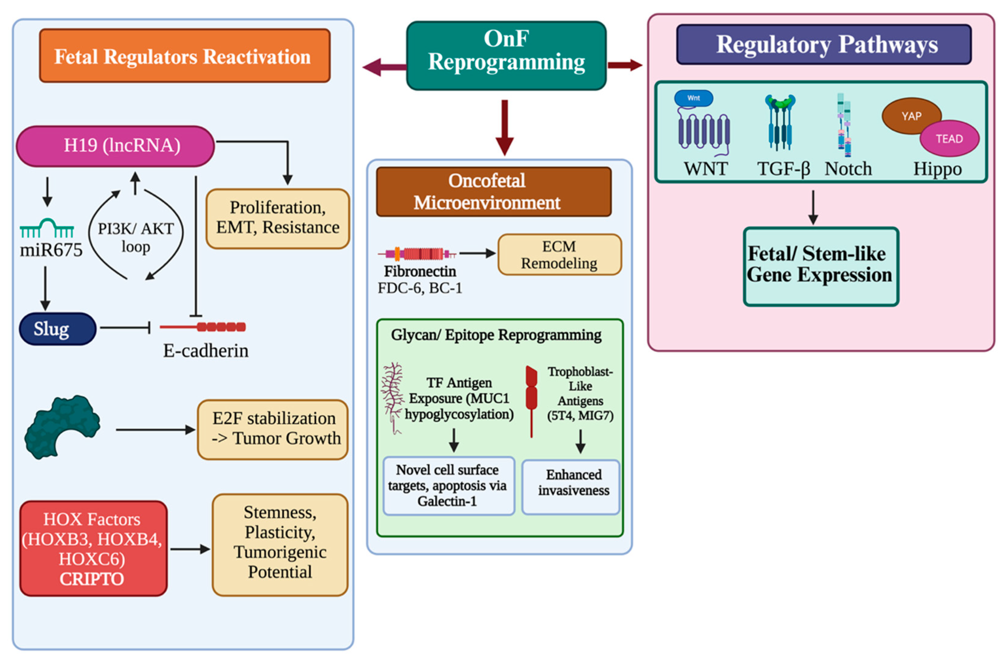
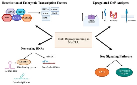
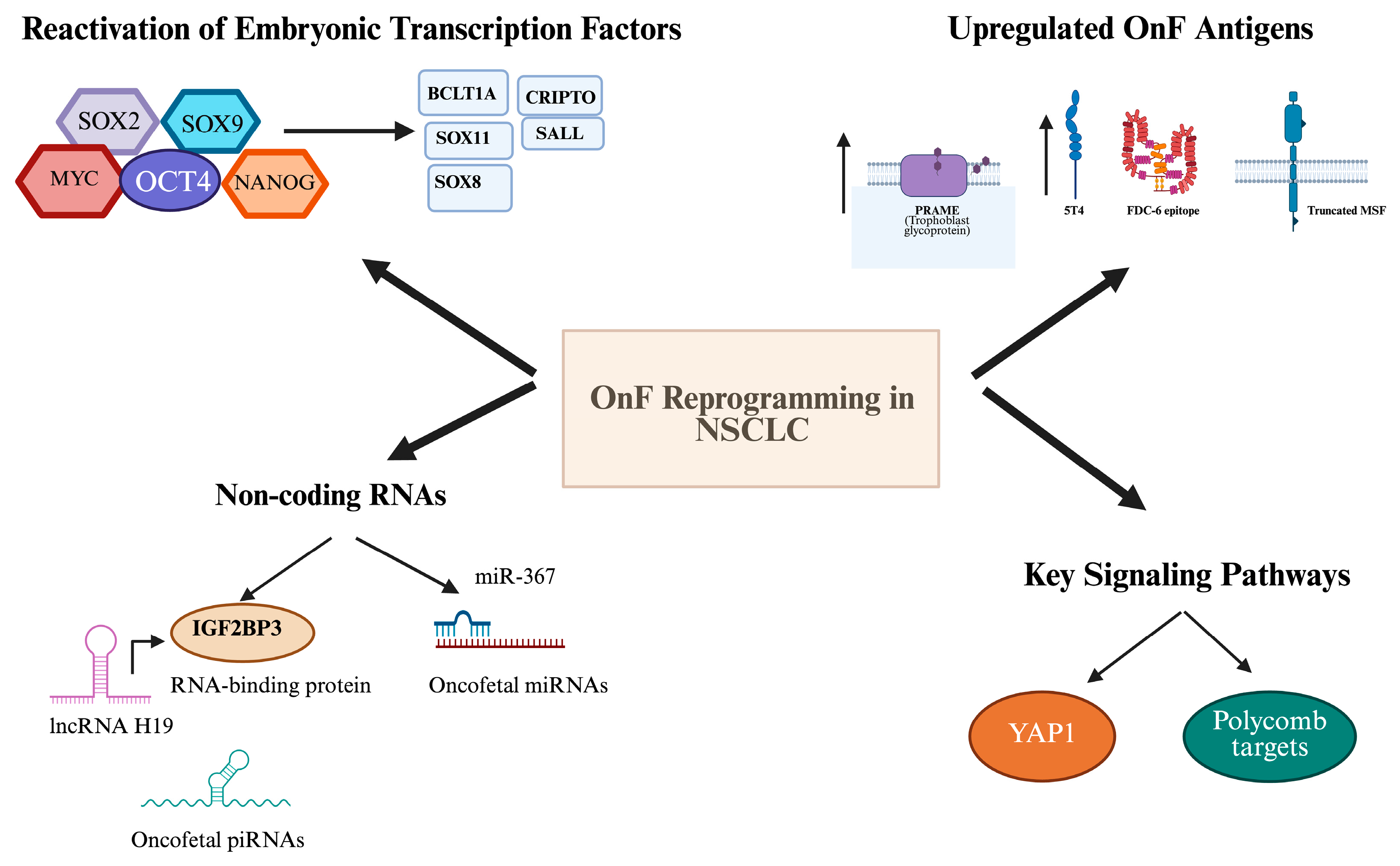
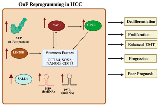
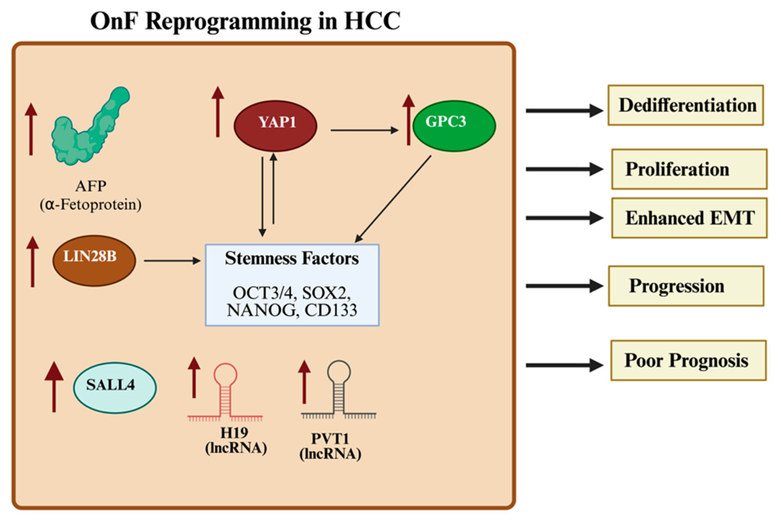
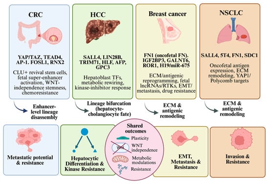
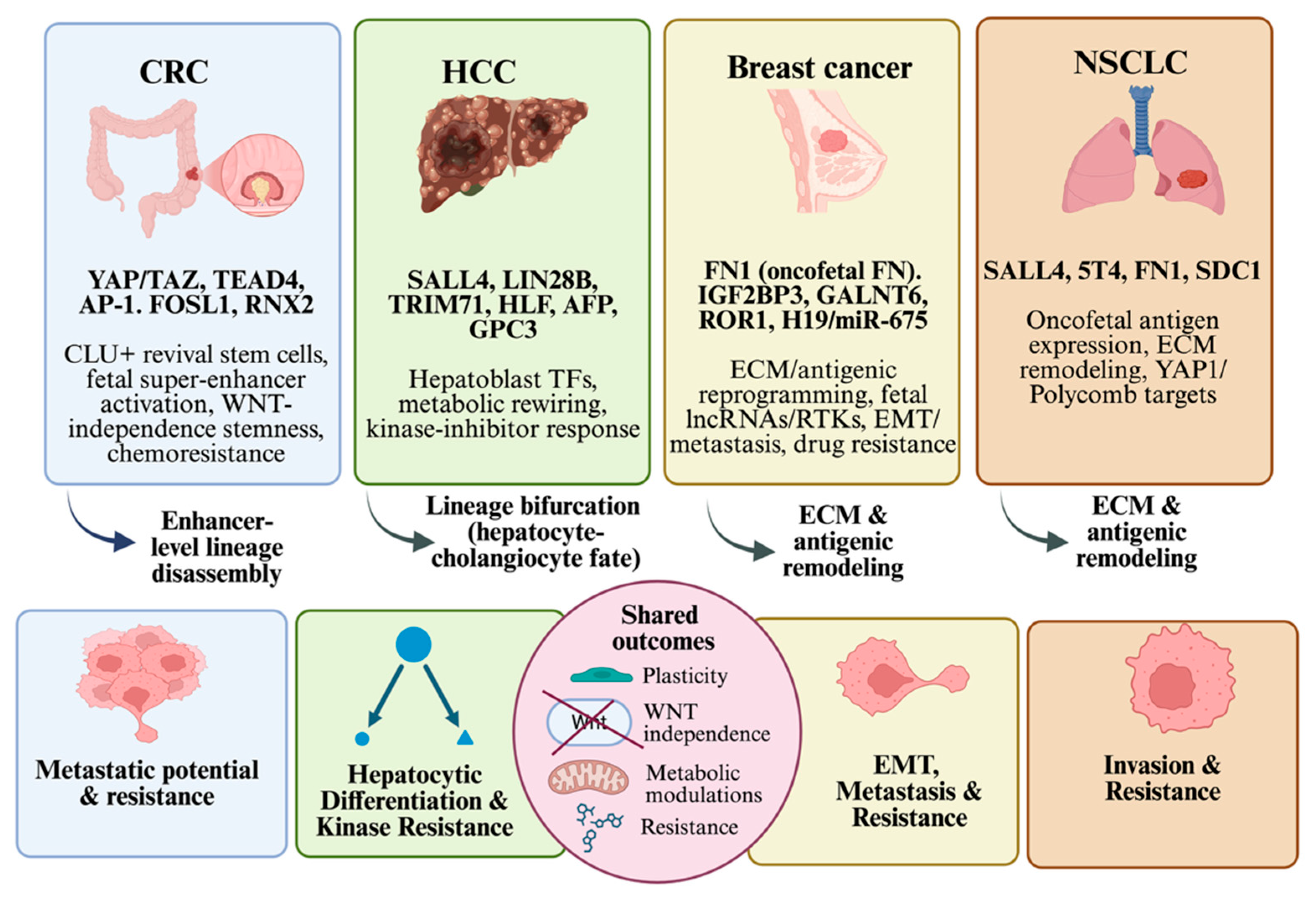

Disclaimer/Publisher’s Note: The statements, opinions and data contained in all publications are solely those of the individual author(s) and contributor(s) and not of MDPI and/or the editor(s). MDPI and/or the editor(s) disclaim responsibility for any injury to people or property resulting from any ideas, methods, instructions or products referred to in the content. |
© 2026 by the authors. Licensee MDPI, Basel, Switzerland. This article is an open access article distributed under the terms and conditions of the Creative Commons Attribution (CC BY) license.