Article

Molecular Autopsy by Exome Sequencing Identifies in Fraternal Twins a CARD11 p.Ser995Leu Variant Within GUK Domain


by
Juan Fernández-Cadena
1,2,†,
Edwin W. Naylor
3,
Heidi Reinhard
4 and
Arindam Bhattacharjee
1,*,†
1
Department of Pediatrics, Brigham and Women’s Hospital, Harvard Medical School, Boston, MA 02115, USA
2
Omics Science Laboratory, Faculty of Health Science, Universidad Espíritu Santo, Samborondón 092301, Ecuador
3
NCGM Laboratory, Apex, NC 27502, USA
4
District 11 Associate Medical Examiner, Miami, FL 33136, USA
*
Author to whom correspondence should be addressed.
These authors contributed equally to this work.
Int. J. Transl. Med. 2026, 6(1), 5; https://doi.org/10.3390/ijtm6010005
Submission received: 5 December 2025 / Revised: 12 January 2026 / Accepted: 19 January 2026 / Published: 28 January 2026

Abstract

Background: We describe the post-mortem analysis of a CARD11 variant allele, p.Ser995Leu, identified in fraternal twins who died in early infancy with no identifiable cause of death. CARD11 variants through varied inheritance models can alter immune function through loss- or gain-of-function mechanisms, involving distinct protein domains; yet the significance of GUK domain variants remains poorly characterized. Twin autopsies showed non-specific findings, such as pulmonary macrophage accumulation and splenic white pulp expansion, but without infection or structural abnormalities. Methods: Whole-exome sequencing, performed as part of molecular autopsies, identified the shared CARD11 p.Ser995Leu variant, previously classified as a variant of uncertain significance (VUS). We assessed evolutionary conservation across CARD family proteins and species and predicted functional impact using in silico tools, which estimate the likelihood that a variant is deleterious. AlphaFold-based structural modeling emphasized qualitative biophysical assessment. Using epidemiological data, population allele frequency, and Bayesian ACMG variant classification, we assessed competing hypotheses under an autosomal dominant model. Results: The p.Ser995Leu substitution affects a conserved, surface-exposed β-sheet within the GUK domain. While CADD scores exceeded 20, other predictive algorithms offered only partial support of pathogenicity. Structural modeling suggested a potential GUK domain destabilization. Integrating genetic, pathologic, immunologic, and probabilistic modeling, we propose a biologically plausible model in which the variant, like other GUK variants, may alter NF-κB or other signaling pathways and is likely pathogenic. Conclusions: While the CARD11 p.Ser995Leu variant’s contribution to disease is uncertain without functional validation or parental testing, and phenotypic findings are non-specific, the presence of an ultra-rare GUK domain variant in both twins, combined with in silico and statistical modeling, supports its interpretation as likely pathogenic or high risk. The results highlight the challenges of data-limited post-mortem variant interpretation.

1. Introduction

Sudden Infant Death Syndrome (SIDS) refers to the sudden and unexpected death of an infant younger than one year, for which the cause of death remains unexplained despite a thorough investigation, including a complete autopsy, review of the circumstances of death, and clinical history [1]. The broader term Sudden Unexpected Infant Death (SUID) encompasses SIDS, deaths from other ill-defined or unknown causes, and accidental suffocation and strangulation in bed [2]. SUID remains a significant public health challenge, accounting for over 3400 infant deaths annually in the United States, although reporting practices vary by jurisdiction [3,4]. Increasingly, genomic sequencing, including whole-exome (WES) and whole-genome sequencing (WGS), has helped uncover underlying genetic vulnerabilities in infants with SUID. A recent study by Bard et al. applied WGS to 144 infants with SUID and 573 healthy adult controls [5]. Variants of interest were identified in 88 genes in 64.6% of the SUID cohort, including many related to cardiac, neurologic, metabolic, immune, and oxidative stress pathways. Forty-three genes were related to cardiomyopathy and arrhythmias, while others were linked to neurologic, metabolic, and two genes related to immune dysfunction, supporting a multifactorial pathogenesis consistent with the triple risk model (developmental vulnerability, critical period, external stressor).
Although not previously reported in SUID cohorts, the question arises whether missense variants in CARD11 (Caspase Activation and Recruitment Domain 11) may contribute to an impaired pathogen defense in infancy, a common finding in SUID autopsies. CARD11 already stands out as a key regulator of antigen receptor signaling in T and B cells, where pathogenic variants can lead to immunodeficiency or immune dysregulation syndromes, including severe atopic disease and inflammatory skin conditions [6]. In this context, the identification of a CARD11 gene variant in fraternal twins raises the possibility of an underlying immune-related dysfunction, including a susceptibility to infections [7].
Fraternal twins originate from two separate ova and, like ordinary siblings, share approximately 50% of their segregating variants, so sharing a variant—even a rare one—can occur by chance [8]. However, when both such twins died from the same unexplained fatal outcome, the interpretive challenge is not merely that the twins share a variant. Rather, the key question is whether the joint occurrence of an ultra-rare variant and an identical lethal phenotype can be explained by inheritance, random (de novo) events, or other non-genetic circumstances. Genetic autopsy can illuminate causal mechanisms in such cases, but interpretation remains challenging because siblings share thousands of variants, creating a high background rate of coincidental overlap [9]. Consequently, probabilistic modeling is required to determine whether a rare variant may contribute to vulnerability, and furthermore if the observed combination of the variant shared in fraternal twins and concordant SUID is plausibly stochastic or caused by the variant.
CARD11, located on 7p22.2, encodes a scaffold protein belonging to the membrane-associated guanylate kinase (MAGUK) family and forms the CARD11-BCL10-MALT1 (CBM) complex, which is essential for NF-κB, JNK, and mTOR pathway activation downstream of T cell and B cell receptors [10]. The protein contains multiple functional domains, including a CARD domain, coiled-coil (CC) region, autoinhibitory domain (ID), and a MAGUK module containing PDZ, SH3, and guanylate kinase-like (GUK) subdomains [6,11,12,13]. In the resting state, CARD11 remains inactive due to autoinhibitory interactions, but, upon receptor engagement, conformational changes allow it to oligomerize and recruit signaling partners such as BCL10 and MALT1 [11].
According to ClinGen gene-disease validity classification, CARD11 has been curated and validated as causally associated with three conditions: BENTA syndrome (autosomal dominant), CADINS (autosomal dominant), and Immunodeficiency 11A (autosomal recessive). These differing inheritance modes are relevant for interpreting loss-of-function and missense variants in a gene-specific context. In vitro studies using CARD11-deficient T cells have shown an impaired NF-κB and mTORC1 activation, consistent with dominant-negative (where a variant form of the protein interferes with the wild-type), loss-of-function, or gain-of-function effects [11,12]. Clinically, CARD11 mutations may lead to immune dysregulation, T cell proliferation defects, and altered cytokine profiles. Gain-of-function mutations, often in the CARD or CC domains, result in BENTA syndrome (B cell Expansion with NF-κB and T cell Anergy), a lymphoproliferative disorder with an increased risk of lymphoma [12,13,14,15]. In contrast, biallelic loss-of-function mutations lead to Immunodeficiency 11A, a form of severe combined immunodeficiency (SCID) [16], and heterozygous dominant-negative mutations cause Immunodeficiency 11B or CARD11-associated atopy with dominant interference of NF-κB signaling (CADINS), characterized by elevated IgE, severe atopy, and recurrent infections [17,18,19]. Notably, while most pathogenic variants cluster in the CARD or coiled-coil domains, the GUK domain is an important structural region, with variants in this domain also impairing NF-κB signaling and mTORC1 activation.
Importantly, some CARD11 variants have been reported in patients without classic features of immunodeficiency, suggesting that clinically relevant effects may be underrecognized in individuals lacking overt phenotypes [10,20,21]. In such contexts, where clinical or segregation data may be limited (such as in newborn screening or post-mortem analyses), modeling, conservation analysis, and bioinformatic approaches are valuable tools for interpreting rare or uncertain variants, especially those identified in conserved, surface-exposed regions that may destabilize protein folding or interactions [22,23].
This study evaluates an ultra-rare variant identified in fraternal twins who died months apart, for whom traditional autopsies were inconclusive. As CARD11-related variants are poorly characterized in neonatal and post-mortem settings, and immune phenotypes span a broad clinical spectrum, we investigated the rare heterozygous missense (VUS) variant p.Ser995Leu, identified through post-mortem in the highly conserved GUK domain of CARD11. The GUK region is implicated in protein–protein interactions in immune signaling via the CBM complex and could represent a biologically plausible contributor to immune-related vulnerability. The study evaluated the relative likelihood of (i) recurrent SUID, (ii) two identical de novo mutations, and (iii) the inheritance of a rare CARD11 missense variant in fraternal twins by integrating these analyses with a Bayesian ACMG classification. The findings inform variant interpretation and risk assessment in molecular autopsy investigations of ultra-rare pediatric deaths.

2. Materials and Methods

2.1. Autopsy Procedures and Additional Testing

A complete autopsy was performed, including external and internal examinations, along with a histological analysis of organs such as the heart, lungs, liver, kidneys, brain, spleen, thymus, gastrointestinal tract, adrenal glands, larynx, trachea, pancreas, prostate, stomach, and esophagus. Microscopic evaluation was carried out on tissue sections from these organs to assess pathological changes. A Gram stain and culture of lung tissue to evaluate potential pathogens, with common post-mortem isolates such as Escherichia coli or Staphylococcus aureus, was taken into account during interpretation. PCR testing was performed to assess respiratory viruses, including rhinovirus/enterovirus and COVID-19. Toxicology testing was conducted on thoracic blood to assess alcohol, drug abuse, and common medications. Additionally, biochemical metabolic screening included acylcarnitine profile, 17-hydroxyprogesterone (17-OHP), thyroid-stimulating hormone (TSH), and galactose testing.

2.2. Whole-Exome Sequence Methodology

Post-mortem Dried Blood Spot (DBS) specimens were provided by the Medical Examiner to a CLIA/CAP certified molecular diagnostics laboratory (NCGM, NC), and genomic DNA was extracted from DBS, which is used routinely in genomic sequencing. All steps from extractions to reporting were performed at NCGM. DBS DNA was enriched for the complete coding regions and splice site junctions of the whole exome using a Twist v2.0 bait-capture system (Twist Bioscience, San Francisco, CA, USA). Paired End Sequencing was performed (2 × 150 bp) on an Illumina NextSeq 2000 platform (Illumina, San Diego, CA, USA). Data were filtered and analyzed to identify variants of interest and interpreted in the context of clinically relevant transcripts. Data were aligned to GRCh37 using BWA-MEM (v0.7.10; Wellcome Trust Sanger Institute, Hinxton, UK), duplicate reads were removed using Picard tools (Broad Institute, Cambridge, MA, USA; https://broadinstitute.github.io/picard/, accessed on 18 January 2026), and variants were called using a GATK HaplotypeCaller (v3.3; Broad Institute, Cambridge, MA, USA). Variants were annotated using custom scripts and the Ensembl Variant Effect Predictor (VEP v75; European Bioinformatics Institute, Hinxton, UK). Enrichment and analysis focused on the coding sequence of the indicated transcripts, 5–10 bp flanking intronic sequence, and other specific genomic regions demonstrated to be causative of disease at the time of assay design. Sequence and copy number variants are reported according to the Human Genome Variation Society (HGVS) nomenclature. Analysis was limited to genes relevant to the phenotype provided using human phenotype ontology terms to identify the most likely candidates relevant to the patient’s presentation, which is consistent with standard clinical practice. Parental genotyping was not possible due to the inability to recontact the parents and the lack of parental consent, which limited the assessment of variant inheritance and precluded the confirmation of a de novo event. This constraint significantly impacts the strength of causality interpretation in a diagnostic setting. The tools and databases employed for analysis included ClinVar, OMIM, HGMD, UCSC genome browser, UniProt, Ensembl, dbSNP, gnomAD, ExAC, PubMed (NCBI), DbGap, Kaviar, and various bioinformatics approaches, predictive tools, as well as disease-specific databases when available. As no ClinGen Variant Curation Expert Panel (VCEP)-specific guidance exists for CARD11, American College of Medical Genetics and Genomics (ACMG) and Association of Molecular Pathology (AMP) rules were applied, using the 2015 framework in conjunction with general recommendations from the ClinGen Sequence Variant Interpretation Working Group [24].

2.3. Variant Interpretation and ACMG/AMP Classification

Variants were classified according to the American College of Medical Genetics and Genomics and the Association for Molecular Pathology (ACMG/AMP) guidelines. Evidence codes were applied following the 2015 ACMG/AMP framework, using established pathogenicity categories and criteria such as
  • Pathogenic Strong (PS)
  • PS3: Well-established in vitro or in vivo functional studies supportive of a damaging effect on the gene or gene product.
  • Pathogenic Moderate (PM)
  • PM1: Located in a mutational hot spot and/or critical and well-established functional domain (e.g., active site of an enzyme) without benign variation.
  • PM2: Absent from controls, or at extremely low frequency if recessive.
  • Pathogenic Supporting (PP)
  • PP2: Missense variant in a gene that has a low-rate or benign missense variants.
  • PP3: Multiple lines of computational evidence support a deleterious effect on the gene or gene product (e.g., high CADD, strong conservation).

2.4. In Silico Prediction Tools

To further characterize the CARD11 p.Ser995Leu variant, we evaluated it using a combination of sequence-based, predictive, and protein structural tools to assess potential functional impact, including SIFT (version 6.2.1; https://sift.bii.a-star.edu.sg/, accessed on 18 January 2026), REVEL (version 1.3), MutationTaster (version 2; https://www.mutationtaster.org/), phyloP100, and Combined Annotation Dependent Depletion (CADD v1.7; University of Washington, Seattle, WA, USA) scores. CADD was evaluated under both standard thresholds and gene-specific mutation significance cutoffs (MSC) [25,26,27,28,29,30]. GERP++ scores were obtained from the UCSC Genome Browser (https://genome.ucsc.edu/, accessed on 11 September 2025) to assess nucleotide-level conservation [31]. ConSurf analysis (https://consurf.tau.ac.il/, accessed on 18 January 2026) was conducted using multiple sequence alignments of 142 homologous sequences under the Jones–Taylor–Thornton substitution model to estimate residue-level evolutionary constraint [32]. AlphaMissense (HegeLab; https://alphamissense.hegelab.org/) scores were included as a machine learning-based computational predictor of missense variant effects that integrates structural and conservation features [33]. In addition, ClinVar annotations were reviewed when available, and variants were evaluated according to ACMG/AMP guidelines [24]. In addition to the p.Ser995Leu variant identified in this study, ten additional CARD11 missense variants previously reported in the literature and with available NF-κB functional characterization were included in the analysis (p.Arg848Cys, p.Glu1028Lys, p.Arg1104Gln, p.Cys971Trp, p.Arg974Cys, p.Arg975Trp, p.Arg1085Ser, p.Phe1086Ser, p.Trp1125Ser, and p.Asp1152Asn). The p.Arg848Cys and p.Asp1152Asn variants are not localized within the GUK domain. Furthermore, a broader set of 264 missense variants within the GUK domains of CARD10, CARD11, and CARD14 were assembled from gnomAD and analyzed based on allele frequency, CADD score, and AlphaMissense pathogenicity predictions. This enabled a comparison of concordance and discordance between predictive tools across the CARD family independent of ClinVar annotations.

2.5. Protein Structural Modeling

AlphaFold (https://alphafold.ebi.ac.uk/, accessed on 18 January 2026; European Bioinformatics Institute, Hinxton, UK) was used to obtain the wild-type CARD11 structure (model: AF-Q9BXL7-F1), which was visualized in PyMOL v3.1.4.1 (Schrödinger, LLC, New York, NY, USA) to examine the spatial localization and surface exposure of residue Ser995 [29,30]. The unmodified AlphaFold model was then submitted to DynaMut, where the p.Ser995Leu substitution was computationally introduced. DynaMut (http://biosig.unimelb.edu.au/dynamut/, accessed on 18 January 2026) predicted the resulting changes in protein stability (ΔΔG), vibrational entropy (ΔΔS_vib), and interatomic interactions such as hydrogen bonds and hydrophobic contacts. Normal Mode Analysis (NMA) was also performed to evaluate mutation-induced changes in protein flexibility [19,30].

2.6. Probability Modeling of Shared Variant and SUID Risk

To assess whether the shared CARD11 p.Ser995Leu variant in both fraternal twins could be explained by chance, we evaluated three potential explanations: We estimated probabilities for three scenarios in fraternal twins: (1) Independent SUID using U.S. annual birth and SUID data; (2) two independent de novo occurrences of the same variant using a per-site mutation rate; and (3) inheritance from a carrier parent using an allele frequency and autosomal dominant transmission (25%). Independence was assumed for modeling purposes. For genetic probability calculations, we used autosomal dominant transmission probabilities using the gnomAD allele frequency for the CARD11 variant (AF = 2.484 × 10⁻⁶). De novo mutation rates used a standard per-base estimate of 1 × 10⁻⁸ per gamete. For Bayesian Variant Classification, we used the Bayesian ACMG framework using standard log-odds weightings for evidence categories [34,35] and custom phenotype/evidence-informed likelihood ratios. Posterior pathogenicity probability was calculated from prior odds × Bayes factor. Detailed formulas, assumptions, and conversions are provided in the Supplementary Materials under Summary of Bayesian ACMG Framework Analysis.

3. Results

3.1. Summary of Clinical Course, Microbiology, and Autopsy Findings for Each Twin

The cases described in this report involve fraternal twin siblings.

3.1.1. Male Twin

The male twin was found unresponsive by the father at 3 months and 20 days of age. He was transported to the hospital where, despite medical intervention, he was pronounced deceased. At autopsy, the body was determined to be well developed, well nourished, and of a 12-pound male infant. The pleural surfaces of the lungs were smooth, glistening, mottled from pink-tan to maroon-purple, and had a honeycomb pattern. Multiple petechial hemorrhages were scattered over the surface of all the lobes of both lungs. The pulmonary parenchyma was congested and exuded red fluid. The conclusion of the autopsy was a non-dysmorphic infant with pulmonary congestion and edema and visceral pleural petechiae. A microscopic examination of the tissues revealed no significant pathologic changes in all tissues except for the lungs and spleen. The lungs had atelectasis, congestion, and foamy macrophages. The spleen had subtle white pulp expansion. The Gram stain of the right lung revealed few white blood cells and Gram-negative rods. Bacterial cultures of the right lung had a moderate growth of Escherichia coli and Staphylococcus aureus. Respiratory viral PCR performed on a nasopharyngeal swab detected rhinovirus/enterovirus. While bacterial cultures did grow out two different types of bacteria, the history and microscopic examination did not indicate that the decedent was suffering from an active infection. Rhinovirus was detected, but there was no evidence of viral pneumonia. No anatomic, microscopic, metabolic, toxicologic, or circumstantial cause of death was identified. There was no phenotypic evidence of opportunistic infection or atopic dermatitis.

3.1.2. Female Twin

The female twin was found unresponsive by the mother at 9 months and 17 days of age. She was transported to the hospital, where her condition deteriorated, and she was pronounced deceased. She had a history of gastroesophageal reflux and had been treated for a diaper rash. COVID-19 was not detected by PCR in a nasopharyngeal swab in the hospital. She initially presented with Upper Respiratory Infection (URI)-like symptoms, including Rhinovirus/Enterovirus + Respiratory Viral Panel infections. At autopsy, the body was that of a well-developed, well-nourished, non-dysmorphic, 18-pound female infant who was appropriately developed. She had visceral pleural petechiae and pulmonary consolidation. The pleural surfaces were smooth, mottled from maroon to red-tan, and glistening. There were scattered petechial hemorrhages on the surface of the lungs.
The pulmonary parenchyma of the upper lobe of the left lung was brown-tan and soft, while that of the remaining lobes was firm, consolidated, and exhibited a variegated coloration ranging from maroon-red to red-tan. A microscopic examination of the lungs showed vascular congestion, intra-alveolar hemorrhages, abundant intra-alveolar pigmented macrophages, and foci of bacterial overgrowth and aspirated material. No anatomic, microscopic, metabolic, toxicologic, or circumstantial cause of death was identified. There was no phenotypic evidence of opportunistic infection or atopic dermatitis. There was no evidence of hypertrophic cardiomyopathy.

3.2. Molecular Autopsy Genomic Results

DBS specimens were sent by the Medical Examiner in Florida for molecular autopsy analysis using WES (performed at NCGM), along with a summary of autopsy findings and history. The WES analysis indicated that both twins shared a heterozygous missense variant in CARD11 (Chr7:2952956) c.2984C>T/p.Ser995Leu, located in exon 22. The reference transcript and protein used for annotation were NM_032415.7 and NP_115791.3, respectively. This variant had not been previously reported as pathogenic or benign and is currently classified as a variant of uncertain significance (VUS). It was observed at an allele frequency of 0.00000248, as shown in Table 1 (gnomAD v4.1.0). The substitution of serine (a polar residue) with leucine (a hydrophobic residue) represents a significant physicochemical change that affects secondary structure, polarity, and charge. Moreover, CARD11 showed a high missense constraint (Z = 3.78), suggesting that amino acid substitutions are generally not tolerated across the gene, reinforcing the likely functional relevance of changes that affect conserved domains such as the GUK domain, where this alteration is located.
The female twin also harbored an MYH6 variant, c.4723G>A (p.Glu1575Lys), observed at a frequency of 0.00001363 in gnomAD and previously reported as a risk gene [5]. Although the conservation analysis for this missense variant supported functional constraint, the absence of a cardiac phenotype at autopsy and the fact that the variant was present only in the female twin led us to consider it incidental and not contributory in this case.

3.3. In Silico Characterization of the CARD11 p.Ser995Leu Variant

3.3.1. Overview of GUK Domain Variants

The GUK (guanylate kinase-like) domain of CARD11 (residues 947–1140) interacts intermolecularly and intramolecularly with other domains and proteins, and is essential for the conformational activation of the CBM complex [6]. For a comparison of p.Ser995Leu to other similar GUK variants, we identified ten other missense GUK variants in the literature with available functional characterization and/or associated phenotypic information within the GUK domain. The previously reported p.Glu947Lys variant was not included in this comparative analysis, as the present study was restricted to missense variants annotated within coding exons; p.Glu947Lys lies at a boundary region and has been described as having splicing effects. Of these, only p.Trp1125Ser was present in the homozygous state. Table 1 and Supplementary Table S1 summarize the missense variants, their in silico predictions including AlphaMissense and CADD scores, population frequency from gnomAD v4.1.0, clinical classification from ClinVar, ACMG/AMP-based variant classification, and functional evidence data further described below [24].

3.3.2. Functional Score and Evolutionary Conservation and Evidence of 11 GUK Missense Variants

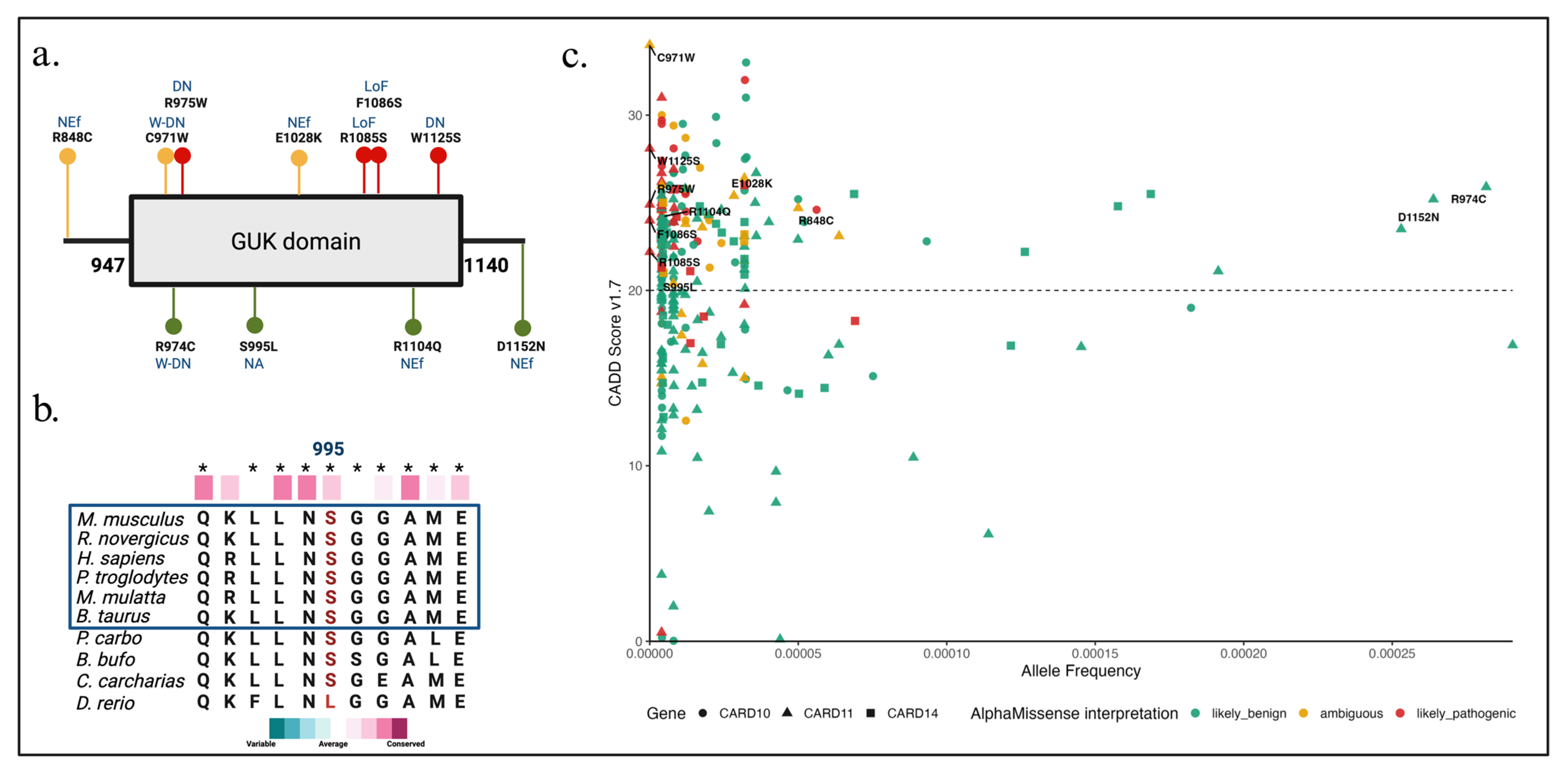
Eleven missense variants in the GUK domain of CARD11 and nearby regions were analyzed in this study, as shown in Figure 1a. Variants are color-coded by AlphaMissense classification, with likely pathogenic (red), ambiguous (orange), and likely benign (green) distributed along the domain. Residue p.Ser995Leu, identified in this study, is located in this region. Multiple sequence alignment (Figure 1b) includes zebrafish (Danio rerio), which harbors leucine at the homologous position, suggesting an evolutionary divergence and possible tolerance at this site. All eleven CARD11 variants, including p.Ser995Leu, had CADD scores above 20, low allele frequencies, and met the PM2 (absent from controls, or at extremely low frequency if recessive) ACMG/AMP criterion (Table 1). The AlphaMissense score (0.12) for p.Ser995Leu suggested a benign classification.

3.3.3. In Silico Predictions, Functional Assays of GUK Domain Variants

The 10 CARD11 missense variants located near and within the GUK domain and previously reported in the literature with available functional characterization were further analyzed (Table 1). Many had destabilizing ΔΔG stability values. However, only 4 out of these 10 variants had >0.564 (likely pathogenic) AlphaMissense scores (p.Arg975Trp, p.Arg1085Ser, p.Phe1086Ser, and p.Trp1125Ser). However, even within this concordant subset, impaired NF-κB activation as measured by functional assays varied. Not all in silico predictors yielded uniform results. Thus, functional data did not uniformly indicate a loss of NF-κB activation across this set. The p.Ser995Leu variant analyzed in this study showed a similarly discordant in silico profile, characterized by an elevated CADD score alongside a benign AlphaMissense classification and low conservation metrics. Other algorithms, such as MutationTaster, PhyloP100, and GERP++, showed variable predictions across these variants (Supplementary Table S1).

3.3.4. CADD and AlphaMissense Scores for the 264 GUK Missense Variants

To further investigate variant characteristics within the GUK domain, we analyzed 264 missense variants across the CARD family genes: CARD10, CARD11, and CARD14. These variants were plotted according to their AlphaMissense classification and CADD score against the allele frequency from gnomAD (Figure 1c). A CADD threshold of 20 was applied to identify variants predicted to be highly deleterious, and, although not explicitly shown, the gene-specific CADD mutation significance cutoff (MSC) for CARD11 was achieved at 21. A substantial number of variants across all three genes exceeded this threshold; however, their AlphaMissense classifications varied considerably, ranging from likely benign to likely pathogenic. Supplementary Figures S1–S3 provide a more comprehensive assessment of how the two predictors differ in their interpretation of GUK domain variants. In general, CADD classified a markedly larger proportion of variants as potentially deleterious, whereas AlphaMissense more frequently assigned benign or uncertain labels, resulting in a substantial variant-level discordance across CARD10, CARD11, and CARD14. This reflects a methodological difference whereby CADD relies on evolutionary conservation, while AlphaMissense evaluates predicted structural effects.
Taken together, these findings reveal a complex landscape of variant classification within the conserved GUK domain of CARD11 and across its paralogs, CARD10 and CARD14.

3.4. Protein Structural Modeling of CARD11 GUK Domain Variants

3.4.1. Protein Structure Predictions and Alpha Missense Scores for 11 GUK Missense Variants

From a structural perspective (Table 1), GUK missense variant predictions indicate a destabilizing impact on both protein stability and protein–protein binding affinity. Several missense variants within the GUK domain of CARD11 displayed a range of predicted structural impacts and classification outputs. The variant p.Ser995Leu showed a ΔΔG of −0.323, while other GUK domain variants—including p.Arg848Cys (−0.037), p.Arg1104Gln (−0.15), and p.Glu1028Lys (−0.31)—also exhibited a predicted destabilization. All four variants were located at conserved positions, with phyloP100 scores exceeding 3.9. The AlphaMissense classification differed across these variants: p.Arg848Cys and p.Glu1028Lys were labeled ambiguous, while p.Arg1104Gln was classified as likely benign. The functional data available for p.Arg848Cys indicated a reduced NF-κB activation in cell-based assays, whereas no measurable signaling effects were reported for p.Arg1104Gln and p.Glu1028Lys. These variant-specific differences in predicted and experimental parameters were observed within the same conserved protein domain, highlighting the complexity of predicting functional impact based solely on domain location. These findings highlight the structural context of Ser995 and its proximity to other GUK domain variants (Figure S1). Structural analysis (Figure S4a,b) indicates that the p.Ser995Leu substitution disrupts a local hydrogen-bonding interaction and introduces a new hydrophobic interaction. Residue 995 lies on the protein surface (Figure S4c), consistent with solvent exposure and a potential involvement in intermolecular interfaces. Normal-mode modeling (Figure S4d) shows that the overall mobility of the protein remains similar between wild-type and p.Ser995Leu (S995L), although the variant produces a modest increase in flexibility within a mid-protein region (~residues 400–600) and a localized alteration in dynamics at the substitution site.

3.4.2. Probability Modeling of CARD11 p.Ser995Leu Occurrence

We first evaluated the epidemiologic baselines relevant to assessing whether the shared variant and concordant SUID could plausibly arise by chance. The baseline probability of SUID is approximately 1 in 1090 infants based on 3400 events per year in the USA (see Supplement Table S2). Assuming an independence between siblings, the expected frequency of both members of a fraternal twin pair experiencing SUID can be approximated as the product of their individual risks (8.4 × 10⁻⁷, or 1 in 1.2 million twin pairs). Given ~38,500 fraternal twin births per year [39], this translates to roughly one such event every 30 years, underscoring the rarity of simultaneous SUID in fraternal twins. Indeed, empirical evidence of both twins dying of SIDS is uncommon [40], and 90% of SUID cases in the United States occur under 4 months of age, with a peak at 1 to 2 months [41]. Therefore, the actual risk in this coincidental case may be much higher (~1 in 120 million twin pairs).
We next evaluated the probability of both fraternal twins independently acquiring the de novo variant in an autosomal dominant model of inheritance. Using per-site mutation rate estimates (~1 × 10⁻⁸ per gamete per generation), both twins having an identical de novo substitution is ~1 × 10⁻¹⁶, or about 1 in 10 quadrillion sibling pairs—effectively negligible at human population scales. While twin SUID recurrence is rare epidemiologically, same-site de novo mutational coincidence is vastly rarer and cannot reasonably explain a shared candidate variant incidence or dependent causality in fraternal twins.
Neither twin exhibited the environmental or forensic features associated with SUID, which increases the relative weight of a genetic contribution. Autopsy information further informs the relative probabilities. One twin exhibited dermatologic and infectious features consistent with CARD11-associated immune dysregulation. The GUK domain—the location of the variant—contains multiple previously reported infant missense variants, including one described by Meshaal et al. [42], providing a precedent for severe early-life immune failure. Considering the autopsy findings and clinical data alongside the modeled probability, the shared variant in both fraternal twins aligns more closely with an inherited event than two independent de novo occurrences.
To integrate variant information and autopsy findings without double-counting, we applied the Bayesian ACMG framework (see Supplementary Materials, Section Summary of Bayesian ACMG Framework Analysis). Evidence included the extreme rarity in controls (PM2), location within a functionally critical domain with known pathogenic missense variants (PM1), gene-level constraint against benign missense variation (PP2), computational pathogenicity (PP3), phenotype consistency (autopsy pathology and findings), and a precedent from the prior literature documenting multiple infant deaths associated with a GUK domain CARD11 variant. Using a conservative 10% prior probability for a rare missense variant in a constrained immune gene, the posterior probability of pathogenicity reached 90%, meeting the ACMG likely pathogenic threshold (>90%).
Taken together, statistical modeling shows that (i) SUID in both fraternal twins is rare but epidemiologically measurable but unlikely; (ii) two independent de novo occurrences of the same CARD11 variant are extremely unlikely at the population scale; and (iii) the inheritance of a rare pathogenic allele from an asymptomatic parent occurs at a frequency that strongly favors a shared genetic etiology over coincidental SUID.

4. Discussion

The heterozygous missense p.Ser995Leu variant in CARD11 was identified in two fraternal twins who died in early infancy without any clear indication on autopsy results. Despite the absence of overt immunologic symptoms, phenotype-driven testing led to the identification of a rare variant in an evolutionarily conserved and structurally constrained region of CARD11, supporting the hypothesis of a previously unrecognized functional consequence. Notably, other reported variants in the GUK domain (Figure 1; Table 1) have manifested without classic immunodeficiency features, highlighting the possibility of subtle or incomplete clinical phenotypes that may be underestimated in diagnostic and autopsy settings [12,17,22].
An evolutionary analysis further reflects this constraint: the CARD11 GUK domain is well conserved within mammals yet markedly divergent in zebrafish (~28% amino acid identity), whereas the upstream CARD (~85%) and coiled-coil (~78%) domains are more highly conserved across these lineages. This pattern highlights domain-specific evolutionary pressures—while the CARD and coiled-coil regions are ancient and essential for NF-κB scaffold activation, the C-terminal MAGUK module (PDZ, SH3, and GUK) diversified later, likely paralleling the increasing complexity of adaptive immune signaling in higher vertebrates [43,44].
Unlike mammals, where CARD11 participates in adaptive immune signaling, zebrafish lack mature T and B cells and organized lymphoid structures during early development, relying instead primarily on innate and humoral immunity [45,46]. The absence of these adaptive immune components reduces the selective pressure to maintain functionally specialized domains like GUK and is one likely explanation for its poor conservation in zebrafish. While zebrafish encode a leucine at the position homologous to human Ser995, this substitution occurs in a domain with limited or no functional relevance in zebrafish and therefore does not indicate biological tolerance in humans. Accordingly, cross-species conservation analyses must be contextualized within the framework of immune system evolution. For example, when applying conservation-based pathogenicity criteria such as PP3 under ACMG/AMP guidelines, the emphasis should be placed on conservation across mammals, where domain architecture and immune function are more directly comparable [24].
Using the Bayesian adaptation of the ACMG/AMP framework (see Supplementary Materials Section, Summary of Bayesian ACMG Framework Analysis), the combined criteria translate qualitative criteria into likelihood ratios, and the combined evidence (PM1 + PM2 + PP3 + PP2) yields a posterior probability of ~0.90, which corresponds to the 90% threshold for a likely pathogenic classification as defined by Tavtigian et al. [34]. This supports the classification of p.Ser995Leu as a likely pathogenic variant, without case-level risk.
The occurrence of an ultra-rare CARD11 GUK domain variant in both fraternal twins with identical fatal outcomes constitutes a risk signal, even without parental genotyping. Concordant lethal outcomes in siblings are statistically improbable by chance alone, supporting a probabilistic interpretation of pathogenicity, consistent with the Bayesian adaptation of the ACMG/AMP framework [34] rather than relying solely on fixed category labels [7,9]. Importantly, elevated risk does not equate to causality: although similar GUK domain variants have been observed in affected individuals and are biologically plausible, the absence of functional assays or segregation data prevents a definitive causal attribution [24]. Furthermore, prior pedigrees showing incomplete penetrance for GUK domain variants caution against dismissing pathogenicity simply because the parents appear unaffected [10,12]. These considerations align with principles anticipated for future ACMG/AMP guideline updates, which are expected to incorporate more quantitative, probabilistic, and gene-aware frameworks modeled on Bayesian and points-based approaches [34,35]. Such frameworks emphasize graded evidence of strength, variant-specific penetrance, and explicit communication of uncertainty, supporting risk-based interpretation in molecular autopsy. Within this paradigm, a variant recurrent in both twins with consistent pathology should be reported as a high-risk variant with an increased probability of pathogenicity, even when conclusive proof remains pending.
The twins lacked classic features of CARD11-associated disorders such as BENTA (e.g., persistent lymphocytosis and lymphadenopathy), CADINS (e.g., eczema, atopy, IgE elevation), or autosomal recessive CARD11 deficiency (e.g., SCID features, T cell activation defects). Instead, they presented with sudden unexpected death and post-mortem findings of diffuse pulmonary congestion, edema, visceral pleural petechiae, and mild white pulp expansion in the spleen. Although overt signs of CARD11-related atopy were absent, the presence of a diaper rash may reflect early dermatologic involvement—paralleling cases like the p.Glu947Lys variant described by Meshaal et al. [42], which was associated with atopy, elevated IgE, and infections, but showed a variable expression among family members, including some with mild or severe phenotypes. The incomplete clearance of rhinovirus and E. coli in the twins may further suggest a partial immune dysfunction consistent with an autosomal dominant CADINS phenotype effect. Given that the p.Ser995Leu variant lies in the GUK domain, implicated in dominant-negative mechanisms in CADINS, these findings highlight the potential for incomplete or variable penetrance or subclinical manifestations.
Although neither twin exhibited overt phenotypic signs of immunodeficiency, the presence of pulmonary petechiae, macrophage accumulation, white pulp expansion, and incomplete immune clearance of low-virulence pathogens suggest a subtle but consequential immune dysregulation. These findings are consistent with the known effects of CARD11 GUK domain mutations that impair NF-κB signaling without producing overt immunodeficiency. The p.Ser995Leu variant may thus represent a hypomorphic or dominant-negative allele insufficient to cause chronic illness but contributing to an immune-mediated susceptibility to sudden death during mild infection.
Across CARD11 GUK domain variants, conservation-based metrics (PhyloP100, ConSurf, and GERP) and AlphaMissense scores showed a limited concordance with functional assay results and pathogenicity classifications. This pattern reflects the underlying biology of CARD11, in which disease mechanisms frequently involve signaling dysregulation, dominant-negative effects, or gain-of-function rather than the disruption of deeply conserved residues. In contrast, structure- and function-aware predictors, including ΔΔG stability, MutationTaster, and CADD, showed a better concordance with experimentally defined functional impairment, consistent with the importance of structural integrity and signalosome assembly for CARD11 activity. Accordingly, ACMG/AMP criterion PP3 was applied conservatively at supporting strength based on convergent evidence from CADD, stability predictions, and MutationTaster, while conservation-driven metrics and AlphaMissense were considered biologically informative but insufficiently discriminative. These discrepancies are consistent with recent evaluations showing that deep learning-based pathogenicity models like AlphaMissense may perform suboptimally in gene-specific contexts involving immune signaling scaffolds and non-loss-of-function mechanisms [47].
AlphaMissense predictions for GUK domain variants illustrate both the strengths and limitations of the current in silico tools. For p.Ser995Leu, the low AlphaMissense score (0.12) contrasts with its elevated CADD score, suggesting that the model may underestimate pathogenicity for certain GUK missense substitutions. Yet, for other variants in the same region, AlphaMissense aligns well with functional data: p.Arg975Trp and p.Trp1125Ser receive high pathogenicity scores (≥ 0.95), and both demonstrate an impaired NF-κB activation [11], while p.Cys971Trp shows a partial loss of function consistent with its intermediate score (0.47). However, variants with context-dependent or subtle effects such as p.Arg974Cys may be undercalled as benign, despite experimental evidence of partial impairment. These patterns indicate that AlphaMissense generally performs well but may lack sensitivity for variants with intermediate or domain-specific effects, a tendency also noted in recent evaluations showing AlphaMissense may undercall pathogenic variants with CADD >20 as benign [48,49]. The clustering of deleterious missense variants within this region supports the notion that subtle structural perturbations can impair CARD11 signaling, underscoring the relevance of functional assays or MAVE (multiplexed assays of variant effect) or other predictions in variant interpretation.
Monogenic atopic immunodeficiencies, such as those involving CARD11 variants, blur the boundaries between atopy and severe immune deficiency, complicating diagnosis and variant interpretation [50]. In this context, this study is limited by the absence of a direct functional validation of the CARD11 p.Ser995Leu variant and the lack of parental or extended family genotyping, constraining definitive causal attribution in this individual case. Future work should include functional assays in CARD11-deficient systems expressing wild-type versus p.Ser995Leu CARD11 to assess CBM complex assembly, NF-κB, and mTORC1 signaling dynamics (e.g., reporter assays, phosphorylation readouts, and co-immunoprecipitation approaches). Where feasible, family-based genotyping would clarify inheritance versus de novo status and enable the assessment of subtle immunologic or dermatologic phenotypes in carriers. Accordingly, these findings should be interpreted as hypothesis-generating, providing a biologically grounded framework for future functional and genetic validation rather than a definitive pathogenic classification. Molecular autopsies should adopt similar principles, integrating genetic, pathological, and probabilistic evidence to guide counseling and surveillance for families. This limitation is inherent in molecular autopsy investigations, which differ from IRB-approved studies or clinical diagnostic settings in that follow-up consent and family sampling are often not feasible due to jurisdictional and procedural barriers. Integrative genomic approaches, including emerging multiplexed assays of variant effect (MAVE), may ultimately improve the resolution for variants that show limited concordance across current in silico predictors [51], supporting a shift toward risk-based interpretation in preventive genomics and newborn screening contexts [52,53].
Optimization in AlphaMissense and other tools, including newer AI tools, may improve pathogenicity prediction. One example of the importance of a CARD11 GUK variant involves a patient with a homozygous missense variant (p.Glu947Lys) with splicing impact in the Meshaal et al. study [42], who was admitted to the Neonatal Intensive Care Unit (NICU) with respiratory distress, and whose relatives (four maternal uncles) died between 4 and 18 months due to infections. The CARD11 p.Glu947Lys (c.2839G>A) and p.Ser995Leu (c.2984C>T) variants are rare missense variants located within the highly conserved GUK domain, a region known to harbor dominant-negative variants associated with CARD11-related atopy and immunodeficiency. The p.Glu947Lys variant has been classified as likely pathogenic in Meshaal et al. [42], based on its absence from population databases, co-segregation with disease in multiple affected family members (including a sibling with severe eczema, failure to thrive, eosinophilia, and diarrhea, and uncles with asthma and elevated IgE), and the established pathogenicity of other missense variants in the same domain. This supports that the absence or presence of the phenotype or genotype in family members does not exclude or include pathogenicity. CARD11 variants demonstrate variable penetrance and expressivity across ClinGen-recognized phenotypes, including severe immunodeficiency, BENTA disease, and CADINS syndrome. Variability in functional assays, such as those measuring mTORC1-mediated substrate phosphorylation, which may differ between cell types and subcellular localizations, underscores the risk of oversimplifying variant interpretation when penetrance is incomplete [54,55]. In molecular autopsy, tissue-level findings can be more informative than isolated cell-based assays.
Thus, both heterozygous and homozygous variants in the GUK domain may exert clinically significant effects that are easily underestimated. Infection-related arousal failure is a plausible contributor to SIDS pathogenesis. Terminal apnea, often triggered during sleep, may result from an inadequate autonomic or respiratory arousal response to hypoxia or airway obstruction. Mild infections, especially of the upper respiratory tract, have been shown to impair arousal and increase SIDS risk—especially when combined with a prone sleeping positioning. The conditions become fatal as the child suffers from a terminal apnea event, leading to irreversible hypoxia and cardiac arrest. In some infants, the terminal event may instead reflect sudden cardiac arrest, exaggerated cytokine responses, or catastrophic metabolic decompensation [56].
Ultra-rare pediatric SUID-like cases, such as this study, expose a major gap in the current ACMG/AMP guidelines, which prioritizes functional validation, which is often impractical, or a reliance on historical databases like HGMD that cannot accumulate evidence for variants that appear once every few decades. In our case, the joint probability of two independent de novo CARD11 variants and coincident SUID is approximately 10−23—effectively zero—making random occurrence implausible. Yet, under current guidelines, this extreme improbability is treated as “supporting” evidence, while functional data are weighted as “strong.” This imbalance means that biologically compelling variants remain classified as VUS indefinitely, erasing clinically meaningful insights and preventing progress in understanding lethal immune disorders. A Bayesian approach that integrates prior probabilities, biological plausibility, and mechanistic contexts achieved a posterior pathogenicity probability >90%, which could allow these cases to be considered, ensuring that evidence from statistical reasoning is not lost simply because wet-lab functional validation is infeasible. Without such a shift, these cases will continue to remain classified as VUS, despite the strong probabilistic evidence supporting increased disease risk. These results demonstrate that the inheritance of an ultra-rare CARD11 variant conferring a substantially increased risk is more plausible than coincidental SUID and far more plausible than dual de novo mutations. The findings underscore the critical role of statistical and mechanistic reasoning in translational genetics, particularly for variants that appear only once in several decades.
The pulmonary and splenic findings were non-specific and commonly reported in SUID, and do not explain sudden death in fraternal twins at different ages following minor stressors, suggesting a shared vulnerability rather than exposure. Thus, the proposed genetic contribution is biologically plausible but non-diagnostic.
CARD11 GUK domain variants show a wide phenotypic variability, with rare infant fatal presentations. Although these twins lack features of classic CARD11 syndromes, they may represent an extreme, context-dependent immune vulnerability to inflammatory stress.

5. Conclusions

The CARD11 p.Ser995Leu variant, identified in two fraternal twins who died in early infancy, represents a strong candidate risk variant with a plausible etiologic contribution, despite its current classification as a VUS. Its rarity, localization within a structurally constrained and conserved protein domain, and predicted impact on protein stability support its potential contribution to immune dysregulation. The twins’ shared autopsy findings, including pulmonary petechiae, alveolar macrophage accumulation, and white pulp expansion in the spleen, with no clear infectious, metabolic, or anatomic explanation, may suggest a subtle immune vulnerability, though causality remains unconfirmed. To our knowledge, no other studies besides Meshaal et al. [42] and this study have linked CARD11 variants to infant death.
Given that these two children died suddenly without alternative causes and share only these variants, suspicion arises of a subtle immune defect due to the variant impairing infection control or immune homeostasis. These findings support the broader inclusion of immune genes in workflows for unexplained early-life mortality and highlight the importance of recurrence risk assessment and genetic counseling strategies for at-risk families.
This case highlights an urgent need to modernize variant interpretation frameworks for ultra-rare pediatric lethal disorders. The traditional exclusion of probabilistic evidence leads to misclassification as VUS, whereas a Bayesian-integrated approach yields a likely pathogenic interpretation. The statistical, genetic, and phenotypic evidence supports the inheritance of this variant of a likely pathogenic CARD11 variant as the most plausible risk factor in these fraternal twins.

Supplementary Materials

The following supporting information can be downloaded at https://www.mdpi.com/article/10.3390/ijtm6010005/s1. Supplementary Results—Variant load in HPO-guided panels; variant-level concordance analysis between CADD and AlphaMissense; summary of predicted structural and dynamic effects of the p.Ser995Leu variant; summary of Bayesian ACMG framework analysis; Figure S1: Identification of discordant variant classifications between CADD and AlphaMissense predictions; Figure S2: Comparison of AlphaMissense and CADD predictions across CARD genes; Figure S3: Correlation between AlphaMissense and CADD scores across CARD gene variants; Figure S4: Modeled structural context and predicted dynamics of the CARD11 p.Ser995Leu variant; Table S1: Functional prediction scores for CARD11 missense variants. Table S2: Event-Level Probability Comparison for SUID, De Novo, and Inherited Variants in Fraternal Twins.

Author Contributions

Conceptualization, A.B. and E.W.N.; methodology, A.B. and J.F.-C.; software, A.B. and J.F.-C.; formal analysis, A.B.; investigation, A.B. and J.F.-C.; resources, H.R.; data curation, A.B.; writing, original draft preparation, A.B. and E.W.N.; writing, review and editing, A.B. and J.F.-C.; visualization, J.F.-C.; supervision, A.B.; project administration, A.B. All authors have read and agreed to the published version of the manuscript.

Funding

This research received no external funding.

Institutional Review Board Statement

The Mass General Brigham Institutional Review Board determined that this project does not constitute Human Subjects Research, as it involved exclusively de-identified post-mortem genetic data generated during the official medical examiner investigation. No living individuals were involved, no identifiable or HIPAA-protected information was used, and therefore Institutional Review Board approval and informed consent were not required.

Informed Consent Statement

Not applicable. This study did not involve living individuals, and the post-mortem samples were analyzed in a fully de-identified manner as part of a clinical molecular autopsy conducted under the authority of the Medical Examiner. Accordingly, informed consent was not required.

Data Availability Statement

The data supporting the findings of this study are not publicly available due to ethical and privacy restrictions related to post-mortem genomic information. The raw genomic data is not available for public sharing, as whole-exome sequencing was performed by an external clinical laboratory, and only final reports were provided to the authors. No raw sequencing files (e.g., FASTQ, BAM, or VCF) are available.

Acknowledgments

During the preparation of this manuscript, the authors used ChatGPT (OpenAI, GPT-5, January 2026 version) for figure generation, grammatical correction, and text refinement, in accordance with MDPI guidance on AI-assisted tools. The authors have reviewed and edited all AI-generated content and take full responsibility for the accuracy and integrity of the final publication.

Conflicts of Interest

The authors declare no conflicts of interest.

Abbreviations

ACMG/AMPAmerican College of Medical Genetics and Genomics/Association of Molecular Pathology
BENTAB cell Expansion with NF-κB and T cell Anergy
CBMCARD11–BCL10–MALT1 Complex
CADDCombined Annotation Dependent Depletion
ConSurfConservation Surface Analysis Tool
DBSDried Blood Spot
GERP++Genomic Evolutionary Rate Profiling
GUKGuanylate Kinase Domain
HPOHuman Phenotype Ontology
LoFLoss of Function
mCSMMutation Cutoff Scanning Matrix
MAVEMultiplexed Assays of Variant Effect
NF-κBNuclear Factor Kappa-Light-Chain-Enhancer of Activated B cells
REVELRare Exome Variant Ensemble Learner
SIFTSorting Intolerant from Tolerant
VUSVariant of Uncertain Significance

References

  1. Perrone, S.; Lembo, C.; Moretti, S.; Prezioso, G.; Buonocore, G.; Toscani, G.; Marinelli, F.; Nonnis-Marzano, F.; Esposito, S. Sudden Infant Death Syndrome: Beyond Risk Factors. Life 2021, 11, 184. [Google Scholar] [CrossRef]
  2. Lavista Ferres, J.M.; Anderson, T.M.; Johnston, R.; Ramirez, J.-M.; Mitchell, E.A. Distinct Populations of Sudden Unexpected Infant Death Based on Age. Pediatrics 2020, 145, e20191637. [Google Scholar] [CrossRef] [PubMed]
  3. Goldstein, R.D.; Blair, P.S.; Sens, M.A.; Shapiro-Mendoza, C.K.; Krous, H.F.; Rognum, T.O.; Moon, R.Y. Inconsistent classification of unexplained sudden deaths in infants and children hinders surveillance, prevention and research: Recommendations from The 3rd International Congress on Sudden Infant and Child Death. Forensic Sci. Med. Pathol. 2019, 15, 622–628. [Google Scholar] [CrossRef]
  4. CDC. Sudden Unexpected Infant Death and Sudden Infant Death Syndrome; Data and Statistics for SUID and SIDS; CDC: Atlanta, Georgia, 2024. Available online: https://www.cdc.gov/sudden-infant-death/data-research/data/index.html (accessed on 13 June 2025).
  5. Bard, A.M.; Clark, L.V.; Cosgun, E.; Aldinger, K.A.; Timms, A.; Quina, L.A.; Ferres, J.M.L.; Jardine, D.; Haas, E.A.; Becker, T.M.; et al. Known pathogenic gene variants and new candidates detected in sudden unexpected infant death using whole genome sequencing. Am. J. Med. Genet. Pt. A 2024, 194, e63596. [Google Scholar] [CrossRef] [PubMed]
  6. Bedsaul, J.R.; Carter, N.M.; Deibel, K.E.; Hutcherson, S.M.; Jones, T.A.; Wang, Z.; Yang, C.; Yang, Y.-K.; Pomerantz, J.L. Mechanisms of Regulated and Dysregulated CARD11 Signaling in Adaptive Immunity and Disease. Front. Immunol. 2018, 9, 2105. [Google Scholar] [CrossRef]
  7. Obel, N.; Christensen, K.; Petersen, I.; Sørensen, T.I.A.; Skytthe, A. Genetic and Environmental Influences on Risk of Death due to Infections Assessed in Danish Twins, 1943–2001. Am. J. Epidemiol. 2010, 171, 1007–1013. [Google Scholar] [CrossRef] [PubMed]
  8. Boomsma, D.; Busjahn, A.; Peltonen, L. Classical twin studies and beyond. Nat. Rev. Genet. 2002, 3, 872–882. [Google Scholar] [CrossRef]
  9. Tomassini, L.; Ricchezze, G.; Fedeli, P.; Lancia, M.; Gambelunghe, C.; De Micco, F.; Cingolani, M.; Scendoni, R. New Insights on Molecular Autopsy in Sudden Death: A Systematic Review. Diagnostics 2024, 14, 1151. [Google Scholar] [CrossRef]
  10. Stepensky, P.; Keller, B.; Buchta, M.; Kienzler, A.K.; Elpeleg, O.; Somech, R.; Cohen, S.; Shachar, I.; Miosge, L.A.; Schlesier, M.; et al. Deficiency of caspase recruitment domain family, member (CARD11), causes profound combined immunodeficiency in human subjects. J. Allergy Clin. Immunol. 2013, 131, 477–485. [Google Scholar] [CrossRef]
  11. Ma, C.A.; Stinson, J.R.; Zhang, Y.; Abbott, J.K.; Weinreich, M.A.; Hauk, P.J.; Reynolds, P.R.; Lyons, J.J.; Nelson, C.G.; Ruffo, E.; et al. Corrigendum: Germline hypomorphic CARD11 mutations in severe atopic disease. Nat. Genet. 2017, 49, 1661. [Google Scholar] [CrossRef]
  12. Dorjbal, B.; Stinson, J.R.; Ma, C.A.; Weinreich, M.A.; Miraghazadeh, B.; Hartberger, J.M.; Frey-Jakobs, S.; Weidinger, S.; Moebus, L.; Franke, A.; et al. Hypomorphic caspase activation and recruitment domain 11 (CARD11) mutations associated with diverse immunologic phenotypes with or without atopic disease. J. Allergy Clin. Immunol. 2019, 143, 1482–1495. [Google Scholar] [CrossRef]
  13. Lu, H.Y.; Bauman, B.M.; Arjunaraja, S.; Dorjbal, B.; Milner, J.D.; Snow, A.L.; Turvey, S.E. The CBM-opathies-A Rapidly Expanding Spectrum of Human Inborn Errors of Immunity Caused by Mutations in the CARD11-BCL10-MALT1 Complex. Front. Immunol. 2018, 9, 2078. [Google Scholar] [CrossRef]
  14. Kniffin, C.; OMIM. OMIM Entry–616452: B-Cell Expansion with NFKB and T-Cell Anergy (BENTA); OMIM: Baltimore, MD, USA, 2024; Available online: https://omim.org/entry/616452 (accessed on 12 June 2025).
  15. Zhao, P.; Hu, Y.; Sun, D.; Meng, Q.; Zhang, L.; Zhang, X.; Tan, L.; Zhang, Y.; Ding, Y.; He, X. A novel CARD11 germline mutation in a Chinese patient of B cell expansion with NF-κB and T cell anergy (BENTA) and literature review. Front. Immunol. 2022, 13, 943027. [Google Scholar] [CrossRef] [PubMed]
  16. Modena, M.; Giannoni, A.; Aimo, A.; Aretini, P.; Botto, N.; Vittorini, S.; Scatena, A.; Bonuccelli, D.; Di Paolo, M.; Emdin, M. Whole-exome sequencing to identify causative variants in juvenile sudden cardiac death. Hum. Genom. 2024, 18, 102. [Google Scholar] [CrossRef]
  17. Hara, H.; Yokosuka, T.; Hirakawa, H.; Ishihara, C.; Yasukawa, S.; Yamazaki, M.; Koseki, H.; Yoshida, H.; Saito, T. Clustering of CARMA1 through SH3–GUK domain interactions is required for its activation of NF-κB signalling. Nat. Commun. 2015, 6, 5555. [Google Scholar] [CrossRef]
  18. Hernández González, J.E.; De Araujo, A.S. Alchemical Calculation of Relative Free Energies for Charge-Changing Mutations at Protein–Protein Interfaces Considering Fixed and Variable Protonation States. J. Chem. Inf. Model. 2023, 63, 6807–6822. [Google Scholar] [CrossRef]
  19. Greil, J.; Rausch, T.; Giese, T.; Bandapalli, O.R.; Daniel, V.; Bekeredjian-Ding, I.; Stütz, A.M.; Drees, C.; Roth, S.; Ruland, J.; et al. Whole-exome sequencing links caspase recruitment domain 11 (CARD11) inactivation to severe combined immunodeficiency. J. Allergy Clin. Immunol. 2013, 131, 1376–1383. [Google Scholar] [CrossRef] [PubMed]
  20. Verschuren, J.; Wozniak, S. Genetic dysfunctions leading to the pathogenic cascade of atopic dermatitis. Med. J. Cell Biol. 2023, 11, 83–86. [Google Scholar] [CrossRef]
  21. Snow, A.L.; Xiao, W.; Stinson, J.R.; Lu, W.; Chaigne-Delalande, B.; Zhang, L.; Pittaluga, S.; Matthews, H.F.; Schmitz, R.; Jhavar, S.; et al. Congenital B cell lymphocytosis explained by novel germline CARD11 mutations. J. Exp. Med. 2012, 209, 2247–2261. [Google Scholar] [CrossRef] [PubMed]
  22. AlYafie, R.S.; Adeli, M.; Velayutham, D.; Bougarn, S.; Ata, M.; Al-Ali, F.; Chin-Smith, E.; Bauman, B.M.; Snow, A.L.; Boisson, B.; et al. A novel CARD11 heterozygous missense variant in a CADINS patient. J. Allergy Clin. Immunol. Glob. 2025, 4, 100461. [Google Scholar] [CrossRef]
  23. Pires, D.E.V.; Ascher, D.B.; Blundell, T.L. mCSM: Predicting the effects of mutations in proteins using graph-based signatures. Bioinformatics 2014, 30, 335–342. [Google Scholar] [CrossRef] [PubMed]
  24. Richards, S.; Aziz, N.; Bale, S.; Bick, D.; Das, S.; Gastier-Foster, J.; Grody, W.W.; Hegde, M.; Lyon, E.; Spector, E.; et al. Standards and guidelines for the interpretation of sequence variants: A joint consensus recommendation of the American College of Medical Genetics and Genomics and the Association for Molecular Pathology. Genet. Med. 2015, 17, 405–424. [Google Scholar] [CrossRef]
  25. Sim, N.L. SIFT web server: Predicting effects of amino acid substitutions on proteins. Nucleic Acids Res. 2012, 40, 452–457. [Google Scholar] [CrossRef]
  26. Ioannidis, N.M. REVEL: An ensemble method for predicting the pathogenicity of rare missense variants. Am. J. Hum. Genet. 2016, 99, 877–885. [Google Scholar] [CrossRef]
  27. Schwarz, J.M. MutationTaster2: Mutation prediction for the deep-sequencing age. Nat. Methods 2014, 11, 361–362. [Google Scholar] [CrossRef] [PubMed]
  28. Pollard, K.S. Detection of nonneutral substitution rates on mammalian phylogenies. Genome Res. 2010, 20, 110–121. [Google Scholar] [CrossRef] [PubMed]
  29. Schubach, M. CADD v1.7: Using protein language models, regulatory CNNs and other nucleotide-level scores to improve genome-wide variant predictions. Nucleic Acids Res. 2024, 52, D1143–D1154. [Google Scholar] [CrossRef]
  30. Itan, Y.; Shang, L.; Boisson, B.; Ciancanelli, M.J.; Markle, J.G.; Martinez-Barricarte, R.; Scott, E.; Shah, I.; Stenson, P.D.; Gleeson, J.; et al. The mutation significance cutoff: Gene-level thresholds for variant predictions. Nat. Methods 2016, 13, 109–110. [Google Scholar] [CrossRef]
  31. Davydov, E.V. Identifying a high fraction of the human genome to be under selective constraint using GERP++. PLoS Comput. Biol. 2010, 6, 1001025. [Google Scholar] [CrossRef]
  32. Yariv, B. Using evolutionary data to make sense of macromolecules with a ‘face-lifted’ ConSurf. Protein Sci. 2023, 32, e4582. [Google Scholar] [CrossRef]
  33. Tordai, H.; Torres, O.; Csepi, M.; Padányi, R.; Lukács, G.L.; Hegedűs, T. Analysis of AlphaMissense data in different protein groups and structural context. Sci. Data 2024, 11, 495. [Google Scholar] [CrossRef]
  34. Tavtigian, S.V.; Greenblatt, M.S.; Harrison, S.M.; Nussbaum, R.L.; Prabhu, S.A.; Boucher, K.M.; Biesecker, L.G. Modeling the ACMG/AMP variant classification guidelines as a Bayesian classification framework. Genet. Med. 2018, 20, 1054–1060. [Google Scholar] [CrossRef] [PubMed]
  35. Tavtigian, S.V.; Harrison, S.M.; Boucher, K.M.; Biesecker, L.G. Fitting a naturally scaled point system to the ACMG/AMP variant classification guidelines. Hum. Mutat. 2020, 41, 1734–1737. [Google Scholar] [CrossRef]
  36. Hu, Y.; Han, L.; Xu, W.; Li, T.; Zhao, Q.; Lu, W.; Sun, J.; Wang, Y. CARD11 regulates the thymic Treg development in an NF-κB-independent manner. Front Immunol. 2024, 15, 1364957. [Google Scholar] [CrossRef]
  37. Watt, S.; Purdie, K.; Den Breems, N.; Dimon, M.; Arron, S.; McHugh, A.; Xue, D.; Dayal, J.; Proby, C.; Harwood, C.; et al. Novel CARD11 Mutations in Human Cutaneous Squamous Cell Carcinoma Lead to Aberrant NF-κB Regulation. Am. J. Pathol. 2015, 185, 2354–2363. [Google Scholar] [CrossRef] [PubMed]
  38. Nguyen, A.; Lu, H.; Turvey, S.; Snow, A. Autosomal Recessive Inflammatory Skin Disease Caused by a Novel Biallelic Loss-of-Function Variant in CARD11. J. Clin. Immunol. 2023, 43, 709–713. [Google Scholar] [CrossRef] [PubMed]
  39. Osterman, M.; Hamilton, B.; Martin, J.; Driscoll, A.; Valenzuela, C. Births: Final Data for 2023; Report No.: 74{1); National Center for Health Statistics, Centers for Disease Control and Prevention (CDC): Hyattsville, MD, USA, 2025; pp. 1–48. Available online: https://stacks.cdc.gov/view/cdc/200000 (accessed on 12 August 2025).
  40. Malloy, M.H.; Freeman, D.H. Sudden Infant Death Syndrome Among Twins. Arch. Pediatr. Adolesc. Med. 1999, 153, 736. [Google Scholar] [CrossRef]
  41. Parks, S.E.; Erck Lambert, A.B.; Shapiro-Mendoza, C.K. Racial and Ethnic Trends in Sudden Unexpected Infant Deaths: United States, 1995–2013. Pediatrics 2017, 139, e20163844. [Google Scholar] [CrossRef]
  42. Meshaal, S.; El Hawary, R.; Abd Elaziz, D.; Eldash, A.; Darwish, R.; Erfan, A.; Lotfy, S.; Saad, M.M.; Chohayeb, E.; Alkady, R.; et al. Novel homozygous CARD11 variants in two patients with combined immunodeficiency and atopic skin disease. Egypt. J. Med. Hum. Genet. 2024, 25, 19. [Google Scholar] [CrossRef]
  43. Te Velthuis, A.J.; Admiraal, J.F.; Bagowski, C.P. Molecular evolution of the MAGUK family in metazoan genomes. BMC Evol. Biol. 2007, 7, 129. [Google Scholar] [CrossRef]
  44. The Evolution of Adaptive Immunity in Vertebrates. In Advances in Immunology; Elsevier: Amsterdam, The Netherlands, 2011; pp. 125–157. Available online: https://linkinghub.elsevier.com/retrieve/pii/B9780123876645000042 (accessed on 12 August 2025).
  45. Trede, N.S.; Langenau, D.M.; Traver, D.; Look, A.T.; Zon, L.I. The Use of Zebrafish to Understand Immunity. Immunity 2004, 20, 367–379. [Google Scholar] [CrossRef]
  46. Robertson, T.F.; Hou, Y.; Schrope, J.; Shen, S.; Rindy, J.; Sauer, J.-D.; Dinh, H.Q.; Huttenlocher, A. A tessellated lymphoid network provides whole-body T cell surveillance in zebrafish. Proc. Natl. Acad. Sci. USA 2023, 120, e2301137120. [Google Scholar] [CrossRef]
  47. Molotkov, I.; Mardis, E.R.; Artomov, M. Making sense of missense: Challenges and opportunities in variant pathogenicity prediction. Dis. Models Mech. 2024, 17, dmm052218. [Google Scholar] [CrossRef] [PubMed]
  48. Curtis, D. Assessment of ability of AlphaMissense to identify variants affecting susceptibility to common disease. Eur. J. Hum. Genet. 2024, 32, 1419–1427. [Google Scholar] [CrossRef]
  49. Kurtovic-Kozaric, A.; Delalic, L.; Mutapcic, B.; Comor, L.; Siciliano, E.; Kiel, M.J. Comprehensive evaluation of AlphaMissense predictions by evidence quantification for variants of uncertain significance. Front. Genet. 2024, 15, 1487608. [Google Scholar] [CrossRef] [PubMed]
  50. Béziat, V.; Li, J.; Lin, J.-X.; Ma, C.S.; Li, P.; Bousfiha, A.; Pellier, I.; Zoghi, S.; Baris, S.; Keles, S.; et al. A recessive form of hyper-IgE syndrome by disruption of ZNF341-dependent STAT3 transcription and activity. Sci. Immunol. 2018, 3, eaat4956. [Google Scholar] [CrossRef]
  51. Meitlis, I.; Allenspach, E.J.; Bauman, B.M.; Phan, I.Q.; Dabbah, G.; Schmitt, E.G.; Camp, N.D.; Torgeson, T.R.; Nickerson, D.A.; Bamshad, M.J.; et al. Multiplexed Functional Assessment of Genetic Variants in CARD11. Am. J. Hum. Genet. 2020, 107, 1029–1043. [Google Scholar] [CrossRef]
  52. Gold, N.B.; Adelson, S.M.; Shah, N.; Williams, S.; Bick, S.L.; Zoltick, E.S.; Gold, J.I.; Strong, A.; Ganetzky, R.; Roberts, A.E.; et al. Perspectives of Rare Disease Experts on Newborn Genome Sequencing. JAMA Netw. Open 2023, 6, e2312231. [Google Scholar] [CrossRef]
  53. Yeh, J.M.; Stout, N.K.; Chaudhry, A.; Christensen, K.D.; Gooch, M.; McMahon, P.M.; O’Brien, G.; Rehman, N.; Blout Zawatsky, C.L.; Green, R.C.; et al. Universal newborn genetic screening for pediatric cancer predisposition syndromes: Model-based insights. Genet. Med. 2021, 23, 1366–1371. [Google Scholar] [CrossRef]
  54. Fernandes, S.A.; Angelidaki, D.-D.; Nüchel, J.; Pan, J.; Gollwitzer, P.; Elkis, Y.; Artoni, F.; Wilhelm, S.; Kovacevic-Sarmiento, M.; Demetriades, C. Spatial and functional separation of mTORC1 signalling in response to different amino acid sources. Nat. Cell Biol. 2024, 26, 1918–1933. [Google Scholar] [CrossRef]
  55. Figlia, G.; Müller, S.; Hagenston, A.M.; Kleber, S.; Roiuk, M.; Quast, J.-P.; Ten Bosch, N.; Carvajal Ibañez, D.; Mauceri, D.; Martin-Villalba, A.; et al. Brain-enriched RagB isoforms regulate the dynamics of mTORC1 activity through GATOR1 inhibition. Nat. Cell Biol. 2022, 24, 1407–1421. [Google Scholar] [CrossRef] [PubMed]
  56. Opdal, S.H.; Rognum, T.O. Gene variants predisposing to SIDS: Current knowledge. Forensic Sci. Med. Pathol. 2011, 7, 26–36. [Google Scholar] [CrossRef] [PubMed]
Figure 1. Schematic representation and functional annotation of CARD variants. (a) shows a schematic representation of the GUK (guanylate kinase-like) domain of CARD11, with missense variants mapped along the domain (positions 947–1140). Variants are color-coded according to AlphaMissense (in silico) predictions: green for likely benign, orange for ambiguous, and red for likely pathogenic. Functional classification of each variant is also indicated using the following codes: DN (dominant-negative), LoF (loss of function), NEf (no effect), W-DN (weak dominant-negative), and NA (not available). Detailed functional evidence, ACMG interpretation, and supporting references for each variant are provided in Table 1 and expanded in Table S1. (b) displays a multiple sequence alignment of the region surrounding residue S995 across nine vertebrate species (Mus musculus, Rattus norvegicus, Homo sapiens, Pan troglodytes, Macaca mulatta, Bos taurus, Phalacrocorax carbo, Bufo bufo, Carcharodon carcharias, and Danio rerio), with the serine at position 995 highlighted in red and background shading indicating conservation strength (darker pink for higher conservation). The blue box delineates the subset of mammalian species, and the asterisk (*) denotes residues that are highly conserved within this group. (c) presents a scatterplot of CADD score (v1.7) versus allele frequency for CARD10, CARD11, and CARD14 variants. Each point represents a variant, with shape indicating gene (● CARD10, ▲ CARD11, ■ CARD14) and color indicating AlphaMissense classification (green: likely benign; orange: ambiguous; red: likely pathogenic). A horizontal dashed line marks the CADD threshold of 20 for high deleteriousness, and selected variants are annotated by their amino acid changes.
Ijtm 06 00005 g001
Table 1. Summary of CARD11 missense variants in the GUK domain: in silico predictions, variant classification, and functional evidence.
VariantCADD ScoreAlphaMissense ScoreΔΔG Stability (kcal/mol)Allele FrequencyFunctional NF-κB evidenceClinvar ClassificationACMG ClassificationPublication
p.Ser995Leu21.100.12−0.320.000002484Not availableUncertain significanceLikely pathogenic This study
p.Arg848Cys250.35−0.040.0000606Normal NF-κB signalingConflicting classifications of pathogenicity: Uncertain significance (1); Likely benign (1)Likely pathogenic Hu et al. 2024 [36]
p.Cys971Trp340.47−0.720Partial NF-κB impairment (weak dominant-negative)Uncertain significancePathogenic AlYafie et al. 2025 [22]
p.Arg974Cys25.900.23−0.770.000205Partial NF-κB impairmentConflicting classifications of pathogenicity: Uncertain significance (2); Likely benign (1)Pathogenic Dorjbal et al. 2019 [12]
p.Arg975Trp24.900.95−1.020.0000031Partial NF-κB impairment (weak dominant-negative)Conflicting classifications of pathogenicity: Likely pathogenic (1); Uncertain significance (1)Pathogenic Dorjbal et al. 2019 [12]
p.Glu1028Lys25.400.51−0.310.0000136Normal NF-κB activationNot reported in ClinvarVUS - not enough evidenceDorjbal et al. 2019 [12]
p.Arg1085Ser22.200.85−1.520.00000062Loss of NF-κB activationNot reported in ClinvarPathogenic Watt et al. 2015 [37]
p.Phe1086Ser270.96−3.100Loss of NF-κB activationNot reported in ClinvarPathogenic Watt et al. 2015 [37]
p.Arg1104Gln24.200.21−0.150.00000372Normal NF-κB signalingNot reported in ClinvarLikely pathogenic Hu et al. 2024 [36]
p.Trp1125Ser27.500.96−3.110Loss of NF-κB activationUncertain significancePathogenic Nguyen et al. 2023 [38]
p.Asp1152Asn23.500.090.260.000419Normal NF-κB signalingLikely benignBenignDorjbal et al. 2019 [12]
Note: The interpretation of in silico predictors is as follows: CADD scores ≥ 20 are considered highly deleterious (top 1%). AlphaMissense scores < 0.34 are likely benign (green), scores between 0.34 and 0.564 are considered ambiguous (beige), and scores > 0.564 are likely pathogenic. ΔΔG Stability values > 0 suggest stabilizing effects, values between 0 and −2 indicate moderate destabilization (beige), and values < −2 reflect highly destabilizing changes (pink). ClinVar classifications are shown as reported in ClinVar. ACMG/AMP classifications are color-coded as green for benign, beige for VUS, pink for likely pathogenic, and red for pathogenic.
Disclaimer/Publisher’s Note: The statements, opinions and data contained in all publications are solely those of the individual author(s) and contributor(s) and not of MDPI and/or the editor(s). MDPI and/or the editor(s) disclaim responsibility for any injury to people or property resulting from any ideas, methods, instructions or products referred to in the content.