


Abstract
Clays, historically employed in traditional medical practices, have recently gained prominence within contemporary biomedical science, especially in the context of wound healing, due to advancements in nanotechnology and materials science. This review article investigates the physicochemical characteristics, biological processes, and therapeutic functions of clay materials—including bentonite, halloysite nanotubes, palygorskite, sepiolite, and synthetic clays like Laponite—in diverse wound-healing applications. These materials play a crucial role in the wound-healing process, including stopping bleeding, controlling inflammation, protecting against infection, and rebuilding tissue. They work through both passive and active methods. The layered or fibrous structure of these materials allows for efficient drug loading, controlled release, and mechanical support when used in hydrogels, films, and advanced drug delivery systems. Preclinical and initial clinical investigations have substantiated the biocompatibility, antimicrobial properties, and regenerative capabilities of these systems, although they have also revealed several challenges concerning toxicity, regulatory categorization, and standardization. Consequently, clay-based systems present a potentially valuable multifunctional platform for advancing next-generation wound-treatment therapies, necessitating additional translational and clinical research.
1. Introduction
Wound healing, a complex biological process, involves a series of interconnected stages—hemostasis, inflammation, proliferation, and remodeling—that work together to restore tissue integrity after an injury [1]. Conventional wound dressings, such as gauze, hydrogels, and films, are widely employed; however, they often fall short in providing complete support, especially concerning infection control, the stimulation of angiogenesis, and the regulated release of therapeutic agents [1,2]. Consequently, research into novel wound-care materials has broadened to encompass nanotechnology and platforms derived from natural minerals.
Recent studies have significantly advanced our understanding of clay nanoparticles, suggesting they could be useful in biomaterial engineering and regenerative medicine [1,2,3].
Unlike many conventional nanomaterials, clay nanoparticles demonstrate exceptional biocompatibility, maintaining their suitability even at high concentrations; their degradation products are non-toxic, easily absorbed, and foster osteogenic cell activity [4,5,6,7,8]. A multitude of investigations have demonstrated that clay nanoparticles can directly and positively influence cellular behaviors, encompassing adhesion, proliferation, and differentiation, often in a concentration-dependent manner [4,5,6,9,10,11,12]. Clay nanoparticles, beyond their biological functions, have long been recognized for their ability to improve the mechanical strength and flow properties of polymeric hydrogels and scaffolds [9,10,11,12,13,14]. Their function as modifiers within drug delivery systems is similarly well documented, facilitating the controlled and prolonged release of active pharmaceutical ingredients [15,16]. These characteristics, taken together, underscore the considerable promise of clays in the creation of multifunctional bioactive scaffolds.
Clay minerals, also known as sheet silicates or phyllosilicates, constitute a class of inorganic, layered nanomaterials, conventionally characterized as “minerals which impart plasticity to clay, and which harden upon drying or firing” [17]. Historical and archaeological findings suggest that clays were fundamental to early medical practices, including geophagy and external treatments for wounds and bleeding [18,19]. Today, clays are still widely used in the pharmaceutical industry. They are key ingredients in both oral medications, like antacids and drugs that protect the gastrointestinal tract, and topical treatments, such as those for skin conditions and inflammation [20]. Excipients are also important in formulations, serving as disintegrants, binders, emulsifiers, and agents that control drug release [21,22,23,24].
Bentonite, a natural clay primarily made of montmorillonite, has become a key focus in wound-healing research. The exceptional swelling capacity, robust adsorption characteristics, and the ability to generate stable colloidal suspensions are the primary reasons for their widespread application (Figure 1) [3,4,5]. Moreover, bentonite’s versatility has been evidenced in nanocomposite systems that integrate silver nanoparticles, antibiotics such as levofloxacin, and naturally occurring substances such as propolis, thus enhancing its therapeutic potential without compromising biocompatibility [6,7,8].
Figure 1.
Schematic illustration of a clay-based wound-healing system using a bentonite-integrated hydrogel dressing (The horizontal arrows at the top indicate the sequential progression of wound healing through hemostasis, inflammation, proliferation, and remodeling. The curved arrow from montmorillonite to the wound dressing represents incorporation of drug-loaded clay into the dressing matrix. The downward arrow indicates the therapeutic contribution of the clay-containing dressing to the wound-healing process. Arrows extending from the dressing into the wound bed illustrate controlled drug release, diffusion, and distribution of therapeutic molecules within the injured tissue).
The utilization of clay minerals in wound management transcends a simple revival of historical methods; it embodies a contemporary, multidisciplinary strategy that synthesizes materials science, nanotechnology, and biomedicine. Contemporary developments have facilitated the employment of clays for controlled drug release, as antimicrobial agents via metal-ion functionalization, and as scaffold materials within regenerative medicine [9,10].
Several recent reviews have discussed nanoclays in biomedical applications and wound management [25,26]. However, most have focused either on specific clay families or on general nanoclay-based biomaterials. In contrast, the present review provides an integrated overview of both natural and synthetic clay minerals used in wound healing, with particular emphasis on antibacterial mechanisms, advanced drug delivery systems, multifunctional wound dressings, and translational challenges associated with clinical implementation. Furthermore, recent developments reported during the last few years are critically summarized to provide an updated perspective on the field.
The move toward natural and sustainable biomaterials in health care further underscores the relevance of clay-based systems in current therapeutic landscapes. This review offers a critical assessment of contemporary research and technological progress concerning the application of clays, with a focus on bentonite, in the context of wound healing. This study will provide a detailed examination of the physical and chemical properties of these substances. It will also explore the biological processes that explain their therapeutic effects, their use in delivery systems, and an evaluation of their effectiveness in both laboratory and clinical settings. In addition, the analysis will include a discussion of the relevant regulatory requirements and potential areas for future research.
2. Overview of Clay Minerals
Clay minerals, a specific group of phyllosilicates, are hydrated aluminosilicates. They have layered tetrahedral and octahedral sheets, which give them unique physical and chemical properties. These properties make them very useful in medicine. Clay minerals usually form through weathering or hydrothermal alteration of rocks that contain silicates. They are known for their large surface area, ability to swell, and capacity to exchange ions [23].
Structurally, clay minerals have either 1:1 (T:O) or 2:1 (T:O:T) layers. In this structure, “T” refers to a tetrahedral sheet, usually made of Si4+ ion s, and “O” refers to an octahedral sheet, typically made of Al3+ or Mg2+ ions. Clays with 1:1 layers, like kaolinite, have a structure made of one tetrahedral sheet and one octahedral sheet. This specific structure leads to a low ability to exchange cations and very little swelling.
In contrast, 2:1 clays, such as montmorillonite (Figure 2), which is the main component of bentonite, have a structure made up of two tetrahedral sheets surrounding an octahedral sheet. The structural arrangement allows for considerable space between layers, a higher charge density, and improved ion exchange properties [24,25,26,27]. Consequently, these characteristics favor the absorption of water and various cations by 2:1 clays, making them suitable for drug delivery and moisture control in wound care [28].
Figure 2.
Layer structures and key properties of common clay minerals. (Triangular units represent tetrahedral silicate sheets, while horizontal plate-like units represent octahedral sheets. Orange and brown triangles distinguish tetrahedral sheets associated with different layer arrangements, whereas teal and gray plates represent octahedral sheets with differing dominant cation composition. The stacking sequence illustrates 1:1 and 2:1 clay mineral structures, and the variable separation between adjacent layers indicates differences in interlayer accessibility and swelling behavior. The arrows at the bottom show the general increase in layer charge per formula unit and swelling tendency across the mineral groups).
Example of 1:1 type: kaolinite (electrically neutral, non-swelling).
Example of 2:1 type: talc/pyrophyllite (high CEC, non-swelling) → illite → vermiculite (high CEC, restricted swelling) → smectite (high CEC, complete delamination).
The arrows beneath indicate the increasing trends in layer charge (charge per formula unit) and swelling capacity.
In addition to planar clays, fibrous clays such as sepiolite and palygorskite, and tubular clays like halloysite, possess non-planar morphologies and distinct interaction profiles with biological molecules and polymers. Halloysite nanotubes (HNTs), for instance, possess a positively charged internal lumen and a negatively charged exterior, thereby facilitating dual-mode drug loading and targeted delivery [28,29,30]. Recent studies have provided new insights into the potential uses of clay nanoparticles and composites in creating biomaterials and in regenerative medicine [6,7,8].
Unlike many other nanomaterials, clay nanoparticles show better biocompatibility, remaining safe even at high doses. Moreover, their breakdown products are not harmful, are easily absorbed, and may even help osteogenic cells work better [28,29,30,31,32,33,34]. Furthermore, a growing body of evidence suggests that these materials directly influence cell adhesion, proliferation, and differentiation in a concentration-dependent manner [33,34,35,36].
In addition to their bioactivity, clay minerals are well known for enhancing the mechanical and rheological properties of polymer matrices, such as hydrogels, films, and cryogels. Their ability to stabilize these formulations, modify drug release profiles, and interact favorably with both natural and synthetic polymers is of value in wound-dressing development [22,37,38]. Therefore, the use of clays in smart and bioresponsive systems is gaining more attention.
Historically, clays have been used in medicine since ancient times. Early uses, such as wound healing, detoxification (geophagy), and reducing inflammation, are well documented in Egyptian, Greek, and Roman medical practices [38,39,40].
Today, clays are still important in modern pharmaceutical science. They are used as active ingredients, providing therapeutic effects, and as excipients. As excipients, they act as disintegrants, thickeners, binders, or drug release modifiers in many different formulations [40,41,42,43].
In essence, the multifaceted roles of clay minerals in biomedical contexts stem from their structural heterogeneity and chemical adaptability. These inherent characteristics are crucial for their increasing use as therapeutic agents in wound healing and regenerative medicine.
3. Mechanisms of Action in Wound Healing
The process of wound healing unfolds through a precisely coordinated biological sequence, encompassing four interconnected phases: hemostasis, inflammation, proliferation, and remodeling. These stages necessitate a finely tuned equilibrium of biochemical signals, cellular actions, and extracellular matrix (ECM) dynamics to facilitate tissue restoration without the complications of scarring or infection [23]. Clay minerals, due to their unique physicochemical and biointeractive characteristics, can positively influence this cascade at various levels, thus expediting and improving tissue repair.
A key mechanism through which clay-based materials exert their influence is by promoting hemostasis. Specific clays, especially smectites like bentonite and fibrous clays such as sepiolite, exhibit a high surface area and absorbent capacity, which facilitates the rapid uptake of exudates and the concentration of clotting factors at the wound site. The swelling characteristics of clays, in turn, facilitate mechanical compression, thereby encouraging plate-like aggregation and the formation of fibrin [24,25]. Moreover, research indicates that negatively charged silicate surfaces can initiate coagulation pathways through the absorption of calcium ions and clotting proteins, thus functioning as pro-coagulant scaffolds [22].
A further significant attribute is the antibacterial efficacy of clay minerals, which stem from several interrelated factors. The ion exchange capacity of clays allows for the release of antimicrobial metal ions—such as silver, copper, and zinc—that are either present or introduced via modification [31,39]. These ions compromise bacterial cell walls, disrupt metabolic pathways, and instigate oxidative stress through the generation of reactive oxygen species (ROS) [41].
Furthermore, the physical arrangement of clay particles can compromise microbial membranes via direct interaction, a process especially evident in fibrous clays and exfoliated nanoclays [27]. It is important to note that the antimicrobial properties of clays are not consistent across all formulations, thereby highlighting the significance of mineralogical purity and ion composition [26].
Clays also influence the inflammatory response, a crucial stage in wound healing. Excessive or prolonged inflammation impedes tissue regeneration and elevates the likelihood of chronic wounds. Clay nanoparticles, specifically halloysite nanotubes and montmorillonite, have demonstrated the capacity to diminish inflammatory cytokine expression and macrophage activation, both in vitro and in vivo [28,32]. This immunomodulatory action could be attributed to the adsorption of inflammatory mediators and the creation of a more neutral wound microenvironment facilitated by buffering [22].
During the proliferation and remodeling phases, clays further aid in the process by acting as scaffolds that facilitate fibroblast adhesion, keratinocyte migration, and neovascularization. Their elevated surface area and surface hydroxyl groups facilitate interactions with growth factors and ECM proteins, while their mechanical reinforcement of polymeric matrices ensures structural stability during tissue ingrowth [33,34]. Empirical findings indicate that clays encourage collagen deposition and re-epithelialization, potentially through both biochemical signaling and mechanical stimuli [35].
Ultimately, clay minerals have been widely utilized as drug delivery platforms, thereby augmenting the therapeutic efficacy of wound dressings. The interlayer and surface sites of these materials serve as reservoirs for antibiotics, anti-inflammatories, and bioactive molecules, thereby enabling a controlled release profile [36]. This controlled delivery is critical for sustaining therapeutic drug concentrations while simultaneously mitigating cytotoxic effects or the development of antibiotic resistance [37].
These mechanisms—hemostatic action, antibacterial activity, immunomodulation, scaffold support, and controlled drug release—collectively highlight the distinctive and multifaceted contributions of clay minerals to wound healing. Their incorporation into sophisticated wound-care systems exemplifies a fusion of conventional materials and contemporary biomedical engineering principles.
4. Types of Clays Used in Wound Healing
4.1. Bentonite (Montmorillonite)
Recent in vivo studies demonstrated that a novel bentonite (Figure 3) complex accelerates burn wound healing by promoting collagen synthesis, angiogenesis, and re-epithelialization, while concurrently downregulating inflammatory markers such as COX 2 and TNF α in a Yucatan minipig model [44]. A rabbit skin wound model corroborated these findings, showing accelerated wound closure and reduced edema compared to controls [45]. Moreover, bentonite has been formulated in carbopol gels enriched with peptides to deliver carnosine-loaded bentonite to burn wounds, resulting in improved epithelialization, angiogenesis, and fibroblast migration [46]. These studies reinforce bentonite’s multifunctional role as an anti-inflammatory scaffold capable of delivering therapeutics effectively.
Figure 3.
Three-dimensional structural model of bentonite (montmorillonite), a 2:1 phyllosilicate composed of two tetrahedral silica sheets sandwiching an octahedral sheet rich in Al3+/Mg2+. The diagram highlights the origin of permanent negative surface charge via isomorphous substitution in the octahedral layer and the presence of exchangeable interlayer cations (e.g., Na+, Ca2+). pH-dependent edge charges are also illustrated, emphasizing the amphoteric nature of the sheet edges.
4.2. Halloysite Nanotubes (HNTs)
Halloysite nanotubes (HNTs), a naturally occurring clay mineral, are characterized by a unique tubular structure and dual-charged surfaces, properties that make them especially appealing for biomedical applications.
Integration in composite films: HNTs has been successfully incorporated into starch–gelatin films loaded with plant extracts or zinc oxide (ZnO), and several beneficial effects have been observed, including promotion of fibroblast proliferation, improved mechanical strength and sustained antimicrobial release.
Hydrogel applications: Chitosan/halloysite hydrogels were found to significantly improve the drug loading efficiency (up to 74%) and sustained drug release over a period of 20 days. In addition, good biocompatibility and hemostatic properties were also observed, indicating the potential of these systems for wound-care applications.
Broader applications: The potential of HNTs in tissue engineering and advanced wound-dressing platforms is also promising. A detailed discussion on these applications can be found in recent reviews on this topic [47,48].
4.3. Palygorskite and Sepiolite (Fibrous Clays)
Although less studied than bentonite and HNTs, fibrous clays like palygorskite and sepiolite (Figure 4) exhibit morphological advantages for wound healing. Sepiolite-based nanofibrous mats incorporating silver nanoparticles demonstrated rapid clot formation and strong antibacterial effects against common pathogens [49]. Additionally, palygorskite films developed via tape-casting showed impressive structural durability and favorable healing responses in ex vivo skin models. Sepiolite-containing hydrogels, combined with mineral-rich spring water, further enhanced fibroblast motility and supported chronic wound-repair scenarios.
Figure 4.
Molecular structure and channel arrangement of fibrous clay minerals—palygorskite and sepiolite. The figure displays atomic-level 2D structures, showing the arrangement of Si, Mg, O, and various water species (structural OH, bound H2O, and zeolitic water), and a 3D schematic emphasizing the internal channel morphology. Differences in tunnel dimensions between palygorskite (6.4 Å × 3.7 Å) and sepiolite (10.6 Å × 3.7 Å) are highlighted, illustrating their potential for adsorptive and therapeutic applications in wound-healing materials.
4.4. Laponite and Other Synthetic Clays
Laponite, a synthetic smectite analogue, offers precise compositional control and excellent biocompatibility. Its incorporation into injectable hydrogels with gelatin or hyaluronic acid enabled controlled release of ions such as Mg2+, which stimulated fibroblast proliferation and improved scaffold integration [50]. Earlier work produced Laponite–mafenide composite films with sustained antibiotic release while reducing cytotoxicity, highlighting its potential in infection-resistant wound dressings [51,52]. Laponite’s modular nature and favorable safety profile aligns it well with injectable and topical wound therapies.
Collectively, both natural and synthetic clays offer diverse structural morphologies and unique physicochemical properties that are highly relevant to wound healing. From the high adsorption capacity of bentonite to the ion-releasing capabilities of Laponite, each clay type contributes distinct therapeutic benefits, such as antimicrobial activity, hemostasis, and controlled drug delivery. Table 1 provides a comparative overview of the major clay minerals discussed in this chapter, highlighting their key roles and recent biomedical applications. Table 2 provides a detailed overview of representative clay-based wound-healing systems, summarizing the clay morphology, formulation matrix and preparation strategy, as well as their key therapeutic functions and recent in vitro and in vivo applications.
Table 1.
Summary of clay types and key applications.
Table 2.
Clays and composites used in wound healing (* Representative recent study selected to illustrate the corresponding clay-based wound-healing system and its therapeutic application. ↑ indicates an increase or enhancement, whereas ↓ indicates a decrease or reduction in the specified parameter or biological effect).
4.5. Kaolinite
Kaolinite is a naturally occurring 1:1 layered aluminosilicate clay mineral characterized by low swelling capacity, good biocompatibility, chemical stability, and high adsorptive properties. Although it has received less attention in wound healing compared with bentonite or halloysite nanotubes, kaolinite has been investigated as a component of wound-dressing materials due to its ability to absorb wound exudate, support hemostasis, and improve the mechanical stability of polymeric matrices. Recent studies have demonstrated that kaolinite-containing composite hydrogels and wound dressings exhibit favorable water uptake, adhesive properties, antibacterial activity, and wound-healing potential, particularly when combined with antimicrobial agents or polymers such as poly(vinyl alcohol) (PVA). Furthermore, kaolinite has been recognized as a promising clay mineral for biomedical and pharmaceutical applications because of its safety profile and ability to enhance the functional performance of advanced wound-care systems. Nevertheless, compared with bentonite, halloysite, and Laponite, further in vivo and clinical studies are required to fully establish its therapeutic efficacy in wound healing [25,76,77,78].
4.6. Vermiculite
Vermiculite is a naturally occurring 2:1 phyllosilicate clay mineral of the hydrobiotite group. It is structurally related to the smectites, yet is characterized by its high layer charge and characteristically restricted interlayer swelling [79]. With its large specific surface area, elevated cation exchange capacity (CEC) and thermal stability, vermiculite is a widely exploited raw material for the preparation of nanofillers and polymer nanocomposites [80]. Both organic modification, by intercalation of quaternary ammonium salts or surfactants, and inorganic modification, by pillaring with metal oxide clusters or exchange with multivalent cations, have been used to tune the interlayer spacing and surface chemistry of vermiculite. This improves its compatibility with the polymer matrices and enhances drug adsorption capacity [43,81,82]. The incorporation of exfoliated vermiculite nanoplatelets in PVA or chitosan films have shown enhanced mechanical reinforcement and controlled release of antimicrobial agents, thus indicating potential for wound-dressing applications [83]. Furthermore, the biocompatibility of vermiculite-based materials at relevant concentrations has been demonstrated in several cytotoxicity studies, corroborating its candidacy as a safe nanofiller in biomedical composites [20,84,85,86,87]. Despite these properties, vermiculite has been underexplored in wound-healing research compared to montmorillonite or halloysite, and dedicated studies to evaluate its hemostatic, antimicrobial and tissue-regenerative performance are warranted [88,89,90].
5. Clay-Based Delivery Systems
The design of effective drug delivery systems (DDSs) for wound healing remains a cornerstone of modern regenerative therapy. Clay minerals, owing to their layered nanostructure, high surface reactivity, and controllable release behavior, have emerged as versatile platforms for both topical and implantable therapeutic systems. Their ability to carry, protect, and release biologically active agents under physiological conditions positions them at the intersection of materials science and biomedicine [44].
5.1. Intercalation and Adsorption-Based Drug Loading
The interlayer galleries and external surface sites of clay minerals allow for effective immobilization of diverse therapeutic agents through mechanisms such as cation exchange, hydrogen bonding, van der Waals interactions, and electrostatic forces. For example, montmorillonite has been extensively employed to intercalate antibiotics such as gentamicin, ciprofloxacin, and chlorhexidine, providing prolonged release profiles while maintaining their bactericidal efficacy over extended periods [83]. Halloysite nanotubes, with their positively charged lumen and negatively charged outer surface, offer a unique dual-loading mechanism for both hydrophilic and hydrophobic agents. This duality enables complex payloads to be delivered in a site-specific and sustained manner. Furthermore, intercalated drugs within clay structures benefit from enhanced chemical stability, reduced photodegradation, and improved solubility.
5.2. Hydrogel–Clay Composites
Hydrogels are widely used in wound healing due to their ability to mimic native extracellular matrix (ECM), retain moisture, and conform to wound topography. Incorporating clay nanoparticles into these hydrogels enhances their mechanical properties, rheological behavior, and biofunctionality. For instance, gelatin–Laponite hydrogels have demonstrated improved tensile strength, injectability, and bioresorbability, making them suitable for deep wounds and surgical sites [84]. Chitosan–montmorillonite and alginate–halloysite hydrogels have been engineered to co-load growth factors (e.g., VEGF, EGF) and antimicrobial agents (e.g., silver, triclosan), resulting in accelerated angiogenesis and infection control in chronic wound models. Notably, Laponite releases beneficial Mg2+ ions during degradation, further promoting fibroblast proliferation and ECM deposition [43]. The synergy between polymeric matrix and clay enhances both physical structure and controlled drug diffusion kinetics.
5.3. Film, Spray, and Membrane-Based Delivery
Beyond hydrogels, clay-based DDSs have been successfully implemented in solid film matrices, aerosolized sprays, and electrospun fiber membranes. These systems enable direct application, ease of storage, and reduced dosing frequency. For example, bentonite–PVA films doped with silver nanoparticles demonstrated sustained release of Ag+ ions for up to 72 h while maintaining flexibility and breathability [12]. Such films also offer oxidative stability and barrier properties suitable for post-surgical dressings. Sprayable clay–polymer emulsions loaded with essential oils or antibiotics allow uniform coating over irregular surfaces, such as burns or traumatic lacerations. Electrospun nanofibrous membranes with sepiolite or halloysite enhance surface area for cell adhesion and can be functionalized with peptides or enzymes to promote targeted regeneration [87]. These designs ensure that drug release and wound protection occur simultaneously and adaptively.
5.4. Stimuli-Responsive and Smart Systems
Smart wound dressings represent a new generation of materials capable of sensing wound conditions and adjusting drug release accordingly. Stimuli such as pH shifts, temperature fluctuations, or oxidative stress—common in infected or chronic wounds—can be harnessed to trigger the release of therapeutic agents embedded in clay-based carriers. For example, halloysite–poly(N-isopropylacrylamide) (PNIPAm) hydrogels exhibit thermoresponsive behavior, releasing encapsulated antimicrobial peptides at elevated wound temperatures [88]. Similarly, montmorillonite-containing hydrogels loaded with anti-inflammatory drugs were shown to selectively release their cargo in acidic environments typical of infected wounds. This precision prevents overexposure of healthy tissue to cytotoxic agents and supports the natural healing timeline. Advanced smart systems are being integrated with wearable electronics for real-time monitoring and dosing adjustment.
5.5. Co-Delivery and Synergistic Therapeutics
Chronic wounds often involve a combination of infection, oxidative stress, poor vascularization, and impaired cell proliferation. Clay-based platforms provide an ideal scaffold for co-delivering multiple agents to address these complexities. Recent systems have demonstrated simultaneous delivery of antimicrobials (e.g., silver ions or gentamicin) and antioxidants (e.g., curcumin, quercetin) from layered montmorillonite carriers, yielding enhanced wound contraction and re-epithelialization in diabetic wound models [89]. Sequential release systems have also been engineered: halloysite nanotubes were loaded with EGF for immediate release and doxycycline for delayed antimicrobial effect, thereby coordinating growth promotion and infection control within the same scaffold. These co-delivery platforms offer tailored, multi-phase healing support that conventional dressings cannot replicate.
6. Antibacterial Nanoclays for Wound Healing
For this review, nanoclay will be used as a term to refer to clay minerals with at least one structural dimension on the nanometer scale, including layered nanoplatelets (montmorillonite and Laponite, for example), nanotubes (halloysite nanotubes) and fibrous nanostructures (sepiolite and palygorskite). The term “nanoclay” is based not only on average particle size, but also on the existence of nanoscale structural features such as interlayer galleries, nanotubular lumens and internal channels, which can serve as nanoreactors or reservoirs for the adsorption, intercalation and controlled release of bioactive molecules.
Depending on the chemical properties, some natural clays have antimicrobial and healing effects with light modifications. For this reason, clay-based nanomaterials have gained increasing interest for the design of multifunctional wound-care systems. It is already known that nanoclays can naturally be found as kaolinite, montmorillonite, halloysite nanotubes (HNTs), while palygorskite and sepiolite have demonstrated antimicrobial activity through different complementary mechanisms. The primary mechanisms include direct contact between nanoclay particles and bacterial cell surfaces. The surface of the nanoclays is charged, and it stimulates adhesion to microbe membranes, which can result in membrane destabilization, increased permeability and leakage of the intracellular context, causing direct damage to the cell [90,91,92].
Furthermore, except for physical disruption, nanoclays have shown antibacterial activity through ion-mediated mechanisms. According to the literature, many clay materials contain metal ions such as Fe2+, Cu2+, Zn2+, and Cu2+, which can be released to the surrounding environment. The ions penetrate bacterial cells and cause the generation of reactive oxygen species (ROS) like hydroxyl radicals. The oxidative stress in the cell disrupts cellular components, including DNA, proteins and lipids, which affect the bacterial viability [87,90,91]. It was shown by Williams et al. that some clays have lethal characterization against bacterial pathogens [92]. They showed the movement of metal precipitations toward E. coli poles and formation of intracellular voids. The interactions with E. coli showed that nanoclay particles absorb on the bacterial surface and intracellular change is caused primarily through ion-mediated oxidative mechanisms. As shown in the study, Fe2+ ions break down membrane-associated regulatory systems, which leads to the formation of hydroxyl radicals that result in cellular destruction [50,93]. In addition, modified clays with Ag+, Zn2+, or Cu2+ showed strong antimicrobial activity in the in vitro condition, and the fibroblast viability of the samples was 85% [94,95,96,97,98,99,100]. The results from these related studies highlight that nanoclays act by chemical toxicity and cell stress mechanisms rather than pure mechanical damage. A summary of nanoclays that have been studied for wound-healing experiments is provided in Table 3. Recent studies have demonstrated that nanoclay-based materials are effective against relevant wound pathogens such as Staphylococcus aureus (including MRSA), Escherichia coli, Pseudomonas aeruginosa, Klebsiella pneumoniae and Enterococcus, which are associated with acute and chronic wound infections. For example, Laponite-based hydrogels loaded with silver sulfadiazine exhibited high antibacterial and antibiofilm activity against S. aureus, E. coli and P. aeruginosa. They also showed sustained drug release, which can be effective in wound treatment [73,101,102,103,104,105,106,107,108,109]. The nanomaterial-integrated hydrogels incorporating metallic nanoparticles (AgNPs, ZnO) showed broad-spectrum antimicrobial activity and support for controlling infection in wound environments [110,111]. The antimicrobial activity of nanocomposites/nanoclays in wound healing depends on multimodal and synergistic mechanisms, which significantly support enhancement of nanocomposite systems. The antimicrobial activity mechanisms of nanoclays include electrostatic interaction and membrane disruption, metal-ion-mediated toxicity, ROS generation, and antibiofilm activity [27,97,98,99,100,101,102,103,104,105,106,107,108,109,110,111,112,113,114]. Regarding electrostatic interaction and membrane disruption, nanoclay surfaces are negatively charged because of isomorphic substitution within their lattice structure. This helps with strong interactions with the bacterial cell membranes, especially in Gram-positive bacteria. These types of interactions caused disruption of membrane integrity, with membrane permeability increasing as a result in intracellular components (proteins, DNA) and leaking to the environment [113,114,115]. For example, Nascimento et al. described that Laponite hydrogels enhanced antimicrobial activity against S. aureus, which is attributed to membrane destabilization [116]. The plate-like (montmorillonite), tubular (halloysite) or fibrous (sepiolite) nanoclay morphologies directly interact with bacterial cells, which causes structural deformation [117]. The disruption of protein function-induced oxidative stress is caused by nanoclays that are characterized by high cation exchange capacity. Therefore, nanoclays act as a reservoir for antimicrobial ions like Ca2+, Mg2+, Al3+, Zn2+, Fe2+ and Cu2+, which produce antibacterial activity [114,115,116,117,118,119]. It has been reported that halloysite nanotubes attach to bacterial surfaces and cause morphological changes that lead to reduced viability [118]. This effect is important in wound dressings, where direct contact between the material and bacteria occurs [116,117,118,119,120,121,122].
Table 3.
Antibacterial nanoclay composites for wound healing.
Biofilm formation is a major problem in chronic wound infections, especially for Staphylococcus aureus, Staphylococcus epidermidis and Pseudomonas aeruginosa. The antibiofilm activity of the nanoclays is developed through inhibition of initial bacterial adhesion and disruption of the extracellular polymeric substances, which induces penetration of antimicrobial agents [116,119,120]. As reported by do Nascimento et al., Laponite hydrogels loaded with antimicrobial agents exhibited major antibiofilm activity, with high inhibition compared to the conventional treatments [116,117]. Nanomaterial-integrated hydrogels have exhibited synergistic antimicrobial, anti-inflammatory and regenerative effects, making them highly suitable for advanced wound care [106,117,118,119,120,121,122,123,124,125].
In conclusion, the effectiveness of nanoclay or nanocomposites against clinically relevant pathogens like Staphylococcus aureus, Escherichia coli and Pseudomonas aeruginosa, combined with strong antibiofilm activity, has been demonstrated. According to the literature, nanoclays showed the most effective activity when it was integrated into hybrid systems. It improves dispersion, enhances mechanical properties, and stabilizes nanoparticles in hybrid systems.
6.1. Comparison with Conventional Wound Dressings
Results from recent studies show that clay-based wound systems cannot be discussed as a single treatment class. The antibacterial activity performance of these systems depends strongly on morphology, polymer matrix, functional addition, and dose [44,59,61,62,63,64,68,69,71,72,73,116]. Compared with conventional wound dressings, such as gauze, passive films and simple hydrogels, clay-based hybrid systems have additional properties, such as exudate adsorption, hemostatic activity, sustained antimicrobial release and tissue regeneration. On the other hand, unlike conventional dressings, clay-based systems require careful clinical investigation of the purity of the clay, particle size, ion release, metal nanoparticles and cytotoxicity. A comparative interpretation of recent research is summarized in Table 4. Bentonite/montmorillonite-based systems are applied to burns and wounds in which fluid adsorption, swelling, and anti-inflammatory activity are required. For example, modification of silk fibroin dressings with carnosine–bentonite nanosheets speeds up the repair of the second-degree burns. In bentonite-containing systems, improved burn wound healing was observed, and these systems are mainly useful for the regeneration of epithelial cells [44,116,117,118,119,120,121,122].
Table 4.
Comparative quantitative performance metrics of selected clay-based wound-healing systems.
Halloysite nanotube-based systems demonstrated strong wound-related outcomes, including biocompatible hydrogel behavior, and they showed good antibacterial and hemostatic performance and wound closure within 10 days in the case of Au@HNT–chitin hydrogel [59,61]. The palygorskite-based films exhibited a hemostatic barrier and regeneration of epithelial cell function. The films and hydrogels were designed based on sepiolite systems from the literature, which were associated with improved wound-dressing properties [62,63,64,68,69]. Laponite-containing systems should be promising agents for chronic, infected or irregular wounds, which require sustained drug release. Laponite-based systems are suitable for designing sprays, injections or antibiotic-eluting smart dressings [71,72,73]. The toxicity risk is actually low for purified pharmaceutical-grade clays, but it is important to determine whether metallic nanoparticles, high ion release, poorly purified minerals or cationic polymers are incorporated into the systems [42,72,95,102].
6.2. Clinical, Regulatory, and Safety Perspectives
Clinical translation is required to evaluate the preclinical efficacy of the clay-based wound-healing systems that showed promising antimicrobial, hemostatic, and anti-inflammatory effects [122,123,124,125]. According to the clinical perspectives, these materials should be tested based on the wound type, wound depth, infection status and presence/absence of treatment agents [126,127,128,129]. In general, clay-containing dressings are designed to maintain moisture and create a barrier to provide fast regeneration of wounds. Clay-based systems incorporate antibiotics, silver, copper or bioactive agents [26,105,130,131,132]. In this case, the designed wound materials should be evaluated not only for wound closure and antimicrobial activity, but also for patient tolerance, pain reduction, and compatibility with current wound-care practice. In the United States, wound-dressing materials that cover wounds, absorb exudate and maintain moisture in the skin environment are generally considered medical devices [127,128,129]. Similarly, regarding the European Medical Device Regulation, wound condition, duration of contact and the product’s incorporation of a medicinal substance are examined in terms of the pharmacological efficacy [131]. Furthermore, a safety evaluation has to be carried out for risk-based approaches. The following key biological tests are mandatory for clay-based systems in contact with wounds: cytotoxicity, skin irritation, hemocompatibility, systemic toxicity, genotoxicity and local tissue response. In safety and risk evaluation processes, clay-specific parameters are also important, such as purity of clay, particle size distribution, swelling behavior, cation exchange capacity, endotoxins, and residual impurities and batch-to-batch reproducibility. Assessing these parameters is mandatory because natural clays may contain trace metals or several mineral compositions. Pharmaceutical-grade purification and physicochemical standardization are important before clinical testing [132]. Furthermore, antibiotic-incorporated systems also need to be studied in order avoid any therapeutic exposure, which can cause antimicrobial resistance. For this reason, MIC values, inhibition-zone diameter in relation to the release kinetics, cytotoxic concentration and therapeutic index should be included in the reports. Manufacturing and sterilization involve other translational challenges, including gamma irradiation, ethylene oxide, autoclaving and aseptic processing, which may change clay dispersion, hydrogel structure, drug release, swelling capacity and strength. For this reason, the final sterilized product should again be investigated for antimicrobial activity, release behavior, mechanical integrity and biocompatibility. In conclusion, clay-based wound dressings required standardized preclinical testing, comparison with commercial dressings, long-term safety investigation and well-designed clinical studies before clinical implementation [26,128,129,130,131,132].
6.3. Limitations and Future Directions
From a translational perspective, the clinical maturity of clay-based wound-healing systems is still heterogeneous. While some clay-containing formulations, particularly bentonite-based formulations and pharmaceutical-grade clay materials, have been used clinically or commercially, most of the advanced nanocomposite systems based on halloysite nanotubes, sepiolite, palygorskite and Laponite are still at the preclinical stage, with the majority of the data coming from in vitro and animal studies. Future efforts should be directed towards clinical validation, long-term safety assessment, and regulatory standardization to promote their successful translation into routine wound-care practice.
Despite the promising therapeutic potential of clay-based wound-healing systems, several limitations still exist. Most of the existing studies are limited to in vitro experiments and small animal models, and well-designed clinical trials are still lacking. Differences in mineral composition, purification protocols and physicochemical properties may pose a concern for reproducibility and therapeutic efficacy. Furthermore, the long-term safety of functionalized clay systems, in particular those containing metallic nanoparticles or bioactive additives, remain to be further investigated [25,26,43,108]. Future studies should be directed towards the establishment of standardized characterization protocols, scalable manufacturing strategies, optimization of drug–clay interactions and extensive clinical validation. The incorporation of clay minerals in smart, stimuli-responsive and personalized wound-dressing matrices is a very promising avenue for future development.
7. Conclusions
Clay-based nanomaterials, particularly those derived from bentonite, halloysite, palygorskite, sepiolite, and synthetic analogues like Laponite, have emerged as promising multifunctional platforms in modern wound care. Their unique physicochemical properties, including high surface area, ion exchange capacity, swelling ability, and structural tunability, have enabled applications ranging from passive moisture control to active therapeutic delivery. As wound healing requires a multifaceted approach involving hemostasis, inflammation resolution, microbial control, and tissue regeneration, the integration of clay minerals into biomedical systems aligns closely with these clinical demands.
Over the past decade, advances in nanotechnology and material engineering have expanded the utility of clays beyond traditional use, enabling their incorporation into injectable hydrogels, nanocomposite films, electrospun membranes, and stimuli-responsive delivery systems. These innovations have demonstrated strong preclinical efficacy, including enhanced re-epithelialization, neovascularization, antimicrobial action, and cytokine modulation in both acute and chronic wound models. Additionally, early-phase clinical applications have confirmed favorable outcomes in terms of safety, patient tolerance, and dressing functionality.
Nevertheless, the clinical translation of clay-based systems still faces significant challenges. Issues of long-term biocompatibility, potential metal-ion toxicity, lack of standardization in characterization and testing, and regulatory ambiguity must be systematically addressed. Current studies support the general safety of purified clay minerals at therapeutic concentrations, but the addition of functional agents—such as silver or copper—requires cautious dose optimization. Furthermore, achieving pharmaceutical-grade consistency in large-scale production will be essential for regulatory approval and commercial viability.
Looking forward, clay-based materials are positioned at the convergence of bioinspired design, nanomedicine, and regenerative therapy. Their affordability, adaptability, and proven therapeutic potential make them compelling candidates for next-generation wound management solutions. Future work should focus on multidisciplinary collaborations to standardize manufacturing protocols, optimize drug–clay interactions, and validate efficacy through large-scale clinical trials. As these goals are achieved, clay nanomaterials are poised to become integral components of smart, bioactive wound dressing technologies.
Author Contributions
A.A. (Alibala Aliyev): writing (original draft, review and editing), conceptualization; A.I.: writing (original draft, review and editing), project administration; U.H.: editing, supervision; S.B.: editing and review of original draft; A.A. (Altunay Aliyeva): editing and review of original draft. All authors have read and agreed to the published version of the manuscript.
Funding
This work was carried out with the financial support of the Azerbaijan Science Foundation. Grant No. AEF-MGC-2025-1(54)-20/173-M-17.
Institutional Review Board Statement
Not applicable.
Informed Consent Statement
Not applicable.
Data Availability Statement
No new data were created or analyzed in this study. Data sharing is not applicable to this article.
Conflicts of Interest
The authors declare that they have no known competing financial interests or personal relationships that could have appeared to influence the work reported in this paper.
References
- Dawson, J.I.; Oreffo, R.O.C. Clay: New Opportunities for Tissue Regeneration and Biomaterial Design. Adv. Mater. 2013, 25, 4069–4086. [Google Scholar] [CrossRef] [Scilit] [PubMed]
- Chimene, D.; Alge, D.L.; Gaharwar, A.K. Two-Dimensional Nanomaterials for Biomedical Applications: Emerging Trends and Future Prospects. Adv. Mater. 2015, 27, 7261–7284. [Google Scholar] [CrossRef] [Scilit] [PubMed]
- Kerativitayanan, P.; Carrow, J.K.; Gaharwar, A.K. Nanomaterials for Engineering Stem Cell Responses. Adv. Healthc. Mater. 2015, 4, 1600–1627. [Google Scholar] [CrossRef] [Scilit] [PubMed]
- Gaharwar, A.K.; Mihaila, S.M.; Swami, A.; Patel, A.; Sant, S.; Reis, R.L.; Marques, A.P.; Gomes, M.E.; Khademhosseini, A. Bioactive Silicate Nanoplatelets for Osteogenic Differentiation of Human Mesenchymal Stem Cells. Adv. Mater. 2013, 25, 3329–3336. [Google Scholar] [CrossRef] [Scilit] [PubMed]
- Mihaila, S.M.; Gaharwar, A.K.; Reis, R.L.; Khademhosseini, A.; Marques, A.P.; Gomes, M.E. The Osteogenic Differentiation of SSEA-4 Sub-Population of Human Adipose Derived Stem Cells Using Silicate Nanoplatelets. Biomaterials 2014, 35, 9087–9099. [Google Scholar] [CrossRef] [Scilit] [PubMed]
- Xavier, J.R.; Thakur, T.; Desai, P.; Jaiswal, M.K.; Sears, N.; Cosgriff-Hernandez, E.; Kaunas, R.; Gaharwar, A.K. Bioactive Nanoengineered Hydrogels for Bone Tissue Engineering: A Growth-Factor-Free Approach. ACS Nano 2015, 9, 3109–3118. [Google Scholar] [CrossRef] [Scilit] [PubMed]
- Gaharwar, A.K.; Avery, R.K.; Assmann, A.; Paul, A.; McKinley, G.H.; Khademhosseini, A.; Olsen, B.D. Shear-Thinning Nanocomposite Hydrogels for the Treatment of Hemorrhage. ACS Nano 2014, 8, 9833–9842. [Google Scholar] [CrossRef] [Scilit] [PubMed]
- Thompson, D.W.; Butterworth, J.T. The Nature of Laponite and Its Aqueous Dispersions. J. Colloid Interface Sci. 1992, 151, 236–243. [Google Scholar] [CrossRef] [Scilit]
- Paul, A.; Manoharan, V.; Krafft, D.; Assmann, A.; Uquillas, J.A.; Shin, S.R.; Hasan, A.; Hussain, M.A.; Memic, A.; Gaharwar, A.K.; et al. Nanoengineered Biomimetic Hydrogels for Guiding Human Stem Cell Osteogenesis in Three-Dimensional Microenvironments. J. Mater. Chem. B 2016, 4, 3544–3554. [Google Scholar] [CrossRef] [Scilit] [PubMed]
- Kerativitayanan, P.; Tatullo, M.; Khariton, M.; Joshi, P.; Perniconi, B.; Gaharwar, A.K. Nanoengineered Osteoinductive and Elastomeric Scaffolds for Bone Tissue Engineering. ACS Biomater. Sci. Eng. 2017, 3, 590–600. [Google Scholar] [CrossRef] [Scilit] [PubMed]
- Su, D.; Jiang, L.; Chen, X.; Dong, J.; Shao, Z. Enhancing the Gelation and Bioactivity of Injectable Silk Fibroin Hydrogel with Laponite Nanoplatelets. ACS Appl. Mater. Interfaces 2016, 8, 9619–9628. [Google Scholar] [CrossRef] [Scilit] [PubMed]
- Ambre, A.H.; Katti, D.R.; Katti, K.S. Nanoclays Mediate Stem Cell Differentiation and Mineralized ECM Formation on Biopolymer Scaffolds. J. Biomed. Mater. Res. Part A 2013, 101A, 2644–2660. [Google Scholar] [CrossRef] [Scilit] [PubMed]
- Hong, S.; Sycks, D.; Chan, H.F.; Lin, S.; Lopez, G.P.; Guilak, F.; Leong, K.W.; Zhao, X. 3D Printing of Highly Stretchable and Tough Hydrogels into Complex, Cellularized Structures. Adv. Mater. 2015, 27, 4035–4040. [Google Scholar] [CrossRef] [Scilit] [PubMed]
- Mehrali, M.; Thakur, A.; Pennisi, C.P.; Talebian, S.; Arpanaei, A.; Nikkhah, M.; Dolatshahi-Pirouz, A. Nanoreinforced Hydrogels for Tissue Engineering: Biomaterials That Are Compatible with Load-Bearing and Electroactive Tissues. Adv. Mater. 2017, 29, 1603612. [Google Scholar] [CrossRef] [Scilit] [PubMed]
- Dawson, J.I.; Kanczler, J.M.; Yang, X.B.; Attard, G.S.; Oreffo, R.O.C. Clay Gels for the Delivery of Regenerative Microenvironments. Adv. Mater. 2011, 23, 3304–3308. [Google Scholar] [CrossRef] [Scilit] [PubMed]
- Gibbs, D.M.R.; Black, C.R.M.; Hulsart-Billström, G.; Shi, P.; Scarpa, E.; Oreffo, R.O.C.; Dawson, J.I. Bone Induction at Physiological Doses of BMP through Localization by Clay Nanoparticle Gels. Biomaterials 2016, 99, 16–23. [Google Scholar] [CrossRef] [Scilit] [PubMed]
- Guggenheim, S.; Adams, J.M.; Bain, D.C.; Bergaya, F.; Brigatti, M.F.; Drits, V.A.; Formoso, M.L.L.; Galán, E.; Kogure, T.; Stanjek, H. Definition of Clay and Clay Mineral: Joint Report of the AIPEA Nomenclature and CMS Nomenclature Committees. Clays Clay Miner. 1995, 43, 255–256. [Google Scholar] [CrossRef] [Scilit]
- Carretero, M.I.; Gomes, C.S.F.; Tateo, F. Clays, Drugs, and Human Health. In Handbook of Clay Science; Bergaya, F., Lagaly, G., Eds.; Elsevier: Amsterdam, The Netherlands, 2013; Volume 5, pp. 717–741. [Google Scholar] [CrossRef] [Scilit]
- Aliyev, A.; Israyilova, A.; Hasanova, U.; Gakhramanova, Z.; Ahmadova, A. Nanotechnology in Wound Healing: A New Frontier in Regenerative Medicine. Micro 2025, 5, 60. [Google Scholar] [CrossRef] [Scilit]
- Carretero, M.I.; Pozo, M. Clay and Non-Clay Minerals in the Pharmaceutical and Cosmetic Industries. Part II. Active Ingredients. Appl. Clay Sci. 2010, 47, 171–181. [Google Scholar] [CrossRef] [Scilit]
- Carretero, M.I.; Pozo, M. Clay and Non-Clay Minerals in the Pharmaceutical Industry. Part I. Excipients and Medical Applications. Appl. Clay Sci. 2009, 46, 73–80. [Google Scholar] [CrossRef] [Scilit]
- Carretero, M.I. Clay Minerals and Their Beneficial Effects upon Human Health: A Review. Appl. Clay Sci. 2002, 21, 155–163. [Google Scholar] [CrossRef] [Scilit]
- Sandri, G.; Bonferoni, M.; Rossi, S.; Ferrari, F.; Aguzzi, C.; Viseras, C.; Caramella, C. Clay minerals for tissue regeneration, repair, and engineering. In Wound Healing Biomaterials; Woodhead Publishing: London, UK, 2016; Volume 2, pp. 385–402. [Google Scholar] [CrossRef] [Scilit]
- Xie, W.; Yuan, Y.; Liang, X.; Liu, Q.; Liao, J.; Chen, Y.; Yang, H. Nanoclay Mediated Two-Pronged Strategy for Infected-Wound Healing. Nano Lett. 2024, 24, 14812–14820. [Google Scholar] [CrossRef] [Scilit] [PubMed]
- Tian, G.; Wang, Z.; Huang, Z.; Xie, Z.; Xia, L.; Zhang, Y. Clays and Wound Healing. Materials 2024, 17, 1691. [Google Scholar] [CrossRef] [Scilit] [PubMed]
- Mascarenhas-Melo, F.; Peixoto, D.; Aleixo, C.; Gonçalves, M.B.S.; Raza, F.; Pawar, K.D.; Veiga, F.; Liu, M.; Paiva-Santos, A.C. Nanoclays for Wound Management Applications. Drug Deliv. Transl. Res. 2023, 13, 924–945. [Google Scholar] [CrossRef] [Scilit] [PubMed]
- Gaskell, E.E.; Hamilton, A.R. Antimicrobial Clay-Based Materials for Wound Care. Future Med. Chem. 2014, 6, 641–655. [Google Scholar] [CrossRef] [Scilit] [PubMed]
- Gomes, C.S.F. Healing and Edible Clays: A Review of Basic Concepts, Benefits and Risks. Environ. Geochem. Health 2018, 40, 1739–1765. [Google Scholar] [CrossRef] [Scilit] [PubMed]
- Tan, Y.; Yang, Q.; Zheng, M.; Sarwar, M.T.; Yang, H. Multifunctional Nanoclay-Based Hemostatic Materials for Wound Healing: A Review. Adv. Healthc. Mater. 2024, 13, e2302700. [Google Scholar] [CrossRef] [Scilit] [PubMed]
- Same, S.; Akbari Nakhjavani, S.; Samee, G.; Navidi, G.; Jahanbani, Y.; Davaran, S. Halloysite Clay Nanotube in Regenerative Medicine for Tissue and Wound Healing. Ceram. Int. 2022, 48, 31065–31079. [Google Scholar] [CrossRef] [Scilit]
- Madanayake, N.H.; Adassooriya, N.M. Healing Clays: Structure and Functions. In Clay Mineral Cements in Sandstones; Wiley: Hoboken, NJ, USA, 2022; pp. 253–260. [Google Scholar] [CrossRef] [Scilit]
- Viseras, C.; Carazo, E.; Borrego-Sánchez, A.; García-Villén, F.; Sánchez-Espejo, R.; Cerezo, P.; Aguzzi, C. Clay Minerals in Skin Drug Delivery. Clays Clay Miner. 2019, 67, 59–71. [Google Scholar] [CrossRef] [Scilit]
- Liu, M.; Zeng, X.; Ma, C.; Yi, H.; Ali, Z.; Mou, X.; Li, S.; Deng, Y.; He, N. Injectable Hydrogels for Cartilage and Bone Tissue Engineering. Bone Res. 2017, 5, 17014. [Google Scholar] [CrossRef] [Scilit] [PubMed]
- Darder, M.; Aranda, P.; Ruiz-Hitzky, E. Bionanocomposites: A New Concept of Ecological, Bioinspired, and Functional Hybrid Materials. Adv. Mater. 2007, 19, 1309–1319. [Google Scholar] [CrossRef] [Scilit]
- Gaharwar, A.K.; Rivera, C.P.; Wu, C.J.; Schmidt, G. Transparent, elastomeric and tough hydrogels from poly(ethylene glycol) and silicate nanoparticles. Acta Biomater. 2011, 7, 4139–4148. [Google Scholar] [CrossRef] [Scilit] [PubMed]
- Ruiz-Hitzky, E.; Darder, M.; Aranda, P. Functional Biopolymer Nanocomposites Based on Layered Solids. J. Mater. Chem. 2005, 15, 3650–3662. [Google Scholar] [CrossRef] [Scilit]
- Massaro, M.; Viseras Iborra, C.; Cavallaro, G.; Colletti, C.G.; García-Villén, F.; Lazzara, G.; Riela, S. Synthesis and Characterization of Nanomaterial Based on Halloysite and Hectorite Clay Minerals Covalently Bridged. Nanomaterials 2021, 11, 506. [Google Scholar] [CrossRef] [Scilit]
- Kumar, A.; Dixit, C.K. Methods for Characterization of Nanoparticles. In Advances in Nanomedicine for the Delivery of Therapeutic Nucleic Acids; Woodhead Publishing: London, UK, 2017; pp. 43–58. [Google Scholar] [CrossRef] [Scilit]
- Grim, R.E. Clay Mineralogy, 2nd ed.; McGraw-Hill: New York, NY, USA, 1968. [Google Scholar]
- Williams, L.B.; Haydel, S.E. Evaluation of the Medicinal Use of Clay Minerals as Antibacterial Agents. Int. Geol. Rev. 2010, 52, 745–770. [Google Scholar] [CrossRef] [Scilit] [PubMed]
- Theng, B.K.G. Formation and Properties of Clay–Polymer Complexes; Elsevier: Amsterdam, The Netherlands, 1979. [Google Scholar]
- Haydel, S.E.; Remenih, C.M.; Williams, L.B. Broad-Spectrum in Vitro Antibacterial Activities of Clay Minerals against Antibiotic-Susceptible and Antibiotic-Resistant Bacterial Pathogens. J. Antimicrob. Chemother. 2008, 61, 353–361. [Google Scholar] [CrossRef] [Scilit] [PubMed]
- Viseras, C.; Cerezo, P.; Sánchez, R.; Salcedo, I.; Aguzzi, C. Current Challenges in Clay Minerals for Drug Delivery. Appl. Clay Sci. 2010, 48, 291–295. [Google Scholar] [CrossRef] [Scilit]
- López-Galindo, A.; Viseras, C.; Cerezo, P. Compositional, Technical and Safety Specifications of Clays to Be Used as Pharmaceutical and Cosmetic Products. Appl. Clay Sci. 2007, 36, 51–63. [Google Scholar] [CrossRef] [Scilit]
- Lee, J.Y.; Suh, H.N.; Choi, K.Y.; Song, C.W.; Hwang, J.H. Regenerative and Anti-Inflammatory Effect of a Novel Bentonite Complex on Burn Wounds. Vet. Med. Sci. 2022, 8, 2422–2433. [Google Scholar] [CrossRef] [Scilit] [PubMed]
- Jahandideh, M.; Hajimehdipoor, H.; Mortazavi, S.A.; Dehpour, A.; Hassanzadeh, G. Evaluation of the Wound Healing Activity of a Traditional Compound Herbal Product Using Rat Excision Wound Model. Iran. J. Pharm. Res. 2017, 16, 153–163. [Google Scholar] [PubMed]
- Zhang, W.; Li, X.; Chen, W.; Huang, X.; Hua, T.; Hu, J.; Zhu, J.; Ye, S.; Li, X. l-Carnosine Loaded on Carboxymethyl Cellulose Hydrogels for Promoting Wound Healing. RSC Adv. 2024, 14, 18317–18329. [Google Scholar] [CrossRef] [Scilit] [PubMed]
- Sandri, G.; Aguzzi, C.; Rossi, S.; Bonferoni, M.C.; Bruni, G.; Boselli, C.; Cornaglia, A.I.; Riva, F.; Viseras, C.; Caramella, C.; et al. Halloysite and Chitosan Oligosaccharide Nanocomposite for Wound Healing. Acta Biomater. 2017, 57, 216–224. [Google Scholar] [CrossRef] [Scilit] [PubMed]
- Boraei, S.B.A.; Eshghabadi, F.; Hosseinpour, R.; Zare, Y.; Munir, M.T.; Rhee, K.Y. Halloysite Nanotubes in Biomedical Applications: Recent Approaches and Future Trends. Appl. Clay Sci. 2024, 243, 107346. [Google Scholar] [CrossRef] [Scilit]
- Jiang, Y.; Yang, Y.; Peng, Z.; Li, Y.; Peng, J.; Zhang, Y. Sustainable Sepiolite-Based Composites for Fast Clotting and Wound Healing. Biomater. Adv. 2023, 146, 213402. [Google Scholar] [CrossRef] [Scilit] [PubMed]
- Gounden, V.; Singh, M. Hydrogels and Wound Healing: Current and Future Prospects. Gels 2024, 10, 43. [Google Scholar] [CrossRef] [Scilit] [PubMed]
- Alberts, A.; Moldoveanu, E.T.; Niculescu, A.G.; Grumezescu, A.M. Hydrogels for Wound Dressings: Applications in Burn Treatment and Chronic Wound Care. J. Compos. Sci. 2025, 9, 133. [Google Scholar] [CrossRef] [Scilit]
- Drobota, M.; Ursache, S.; Aflori, M. Surface Functionalities of Polymers for Biomaterial Applications. Polymers 2022, 14, 2307. [Google Scholar] [CrossRef] [Scilit] [PubMed]
- Singh, M.K.; Palaniappan, S.K.; Arora, G.; Rangappa, S.M.; Siengchin, S. Recent Advances in Applications of Nanocomposites: A Brief Overview. Discov. Mater. 2026, 6, 58. [Google Scholar] [CrossRef] [Scilit]
- Kulkarni, D.; Musale, S.; Panzade, P.; Paiva-Santos, A.C.; Sonwane, P.; Madibone, M.; Choundhe, P.; Giram, P.; Cavalu, S. Surface Functionalization of Nanofibers: The Multifaceted Approach for Advanced Biomedical Applications. Nanomaterials 2022, 12, 3899. [Google Scholar] [CrossRef] [Scilit] [PubMed]
- Sharma, A.; Sharma, M.; Sharma, S.; Sharma, V.; Sharma, S.; Sivanesan, I. Architecting Functional Polymers: Advances in Modular Synthesis, Responsive Design, and Multifaceted Applications. Polymers 2026, 18, 334. [Google Scholar] [CrossRef] [Scilit] [PubMed]
- Donato, L.; Bernardo, P. Polymeric Membrane-Based Systems in Transdermal Drug Delivery. Polymers 2026, 18, 376. [Google Scholar] [CrossRef] [Scilit] [PubMed]
- Bera, R.; Bandyopadhyay, R.; Debnath, B.; Dutta, G.; Sugumaran, A. Review on Various Activator-Assisted Polymer Grafting Techniques for Smart Drug Delivery Applications. RSC Adv. 2025, 15, 23025–23044. [Google Scholar] [CrossRef] [Scilit] [PubMed]
- Rando, G.; Sfameni, S.; Galletta, M.; Drommi, D.; Cappello, S.; Plutino, M.R. Functional Nanohybrids and Nanocomposites Development for the Removal of Environmental Pollutants and Bioremediation. Molecules 2022, 27, 4856. [Google Scholar] [CrossRef] [Scilit] [PubMed]
- Mollaheydaralimoazzem, M.; Sheikholeslam, M.; Poursamar, S.A.; Farzan, M.; Farzan, M.; Rafienia, M. 3D-Printing of Shear-Thinning and Self-Healing Gelatin/Starch/Halloysite Nanotube Hydrogels for Soft Tissue Engineering: An In Vitro and In Vivo Assessment. Int. J. Biol. Macromol. 2025, 315, 144502. [Google Scholar] [CrossRef] [Scilit] [PubMed]
- Abbaszadeh, M.; Arsalani, N.; Kazeminava, F.; Mahoutforoush, A. Incorporating Halloysite Nanotubes@Malva Extract into Starch–Hyaluronic Acid Nanocomposite Films Containing Carbon Quantum Dots for Antibacterial Application. Carbohydr. Polym. Technol. Appl. 2025, 11, 100906. [Google Scholar] [CrossRef] [Scilit]
- Zhao, P.; Feng, Y.; Zhou, Y.; Tan, C.; Liu, M. Gold@Halloysite Nanotubes–Chitin Composite Hydrogel with Antibacterial and Hemostatic Activity for Wound Healing. Bioact. Mater. 2023, 20, 355–367. [Google Scholar] [CrossRef] [Scilit] [PubMed]
- Dutta, J.; Devi, N. Preparation, Optimization, and Characterization of Chitosan–Sepiolite Nanocomposite Films for Wound Healing. Int. J. Biol. Macromol. 2021, 186, 244–254. [Google Scholar] [CrossRef] [Scilit] [PubMed]
- Jiang, Y.; Wang, L.; Qi, W.; Yin, P.; Liao, X.; Luo, Y.; Ding, Y. Antibacterial and Self-Healing Sepiolite-Based Hybrid Hydrogel for Hemostasis and Wound Healing. Biomater. Adv. 2024, 159, 213838. [Google Scholar] [CrossRef] [Scilit] [PubMed]
- Gul, A.; Hussain, T.; Islam, A.; Ara, C. Copper Functionalized, Pro-Angiogenic, and Skin Regenerative Scaffolds Based on Novel Chitosan/APDEMS Modified Sepiolite-Based Formulation. Int. J. Biol. Macromol. 2024, 283, 137538. [Google Scholar] [CrossRef] [Scilit] [PubMed]
- Zhang, Q.; Zhang, H.; Hui, A.; Ding, J.; Liu, X.; Wang, A. Synergistic Effect of Glycyrrhizic Acid and ZnO/Palygorskite on Improving Chitosan-Based Films and Their Potential Application in Wound Healing. Polymers 2021, 13, 3878. [Google Scholar] [CrossRef] [Scilit] [PubMed]
- Freitas, A.K.C.; Puton, B.M.S.; Peres, A.P.D.S.; Cansian, R.L.; Pergher, S.B.C.; Acchar, W. Palygorskite Sheets Prepared via Tape Casting for Wound Healing Applications. Int. J. Appl. Ceram. Technol. 2020, 17, 320–326. [Google Scholar] [CrossRef] [Scilit]
- Yang, Y.; Wang, X.; Li, Y.; Yang, F.; Liu, X.; Wang, A. Dencichine/Palygorskite Nanocomposite Incorporated Chitosan/Polyvinylpyrrolidone Film for Accelerating Wound Hemostasis. Int. J. Biol. Macromol. 2024, 275, 133399. [Google Scholar] [CrossRef] [Scilit] [PubMed]
- García-Villén, F.; Faccendini, A.; Miele, D.; Ruggeri, M.; Sánchez-Espejo, R.; Borrego-Sánchez, A.; Cerezo, P.; Rossi, S.; Viseras, C.; Sandri, G. Wound Healing Activity of Nanoclay/Spring Water Hydrogels. Pharmaceutics 2020, 12, 467. [Google Scholar] [CrossRef] [Scilit] [PubMed]
- Farid, E.; Kamoun, E.A.; Taha, T.H.; El-Dissouky, A.; Khalil, T.E. PVA/CMC/Attapulgite Clay Composite Hydrogel Membranes for Biomedical Applications: Factors Affecting Hydrogel Membranes Crosslinking and Bio-Evaluation Tests. J. Polym. Environ. 2022, 30, 4675–4689. [Google Scholar] [CrossRef] [Scilit]
- Meena, M.; Yadav, I.; Barani, P.K.; Gajjar, D.; Joshi, A.; Seshadri, S.; Dhanka, M. Zingerone Nanoparticle and Laponite-Embedded Natural Gum-Based Injectable Hydrogel for Fast-Track Wound Repair. Colloids Surf. A Physicochem. Eng. Asp. 2025, 724, 137349. [Google Scholar] [CrossRef] [Scilit]
- Ghadiri, M.; Chrzanowski, W.; Rohanizadeh, R. Antibiotic Eluting Clay Mineral (Laponite®) for Wound Healing Application: An In Vitro Study. J. Mater. Sci. Mater. Med. 2014, 25, 2513–2526. [Google Scholar] [CrossRef] [Scilit] [PubMed]
- Pineda-Álvarez, R.A.; Flores-Avila, C.; Medina-Torres, L.; Gracia-Mora, J.; Escobar-Chávez, J.J.; Leyva-Gómez, G.; Shahbazi, M.A.; Bernad-Bernad, M.J. Laponite Composites: In Situ Films Forming as a Possible Healing Agent. Pharmaceutics 2023, 15, 1634. [Google Scholar] [CrossRef] [Scilit] [PubMed]
- Long, L.; Ma, X.; Zhang, H.; Lan, C. Novel Preparation of Laponite-Based Theranostic Silver Nanocomposite for Drug Delivery, Radical Scavenging and Healing Efficiency for Wound Care Management after Surgery. Regen. Ther. 2025, 28, 235–245. [Google Scholar] [CrossRef] [Scilit] [PubMed]
- Ansari, A.; Shaikh, M.S.; Siddique, M.U.M.; Khairnar, M.; Ali, S.S.; Goyal, S.N.; Hasnain, M.S.; Bose, A.; Nayak, A.K. Gellan Gum–Based Wound Dressings. In Application of Gellan Gum as a Biomedical Polymer; Academic Press: Cambridge, MA, USA, 2024; pp. 251–264. [Google Scholar] [CrossRef] [Scilit]
- Tamer, T.M.; Sabet, M.M.; Omer, A.M.; Abbas, E.; Eid, A.I.; Mohy-Eldin, M.S.; Hassan, M.A. Hemostatic and antibacterial PVA/Kaolin composite sponges loaded with penicillin–streptomycin for wound dressing applications. Sci. Rep. 2021, 11, 3428. [Google Scholar] [CrossRef] [Scilit] [PubMed]
- Ovincy, C.; Babel, S.; Baral, S.; Poudel, S.; Jain, S. Clay Therapy in Wound Healing: A Brief Review of the Literature. J. Wound Manag. Res. 2024, 20, 1–8. [Google Scholar] [CrossRef] [Scilit]
- Brigatti, M.F.; Galán, E.; Theng, B.K.G. Structure and Mineralogy of Clay Minerals. In Handbook of Clay Science; Bergaya, F., Lagaly, G., Eds.; Elsevier: Amsterdam, The Netherlands, 2013; Volume 5, pp. 21–81. [Google Scholar] [CrossRef] [Scilit]
- Wang, W.; Wang, A. Vermiculite Nanomaterials: Structure, Properties, and Potential Applications. In Nanomaterials from Clay Minerals: A New Approach to Green Functional Materials; Elsevier: Amsterdam, The Netherlands, 2019; pp. 415–484. [Google Scholar] [CrossRef] [Scilit]
- Teixeira-Neto, É.; Teixeira-Neto, Â.A. Chemical Modification of Clays: Scientific and Technological Challenges for Obtaining New Value-Added Products. Quim. Nova 2009, 32, 809–817. [Google Scholar]
- Aguzzi, C.; Cerezo, P.; Viseras, C.; Caramella, C. Use of Clays as Drug Delivery Systems: Possibilities and Limitations. Appl. Clay Sci. 2007, 36, 22–36. [Google Scholar] [CrossRef] [Scilit]
- Gaharwar, A.K.; Peppas, N.A.; Khademhosseini, A. Nanocomposite Hydrogels for Biomedical Applications. Biotechnol. Bioeng. 2014, 111, 441–453. [Google Scholar] [CrossRef] [Scilit] [PubMed]
- Viseras, C.; Aguzzi, C.; Cerezo, P.; López-Galindo, A. Uses of Clay Minerals in Semisolid Health Care and Therapeutic Products. Appl. Clay Sci. 2007, 36, 37–50. [Google Scholar] [CrossRef] [Scilit]
- Lvov, Y.; Abdullayev, E. Functional Polymer–Clay Nanotube Composites with Sustained Release of Chemical Agents. Prog. Polym. Sci. 2013, 38, 1690–1719. [Google Scholar] [CrossRef] [Scilit]
- Pasbakhsh, P.; Churchman, G.J.; Keeling, J.L. Characterisation of Properties of Various Halloysites Relevant to Their Use as Nanotubes and Microfibre Fillers. Appl. Clay Sci. 2013, 74, 47–57. [Google Scholar] [CrossRef] [Scilit]
- Du, M.; Guo, B.; Jia, D. Newly Emerging Applications of Halloysite Nanotubes: A Review. Polym. Int. 2010, 59, 574–582. [Google Scholar] [CrossRef] [Scilit]
- Aliyev, A.R.; Hasanova, U.A.; Israyilova, A.A. Development of a Bentonite Nanoparticle-Based Transdermal Drug Delivery System for Burn Wound Infection Prevention. J. Inorg. Organomet. Polym. Mater. 2025, 35, 2814–2826. [Google Scholar] [CrossRef] [Scilit]
- Otto, C.C.; Haydel, S.E. Exchangeable Ions Are Responsible for the In Vitro Antibacterial Properties of Natural Clay Mixtures. PLoS ONE 2013, 8, e64068. [Google Scholar] [CrossRef] [Scilit] [PubMed]
- Aliyev, A.; Israyilova, A.; Hasanova, U. Functionalization of Silver-Coated Polypropylene Meshes with Bentonite, Levofloxacin, and Propolis for Enhanced Antibacterial Protection in Wound Care. J. Inorg. Organomet. Polym. Mater. 2025, 35, 9221–9243. [Google Scholar] [CrossRef] [Scilit]
- Williams, L.B.; Metge, D.W.; Eberl, D.D.; Harvey, R.W.; Turner, A.G.; Prapaipong, P.; Poret-Peterson, A.T. What Makes a Natural Clay Antibacterial? Environ. Sci. Technol. 2011, 45, 3768–3773. [Google Scholar] [CrossRef] [Scilit] [PubMed]
- García-Villén, F.; Souza, I.M.S.; de Melo Barbosa, R.; Borrego-Sánchez, A.; Sánchez-Espejo, R.; Ojeda-Riascos, S.; Iborra, C.V. Natural Inorganic Ingredients in Wound Healing. Curr. Pharm. Des. 2020, 26, 621–641. [Google Scholar] [CrossRef] [Scilit] [PubMed]
- Malachová, K.; Praus, P.; Rybková, Z.; Kozák, O. Antibacterial and Antifungal Activities of Silver, Copper and Zinc Montmorillonites. Appl. Clay Sci. 2011, 53, 642–645. [Google Scholar] [CrossRef] [Scilit]
- Malachová, K.; Praus, P.; Pavlíčková, Z.; Turicová, M. Activity of Antibacterial Compounds Immobilised on Montmorillonite. Appl. Clay Sci. 2009, 43, 364–368. [Google Scholar] [CrossRef] [Scilit]
- Das, G.; Kalita, R.D.; Gogoi, P.; Buragohain, A.K.; Karak, N. Antibacterial Activities of Copper Nanoparticle-Decorated Organically Modified Montmorillonite/Epoxy Nanocomposites. Appl. Clay Sci. 2014, 90, 18–26. [Google Scholar] [CrossRef] [Scilit]
- Putri, G.E.; Rilda, Y.; Syukri, S.; Labanni, A.; Arief, S. Enhancing Morphological and Optical Properties of Montmorillonite/Chitosan-Modified Cerium Oxide Nanoparticles for Antimicrobial Applications. Surf. Interfaces 2022, 32, 102166. [Google Scholar] [CrossRef] [Scilit]
- Krajišnik, D.; Uskoković-Marković, S. Chitosan–Clay Mineral Nanocomposites with Antibacterial Activity for Biomedical Application: Advantages and Future Perspectives. Int. J. Mol. Sci. 2024, 25, 10377. [Google Scholar] [CrossRef] [Scilit] [PubMed]
- Massaro, M.; Ciani, R.; Cinà, G.; Colletti, C.G.; Leone, F. Antimicrobial Nanomaterials Based on Halloysite Clay Mineral: Research Advances and Outlook. Antibiotics 2022, 11, 1761. [Google Scholar] [CrossRef] [Scilit] [PubMed]
- Rezanejad Gatabi, Z.; Heshmati, N.; Mirhoseini, M. The Application of Clay-Based Nanocomposite Hydrogels in Wound Healing. Arab. J. Sci. Eng. 2023, 48, 8481–8494. [Google Scholar] [CrossRef] [Scilit]
- Rao, K.M.; Narayanan, K.B.; Uthappa, U.T.; Park, P.H.; Choi, I.; Han, S.S. Tissue Adhesive, Self-Healing, Biocompatible, Hemostasis, and Antibacterial Properties of Fungal-Derived Carboxymethyl Chitosan-Polydopamine Hydrogels. Pharmaceutics 2022, 14, 1028. [Google Scholar] [CrossRef] [Scilit] [PubMed]
- Hwang, H.S.; Lee, C.S. Nanoclay-Composite Hydrogels for Biomedical Applications. Gels 2024, 10, 513. [Google Scholar] [CrossRef] [Scilit] [PubMed]
- Rodríguez Pozo, F.R.; Ianev, D.; Martínez Rodríguez, T. Development of Halloysite Nanohybrids-Based Films: Enhancing Mechanical and Hydrophilic Properties for Wound Healing. Pharmaceutics 2024, 16, 1258. [Google Scholar] [CrossRef] [Scilit] [PubMed]
- Sayyar, Z.; Mahdavinia, G.R.; Khataee, A. Dual-drug (Curcumin/Ciprofloxacin) loading and release from chitosan-based hydrogels embedded with magnetic Montmorillonite/Hyaluronic acid for enhancing wound healing. J. Biol. Eng. 2023, 17, 385. [Google Scholar] [CrossRef] [Scilit] [PubMed]
- Machowska, A.; Klara, J.; Ledwójcik, G.; Wójcik, K. Clindamycin-Loaded Halloysite Nanotubes as the Antibacterial Component of Composite Hydrogel for Bone Repair. Polymers 2022, 14, 5151. [Google Scholar] [CrossRef] [Scilit] [PubMed]
- Mobaraki, M.; Karnik, S.; Li, Y.; Mills, D.K. Therapeutic Applications of Halloysite Nanotubes. Appl. Sci. 2021, 12, 87. [Google Scholar] [CrossRef] [Scilit]
- Alzahrani, A.S.; Alamry, K.A.; Hussein, M.A. Advanced Biopolymer Nanocomposites for Real-Time Biosurveillance and Defense against Antimicrobial Resistance and Viral Threats. RSC Adv. 2025, 15, 32431–32463. [Google Scholar] [CrossRef] [Scilit] [PubMed]
- Flemming, H.C.; van Hullebusch, E.D.; Little, B.J.; Neu, T.R.; Nielsen, P.H.; Seviour, T.; Stoodley, P.; Wingender, J.; Wuertz, S. Microbial Extracellular Polymeric Substances in the Environment, Technology and Medicine. Nat. Rev. Microbiol. 2025, 23, 87–105. [Google Scholar] [CrossRef] [Scilit] [PubMed]
- Ghosh, D.; Deka, D.; Das, G. Leveraging Metal Oxide–Fenugreek Hydrogel Nanocomposites for Enhanced Structural and Biological Properties. Soft Matter 2025, 21, 4069–4077. [Google Scholar] [CrossRef] [Scilit] [PubMed]
- Ferrag, C.; Li, S.; Jeon, K.; Andoy, N.M.; Sullan, R.M.A.; Mikhaylichenko, S.; Kerman, K. Polyacrylamide Hydrogels Doped with Different Shapes of Silver Nanoparticles: Antibacterial and Mechanical Properties. Colloids Surf. B Biointerfaces 2021, 197, 111397. [Google Scholar] [CrossRef] [Scilit] [PubMed]
- Suterio, N.; Bazzo, G.C.; Rauber, G.S.; Silva, A.H.; Caon, T.; Parize, A.L.; Creczynski-Pasa, T.B.; Stulzer, H.K. Laponite® Gel Formulation Containing Simvastatin for Melanoma Treatment. Appl. Clay Sci. 2022, 228, 106651. [Google Scholar] [CrossRef] [Scilit]
- Muñoz-Pérez, E.; Pérez-Valle, A.; Igartua, M.; Santos-Vizcaíno, E.; Hernández, R.M. High Resolution and Fidelity 3D Printing of Laponite and Alginate Ink Hydrogels for Tunable Biomedical Applications. Biomater. Adv. 2023, 149, 213414. [Google Scholar] [CrossRef] [Scilit] [PubMed]
- Dávila, J.L.; d’Ávila, M.A. Laponite as a Rheology Modifier of Alginate Solutions: Physical Gelation and Aging Evolution. Carbohydr. Polym. 2017, 157, 1–8. [Google Scholar] [CrossRef] [Scilit] [PubMed]
- Bazmi Zeynabad, F.; Abbaszad Rafi, A.; Mahkam, M.; Taheri, H. Preparation of New pH-Sensitive Nanocomposites through In Situ Copolymerization of Methacrylic Acid with Ionic Liquid-Modified Laponite Clay. Polym.-Plast. Technol. Eng. 2016, 55, 1604–1612. [Google Scholar] [CrossRef] [Scilit]
- do Nascimento, J.L.; da Costa, M.C.V.; de Macêdo, L.F.; de Macêdo, L.H.C.; de Moura, R.O.; de Mélo, T.J.A.; da Rocha, W.R.V.; de Melo Costa, A.C.F.; Soares-Sobrinho, J.L.; Silva, D.T.C.D. Laponite®-Based Smart Hydrogels for Sustained Topical Delivery of Silver Sulfadiazine: A Strategy for the Treatment of Contaminated or Biofilm-Forming Wounds. Pharmaceutics 2025, 17, 1234. [Google Scholar] [CrossRef] [Scilit] [PubMed]
- Stevanović, M.; Jović, M.; Filipović, N.; Lukač, S.; Tomić, N.; Maneski, L.P.; Stojanović, Z. Multifunctional Nanomaterial-Integrated Hydrogels for Sustained Drug Delivery: From Synthesis and Characterization to Biomedical Application. Gels 2025, 11, 892. [Google Scholar] [CrossRef] [Scilit] [PubMed]
- Saadat, S.; Pandey, G.; Tharmavaram, M. Nano-Interfacial Decoration of Halloysite Nanotubes for the Development of Antimicrobial Nanocomposites. Adv. Colloid Interface Sci. 2020, 275, 102063. [Google Scholar] [CrossRef] [Scilit] [PubMed]
- Shi, R.; Niu, Y.; Gong, M.; Ye, J.; Tian, W.; Zhang, L. Antimicrobial gelatin-based elastomer nanocomposite membrane loaded with ciprofloxacin and polymyxin B sulfate in halloysite nanotubes for wound dressing. Mater. Sci. Eng. C 2018, 93, 534–542. [Google Scholar] [CrossRef] [Scilit] [PubMed]
- Jiang, Z.; Sun, S.; Liu, J.; Sun, X. Recent Advances of Halloysite Nanotubes in Biomedical Applications. Small 2024, 20, e2306169. [Google Scholar] [CrossRef] [Scilit] [PubMed]
- Stavitskaya, A.; Batasheva, S.; Vinokurov, V.; Fakhrullina, G.; Sangarov, V.; Lvov, Y.; Fakhrullin, R. Antimicrobial Applications of Clay Nanotube-Based Composites. Nanomaterials 2019, 9, 708. [Google Scholar] [CrossRef] [Scilit] [PubMed]
- Wounds UK. Best Practice Statement: Making Day-to-Day Management of Biofilm Simple; Wounds UK: London, UK, 2017. [Google Scholar]
- Ghanaim, A.M.; Mohamed, H.I.; El Ansary, A.E. Production and Characterization of Exopolysaccharides from Pseudomonas aeruginosa AG01 with Some Medical Potential Applications. Microb. Cell Fact. 2025, 24, 107. [Google Scholar] [CrossRef] [Scilit] [PubMed]
- Ding, Y.; Ma, R.; Liu, G.; Li, X.; Xu, K.; Liu, P.; Cai, K. Fabrication of a New Hyaluronic Acid/Gelatin Nanocomposite Hydrogel Coating on Titanium-Based Implants for Treating Biofilm Infection and Excessive Inflammatory Response. ACS Appl. Mater. Interfaces 2023, 15, 13783–13801. [Google Scholar] [CrossRef] [Scilit] [PubMed]
- Azimzadeh Asiabi, P.; Ramazani, A.; Khoobi, M.; Amin, M.; Shakoori, M.; Mirmohammad Sadegh, N.; Farhadi, R. Regenerated Silk Fibroin-Based Dressing Modified with Carnosine-Bentonite Nanosheets Accelerates Healing of Second-Degree Burn Wound. Chem. Pap. 2020, 74, 3243–3257. [Google Scholar] [CrossRef] [Scilit]
- Farrokhi, H.; Koosha, M.; Nasirizadeh, N.; Salari, M.; Abdouss, M.; Li, T.; Gong, Y. The Effect of Nanoclay Type on the Mechanical Properties and Antibacterial Activity of Chitosan/PVA Nanocomposite Films. J. Compos. Sci. 2024, 8, 255. [Google Scholar] [CrossRef] [Scilit]
- Ye, J.J.; Li, L.F.; Hao, R.N.; Gong, M.; Wang, T.; Song, J.; Meng, Q.H.; Zhao, N.N.; Xu, F.J.; Lvov, Y.; et al. Phase-Change Composite Filled Natural Nanotubes in Hydrogel Promote Wound Healing under Photothermally Triggered Drug Release. Bioact. Mater. 2023, 21, 284–298. [Google Scholar] [CrossRef] [Scilit] [PubMed]
- Bayraktar, S.; Copur, B.; Gunes, E.; Uysal, A.; Okudan, A. Characterization and Antimicrobial Activity of PVA/Chitosan/Clay/Essential Oil Hydrogel Prepared via Freeze-Thaw. Res. Sq. 2023. [Google Scholar] [CrossRef] [Scilit] [PubMed]
- Toader, G.; Podaru, I.A.; Rusen, E.; Diacon, A.; Ginghina, R.E.; Alexandru, M.; Zorila, F.L.; Gavrila, A.M.; Trica, B.; Rotariu, T.; et al. Nafcillin-Loaded Photocrosslinkable Nanocomposite Hydrogels for Biomedical Applications. Pharmaceutics 2023, 15, 1588. [Google Scholar] [CrossRef] [Scilit] [PubMed]
- U.S. Food and Drug Administration. Product Classification: Hydrogel Wound Dressing and Burn Dressing; Product Code NAE; Regulation Number 878.4022; FDA: Silver Spring, MD, USA, 2026.
- Electronic Code of Federal Regulations. 21 CFR 878.4020—Occlusive Wound Dressing; U.S. Government Publishing Office: Washington, DC, USA, 2008.
- U.S. Food and Drug Administration. Use of International Standard ISO 10993-1, “Biological Evaluation of Medical Devices—Part 1: Evaluation and Testing within a Risk Management Process”. In Guidance for Industry and Food and Drug Administration Staff; FDA: Silver Spring, MD, USA, 2023. [Google Scholar]
- ISO 14971:2019; Medical Devices—Application of Risk Management to Medical Devices. International Organization for Standardization: Geneva, Switzerland, 2019.
- European Parliament and Council of the European Union. Regulation (EU) 2017/745 of the European Parliament and of the Council of 5 April 2017 on Medical Devices. Off. J. Eur. Union 2017, L117, 1–175. [Google Scholar]
- Ghadiri, M.; Chrzanowski, W.; Rohanizadeh, R. Biomedical Applications of Cationic Clay Minerals. RSC Adv. 2015, 5, 29467–29481. [Google Scholar] [CrossRef] [Scilit]
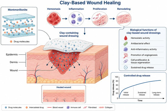
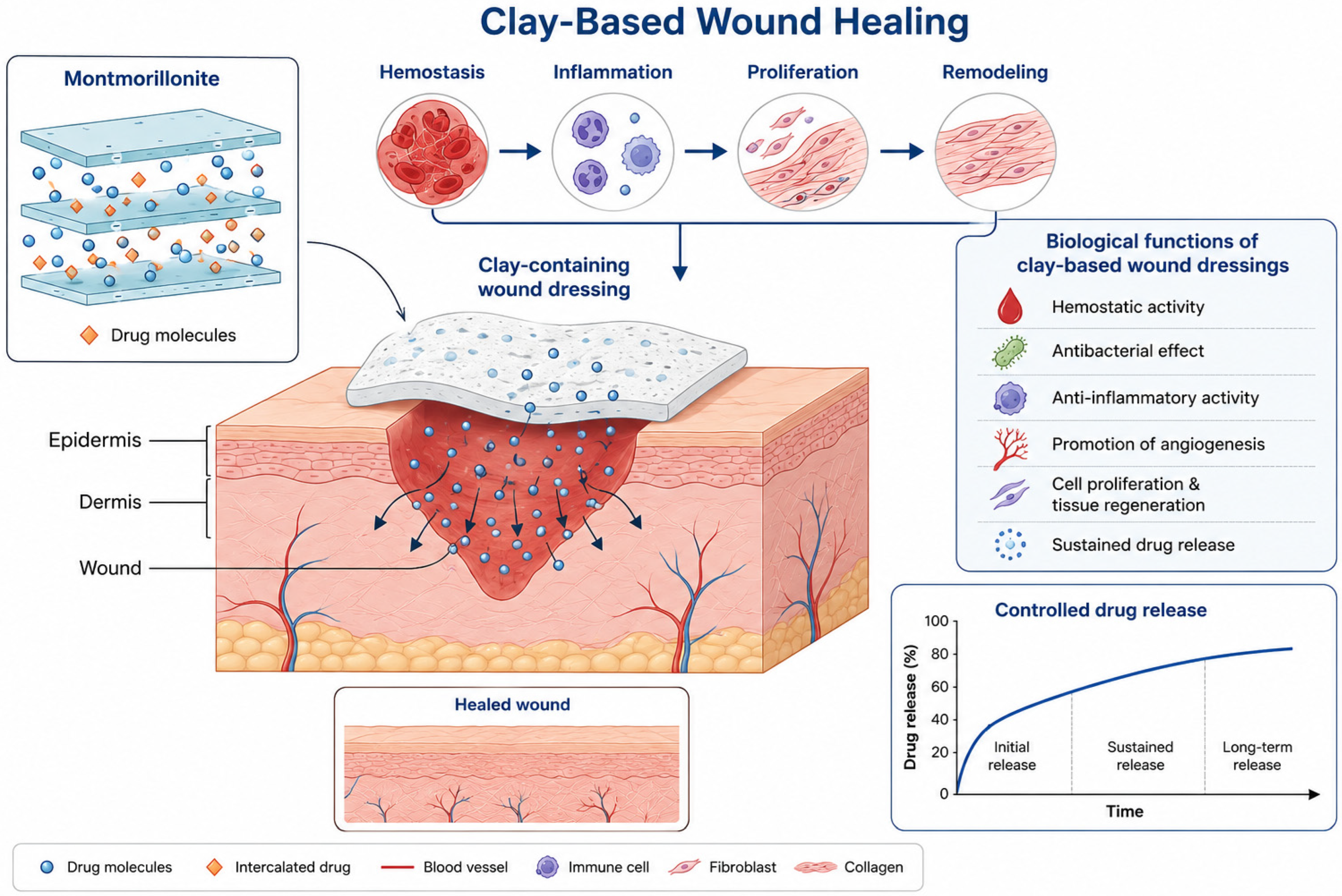
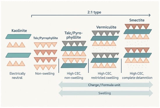
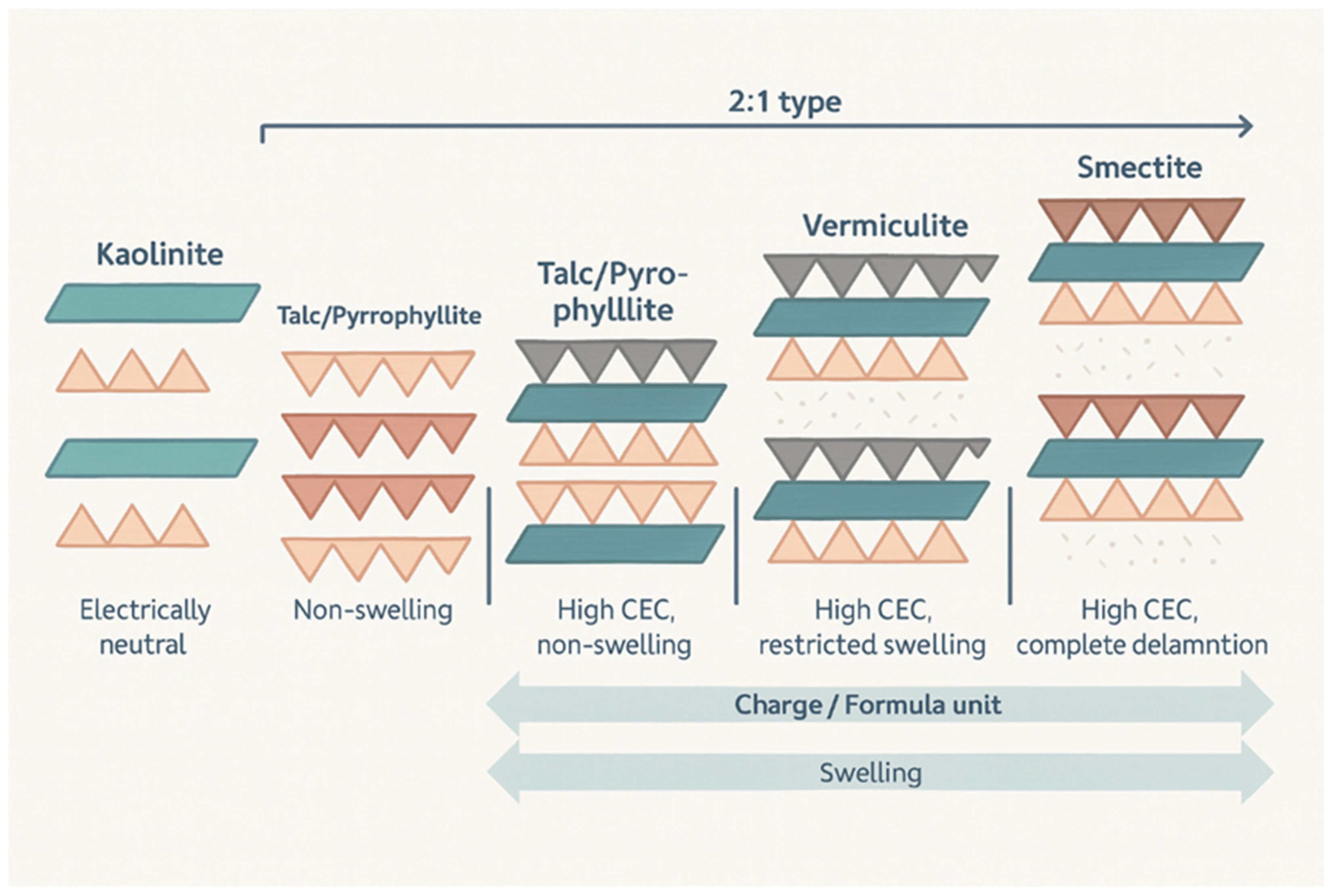
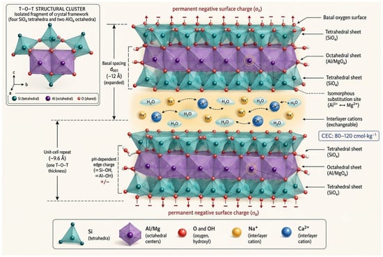
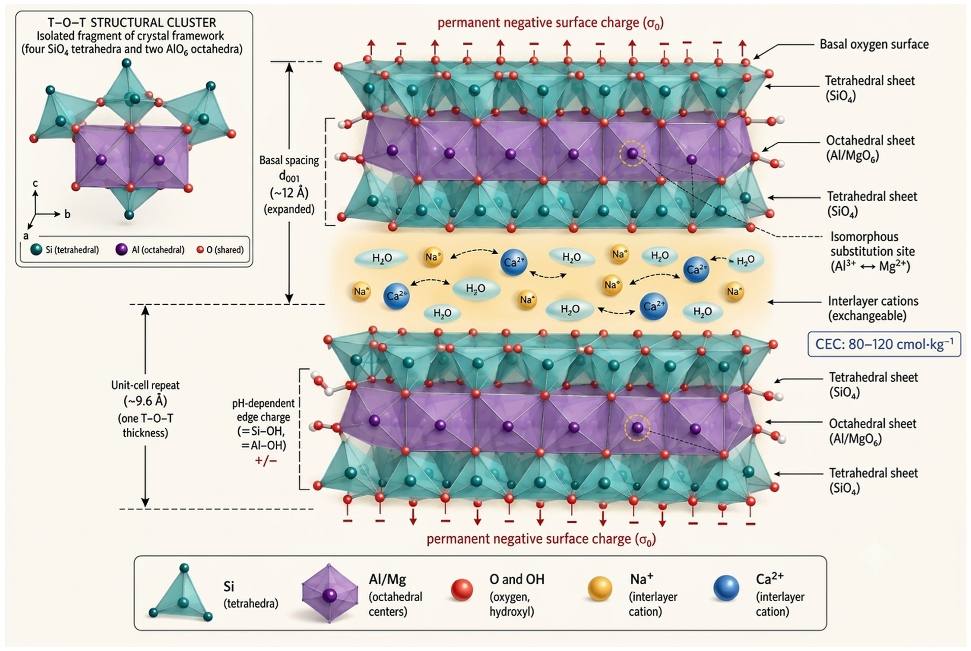
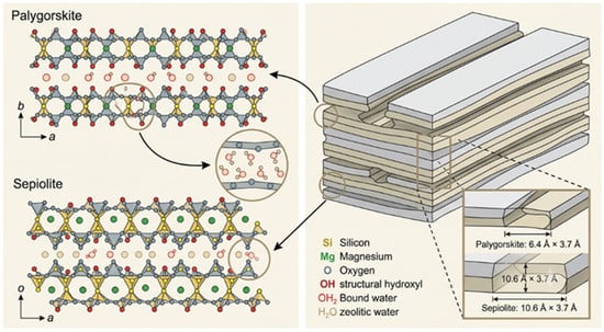
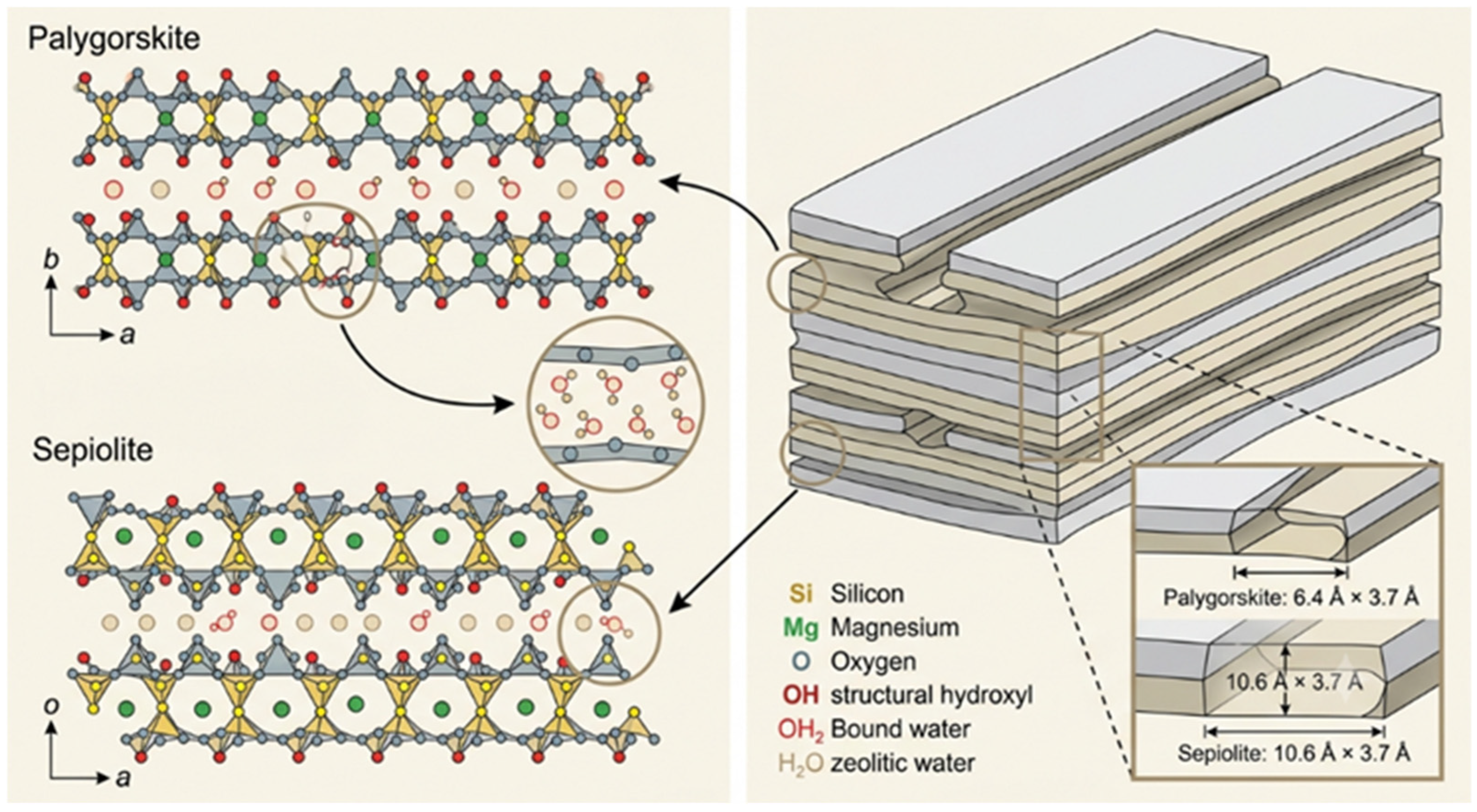

Disclaimer/Publisher’s Note: The statements, opinions and data contained in all publications are solely those of the individual author(s) and contributor(s) and not of MDPI and/or the editor(s). MDPI and/or the editor(s) disclaim responsibility for any injury to people or property resulting from any ideas, methods, instructions or products referred to in the content. |
© 2026 by the authors. Licensee MDPI, Basel, Switzerland. This article is an open access article distributed under the terms and conditions of the Creative Commons Attribution (CC BY) license.