Article

Prevalence of Liver Steatosis and Fibrosis Assessed by Transient Elastography in a High Cardiovascular-Risk Outpatient Cohort Including T1DM and T2DM Patients

by
Alina N. Saidi
1,2,
Willy B. Theel
2,
Diederick E. Grobbee
3,4,
Aart-Jan van der Lely
2,
Femme Dirksmeier-Harinck
1,5,
Marco Alings
4,6,
Ellen van der Zwan-van Beek
7,
Simone P. Rauh
8,
Moniba Rasheed
2 and
Manuel Castro Cabezas
1,2,4,*
1
Centre of Endocrinology, Diabetes and Vascular Medicine, Department of Internal Medicine, Franciscus Gasthuis & Vlietland, Kleiweg 500, 3045 PM Rotterdam, The Netherlands
2
Department of Internal Medicine, Division of Endocrinology, Erasmus University Medical Center, Dr. Molewaterplein 40, 3015 GD Rotterdam, The Netherlands
3
Julius Center for Health Science and Primary Care, University Medical Center Utrecht, Universiteitsweg 100, 3584 CG Utrecht, The Netherlands
4
Julius Clinical, Nieuweroordweg 1, 3704 EC Zeist, The Netherlands
5
Department of Gastroenterology, Franciscus Gasthuis & Vlietland, Kleiweg 500, 3045 PM Rotterdam, The Netherlands
6
Department of Cardiology, Amphia Hospital, Molengracht 21, 4818 CK Breda, The Netherlands
7
Department of Clinical Chemistry, Franciscus Gasthuis & Vlietland, Kleiweg 500, 3045 PM Rotterdam, The Netherlands
8
Department of Science, Franciscus Gasthuis & Vlietland, Kleiweg 500, 3045 PM Rotterdam, The Netherlands
*
Author to whom correspondence should be addressed.
Diabetology 2025, 6(11), 129; https://doi.org/10.3390/diabetology6110129 (registering DOI)
Submission received: 24 June 2025 / Revised: 31 August 2025 / Accepted: 9 October 2025 / Published: 1 November 2025

Abstract

Background: Metabolic dysfunction-associated steatotic liver disease (MASLD) is common in T2DM, likely due to insulin resistance and obesity. Although screening is recommended in high-risk patients, its prevalence in outpatient cardiovascular clinical settings remains unclear. Methods: We analyzed data from 475 patients attending a cardiovascular outpatient clinic: 142 with T2DM, 78 with T1DM, and 255 non-diabetic individuals at elevated cardiovascular risk. Liver steatosis and fibrosis were assessed using vibration-controlled transient elastography (Fibroscan®): steatosis by controlled attenuation parameter (CAP ≥ 275 dB/m), and fibrosis risk by liver stiffness measurement (LSM ≥ 8.1 kPa). Carotid intima-media thickness (cIMT) was also measured. Results: The cohort (47% women, mean age 53 years, BMI 29.8 kg/m2) showed MASLD in 39.2% and fibrosis risk in 18.3%. MASLD was most prevalent in T2DM (57.0%), followed by non-diabetics (35.3%) and T1DM (19.2%) (p < 0.001). Fibrosis risk was also highest in T2DM (22.5%) vs. T1DM (7.7%) and non-diabetics (19.2%) (p = 0.02). CAP values were higher in those with fibrosis risk. T2DM patients with MASLD had higher LSM (7.0 ± 3.0 kPa) compared to those without MASLD (5.1 ± 2.2 kPa; p < 0.001). cIMT was highest in T2DM (0.73 ± 0.12 mm; p = 0.04), but not associated with MASLD or fibrosis. BMI and triglycerides were the strongest predictors of both MASLD and fibrosis. Conclusions: MASLD and risk of significant fibrosis were highest among T2DM patients. Within T2DM, those with MASLD had higher LSM, indicating increased risk of fibrosis. The presence of MASLD and risk of significant fibrosis was not associated with cIMT in this cardiometabolic cohort. BMI and plasma TG were consistent predictors across groups urging for more strict control by body weight reduction and lifestyle interventions.

Graphical Abstract

1. Introduction

Metabolic dysfunction-associated steatotic liver disease (MASLD) encompasses a range of liver disease stages, from isolated steatosis, where lipid droplets accumulate in hepatocytes, to metabolic dysfunction-associated steatohepatitis (MASH), characterized by hepatocyte damage and hepatic inflammation [1]. This can further progress to fibrosis, cirrhosis, and, in some cases, hepatocellular carcinoma (HCC), all in the absence of secondary causes such as excessive alcohol consumption or other liver diseases [1,2,3].
The global prevalence of MASLD is estimated at 30%, with higher rates observed in individuals with obesity and type 2 diabetes (T2DM) [4,5]. As global rates of obesity and T2DM rise, MASLD prevalence is expected to increase, likely becoming the leading indication for liver transplantation in the near future [6]. Despite its growing prevalence, MASLD remains underdiagnosed due to limited awareness among both healthcare providers and patients [7,8,9].
In addition to its hepatic implications, MASLD is closely linked to cardiovascular disease (CVD). Epidemiological studies show that MASLD is associated with increased carotid intima-media thickness (cIMT), a surrogate marker of arterial injury [10,11]. The underlying inflammatory and metabolic mechanisms, such as insulin resistance (IR) and systemic inflammation, contribute to vascular damage thereby increasing CVD risk [10]. Moreover, MASLD’s severity correlates with a higher incidence of atherosclerotic cardiovascular disease (asCVD) risk factors, such as hypertension and diabetes, making early detection and management essential [12,13]. Recent evidence suggests that MASLD contributes to asCVD beyond shared risk factors. In a cohort of 7507 adults in Eastern China, MASLD severity was independently associated with subclinical coronary atherosclerosis, particularly in individuals with severe MASLD or hypertension [14]. Mechanistic and clinical data further highlight links through systemic inflammation, IR, and endothelial dysfunction [11], reinforcing its role as an independent cardiovascular (CV) risk factor. Different scientific organizations, including the American Heart Association (AHA) and the European Association for the Study of the Liver (EASL), have recommended MASLD screening in individuals with cardiometabolic disorders [13,15]. Although liver biopsy is considered the diagnostic gold standard, its invasive nature, cost, and associated risks limit its utility in routine clinical care. To improve accessibility, non-invasive tests (NITs), such as vibration-controlled transient elastography (VCTE), have been developed [1,16]. VCTE is a non-invasive method that simultaneously assesses liver fibrosis and steatosis. Liver stiffness measurement (LSM), obtained from VCTE, stages fibrosis, while the controlled attenuation parameter (CAP) measures hepatic fat content.
While MASLD has been widely studied in T2DM due to its strong association with IR, relatively little is known about MASLD in patients with type 1 diabetes mellitus (T1DM) [17,18]. Furthermore, few studies have explored MASLD and liver fibrosis in high CV risk populations attending cardiometabolic outpatient clinics; groups that are often overlooked despite sharing many metabolic risk factors.
Therefore, this study aimed to evaluate the prevalence of MASLD and risk of significant fibrosis in patients with T1DM and T2DM, compared to a high CV-risk non-diabetic group from a cardiometabolic outpatient clinic in the Netherlands. Secondary objectives include assessing differences in arterial injury between groups defined by MASLD and fibrosis status, and determining independent predictors of MASLD and significant fibrosis using backward stepwise logistic regression.

2. Materials and Methods

This study was conducted at the Endocrinology and Diabetes Vascular Center of Franciscus Gasthuis Hospital in Rotterdam, the Netherlands. Patients attending the outpatient clinic for CV risk management were included. Data were collected from electronic health records (EHRs) of patients enrolled between January 2021 and July 2022, all of whom underwent a standard, protocolized CV assessment, including VCTE. The study received ethical approval from the local Medical Ethical Committee of the Franciscus Gasthuis & Vlietland Hospital and was conducted in accordance with the Declaration of Helsinki.

2.1. Study Population

The study involved 475 adult patients: 142 with T2DM, 78 with T1DM, and 255 non-diabetic individuals, all of whom had cardiometabolic disorders. Patients were eligible if they visited the Endocrinology and Vascular Medicine department of Franciscus Gasthuis Hospital and underwent a FibroScan measurement between 1 January 2021, and 1 October 2022. Patients with previously diagnosed chronic liver disease were excluded, as well as those with conditions associated with secondary liver steatosis or fibrosis, such as HIV, viral hepatitis, hemochromatosis, Wilson’s disease, sarcoidosis, or excessive alcohol consumption (≥3 units per day for men and ≥2 units per day for women).

2.2. Data Collection

MASLD was defined as the presence of at least one cardiometabolic disorder and a controlled attenuation parameter (CAP) ≥ 275 dB/m (S1), in accordance with the 2021 European Association for the Study of the Liver (EASL) Clinical Practice Guidelines, indicating hepatic steatosis [19]. The cardiometabolic disorders considered were: overweight/obesity (BMI ≥ 25 kg/m2), hypertension (blood pressure ≥ 130/85 mmHg or use of antihypertensive medication), hypercholesterolemia (total cholesterol ≥ 5.0 mmol/L or use of lipid-lowering medication), dyslipidemia (LDL-C ≥ 3.0 mmol/L or HDL-C < 1.0 mmol/L), and diabetes (fasting glucose ≥ 7.0 mmol/L, HbA1c > 48 mmol/mol, or use of glucose-lowering medication). All variables were abstracted from the electronic health records. Risk of significant fibrosis was defined as a liver stiffness measurement (LSM) ≥ 8.1 kPa (≥F2) [19]. CVD was defined as the presence of any of the following conditions: transient ischemic attack (TIA), cerebrovascular accident (CVA), peripheral arterial disease (PAD), or coronary artery disease (CAD). All data were encoded with a unique, non-identifiable code to maintain patient confidentiality.

2.3. Laboratory Examination

Blood samples were analyzed for glucose, HbA1c, total cholesterol, HDL cholesterol (HDL-C), LDL cholesterol (LDL-C), triglycerides (TGs), apolipoproteins (apo) B and AI, C-reactive protein (CRP), hemoglobin, thrombocytes, gamma-glutamyl transferase (GGT), creatinine, albumin, estimated glomerular filtration rate (eGFR), alanine aminotransferase (ALT), and aspartate aminotransferase (AST) at the laboratory of Franciscus Gasthuis using standard procedures. All blood tests were conducted in a non-fasting state.

2.4. Vibration-Controlled Transient Elastography

VCTE was performed using Fibroscan™ (Echosens, Paris, France) by trained personnel at the outpatient clinic. At this cardiometabolic clinic, the procedure is routinely applied to screen for hepatic steatosis and fibrosis. Patients were positioned supine with their right arm under their head. The probe (either M- or XL-probe) was placed in the intercostal space between the 10th and 12th ribs along the mid-axillary line. A minimum of 10 valid measurements were required for the VCTE to be considered successful. Reliability was assessed by ensuring the interquartile range (IQR) from the median LSM value was ≤30% [20].

2.5. Carotid Intima Media Thickness

Carotid ultrasound scans were performed using the ART-LAB (Esaote, Genoa, Italy) by trained personnel as described previously in detail [21]. Imaging was conducted for each common carotid artery in three distinct projections. The cIMT was determined as the mean of six measurements, with at least two measurements per carotid artery required for inclusion.

2.6. Data Analysis

Data are presented as mean ± standard deviation (SD) for normally distributed variables, or median and IQR for non-normally distributed variables. Comparisons of normally distributed variables were performed using Student’s t-test, while non-normally distributed variables were analyzed using the Mann–Whitney U test or Kruskal–Wallis. ANOVA with post hoc analyses was used to explore differences between subgroups. Chi-square tests were employed to assess prevalence differences. To identify predictors of MASLD and risk of significant fibrosis, a backward stepwise logistic regression analysis was performed. Predictors included BMI, age, TGs, HDL-C, LDL-C, HbA1c, and sex. Collinearity was assessed using Variance Inflation Factor (VIF) values, with a threshold of VIF < 5 indicating acceptable levels of collinearity. As this study is exploratory in nature, analyses were primarily descriptive, and formal multivariable adjustment was not performed. All statistical analyses were conducted using IBM SPSS Statistics version 28.0.0.0 (IBM SPSS Statistics, New York, NY, USA). A p-value of <0.05 was considered statistically significant.

3. Results

3.1. Baseline Characteristics

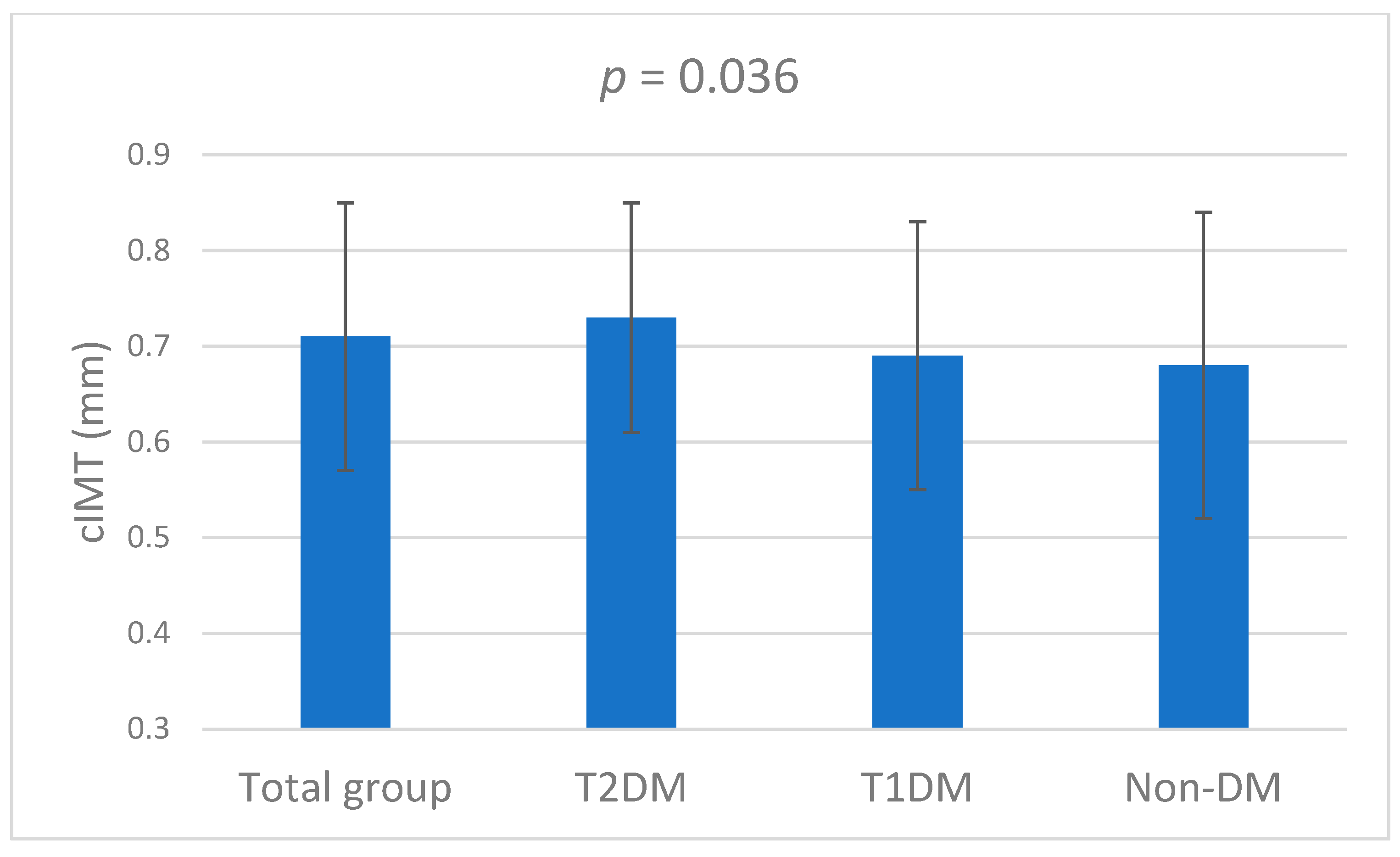
The study population consisted of 475 patients (223 women, mean age 53 ± 14 years, mean BMI 29.8 ± 6.7 kg/m2) (Table 1). Of the total cohort, 39.2% (N = 186) had MASLD, and 18.3% (N = 87) were at risk of significant fibrosis. The prevalence of MASLD was 57.0%, 19.2%, and 35.3% in patients with T2DM, T1DM, and in those without diabetes, respectively (p < 0.001). 22.5%, 7.7%, and 19.2% of the T2DM, T1DM, and non-diabetic patients, respectively, were at risk of significant fibrosis (p = 0.02) (Figure 1). The mean cIMT in the overall cohort was 0.71 mm (±0.14), with T2DM patients showing the highest cIMT (Figure 2). T2DM patients also had the highest glucose levels, HbA1c, and TGs among the three subgroups. Additionally, T2DM patients exhibited significantly higher creatinine levels and reduced eGFR compared to non-diabetic patients. In contrast, T1DM patients had significantly lower BMI and higher HDL-C levels than non-diabetic patients. Non-diabetic patients had the highest LDL-C levels among all subgroups (Table 1). The majority of the cohort was Caucasian (67.7%), with smaller groups of Turkish (6.5%), North African (5.5%), and Hindustani (4.9%) patients. Further details on the ethnicity distribution of the cohort can be found in Supplementary Table S1. Baseline medication use is summarized in Supplementary Table S2.

3.2. Characteristics by MASLD and Fibrosis Status

Across the entire cohort and within each subgroup, BMI was significantly greater in patients with MASLD compared to those without. Additionally, in the overall population, individuals with MASLD showed significantly higher plasma TGs, as well as higher liver enzyme levels relative to those without MASLD (Table 2). Within the T2DM group, patients with MASLD showed significantly higher LSM compared to both T1DM patients and non-diabetic individuals. Although T2DM patients had higher cIMT than T1DM patients and high CV-risk non-diabetic patients, neither MASLD nor risk of significant liver fibrosis appeared to significantly affect cIMT within this group. Additionally, T2DM patients with MASLD showed higher LDL-C levels compared to those without MASLD (Table 2).
In the overall cohort, multivariable regression analysis, adjusted for age, sex, and LDL-C, revealed that MASLD was not significantly associated with cIMT (β = 0.012, 95% CI: −0.025 to 0.049, p = 0.516). Similarly, significant fibrosis did not show a significant association with cIMT (β = 0.044, 95% CI: −0.025 to 0.113, p = 0.211). However, age remained significantly positively associated with cIMT (β = 0.006, p < 0.001).
Subjects at higher risk of significant fibrosis had a higher BMI in the overall cohort as well as in the T2DM and T1DM groups (Table 3). Patients who were at risk of fibrosis also showed increased liver enzyme levels in the total population. Moreover, CAP values were significantly elevated in those with risk of fibrosis across both the entire cohort and the diabetic subgroups (Table 3).

3.3. Predictors of MASLD and Fibrosis

Logistic regression analysis identified BMI as a significant predictor of MASLD across all patient groups (Table 4). In the total cohort, each 1 kg/m2 increase in BMI was associated with a 13.3% increase in the odds of MASLD. Similarly, TGs were significantly associated with MASLD, with a 49.6% increase in the odds for each 1 mmol/L increase. Linearity of continuous predictors was assessed visually, and BMI and TGs showed clear linear trends, supporting their inclusion as continuous variables in the models. In subgroup analyses, BMI remained a crucial predictor of MASLD. In T2DM patients, each 1 kg/m2 increase in BMI raised the odds of MASLD by 13.2%, while in T1DM patients, the odds increased by 22.3%, and by 11.7% in non-diabetic individuals with high CV-risk (Table 4). BMI was also identified as a predictor of risk for significant fibrosis in both T2DM and T1DM patients (Table 5). While BMI showed a positive association with risk of significant fibrosis in the overall cohort, the association did not reach statistical significance (p = 0.08). Additionally, TGs were found to be a strong predictor of risk of significant fibrosis in the overall cohort (Table 5). VIF values between independent variables were all below 5, indicating no significant collinearity.

3.4. MASLD and Fibrosis in CVD Patients

In the entire cohort, 86 patients (18.1%) had a history of CVD. Among these, 35 patients (40.7%) had MASLD, and 18 patients (20.9%) were at risk of significant fibrosis. Chi-square tests revealed no statistically significant differences in the prevalence of MASLD or risk of significant fibrosis between patients with and without CVD (p = 0.75 and p = 0.49, respectively).

4. Discussion

In this cross-sectional study of patients attending a Dutch cardiometabolic outpatient clinic, we observed a high prevalence of MASLD (39.2%) and risk of significant fibrosis (18.3%), indicating a considerable burden of undiagnosed liver disease in this high-risk population. The prevalence of MASLD was highest among patients with T2DM followed by high CV-risk non-diabetic individuals and patients with T1DM. A similar pattern was observed for the risk of significant fibrosis. Notably, T2DM patients with MASLD had a higher LSM, suggesting that MASLD may more strongly predispose this group to fibrosis development. Interestingly, neither MASLD nor risk of significant fibrosis were associated with cIMT. This may be explained that other CV risk factors may be operative in this high-risk population and therefore, MASLD does not differentiate in this respect. Of course, the number of subjects was limited, which may have influenced these results. Our diagnostic model identified BMI and TGs as the strongest predictors of MASLD and risk of significant fibrosis, reinforcing their role as key metabolic risk factors. These data clearly point at the relevance of weight control and lifestyle as main interventions in MASLD.
In line with existing literature, MASLD prevalence was higher in T2DM patients compared to the general population [17,22,23]. Interestingly, MASLD was least prevalent in T1DM patients. This lower prevalence may reflect the unique metabolic profile of T1DM, including absent endogenous insulin, reduced portal insulin levels, and increased GH and reduced IGF-1, fostering a catabolic, fasting-like state that limits hepatic fat accumulation [24]. These distinct metabolic characteristics may confer protection against MASLD development and progression in T1DM. While the small sample size warrants cautious interpretation, similar prevalence rates were observed in another Dutch T1DM cohort [25].
Relative to global data, the MASLD prevalence observed in our cohort was lower than the 65.3% pooled prevalence reported in a recent meta-analysis of T2DM patients worldwide, which included over 2.2 million individuals. The meta-analysis also noted regional variations, with the highest prevalence in Eastern Europe (80.6%) and the Middle East (71.2%) [26]. The lower prevalence in our study may partly reflect differences in diagnostic approaches, as the meta-analysis used estimated rates, whereas we used VCTE, providing more direct assessment of liver steatosis and fibrosis.
Few studies have assessed the prevalence of clinically significant liver fibrosis (defined as ≥8.1 kPa) in the general population, with reported rates ranging from 2% to 13.8% [27,28,29,30,31]. In our cohort, the risk of significant fibrosis was substantially higher than these general population estimates, though still lower than rates reported in T2DM patients from Eastern European studies. For instance, a Romanian study of 424 T2DM patients using VCTE reported a significant fibrosis prevalence of 57.1%, while a Croatian study, applying a lower cutoff value of 7.0 kPa, found a prevalence of 46.6% in T2DM patients [32,33]. These differences likely reflect variations in study populations, diagnostic criteria, VCTE cutoff thresholds, as well as potential influences of healthcare practices, genetic predispositions, and lifestyle factors specific to the Netherlands. Notably, the prevalence of significant fibrosis observed among T1DM patients in our study (7.7%) is consistent with earlier Dutch findings, which reported rates around 6.7% in similar cohorts assessed with VCTE [25].
Previous Dutch MASLD research has primarily focused on general population cohorts, for example, the Lifelines study in Northern Holland, which used the Fatty Liver Index (FLI) for diagnosis [34], and the Rotterdam Study, which assessed an elderly population using ultrasound [35]. De Vries et al. further compared MASLD prevalence between patients with T1DM and T2DM [25]. Our study builds on this work by including non-diabetic patients with high CV risk, thereby offering a more comprehensive and representative overview of MASLD prevalence in routine cardiometabolic clinical practice.
In our cohort, VCTE provided quantitative LSM and CAP values to identify patients at risk of significant fibrosis. Although many studies focus primarily on advanced fibrosis (F3–F4), detecting significant fibrosis is crucial, as this population represents the main target for clinical trials and early intervention. VCTE enables reliable risk stratification in outpatient settings, and while traditional scores such as FIB-4 and NAFLD Fibrosis Score (NFS) can complement it, they may underestimate F2–F3 prevalence. Recent studies have explored combined scoring systems, blood-based biomarkers, and machine learning approaches to improve identification of patients with significant fibrosis, supporting the integration of these tools into routine outpatient screening strategies [36,37,38].
Our findings reveal a substantial burden of undiagnosed liver disease in this cohort, particularly among T2DM patients, reinforcing the importance of incorporating NITs into routine screening. Early detection using these methods could help identify patients at risk of progression, leading to earlier interventions and potentially improving long-term outcomes. These results support current recommendations from several organizations that support non-invasive MASLD assessment in individuals with cardiometabolic risk [13,15,39].
Nonetheless, some limitations should be noted. The cross-sectional design of the current study restricts our ability to establish causality or to evaluate the risk of long-term liver-related morbidity within the cohort. However, the presence of fibrosis has been consistently associated with adverse liver-related outcomes in longitudinal studies [40], underscoring the importance of its identification. Moreover, statistical power was limited by the small T1DM subgroup and the low number of fibrosis events, restricting the ability to draw definitive conclusions for these patients. Additionally, residual confounding could have influenced the observed null associations between cIMT, MASLD, and fibrosis, given the exploratory study design, incomplete covariate data, and small T1DM and T2DM subgroups that restricted fully adjusted analyses.
While VCTE is a reliable and non-invasive method for assessing steatosis and fibrosis, it cannot fully substitute for liver biopsy, the diagnostic gold standard. Some serum markers, such as FLI and routine abdominal ultrasound, were not assessed, and FIB-4 was available for only a subset of participants, possibly limiting detection of differences. Additionally, all blood samples were collected in the non-fasting state, which may particularly influence triglyceride values, although diabetes classification was primarily based on HbA1c and EHR diagnoses. The single-center, high CV-risk nature of the cohort may limit generalizability of our findings to broader populations. Furthermore, baseline medication use was recorded but not adjusted for, which may affect liver and cardiometabolic outcomes.
Future studies with larger cohorts, fasting laboratory assessments, and additional non-invasive markers alongside VCTE are needed to better evaluate MASLD and significant fibrosis.

5. Conclusions

In conclusion, this study highlights a substantial burden of undiagnosed MASLD and the risk of significant fibrosis in cardiometabolic outpatient populations, particularly among patients with T2DM. Elevated LSM values observed in T2DM patients with MASLD suggest an increased risk for fibrosis progression. The absence of an association between MASLD or fibrosis and cIMT may reflect distinct mechanisms driving hepatic and vascular risk in this cohort. BMI and plasma TG were consistent predictors across groups urging for stricter control through body weight reduction and lifestyle interventions.

Supplementary Materials

The following supporting information can be downloaded at https://www.mdpi.com/article/10.3390/diabetology6110129/s1. Table S1: Ethnicity distribution in the total cohort presented as n (%); Table S2: Baseline medication use in the total cohort and subgroups presented as n (%).

Author Contributions

A.N.S.: Writing—Original Draft, Conceptualization, Investigation, Formal analysis, Visualization. W.B.T.: Writing—Review and Editing, Conceptualization, Investigation, Data Curation. D.E.G.: Writing—Review and Editing, Conceptualization. A.-J.v.d.L.: Writing—Review and Editing, Conceptualization. F.D.-H.: Writing—Review and Editing, Conceptualization. M.A.: Writing—Review and Editing, Conceptualization. E.v.d.Z.-v.B.: Writing—Review and Editing, Conceptualization. S.P.R.: Writing—Review and Editing, Conceptualization, Formal Analysis. M.R.: Writing—Review and Editing, Data Curation. M.C.C.: Writing—Review and Editing, Conceptualization, Methodology, Resources, Supervision. All authors have read and agreed to the published version of the manuscript.

Funding

This research received no external funding.

Institutional Review Board Statement

The study was conducted in accordance with the Declaration of Helsinki, and the protocol was approved by the Ethics Committee of Franciscus Gasthuis & Vlietland Hospital (study number 2022-084, approved on 25 April 2023).

Informed Consent Statement

Informed consent for participation was not required according to local legislation and institutional policy, as this retrospective medical record study involved anonymized data and was exempt from the Medical Research Involving Human Subjects Act (WMO).

Data Availability Statement

The data that support the findings of this study are available from the corresponding author upon reasonable request.

Acknowledgments

Preliminary data from this study were presented as a poster at the European Association for the Study of the Liver (EASL) Congress in Milan, 2024.

Conflicts of Interest

Authors Diederick E. Grobbee and Manuel Castro Cabezas were employed by the company Julius Clinical. The remaining authors declare that the research was conducted in the absence of any commercial or financial relationships that could be construed as a potential conflict of interest.

References

  1. Tacke, F.; Horn, P.; Wong, V.W.-S.; Ratziu, V.; Bugianesi, E.; Francque, S.; Zelber-Sagi, S.; Valenti, L.; Roden, M.; Schick, F.; et al. EASL-EASD-EASO Clinical Practice Guidelines on the management of metabolic dysfunction-associated steatotic liver disease (MASLD). J. Hepatol. 2024, 81, 492–542. [Google Scholar] [CrossRef]
  2. Wong, V.W.; Ekstedt, M.; Wong, G.L.; Hagström, H. Changing epidemiology, global trends and implications for outcomes of NAFLD. J. Hepatol. 2023, 79, 842–852. [Google Scholar] [CrossRef] [PubMed]
  3. Osorio-Conles, Ó.; Vega-Beyhart, A.; Ibarzabal, A.; Balibrea, J.M.; Graupera, I.; Rimola, J.; Vidal, J.; de Hollanda, A. A Distinctive NAFLD Signature in Adipose Tissue from Women with Severe Obesity. Int. J. Mol. Sci. 2021, 22, 10541. [Google Scholar] [CrossRef] [PubMed]
  4. Ortiz-Lopez, C.; Lomonaco, R.; Orsak, B.; Finch, J.; Chang, Z.; Kochunov, V.G.; Hardies, J.; Cusi, K. Prevalence of prediabetes and diabetes and metabolic profile of patients with nonalcoholic fatty liver disease (NAFLD). Diabetes Care 2012, 35, 873–878. [Google Scholar] [CrossRef]
  5. Younossi, Z.; Golabi, P.; Paik, J.; Henry, A.; Van Dongen, C.; Henry, L. The global epidemiology of nonalcoholic fatty liver disease (NAFLD) and non-alcoholic steatohepatitis (NASH): A systematic review. Hepatology 2023, 77, 1335–1347. [Google Scholar] [CrossRef]
  6. Wong, R.J.; Aguilar, M.; Cheung, R.; Perumpail, R.B.; Harrison, S.A.; Younossi, Z.M.; Ahmed, A. Nonalcoholic steatohepatitis is the second leading etiology of liver disease among adults awaiting liver transplantation in the United States. Gastroenterology 2015, 148, 547–555. [Google Scholar] [CrossRef]
  7. Loomba, R.; Sanyal, A.J. The global NAFLD epidemic. Nat. Rev. Gastroenterol. Hepatol. 2013, 10, 686–690. [Google Scholar] [CrossRef]
  8. Teng, M.L.; Ng, C.H.; Huang, D.Q.; Chan, K.E.; Tan, D.J.; Lim, W.H.; Yang, J.D.; Tan, E.; Muthiah, M.D. Global incidence and prevalence of nonalcoholic fatty liver disease. Clin. Mol. Hepatol. 2023, 29, S32–S42. [Google Scholar] [CrossRef]
  9. Driessen, S.; de Jong, V.D.; van Son, K.C.; Klompenhouwer, T.; Colardelle, Y.; Alings, M.; Moreno, C.; Anker, S.D.; Castro Cabezas, M.; Holleboom, A.G.; et al. A global survey of health care workers’ awareness of non-alcoholic fatty liver disease: The AwareNASH survey. United Eur. Gastroenterol. J. 2023, 11, 654–662. [Google Scholar] [CrossRef]
  10. Targher, G.; Byrne, C.D.; Tilg, H. MASLD: A systemic metabolic disorder with cardiovascular and malignant complications. Gut 2024, 73, 691–702. [Google Scholar] [CrossRef]
  11. Driessen, S.; Francque, S.M.; Anker, S.D.; Cabezas, M.C.; Grobbee, D.E.; Tushuizen, M.E.; Holleboom, A.G. Metabolic dysfunction associated steatotic liver disease and the heart. Hepatology 2023, 82, 487–503. [Google Scholar] [CrossRef]
  12. Zhou, X.D.; Targher, G.; Byrne, C.D.; Somers, V.; Kim, S.U.; Chahal, C.A.A.; Wong, V.W.; Cai, J.; Shapiro, M.D.; Eslam, M.; et al. An international multidisciplinary consensus statement on MAFLD and the risk of CVD. Hepatol. Int. 2023, 17, 773–791. [Google Scholar] [CrossRef] [PubMed]
  13. Duell, P.B.; Welty, F.K.; Miller, M.; Chait, A.; Hammond, G.; Ahmad, Z.; Cohen, D.E.; Horton, J.D.; Pressman, G.S.; Toth, P.P. Nonalcoholic Fatty Liver Disease and Cardiovascular Risk: A Scientific Statement From the American Heart Association. Arter. Thromb. Vasc. Biol. 2022, 42, e168–e185. [Google Scholar] [CrossRef] [PubMed]
  14. Ma, G.; Xu, G.; Huang, H. Correlation between metabolic dysfunction-associated steatotic liver disease and subclinical coronary atherosclerosis in eastern China. Diabetol. Metab. Syndr. 2025, 17, 16. [Google Scholar] [CrossRef] [PubMed]
  15. Aller, R.; Fernández-Rodríguez, C.; Iacono, O.L.; Bañares, R.; Abad, J.; Carrión, J.A.; García-Monzón, C.; Caballería, J.; Berenguer, M.; Rodríguez-Perálvarez, M.; et al. Consensus document. Management of non-alcoholic fatty liver disease (NAFLD). Clinical practice guideline. Gastroenterol. Hepatol. 2018, 41, 328–349. [Google Scholar] [CrossRef]
  16. Lee, J.; Vali, Y.; Boursier, J.; Spijker, R.; Anstee, Q.M.; Bossuyt, P.M.; Zafarmand, M.H. Prognostic accuracy of FIB-4, NAFLD fibrosis score and APRI for NAFLD-related events: A systematic review. Liver Int. 2021, 41, 261–270. [Google Scholar] [CrossRef]
  17. Younossi, Z.M.; Golabi, P.; de Avila, L.; Paik, J.M.; Srishord, M.; Fukui, N.; Qiu, Y.; Burns, L.; Afendy, A.; Nader, F. The global epidemiology of NAFLD and NASH in patients with type 2 diabetes: A systematic review and meta-analysis. J. Hepatol. 2019, 71, 793–801. [Google Scholar] [CrossRef]
  18. Brouwers, M.; Simons, N.; Kooi, M.E.; de Ritter, R.; van Dongen, M.; Eussen, S.; Bekers, O.; Kooman, J.; van Greevenbroek, M.M.J.; van der Kallen, C.J.H.; et al. Intrahepatic lipid content is independently associated with soluble E-selectin levels: The Maastricht study. Dig. Liver Dis. 2022, 54, 1038–1043. [Google Scholar] [CrossRef]
  19. Berzigotti, A.; Tsochatzis, E.; Boursier, J.; Castera, L.; Cazzagon, N.; Friedrich-Rust, M.; Petta, S.; Thiele, M. EASL Clinical Practice Guidelines on non-invasive tests for evaluation of liver disease severity and prognosis—2021 update. J. Hepatol. 2021, 75, 659–689. [Google Scholar] [CrossRef]
  20. Boursier, J.; Zarski, J.P.; de Ledinghen, V.; Rousselet, M.C.; Sturm, N.; Lebail, B.; Fouchard-Hubert, I.; Gallois, Y.; Oberti, F.; Bertrais, S.; et al. Determination of reliability criteria for liver stiffness evaluation by transient elastography. Hepatology 2013, 57, 1182–1191. [Google Scholar] [CrossRef]
  21. van Breukelen-van der Stoep, D.F.; van Zeben, D.; Klop, B.; van de Geijn, G.J.; Janssen, H.J.; Hazes, M.J.; Birnie, E.; van der Meulen, N.; De Vries, M.A.; Cabezas, M.C. Association of Cardiovascular Risk Factors with Carotid Intima Media Thickness in Patients with Rheumatoid Arthritis with Low Disease Activity Compared to Controls: A Cross-Sectional Study. PLoS ONE 2015, 10, e0140844. [Google Scholar] [CrossRef] [PubMed]
  22. Ciardullo, S.; Perseghin, G. Prevalence of elevated liver stiffness in patients with type 1 and type 2 diabetes: A systematic review and meta-analysis. Diabetes Res. Clin. Pract. 2022, 190, 109981. [Google Scholar] [CrossRef] [PubMed]
  23. Lomonaco, R.; Godinez Leiva, E.; Bril, F.; Shrestha, S.; Mansour, L.; Budd, J.; Portillo Romero, J.; Schmidt, S.; Chang, K.L.; Samraj, G.; et al. Advanced Liver Fibrosis Is Common in Patients With Type 2 Diabetes Followed in the Outpatient Setting: The Need for Systematic Screening. Diabetes Care 2021, 44, 399–406. [Google Scholar] [CrossRef] [PubMed]
  24. Nijenhuis-Noort, E.C.; Berk, K.A.; Neggers, S.; Lely, A.J.V. The Fascinating Interplay between Growth Hormone, Insulin-Like Growth Factor-1, and Insulin. Endocrinol. Metab. 2024, 39, 83–89. [Google Scholar] [CrossRef]
  25. de Vries, M.; Westerink, J.; El-Morabit, F.; Kaasjager, H.; de Valk, H.W. Prevalence of non-alcoholic fatty liver disease (NAFLD) and its association with surrogate markers of insulin resistance in patients with type 1 diabetes. Diabetes Res. Clin. Pract. 2022, 186, 109827. [Google Scholar] [CrossRef]
  26. Younossi, Z.M.; Golabi, P.; Price, J.K.; Owrangi, S.; Gundu-Rao, N.; Satchi, R.; Paik, J.M. The Global Epidemiology of Nonalcoholic Fatty Liver Disease and Nonalcoholic Steatohepatitis Among Patients With Type 2 Diabetes. Clin. Gastroenterol. Hepatol. 2024, 22, 1999–2010. [Google Scholar] [CrossRef]
  27. Roulot, D.; Costes, J.L.; Buyck, J.F.; Warzocha, U.; Gambier, N.; Czernichow, S.; Le Clesiau, H.; Beaugrand, M. Transient elastography as a screening tool for liver fibrosis and cirrhosis in a community-based population aged over 45 years. Gut 2011, 60, 977–984. [Google Scholar] [CrossRef]
  28. Caballería, L.; Pera, G.; Arteaga, I.; Rodríguez, L.; Alumà, A.; Morillas, R.M.; de la Ossa, N.; Díaz, A.; Expósito, C.; Miranda, D.; et al. High Prevalence of Liver Fibrosis Among European Adults With Unknown Liver Disease: A Population-Based Study. Clin. Gastroenterol. Hepatol. 2018, 16, 1138–1145.e5. [Google Scholar] [CrossRef]
  29. Koehler, E.M.; Plompen, E.P.; Schouten, J.N.; Hansen, B.E.; Darwish Murad, S.; Taimr, P.; Leebeek, F.W.; Hofman, A.; Stricker, B.H.; Castera, L.; et al. Presence of diabetes mellitus and steatosis is associated with liver stiffness in a general population: The Rotterdam study. Hepatology 2016, 63, 138–147. [Google Scholar] [CrossRef]
  30. Kim, D.; Cholankeril, G.; Loomba, R.; Ahmed, A. Prevalence of Fatty Liver Disease and Fibrosis Detected by Transient Elastography in Adults in the United States, 2017–2018. Clin. Gastroenterol. Hepatol. 2021, 19, 1499–1501.e1492. [Google Scholar] [CrossRef]
  31. Fabrellas, N.; Alemany, M.; Urquizu, M.; Bartres, C.; Pera, G.; Juvé, E.; Rodríguez, L.; Torán, P.; Caballería, L. Using transient elastography to detect chronic liver diseases in a primary care nurse consultancy. Nurs. Res. 2013, 62, 450–454. [Google Scholar] [CrossRef]
  32. Trifan, A.; Stratina, E.; Nastasa, R.; Rotaru, A.; Stafie, R.; Zenovia, S.; Huiban, L.; Sfarti, C.; Cojocariu, C.; Cuciureanu, T.; et al. Simultaneously Screening for Liver Steatosis and Fibrosis in Romanian Type 2 Diabetes Mellitus Patients Using Vibration-Controlled Transient Elastography with Controlled Attenuation Parameter. Diagnostics 2022, 12, 1753. [Google Scholar] [CrossRef]
  33. Mikolasevic, I.; Rahelic, D.; Turk-Wensween, T.; Ruzic, A.; Domislovic, V.; Hauser, G.; Matic, T.; Radic-Kristo, D.; Krznaric, Z.; Radic, M.; et al. Significant liver fibrosis, as assessed by fibroscan, is independently associated with chronic vascular complications of type 2 diabetes: A multicenter study. Diabetes Res. Clin. Pract. 2021, 177, 108884. [Google Scholar] [CrossRef]
  34. van den Berg, E.H.; Amini, M.; Schreuder, T.C.; Dullaart, R.P.; Faber, K.N.; Alizadeh, B.Z.; Blokzijl, H. Prevalence and determinants of non-alcoholic fatty liver disease in lifelines: A large Dutch population cohort. PLoS ONE 2017, 12, e0171502. [Google Scholar] [CrossRef] [PubMed]
  35. Koehler, E.M.; Schouten, J.N.; Hansen, B.E.; van Rooij, F.J.; Hofman, A.; Stricker, B.H.; Janssen, H.L. Prevalence and risk factors of non-alcoholic fatty liver disease in the elderly: Results from the Rotterdam study. J. Hepatol. 2012, 57, 1305–1311. [Google Scholar] [CrossRef] [PubMed]
  36. Ratziu, V.; Harrison, S.A.; Hajji, Y.; Magnanensi, J.; Petit, S.; Majd, Z.; Delecroix, E.; Rosenquist, C.; Hum, D.; Staels, B.; et al. NIS2+(TM) as a screening tool to optimize patient selection in metabolic dysfunction-associated steatohepatitis clinical trials. J. Hepatol. 2024, 80, 209–219. [Google Scholar] [CrossRef] [PubMed]
  37. Stefanakis, K.; Mingrone, G.; George, J.; Mantzoros, C.S. Accurate non-invasive detection of MASH with fibrosis F2-F3 using a lightweight machine learning model with minimal clinical and metabolomic variables. Metabolism 2025, 163, 156082. [Google Scholar] [CrossRef]
  38. McPherson, S.; Dyson, J.K.; Jopson, L.; Masson, S.; Patel, P.; Anstee, Q.M. Letter: Beyond advanced fibrosis-The critical need for assessing NITs performance in identifying F2-F3 fibrosis. Authors’ reply. Aliment. Pharmacol. Ther. 2024, 60, 976–977. [Google Scholar] [CrossRef]
  39. Vereniging, N.V.v.G.-e.N.I.V.N.L. Richtlijn MASLD/MASH; 2024. Available online: https://richtlijnendatabase.nl/richtlijn/richtlijn_masld_mash/startpagina_masld_mash.html (accessed on 1 September 2025).
  40. Sanyal, A.J.; Van Natta, M.L.; Clark, J.; Neuschwander-Tetri, B.A.; Diehl, A.; Dasarathy, S.; Loomba, R.; Chalasani, N.; Kowdley, K.; Hameed, B.; et al. Prospective Study of Outcomes in Adults with Nonalcoholic Fatty Liver Disease. N. Engl. J. Med. 2021, 385, 1559–1569. [Google Scholar] [CrossRef]
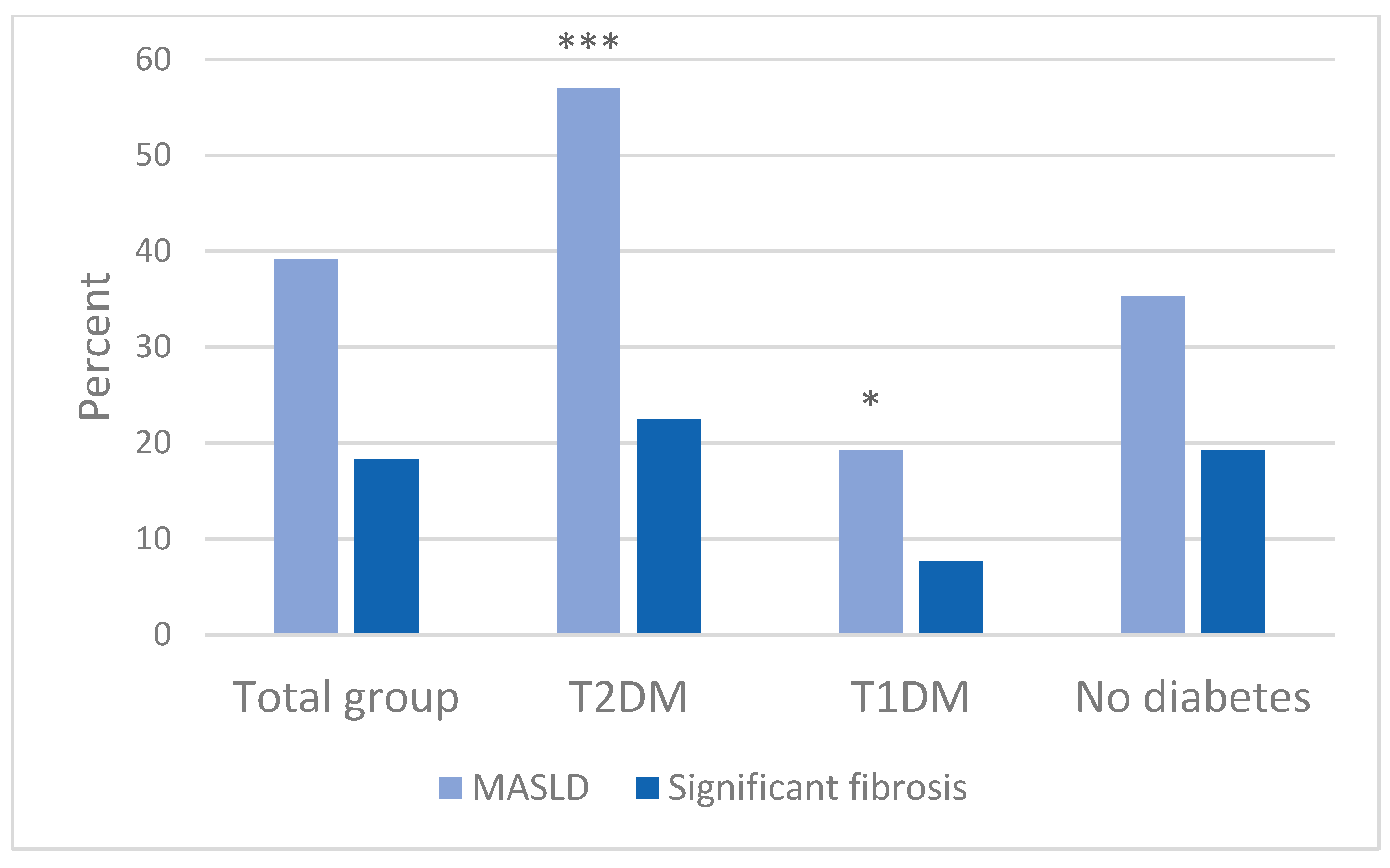
Figure 1. Prevalence of MASLD and risk of significant fibrosis in the total cohort and the among different subgroups. Statistical significance is indicated by asterisks * and ***, corresponding to probability values of <0.05 and <0.001, respectively, in relation to the high CV-risk non-diabetic subgroup.
Diabetology 06 00129 g001
Figure 2. Mean carotid intima-media thickness across the total cohort and subgroups. p-value indicates ANOVA differences between groups. Abbreviations: T2DM, type 2 diabetes mellitus; T1DM, type 1 diabetes mellitus; non-DM, high CV-risk non-diabetics.
Diabetology 06 00129 g002
Table 1. Baseline demographic and clinical characteristics for the overall group and subgroups. Data are expressed as mean ± standard deviation, median (IQR), or n (%), as appropriate.
CharacteristicsTotal
(N = 475)
T2DM
(N = 142)
T1DM
(N = 78)
Non-DM
(N = 255)
p-Value
Age (years)53 ± 1459 ± 12 ***52 ± 1250 ± 14<0.001
M/F252/22386/5639/39127/1280.101
BMI (kg/m2)29.8 ± 6.730.4 ± 6.427.1 ± 5.0 ***30.6 ± 7.3<0.001
cIMT (mm)0.71 ± 0.140.73 ± 0.120.69 ± 0.140.68 ± 0.160.036
Total cholesterol (mmol/L)4.6 (3.8–5.5)4.1 (3.7–5.3)4.3 (3.7–5.0)5.1 (4.5–5.7)0.573
HDL-C (mmol/L)1.3 ± 0.391.2 ± 0.321.5 ± 0.42 ***1.3 ± 0.37<0.001
LDL-C (mmol/L)2.5 ± 0.922.3 ± 0.93 ***2.3 ± 0.75 ***3.0 ± 0.80<0.001
Triglycerides (mmol/L)1.6 (1.0–2.4) 2.1 (1.3–3.1) *1.1 (0.8–1.7) **1.5 (1.1–2.3)<0.001
Glucose (mmol/L)6.2 (5.4–7.9)10.0 (7.4–14) ***8.5 (5.6–11) **5.8 (5.1–6.4)<0.001
HbA1c (mmol/mol)50.9 ± 1559.0 ± 13 ***57.3 ± 12 ***36.4 ± 5.0 <0.001
Apo B (g/L)1.05 ± 0.271.05 ± 0.380.82 ± 0.271.07 ± 0.200.330
Apo AI (g/L)1.54 ± 0.261.49 ± 0.241.39 ± NA1.59 ± 0.270.490
CRP (mg/L)4.0 (2.0–7.0)3.0 (2.0–6.0)8.0 (4.0–25)4.0 (2.0–7.0)0.176
Hemoglobin (mmol/L)8.7 ± 1.18.7 ± 1.18.2 ± 1.0 *8.8 ± 1.10.057
Thrombocytes (109/L)258 ± 87 253 ± 93305 ± 114255 ± 820.099
GGT (U/L)58 (31–106)71 (38–177)23 (14–140)58 (29–97)0.082
ALT (U/L)38 (27–52)38 (28–60)22 (18–39)39 (28–51)0.158
AST (U/L)39 (24–67) 38 (25–72) 29 (14–36)41 (23–67)0.131
Creatinine (μmol/L)74 (65–87) 77 (67–96) **76 (66–92)72 (65–81)0.006
Albumin (g/L)44 (41–46)44 (42–47)46 (40–46)44 (41–46)0.850
eGFR (mL/min/1.73m2)80 (68–88)77 (59–86) ***78 (70–87)82 (75–91)<0.001
FIB-41.86 ± 1.501.94 ± 1.461.73 ± 1.121.85 ± 1.620.789
CAP (dB/m)263 ± 59282 ± 59 ***238 ± 52 *259 ± 58<0.001
LSM (kPa)6.1 ± 3.06.2 ± 2.85.4 ± 1.8 *6.3 ± 3.30.039
Statistical significance is indicated by asterisks *, **, and ***, corresponding to probability values of <0.05, <0.01, and <0.001, respectively. These significance levels are in relation to the high CV-risk non-diabetic subgroup. p-values reflect ANOVA differences between groups. Abbreviations: BMI, body mass index; cIMT, carotid intima media thickness; HDL-C, high-density lipoprotein cholesterol; LDL-C, low-density lipoprotein cholesterol; HbA1c, hemoglobin A1C; CRP, C-reactive protein; GGT, gamma-glutamyl transferase; ALT, alanine aminotransferase; AST, aspartate aminotransferase; eGFR, estimated glomerular filtration rate; FIB-4, fibrosis-4 index; CAP, controlled attenuation parameter; LSM, liver stiffness measurement.
Table 2. Demographic and clinical characteristics by MASLD status in the total population and subgroups.
CharacteristicsTotal
(N = 475)
T2DM
(N = 142)
T1DM
(N = 78)
Non-DM
(N = 255)
MASLD
(N = 186)
No MASLD
(N = 289)
p-ValueMASLD
(N = 81)
No MASLD
(N = 61)
p-ValueMASLD
(N = 15)
No MASLD
(N = 63)
p-ValueMASLD
(N = 90)
No MASLD
(N = 165)
p-Value
Age (years)52 ± 14 54 ± 130.06858 ± 1261 ± 110.09256 ± 1151 ± 120.19745 ± 1452 ± 13<0.001
M/F100/86152/1370.80349/3237/240.9844/1135/280.04447/4380/500.568
BMI (kg/m2)33.1 ± 6.4 27.6 ± 6.0<0.00132.3 ± 6.227.8 ± 5.9<0.00131.9 ± 4.225.9 ± 4.4<0.00134.3 ± 6.828.3 ± 6.7<0.001
cIMT (mm)0.72 ± 0.120.70 ± 0.140.4090.73 ± 0.120.74 ± 0.130.6430.71 ± 0.120.68 ± 0.140.4590.69 ± 0.150.68 ± 0.170.975
Total cholesterol (mmol/L)5.5 ± 7.04.6 ± 1.20.1296.0 ± 9.74.1 ± 1.20.2074.5 ± 1.3 4.4 ± 0.910.6965.1 ± 0.955.1 ± 1.20.958
HDL-C (mmol/L)1.2 ± 0.361.3 ± 0.420.2311.2 ± 0.311.1 ± 0.330.9421.6 ± 0.391.5 ± 0.440.5791.3 ± 0.351.3 ± 0.390.960
LDL-C (mmol/L)2.6 ± 0.902.5 ± 0.930.1552.4 ± 1.02.0 ± 0.870.0422.4 ± 0.872.3 ± 0.720.6343.0 ± 0.703.1 ± 0.870.850
Triglycerides (mmol/L)2.2 ± 1.3 1.8 ± 1.30.0352.5 ± 1.52.2 ± 1.60.3241.1 ± 0.521.3 ± 0.740.4552.0 ± 1.11.9 ± 1.40.611
Glucose (mmol/L)7.1 ± 2.77.6 ± 3.60.4419.8 ± 3.411.5 ± 4.60.2228.1 ± 2.78.8 ± 3.70.8065.9 ± 0.935.9 ± 1.10.911
HbA1c (mmol/mol)51 ± 1451 ± 150.90059 ± 1360 ± 120.68056 ± 7.8 58 ± 130.61237 ± 4.736 ± 5.30.365
ApoB (g/L)1.1 ± 0.301.0 ± 0.230.2091.1 ± 0.390.90 ± 0.310.2070.87 ± 0.360.74 ± NANA1.09 ± 0.211.05 ± 0.200.543
Apo AI (g/L)1.6 ± 0.231.5 ± 0.270.7261.6 ± 0.281.4± 0.170.169NA1.4 ± NANA1.5 ± 0.121.6 ± 0.300.680
CRP (mg/L)7.4 ± 9.38.0 ± 160.7814.2 ± 4.14.7 ± 3.50.68625.0 ± NA10.2 ± 10.1NA8.6 ± 118.7 ± 190.970
Hemoglobin (mmol/L)8.8 ± 1.28.7 ± 1.10.4808.7 ± 1.18.8 ± 1.00.5378.5 ± 0.578.1 ± 1.10.6338.9 ± 1.28.7 ± 1.10.436
Thrombocytes (109/L)266 ± 83252 ± 900.230264 ± 79238 ± 1100.283296 ± 216306 ± 1060.910266 ± 83248 ± 810.194
GGT (U/L)82.4 ± 76106 ± 1410.190102 ± 98173 ± 1880.159NA66.0 ± 73.9NA73.9 ± 62.594.7 ± 1300.202
ALT (U/L)55 ± 3740 ± 260.00356 ± 3745 ± 350.240NA31 ± 21NA53 ± 3840 ± 240.010
AST (U/L)65 ± 5142 ± 30<0.00168 ± 63 44 ± 400.07726 ± 13 38 ± 340.62465 ± 4341 ± 26<0.001
Creatinine (μmol/L)79.9 ± 2181.3 ± 420.69385.0 ± 2491.7 ± 770.48681.5 ± 2081.5 ± 230.99573.9 ± 1576.0 ± 160.371
Albumin (g/L)44.1 ± 3.346.7 ± 410.55744.7 ± 3.242.3 ± 6.3 0.12641.0 ± 1.443.8 ± 3.70.34344.0 ± 3.348.1 ± 470.505
eGFR (mL/min/1.73m2)76.1 ± 15.375.6 ± 15.30.80270.7 ± 16.771.8 ± 18.00.75874.2 ± 13.974.7 ± 15.60.91783.3 ± 10.578.4 ± 12.60.064
FIB-41.68 ± 1.31.98 ± 1.60.1391.82 ± 1.22.10 ± 1.70.4441.40 ± 0.561.79 ± 1.20.4391.60 ± 1.52.01 ± 1.70.182
CAP (dB/m)323 ± 30223 ± 36<0.001325 ± 32227 ± 37<0.001318 ± 31219 ± 35<0.001323 ± 29224 ± 36<0.001
LSM (kPa)6.7 ± 3.05.8 ± 2.9<0.0017.0 ± 3.05.1 ± 2.2<0.0015.8 ± 2.4 5.3 ± 1.60.4366.6 ± 3.16.2 ± 3.40.329
Note: Data are given as mean (standard deviation), median (IQR), or numbers (%) where appropriate. Note: Values shown as mean ± SD. NA = data not available due to small sample size or missing measurements. Abbreviations: T2DM, type 2 diabetes mellitus; T1DM, type 1 diabetes mellitus; non-DM, high CV-risk non-diabetics; BMI, body mass index; cIMT, carotid intima media thickness, HDL-C, high-density lipoprotein cholesterol; LDL-C, low-density lipoprotein cholesterol; HbA1c, hemoglobin A1C; CRP, C-reactive protein; GGT, gamma-glutamyl transferase; ALT, alanine aminotransferase; AST, aspartate aminotransferase; eGFR, estimated glomerular filtration rate; FIB-4, fibrosis-4 index; CAP, controlled attenuation parameter; LSM, liver stiffness measurement.
Table 3. Demographic and clinical characteristics by risk of significant fibrosis status in the total population and subgroups.
CharacteristicsTotal
(N = 475)
T2DM
(N = 142)
T1DM
(N = 78)
Non-DM
(N = 255)
Fibrosis
(N = 87)
No Fibrosis
(N = 388)
p-ValueFibrosis
(N = 32)
No Fibrosis
(N = 110)
p-ValueFibrosis
(N = 6)
No Fibrosis
(N = 72)
p-ValueFibrosis
(N = 49)
No Fibrosis
(N = 206)
p-Value
Age (years)54 ± 13 53 ± 140.45356 ± 1360 ± 110.12456 ± 6.152 ± 120.14452 ± 14 49 ± 140.187
M/F54/33198/1900.06221/1165/450.5063/336/361.00030/1997/1090.075
BMI (kg/m2)31.9 ± 7.6 29.5 ± 6.50.02334.2 ± 7.529.4 ± 5.80.00431.7 ± 6.026.7 ± 4.70.01729.7 ± 7.530.7 ± 7.30.523
cIMT (mm)0.76 ± 0.110.70 ± 0.140.1200.76 ± 0.100.73 ± 0.130.5650.77 ± 0.160.68 ± 0.130.1780.76 ± NA0.68 ± 0.16NA
Total cholesterol (mmol/L)5.0 ± 1.65.0 ± 5.00.9895.0 ± 8.15.2 ± 1.70.9074.2 ± 0.81 4.4 ± 1.00.7285.2 ± 1.65.1 ± 1.00.891
HDL-C (mmol/L)1.2 ± 0.481.3 ± 0.380.1341.1 ± 0.371.2 ± 0.310.7481.5 ± 0.181.5 ± 0.440.9891.1 ± 0.661.3 ± 0.310.506
LDL-C (mmol/L)2.7 ± 1.22.5 ± 0.870.5752.6 ± 1.32.2 ± 0.840.2572.0 ± 0.552.3 ± 0.760.3823.1 ± 1.23.0 ± 0.740.762
Triglycerides (mmol/L)2.3 ± 1.2 1.9 ± 1.40.1682.6 ± 1.22.3 ± 1.60.4251.6 ± 0.781.3 ± 0.700.3602.0 ± 1.11.9 ± 1.30.905
Glucose (mmol/L)8.0 ± 3.07.3 ± 3.30.31710.3 ± 3.410.9 ± 4.30.6919.3 ± 1.18.5 ± 3.80.8006.0 ± 0.765.9 ± 1.10.787
HbA1c (mmol/mol)52 ± 1551 ± 150.65660 ± 1659 ± 120.69057 ± 6.5 57 ± 130.99738 ± 4.2 36 ± 5.10.079
Apo B (g/L)1.1 ± 0.321.0 ± 0.270.4541.2 ± 0.381.0 ± 0.380.3990.61 ± NA0.93 ± 0.27NA1.2 ± 0.171.1 ± 0.200.351
Apo AI (g/L)1.4 ± NA1.5 ± 0.26NA1.4 ± NA1.5 ± 0.25NANA1.4 ± NANANA1.6 ± 0.27NA
CRP (mg/L)6.8 ± 8.68.0 ± 150.6393.9 ± 2.2 4.6 ± 4.30.6238.0 ± NA13 ± 12NA7.9 ± 108.9 ± 170.786
Hemoglobin (mmol/L)8.8 ± 1.18.7 ± 1.1 0.8668.7 ± 0.988.7 ± 1.10.9878.5 ± 0.578.1 ± 1.10.6338.8 ± 1.28.8 ± 1.10.925
Thrombocytes (109/L)240 ± 87 263 ± 870.085240 ± 90259 ± 950.469193 ± 70.7321 ± 1110.143243 ± 88258 ± 800.308
GGT (U/L)96.8 ± 80.296.4 ± 1310.983117 ± 89143 ± 1750.53798 ± NA 58 ± 83NA86.3 ± 76.087.3 ± 1180.967
ALT (U/L)57 ± 4542 ± 240.02167 ± 4743 ± 260.05282 ± NA25 ± 9.6NA51 ± 4542 ± 230.277
AST (U/L)65 ± 5648 ± 350.02591 ± 80 45 ± 370.01866 ± 43 31 ± 280.15651 ± 3650 ± 350.793
Creatinine (μmol/L)76.6 ± 1781.6 ± 380.28381.5 ± 1989.7 ± 600.49081.0 ± 2781.5 ± 220.95872.2 ± 1176.0 ± 160.087
Albumin (g/L)52.4 ± 6143.4 ± 4.10.31544.3 ± 2.943.4 ± 5.5 0.55243.0 ± 4.243.3 ± 3.70.91257.0 ± 7643.3 ± 3.70.334
eGFR (mL/min/1.73m2)75.5 ± 12.575.8 ± 15.70.90171.7 ± 14.771.2 ± 17.80.97367.3 ± 11.975.1 ± 15.10.39180.5 ± 8.780.2 ± 12.70.916
FIB-41.79 ± 1.31.88 ± 1.50.7381.62 ± 1.12.08 ± 1.60.2402.34 ± NA1.72 ± 1.1NA1.90 ± 1.51.83 ± 1.70.840
CAP (dB/m)283 ± 62258 ± 58<0.001310 ± 46275 ± 60<0.001286 ± 62234 ± 500.019266 ± 66257 ± 560.308
LSM (kPa)10.8 ± 3.65.1 ± 1.4<0.00110.5 ± 2.15.0 ± 1.5<0.0019.8 ± 1.3 5.0 ± 1.2<0.00111.1 ± 4.55.2 ± 1.4<0.001
Note: Data are given as mean (standard deviation), median (IQR), or numbers (%) where appropriate. Note: Values shown as mean ± SD. NA = data not available due to small sample size or missing measurements. Abbreviations: T2DM, type 2 diabetes mellitus; T1DM, type 1 diabetes mellitus; non-DM, high CV-risk non-diabetics; BMI, body mass index; cIMT, carotid intima media thickness, HDL-C, high-density lipoprotein cholesterol; LDL-C, low-density lipoprotein cholesterol; HbA1c, hemoglobin A1C; CRP, C-reactive protein; GGT, gamma-glutamyl transferase; ALT, alanine aminotransferase; AST, aspartate aminotransferase; eGFR, estimated glomerular filtration rate; FIB-4, fibrosis-4 index; CAP, controlled attenuation parameter; LSM, liver stiffness measurement.
Table 4. Independent predictors of MASLD from the final backward stepwise logistic regression model in the overall cohort and subgroups.
GroupVariableB (SE)Exp(B)95% CIp-Value
Total cohortConstant−4.745 (0.849)0.009 <0.001
BMI (kg/m2)0.125 (0.026)1.1331.077–1.192 <0.001
Triglycerides (mmol/L)0.403 (0.174)1.4961.064–2.1020.020
T2DMConstant−3.491 (1.403)0.030 0.013
BMI (kg/m2)0.125 (0.026)1.1321.033–1.2400.008
T1DMConstant−7.001 (2.143)0.001 0.001
BMI (kg/m2)0.202 (0.071)1.2231.064–1.4060.005
Non-diabeticsConstant−5.152 (1.513)0.006 <0.001
BMI (kg/m2)0.111 (0.038)1.1171.037–1.2030.004
Variables entered on step 1: Age, BMI, HbA1c, triglycerides, HDL-C, LDL-C, sex. Abbreviations: T2DM, type 2 diabetes mellitus; T1DM, type 1 diabetes mellitus; non-diabetics, high CV-risk non-diabetics.
Table 5. Independent predictors of significant fibrosis from the final backward stepwise logistic regression model in the overall cohort and subgroups.
GroupVariableB (SE)Exp(B)95% CIp-Value
Total cohortConstant−4.337 (1.047)0.013 <0.001
Triglycerides (mmol/L)0.422 (0.208)1.5241.015–2.2890.042
T2DMConstant−4.746 (1.544)0.009 0.002
BMI (kg/m2)0.104 (0.047)1.1101.012–1.217 0.027
T1DMConstant−8.904 (3.238)0.000 0.006
BMI (kg/m2)0.212 (0.097)1.2361.022–1.494 0.029
Non-diabeticsConstant−3.020 (0.724)0.049 <0.001
Variables entered on step 1: Age, BMI, HbA1c, triglycerides, HDL-C, LDL-C, sex. Abbreviations: T2DM, type 2 diabetes mellitus; T1DM, type 1 diabetes mellitus; non-diabetics, high CV-risk non-diabetics.
Disclaimer/Publisher’s Note: The statements, opinions and data contained in all publications are solely those of the individual author(s) and contributor(s) and not of MDPI and/or the editor(s). MDPI and/or the editor(s) disclaim responsibility for any injury to people or property resulting from any ideas, methods, instructions or products referred to in the content.