


Abstract
The biological effects of weak low-frequency magnetic fields (LFMFs) remain controversial, particularly regarding frequency-specific resonance-like responses. Many previous studies tested different frequencies sequentially, potentially introducing uncontrolled environmental variability. This study aimed to evaluate frequency-dependent effects of LFMFs on the growth of juvenile Daphnia magna under strictly synchronized and temperature-controlled conditions. Genetically identical neonates from a single parthenogenetic brood were simultaneously exposed to sinusoidal 50 μT magnetic fields at 20, 25, 30, 35, or 40 Hz using spatially separated Helmholtz coils integrated into a closed-loop thermal stabilization system. Body length was measured after 48, 96, and 144 h of exposure. No significant growth differences were detected after 48 h. After 96 h, a significant biological effect was observed only at 30 Hz. The most pronounced responses occurred after 144 h, with significant growth stimulation at 25, 30, and 35 Hz and a maximal effect at 30 Hz. The frequency–response relationship exhibited a dome-shaped pattern that became less sharply peaked with prolonged exposure. These findings demonstrate duration-dependent and frequency-specific stimulation of juvenile daphnid growth with weak LFMFs. It suggests that exposure time critically influences the manifestation and breadth of resonance-like magnetobiological effects.
1. Introduction
The influence of weak low-frequency magnetic fields (LFMFs) on organisms and cells is currently supported by results of numerous experiments [1,2,3]. Several hypotheses have been proposed to explain the underlying mechanisms [4,5,6]. However, important unresolved issues remain that require careful investigation.
One of the most intriguing and persistent issues concerns the nonlinear, resonance-like responses of living systems to LFMFs of different frequencies [7]. They consist of the fact that with the same amplitude of alternating magnetic influence, biological effects can manifest themselves at certain frequency values and be absent at other, higher and lower frequencies [8,9,10]. It should also be emphasized that the manifestation of resonance-like effects appears to depend strongly on the biological model under investigation [1]. Furthermore, the hypothetical mechanisms proposed to account for these effects can explain only a limited set of the phenomena described in the literature [7].
One possible explanation for these inconsistencies may lie in methodological limitations of previous experimental designs. In the vast majority of studies, different magnetic field frequencies were tested sequentially rather than simultaneously. Under such protocols, short-term exposures were conducted at different times of day, while longer exposures could span different days. This approach introduces numerous uncontrolled environmental and biological variables, including fluctuations in atmospheric pressure, background noise, vibration, and intrinsic physiological and biochemical changes associated with circadian rhythms, feeding status, or stress. Each of these factors may significantly influence the observed biological response. A more rigorous experimental approach involves exposing organisms to multiple magnetic fields of different frequencies simultaneously. The principal advantage of this design is the improved control of confounding influences that may bias the results. However, it requires additional technical resources, including multiple systems for generating LFMFs.
Only a sporadic study has reported simultaneous exposure of test organisms to multiple LFMFs of different frequencies [11]. However, even in these cases, potential local temperature heterogeneity between magnetic field-generating systems cannot be excluded, which may also confound the results. The lack of experiments involving strictly synchronous exposure to different LFMFs under rigorously controlled thermal conditions, therefore, represents a significant methodological gap and raises questions regarding the robustness of reported resonance-like effects.
To address this limitation, we designed an experiment in which biological responses to LFMFs at frequencies of 20, 25, 30, 35, and 40 Hz, all with an identical amplitude of 50 μT, were assessed simultaneously. Such alternating magnetic fields do not occur under natural environmental conditions. Their use in the present research is reasoned by prior theoretical and experimental studies [5,9,10], which predict resonance-like biological responses within this frequency range. Temperature stability was ensured using a closed-loop experimental system with water serving as the heat transfer medium. To minimize biological variability, we used offspring from a single parthenogenetic brood of Daphnia magna. Such offspring emerge synchronously and are genetically identical, providing a highly standardized experimental model. Previous studies have shown that daphnids are sensitive to magnetic fields [12,13,14,15,16]. Growth rate in juvenile daphnids was used as the primary endpoint to assess the effects of LFMFs. This parameter is recognized as a sensitive and integrative indicator of physiological responses to environmental stressors [17,18].
2. Materials and Methods
2.1. Test Animals
A laboratory strain of D. magna originally obtained from Lomonosov Moscow State University has been maintained for four years at the Papanin Institute for Biology of Inland Waters of the Russian Academy of Sciences. The cultivation followed the standard maintenance protocol [19]. The culture was maintained in a hard water medium [20] at a temperature of 22 ± 0.5 °C, with dissolved oxygen levels, pH, and conductivity maintained around 7.5 mg/L, 7.97, and 460 mS/cm, respectively. A 16 h/8 h light/dark cycle was used. Neonates were removed daily to prevent overcrowding, and the culture medium was renewed twice per week. Daphnids were fed daily with a suspension of the green microalga Chlorella vulgaris (3 × 10⁷ cells per 100 cm3 of medium).
2.2. Magnetic Fields Generation
LFMFs were generated using five pairs of Helmholtz coils (0.5 m in diameter), each consisting of 700 turns of copper wire (0.25 mm in diameter). Each coil pair was connected to an individual GZ-53/1 signal generator, which produced a sinusoidal waveform. Signals with frequencies of 20 Hz, 25 Hz, 30 Hz, 35 Hz, and 40 Hz were applied to five pairs of Helmholtz coils. Previous studies have reported biological effects of LFMFs within the 20–40 Hz range [9,10,21]. The magnetic flux density of all generated fields was 50 μT. We selected this intensity based on the studies of Blackman, who demonstrated that fields of comparable strength produce significant biological effects [4,22,23]. The geomagnetic field intensity and inclination at the experimental site were 52.9 μT and 72.05°, respectively.
The Helmholtz coil systems were positioned 1.5 m apart to minimize mutual interference. Direct measurements showed that when a 30 Hz, 50 μT field is generated in one coil pair, the residual field detected at the center of the nearest adjacent coil pair is attenuated by more than two orders of magnitude, to approximately 0.3 μT. At the location of the control samples, the background magnetic field amplitude did not exceed 0.3 μT.
Signal frequency and amplitude were monitored using a Zeeweii DS03D12 digital oscilloscope (Zeeweii Instrument Co., Shenzhen, China). Direct measurements of magnetic field frequency and amplitude within the Helmholtz coils were performed using an NV0599B fluxgate magnetometer (NPO ENT, Saint Petersburg, Russia).
2.3. Temperature-Controlled Exposure System
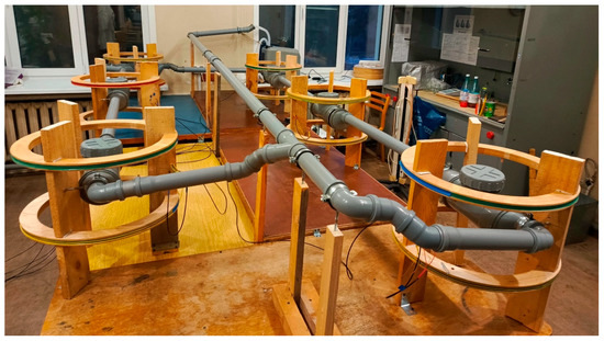
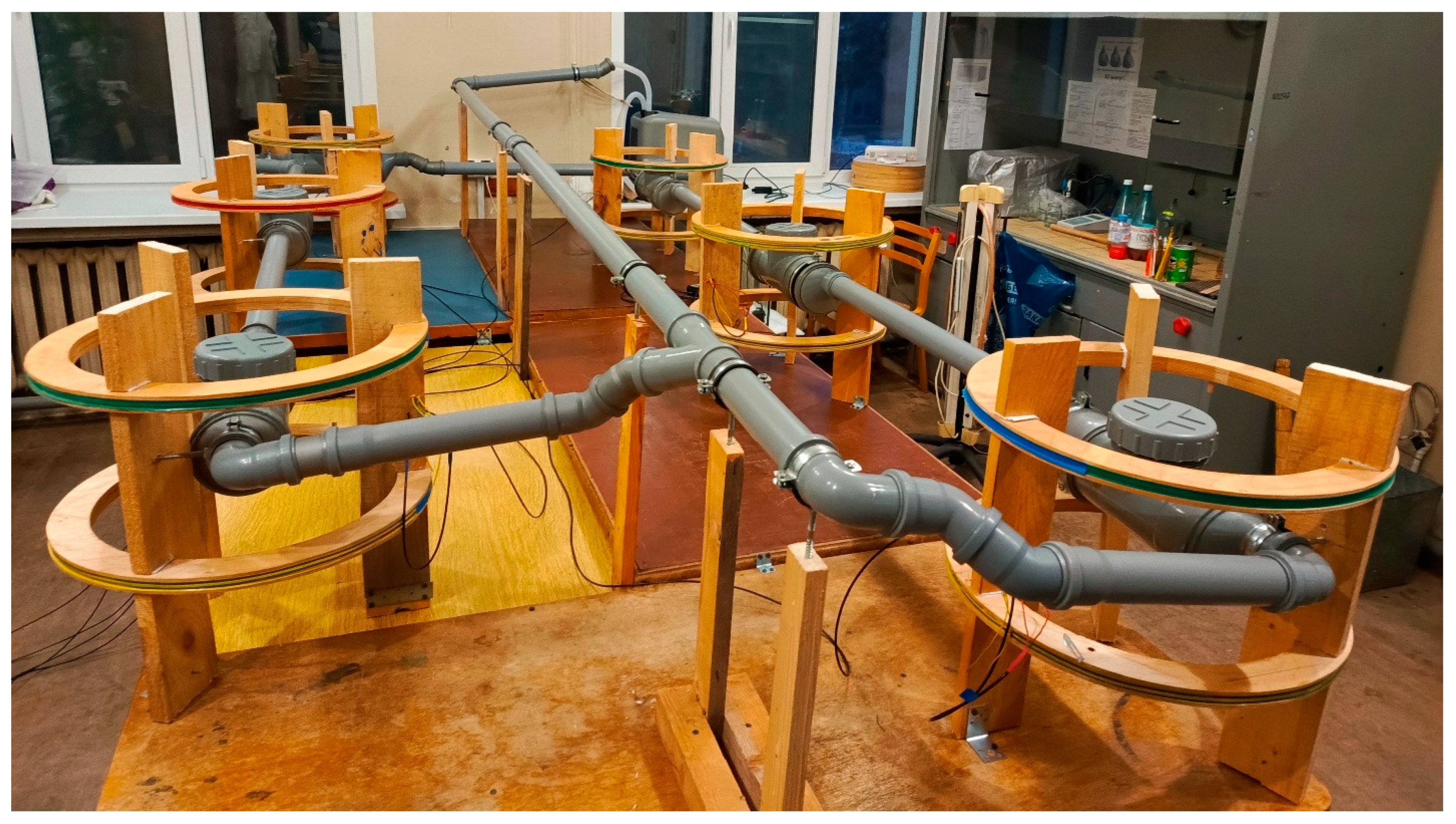
To ensure identical temperature conditions across all experimental groups, we developed and built a closed-loop water circulation system. Water maintained at a constant temperature circulated at a flow rate of 50 L/min through six exposure chambers: five positioned within the working volumes of the Helmholtz coils that generated LFMFs and one control chamber not exposed to magnetic fields (Figure 1).
Figure 1.
Temperature-controlled magnetic exposure system.
Water served as the heat-transfer medium and circulated through pipes 50 mm in diameter, expanding to 110 mm within the central working area of each pair of Helmholtz coils. Temperature regulation was achieved using a Teco TR-15 chiller (Teco, Ravenna, Italy) in combination with electric heaters connected to a thermostatic relay. The temperature was maintained at 22 °C in all groups. Owing to the high heat capacity of water, temperature fluctuations did not exceed ±0.5 °C from the set point. Any temperature oscillations associated with heater or chiller operation occurred synchronously across all experimental groups.
Within the widened section of the temperature-controlled system located at the center of each Helmholtz coil and in the control chamber, an opening accommodated a perforated plastic sleeve containing the experimental vessels with daphnids. The circulating water continuously flowed through the perforations, ensuring uniform thermal conditions for all experimental and control groups.
2.4. Experimental Procedure
Offspring produced by a single D. magna female were isolated and maintained individually in 100 mL containers under standard culture conditions. Newly produced offspring (the first three broods) were removed daily. When females reached the stage of producing their fourth and subsequent broods, neonates (<24 h old) from each brood were counted. If a brood contained 24 or more offspring, it was chosen for the experiments. The genetically identical offspring from a single female were randomly split into six experimental groups. Depending on the total brood size, each group contained 4 to 7 individuals.
The exposure of sibling offspring was performed in six identical wide glass test tubes. To ensure uniformity, a medium was prepared in one common container for all groups. It consisted of cultivating water in which 3 × 10⁸ cells of C. vulgaris were added per 100 cm3. The algal concentration corresponded to ten times the standard daily feeding dose to exclude food limitation during exposure. Each tube was filled with 20 mL of the medium. Each experimental group of daphnids was placed into one of the six test tubes.
Five tubes were randomly positioned within the working volumes of the Helmholtz coils, which generated magnetic fields, and one tube served as a control. The animals were exposed to one of the five LFMFs or to control conditions for 48, 96, and 144 h. Each experiment was performed in nine replicates. Daphnid’s survival in all groups was 100%. Immediately after exposure, we fixed individuals from each group in 4% formalin.
Body length was measured in all fixed specimens as a measure of D. magna growth rate. For morphometric analysis, all individuals were photographed at identical magnification using an MC-2 microscope (Micromed, Saint Petersburg, Russia) equipped with a DCM 500 digital camera (Hangzhou Huaxin IC Technology, Hangzhou, China). Body length measurements were performed on the acquired images using Image-Pro 3 software (Media Cybernetics, Bethesda, MD, USA). The raw data are included in the Supplementary Material.
2.5. Statistical Analysis
Data distributions were tested for normality using the Kolmogorov–Smirnov and Shapiro–Wilk tests. The homogeneity of variances was assessed using Levene’s test. One-way analysis of variance was performed to evaluate the effect of LFMF frequency on the studied parameters. Differences between each experimental group and the control were assessed using Dunnett’s post hoc test.
In addition, to facilitate comparison with previous studies in this field [9,10,21,24,25], the results were expressed as the biological effect of LFMF exposure. This effect was calculated for each experimental replicate using the following formula:
where Mcon and Mexp represent the mean values of the measured parameter in the control and experimental groups, respectively, and SEcon and SEexp denote the corresponding standard errors of the mean. The mean biological effect values calculated across all experimental replicates were then tested against zero using Student’s t-test for normally distributed data and the Wilcoxon test for non-normally distributed data.
3. Results
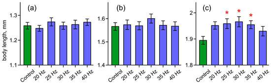
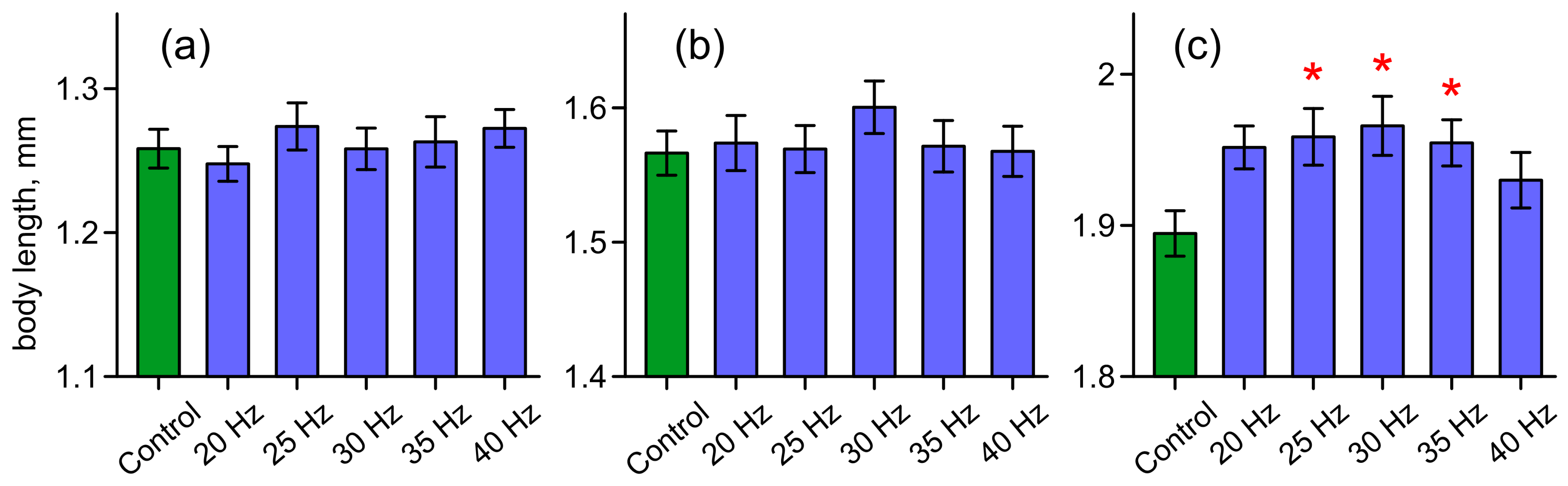
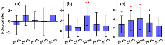
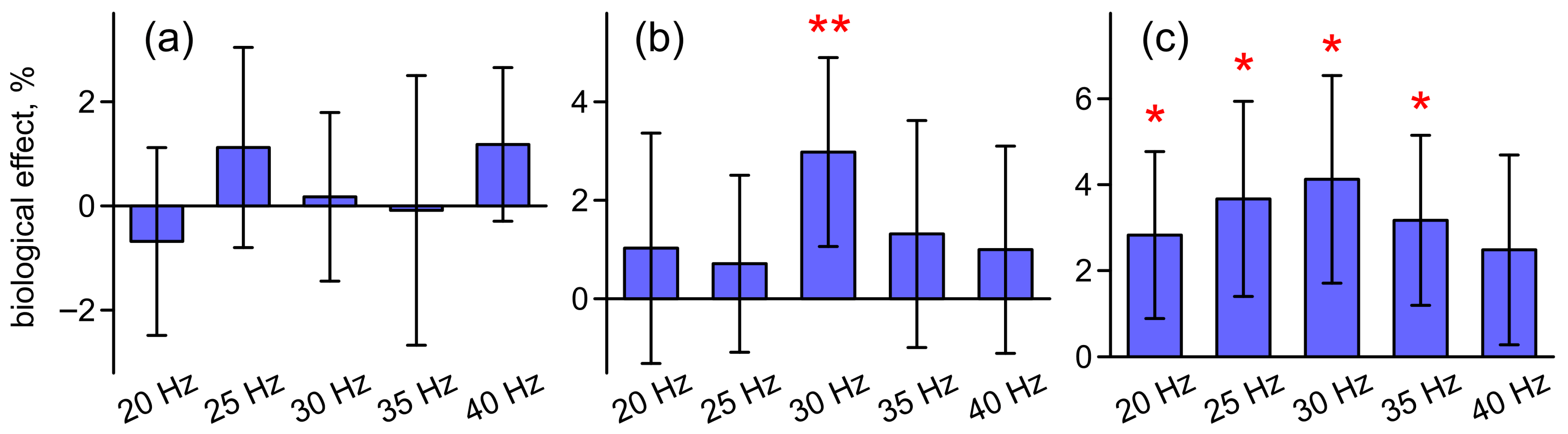
The applied magnetic fields did not affect the growth rate of juvenile Daphnia when the exposure duration was limited to 48 h. There were no significant differences in body length between the experimental groups and the control (Figure 2a), and the calculated biological effect did not differ from zero (Figure 3a).
Figure 2.
Body length of D. magna in the control and LFMF-exposed groups after 48 h (a), 96 h (b), and 144 h (c) of exposure. Values are means ± standard error. Asterisks indicate a significant difference between control and LFMF-exposed groups (* p < 0.05).
Figure 3.
Biological effect of LFMFs after 48 h (a), 96 h (b), and 144 h (c) of exposure, expressed as a percentage of the relative difference between the mean body length of exposed and control D. magna. Positive values indicate an increase in body length in the experimental group relative to the control, whereas negative values indicate a decrease. Error bars represent the sum of the standard errors of the mean in the control and experimental groups. Asterisks denote values significantly different from zero (* p < 0.05, ** p < 0.01).
After 48 h of exposure, a tendency toward increased mean body length was observed in individuals subjected to the 30 Hz LFMF compared with both the control and the other experimental groups (Figure 2b). However, these differences did not reach statistical significance. In contrast, the calculated biological effect for the 30 Hz LFMF (2.98%) was statistically significant (Figure 3b).
The most pronounced effects were revealed after the longest 144-h exposure to LFMFs. Accelerated growth, reflected by increased body length, was observed in all experimental groups relative to the control. However, statistically significant differences were confirmed only for the 25 Hz, 30 Hz, and 35 Hz treatments (Figure 2c).
Analysis of the biological effect revealed a dome-shaped frequency dependence. The maximal effect of 4.12% was observed at the 30 Hz LFMF. Both decreasing the frequency to 25 Hz and 20 Hz, as well as increasing it to 35 Hz and 40 Hz, resulted in a slight reduction in the magnitude of the biological effect (Figure 3c).
4. Discussion
The dependence of the observed effects on exposure duration is most likely related to the specific growth pattern of crustaceans. The body of Daphnia is enclosed within a chitinous carapace that prohibits continuous growth. During molting, the old carapace is shed and replaced by a newly formed, initially soft and extensible exoskeleton. The body length increases at this stage. Once the new carapace hardens, growth effectively ceases during the intermolt period. In juvenile individuals maintained at 20–25 °C, the intermolt interval typically lasts 1–2 days. Thus, a 48-h exposure would encompass only one or two molting events, a 96-h exposure approximately three to four molts, and a 144-h exposure approximately five to six molts [26,27]. Consequently, short exposure durations include relatively few discrete growth events, which may limit the detectability of LFMFs’ effects on linear body dimensions.
The linear growth rate of juvenile daphnids represents an integrative parameter reflecting multiple physiological processes, including food assimilation, hormonal regulation, and energy metabolism. Magnetic fields may influence magnetosensitive molecular primary targets involved in these pathways, according to several currently discussed mechanisms [4,5,6,7]. Such modulation could result in stimulation of somatic growth.
In our study, the most pronounced effect was observed under 30 Hz LFMF. This frequency was the only one to produce a significant effect after 96 h of exposure and yielded the most pronounced response in the 144-h experiment. These findings are consistent with data reported by Ermakov et al. [9], who demonstrated enhanced regeneration in planarians exposed to a 30 Hz 74 μT LFMF. Regenerative processes and somatic growth share common physiological foundations, which suggests that similar mechanisms underlie these effects. Similarly, Belova and Lednev reported stimulation of gravitropic responses in stems of flax, millet, and clover exposed to a 35.6 Hz, 85.6 μT LFMF [24]. In another study, it was shown that exposure of phorbol-12-myristate-13-acetate-activated mouse neutrophils to a 31 Hz, 74.7 μT LFMF significantly reduced the rate of reactive oxygen species production [27].
The biophysical mechanism underlying these effects may involve the interaction of alternating magnetic fields with ions bound at enzyme ion-binding sites, as proposed by Lednev [5]. An ion in the ion-binding center behaves as an oscillator in the static magnetic field. According to this hypothesis, the oscillation frequency of calcium ions is close to 30 Hz, with its precise value depending on the strength of the geomagnetic field [21]. When applying an LFMF with parameters “tuned to the ion oscillations”, changes in the ion-dependent functioning of the enzyme can occur, leading to the observed biological effects [9,10,24,25]. An alternative hypothesis attributes the biological effects of alternating magnetic fields to their influence on radical pair reactions within biochemical oscillators [1]. In this framework, a resonance-like response is expected to occur when the frequency of the LFMF matches the ~30 Hz oscillations of the mitochondrial membrane potential. It is assumed that such frequency alignment may substantially alter the magnetic environment experienced by a subset of radical pairs involved in these processes, thereby modulating mitochondrial function [7].
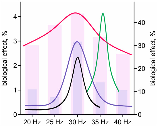
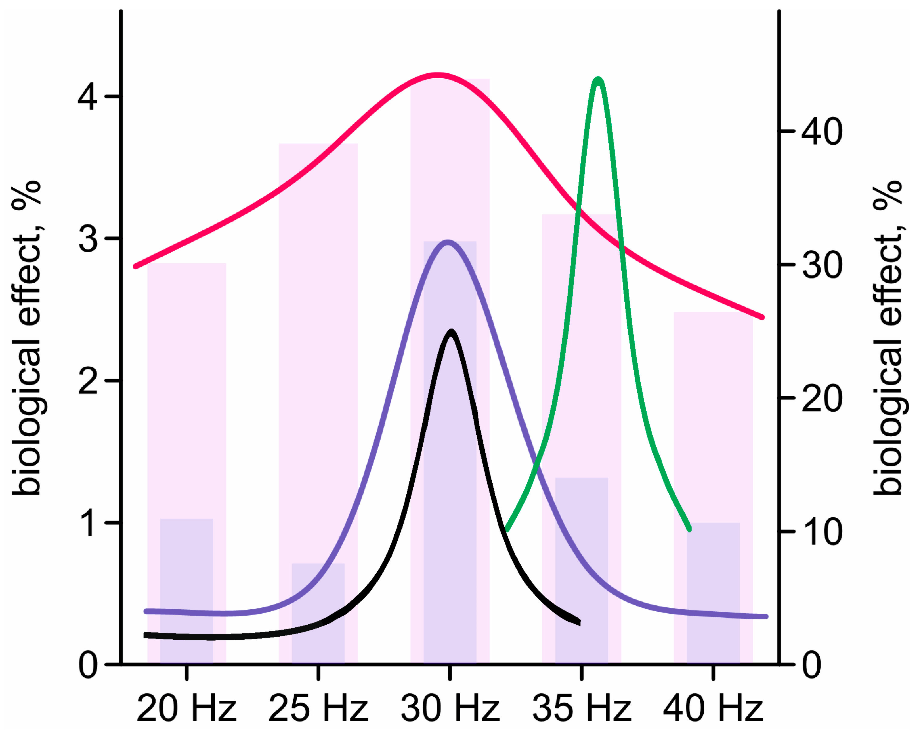
At the same time, our experiments revealed features not entirely consistent with earlier observations. Previous studies on planarians and plant stem segments demonstrated a sharply defined peak of biological effectiveness within the 20–40 Hz range, centered near 30 Hz, with negligible effects at neighboring frequencies [9,10]. A similar pattern was observed in our experiments after 96 h of exposure. However, following 144 h of exposure, biologically significant effects were detected at nearly all tested frequencies, resulting in a markedly flattened frequency–response curve (Figure 4).
Figure 4.
Frequency dependence of the biological effects of LFMFs in the 20–40 Hz range. Bar charts display the mean values obtained in the present study, with fitted curves superimposed. The purple bars and blue line (left y-axis) correspond to a 96-h exposure, and the pink bars with the red line (left y-axis) correspond to a 144-h exposure. For comparison, fitted curves from previously published studies are also included: the effect of a 74 μT LFMF on planarian regeneration rate (right y-axis, black line) [9] and the effect of an 85.6 μT LFMF on the gravitropic response of excised flax stem segments (right y-axis, green line) [10].
The length of exposure may therefore be a crucial factor in determining the outcome. In the studies by Ermakov [9] and Belova-Lednev [10], exposure times ranged from 30 min to 24 h. It is plausible that during relatively short exposures, such as those encompassing only three to four molting cycles in our experiments, compensatory mechanisms mitigate the effects of magnetic fields at the frequencies neighboring to the effective one. With prolonged exposure (144 h in our study), these compensatory responses may become exhausted, allowing magnetobiological effects to manifest across a broader frequency range. A similar expansion of the effective frequency window may also occur for other physiological endpoints as exposure duration increases.
The ecological relevance of the observed increase in body size of daphnids under exposure to the tested alternating magnetic fields is difficult to interpret, as such fields are almost absent in natural environments. Nevertheless, the physiological effects identified here suggest potential biomedical applications. Given the general trend toward enhanced growth and proliferative processes across different taxa [9,10,24,25], such magnetic field conditions may warrant investigation as a therapeutic modality, for example, to promote wound healing. However, further studies are required to evaluate their efficacy and safety.
Supplementary Materials
The following supporting information can be downloaded at: https://www.mdpi.com/article/10.3390/biophysica6020028/s1, raw data (Data.xlsx).
Author Contributions
Conceptualization, V.V.K.; methodology, V.V.K.; investigation, V.V.K.; measurements, D.A.S. and A.A.S.; resources, V.V.K.; data curation, V.V.K., D.A.S. and A.A.S.; writing—original draft preparation, V.V.K.; writing—review and editing, D.A.S. and A.A.S.; visualization, V.V.K.; project administration, V.V.K.; funding acquisition, V.V.K. All authors have read and agreed to the published version of the manuscript.
Funding
This research was funded by the Russian Science Foundation, grant number 25-24-00088 (https://rscf.ru/en/project/25-24-00088/ accessed on 1 April 2026).
Institutional Review Board Statement
No approval of research ethics committees was required for invertebrate species.
Data Availability Statement
The raw data supporting the conclusions of this article are included in the Supplementary Material.
Conflicts of Interest
The authors declare no conflicts of interest.
Abbreviations
The following abbreviation is used in this manuscript:
| LFMF | Low-frequency magnetic field |
References
- Krylov, V.V.; Osipova, E.A. Molecular biological effects of weak low frequency magnetic fields: Frequency–amplitude efficiency windows and possible mechanisms. Int. J. Mol. Sci. 2023, 24, 10989. [Google Scholar] [CrossRef] [Scilit]
- Markov, M.S. Biological effects of extremely low frequency magnetic fields. In Biomagnetic Stimulation; Ueno, S., Ed.; Springer: Boston, MA, USA, 1994; pp. 91–103. [Google Scholar] [CrossRef] [Scilit]
- Tian, H.; Zhu, H.; Gao, C.; Shi, M.; Yang, D.; Jin, M.; Wang, F.; Sui, X. System-level biological effects of extremely low-frequency electromagnetic fields: An in vivo experimental review. Front. Neurosci. 2023, 17, 1247021. [Google Scholar] [CrossRef] [Scilit]
- Blanchard, J.P.; Blackman, C.F. Clarification and application of an ion parametric resonance model for magnetic field interactions with biological systems. Bioelectromagnetics 1994, 15, 217–238. [Google Scholar] [CrossRef] [Scilit]
- Belova, N.A.; Panchelyuga, V.A. Lednev’s model: Theory and experiment. Biophysics 2010, 55, 661–674. [Google Scholar] [CrossRef] [Scilit]
- Binhi, V.N.; Prato, F.S. A physical mechanism of magnetoreception: Extension and analysis. Bioelectromagnetics 2017, 38, 41–52. [Google Scholar] [CrossRef] [Scilit]
- Krylov, V.V. Biological effects of weak magnetic fields: Can the radical-pair mechanism provide a universal explanation? Biol. Rev. 2026, in press. [Google Scholar] [CrossRef] [Scilit] [PubMed]
- Smith, S.D.; McLeod, B.R.; Liboff, A.R.; Cooksey, K. Calcium cyclotron resonance and diatom mobility. Bioelectromagnetics 1987, 8, 215–227. [Google Scholar] [CrossRef] [Scilit] [PubMed]
- Ermakov, A.; Afanasyeva, V.; Ermakova, O.; Blagodatski, A.; Popov, A. Effect of weak alternating magnetic fields on planarian regeneration. Biochem. Biophys. Res. Commun. 2022, 592, 7–12. [Google Scholar] [CrossRef] [Scilit]
- Belova, N.A.; Lednev, V.V. Dependence of gravitropic reaction in segments of flax stems on frequency and amplitude of variable components of a weak combined magnetic field. Biofizika 2000, 45, 1108–1111. [Google Scholar] [PubMed]
- Grinberg, M.; Ilin, N.; Nemtsova, Y.; Sarafanov, F.; Ivanova, A.; Dolinin, A.; Pirogova, P.; Vodeneev, V.; Mareev, E. Response of photosynthesis and electrical reactions of wheat plants upon the action of magnetic fields in the Schumann resonance frequency band. Plant Signal. Behav. 2024, 19, 2294425. [Google Scholar] [CrossRef] [Scilit]
- Krylov, V.V.; Papchenkova, G.A.; Golovanova, I.L. Influence of calcium resonance-tuned low-frequency magnetic fields on Daphnia magna. Int. J. Mol. Sci. 2022, 23, 15727. [Google Scholar] [CrossRef] [Scilit]
- Krylov, V.V.; Bolotovskaya, I.V.; Osipova, E.A. The response of European Daphnia magna Straus and Australian Daphnia carinata King to changes in geomagnetic field. Electromagn. Biol. Med. 2013, 32, 30–39. [Google Scholar] [CrossRef] [Scilit]
- Krylov, V.V.; Osipova, E.A. The response of Daphnia magna Straus to the long-term action of low-frequency magnetic fields. Ecotoxicol. Environ. Saf. 2013, 96, 213–219. [Google Scholar] [CrossRef] [Scilit]
- Krylov, V.V.; Papchenkova, G.A.; Osipova, E.A. The influence of changes in magnetic variations and light–dark cycle on life-history traits of Daphnia magna. Bioelectromagnetics 2020, 41, 338–347. [Google Scholar] [CrossRef] [Scilit]
- Krylov, V.V.; Osipova, E.A. The response of Daphnia magna Straus to long-term exposure to simulated geomagnetic storms. Life Sci. Space Res. 2019, 21, 83–88. [Google Scholar] [CrossRef] [Scilit]
- Lazorchak, J.M.; Smith, M.E.; Haring, H.J. Development and validation of a Daphnia magna four-day survival and growth test method. Environ. Toxicol. Chem. 2009, 28, 1028–1034. [Google Scholar] [CrossRef] [Scilit] [PubMed]
- Suzuki, A.; Kato, Y.; Matsuura, T.; Watanabe, H. Growth evaluation method by live imaging of Daphnia magna and its application to the estimation of an insect growth regulator. J. Appl. Toxicol. 2014, 35, 68–74. [Google Scholar] [CrossRef] [Scilit]
- Peters, R.H. Daphnia Culture. Mem. Ist. Ital. Idrobiol. 1987, 45, 483–495. [Google Scholar]
- ASTM. Standard Practice for Conducting Acute Toxicity Tests with Fishes, Macroinvertebrates and Amphibians; American Society for Testing and Materials: Philadelphia, PA, USA, 1980. [Google Scholar]
- Lednev, V.V.; Srebnitskaya, L.K.; Il’Yasova, Y.N.; Rozhdestvenskaya, S.Y.; Klimov, A.A.; Belova, N.A.; Tiras, K.P. Magnetic parametric resonance in biosystems: Experimental verification of theoretical predictions using regenerating planarians Dugesia tigrina as a test system. Biophysics 1996, 41, 815–825. [Google Scholar]
- Blackman, C.F.; Blanchard, J.P.; Benane, S.G.; House, D.E. Empirical test of an ion parametric resonance model for magnetic field interactions with PC-12 cells. Bioelectromagnetics 1994, 15, 239–260. [Google Scholar] [CrossRef] [Scilit] [PubMed]
- Blackman, C.F.; Blanchard, J.P.; Benane, S.G.; House, D.E. The ion parametric resonance model predicts magnetic field parameters that affect nerve cells. FASEB J. 1995, 9, 547–551. [Google Scholar] [CrossRef] [Scilit] [PubMed]
- Belova, N.A.; Lednev, V.V. Activation and inhibition of gravitropic reaction of plants using weak combined magnetic fields. Biofizika 2000, 45, 1102–1107. [Google Scholar] [PubMed]
- Belova, N.A.; Potselueva, M.M.; Srebnitskaya, L.K.; Znobishcheva, A.V.; Lednev, V.V. The influence of weak magnetic fields on the production of reactive oxygen species in peritoneal neutrophils of mice. Biophysics 2010, 55, 586–591. [Google Scholar] [CrossRef] [Scilit]
- Lavens, P.; Sorgeloos, P. (Eds.) Manual on the Production and Use of Live Food for Aquaculture; FAO Fisheries Technical Paper No. 361; FAO: Rome, Italy, 1996. [Google Scholar]
- Mu, X.; LeBlanc, G.A. Synergistic interaction of endocrine-disrupting chemicals: Model development using an ecdysone receptor antagonist and a hormone synthesis inhibitor. Environ. Toxicol. Chem. 2004, 23, 1085–1091. [Google Scholar] [CrossRef] [Scilit] [PubMed]
Disclaimer/Publisher’s Note: The statements, opinions and data contained in all publications are solely those of the individual author(s) and contributor(s) and not of MDPI and/or the editor(s). MDPI and/or the editor(s) disclaim responsibility for any injury to people or property resulting from any ideas, methods, instructions or products referred to in the content. |
© 2026 by the authors. Licensee MDPI, Basel, Switzerland. This article is an open access article distributed under the terms and conditions of the Creative Commons Attribution (CC BY) license.