
Article

Computational Molecular Docking and Molecular Dynamics Simulations of Potential Inhibitors from Cistus incanus (Cistaceae) Against Ebola Virus

by
Wafa Hourani
1,
Balakumar Chandrasekaran
2,*,
Sankar Muthumanickam
3 and
Pandi Boomi
3
1
Department of Pharmaceutical Sciences, Faculty of Pharmacy, Philadelphia University, P.O. Box 1, Amman 19392, Jordan
2
Department of Pharmaceutical Sciences, Faculty of Pharmacy, Zarqa University, P.O. Box 2000, Zarqa 13111, Jordan
3
Department of Bioinformatics, Alagappa University, Karaikudi 630003, Tamil Nadu, India
*
Author to whom correspondence should be addressed.
Biophysica 2026, 6(2), 29; https://doi.org/10.3390/biophysica6020029
Submission received: 14 February 2026 / Revised: 20 March 2026 / Accepted: 28 March 2026 / Published: 6 April 2026
(This article belongs to the Special Issue Biophysical Insights into Small Molecule Inhibitors)

Abstract

Background/Objectives: Until now, there have been no suitable medicines to treat infections caused by the Ebola virus. Cistus incanus, a traditional medicinal plant, contains several phytocompounds exhibiting antioxidant and anti-inflammatory properties. Methods: In this research, the molecular level interactions of the phytocompounds of Cistus incanus were investigated for their antiviral potential against the active site of VP40 protein of Ebola virus using in silico molecular docking. Further, the potential compounds were assessed for their stability in the protein using molecular dynamics (MD) simulations. Results: Methyl gallate, catechin, and quercetin showed excellent docking scores of −9.8, −8.8, and −7.7 kcal/mol, respectively, and favorable interactions with the target protein. These complexes showed good stability over the 100 ns MD simulation time. In addition, the phytocompounds displayed favorable pharmacokinetics and drug-like properties. Conclusions: Our study offers the antiviral potential of phytocompounds (methyl gallate, catechin, and quercetin) of Cistus incanus, suggesting their suitability as lead candidates for the treatment of Ebola viral infection.

1. Introduction

The Ebola virus (EBOV) is a virulent pathogen that causes acute hemorrhagic fever in humans and animals, with a high case fatality rate of 90% within 7 to 11 days of the occurrence of this infection [1]. EBOV is an enveloped, non-segmented, filamentous, and negative-sense ribonucleic acid (RNA) virus [2]. In the recent outbreak (September 2025), it was revealed that nearly 64 confirmed cases in the Democratic Republic of the Congo (DRC) had 45 fatalities, for a 70% fatality rate, highlighting EBOV as a serious public health and economic threat [3]. It has proven to be a dangerous infection in many African countries, with well-known causative agents that cause a higher incidence of death [4,5]. EBOV is a member of the Filoviridae virus family that consists of four species of EBOV, namely Ivory Coast (CIEBOV), Reston (REBOV), Zaire (ZEBOV), and Sudan (SEBOV), which have been identified to date. SEBOV shares around 70% amino acid sequence identity with EBOV, while ZEBOV plays a critical role in causing the highest mortality rate [6]. Recently, one new species of ebolavirus was found and named Bundibugyo ebolavirus (BEBOV), which forms filamentous virions [7]. The incubation period lasts between 2 and 21 days. An initial person-to-person transmission of EBOV triggers a community-wide spread through body fluids, physical contacts or through contaminated objects [8]. Aside from infecting blood, EBOV also spreads to urine, semen, saliva, breast milk, cerebrospinal fluid (CSF), and aqueous humor [9]. EBOV can be diagnosed using reverse transcription polymerase chain reaction (RT-PCR) tools by collecting samples of tears, feces, amniotic fluids, and skin swabs [10]. Principally, EBOV affects cells of the immune system such as macrophages, dendritic cells, and monocytes, followed by spreading to other cellular types [11]. The fruit-eating bat is the general reservoir of the EBOV virus, while other insectivorous bat (frugivorous bat and Mops condylurus) species are also notable hosts for EBOV [12].
EBOV is an enveloped, negative-sense RNA virus consisting of multiple structural proteins that play crucial roles in the viral life cycle [13]. The seven structural proteins of EBOV, namely VP24, VP30, VP35, VP40, glycoprotein (GP), nucleoprotein (NP), and polymerase (L), represent promising therapeutic targets for the development of antiviral compounds [14]. Viral matrix protein VP40 plays a vital role in the life cycle of the Ebola virus and is considered as a potential target for antiviral treatment [15]. In particular, the matrix protein VP40, with a molecular weight of 40 kDa, is a highly expressed filoviral protein, and is involved in various events of the viral life cycle (maturation of the virus, regulation of RNA transcription and the budding of the matured virus from the host plasma membrane) [16]. Additionally, VP40 is the largest protein found in virions involved in host cell RNA metabolism during the replication process [17]. VP40 is a peripheral protein positioned on the outer edge of the layer just below the plasma membrane, containing 326 amino acids with a distinct N-terminal domain (NTD) and C-terminal domain (CTD) that are connected via a flexible linker; NTD promotes the protein’s oligomerization, whereas CTD is responsible for the interaction with the membrane [18]. Experimental evidence through crystallographic images of the VP40–RNA complex clearly shows that His123-Gly126, Arg134, and Tyr171 accommodate RNA substrate moiety into the binding site of VP40, and these amino acids are mainly involved in the interactions with RNA [19]. These interactions lead to the octamer formation of VP40 and promote viral transcription in early stages of infection [20]. Hence, the interactions between EBOV RNA and VP40 octamer are sufficient for the viral life cycle and considered as a potential target for the design of new anti-EBOV drugs [21,22]. The lack of effective preventive vaccines and approved therapeutics for the Ebola virus disease (EVD) represents a major concern, as outbreaks can pose pandemic threats, and case fatality rates may reach up to 90% [23]. Therefore, there is an urgent need to identify an effective therapeutic strategy for the management of Ebola virus. We speculated that virion production would be decreased if a small molecule inhibits the critical nucleation step in VP40. Hence, our new drug design was based on the discovery of molecules that potentially interfere with the functions of VP40, since this protein is very important for the life cycle of EBOV.
Plant-derived bioactive compounds have played an important role in the field of drug development in the past decades due to few or no side effects during the treatment of infectious diseases. According to the global report on traditional and complementary medicine by the World Health Organization (WHO), nearly 40% of pharmaceutical products are from nature and traditional knowledge. Hence, they have been used to treat viral infections with strong therapeutic efficacy [24,25,26,27,28]. Cistus incanus, commonly known as pink rockrose, was reported to exhibit antiviral effects against human immunodeficiency viruses (HIV) and Filoviruses [29,30]. Cistus incanus (Ci) is mainly available in Mediterranean countries [31], exhibiting a wide range of pharmacological activities such as anti-inflammatory [32], antioxidant/chemopreventive [33], cytotoxic [34], anti-ulcerogenic [35], and anti-bacterial properties [36,37,38]. The Ci plant is rich in polyphenolic compounds that induce antimicrobial and antiviral activities, and the Ci extract inhibits the infection caused by influenza A virus [39,40]. The Ci plant contains numerous other bioactive compounds that prevent these viruses from entering host cells for replication [41]. Hence, we envisioned potential natural phytocompounds that could be applicable in the treatment of EVD.
Datasets of natural products are very useful for in silico research, such as molecular docking and molecular dynamics simulations (MDS) to identify pharmacologically active phytocompounds as potential therapeutic agents [42,43]. Over the last decade, 58 new natural product-based drugs have been released, and many more molecules are currently in clinical trials and the drug approval process [44]. Computer-aided drug design (CADD) provides a rapid, reliable method in the new drug discovery process that helps minimize the use of animal models for the initial screening of novel molecules [45]. Thus, CADD can be employed effectively for the virtual screening of large chemical libraries to identify promising drug candidates. In silico analysis, including the determination of pharmacokinetics, and drug-likeliness properties, is conducted to optimize lead compounds [46]. Molecular docking [47] is a validated tool in the determination of the ligand’s orientation and binding interactions with target proteins and facilitates new drug discovery [48]. Similarly, MDS [49] is helpful in studying the stability of ligand–protein complexes. In this research, the bioactive compounds present in the medicinal plant of Cistus incanus were selected based on reports in the literature to determine the crucial interactions of such bioactive compounds against the protein of EBOV. Prime MM/GBSA and MDS were performed to examine the binding free energy and binding stability of the complexes. Several preclinical compounds fail to progress into clinical trials due to their poor pharmacokinetic properties [50]. Hence, the ADME (absorption, distribution, metabolism, and excretion) predictions of those bioactive compounds were computed to assess their drug-likeness and pharmacokinetic properties.

2. Materials and Methods

2.1. Preparation of Target Protein Structure

The X-ray solved crystal structure of VP40 was retrieved from a protein data bank (PDB) database (https://www.rcsb.org/structure/3TCQ accessed on 11 January 2026) with a resolution of 1.60 Å (PDB ID: 3TCQ) [51]. The Protein Preparation Wizard module in Schrödinger (Schrödinger LLC, New York, NY, USA) was used to prepare the structure by assigning proper bond order, hydrogen atoms, and removing water molecules beyond 5 Å [52]. The prepared structure was minimized and optimized by using the OPLS4 force field [53]. A receptor grid was generated using the Receptor Grid Generation tool in the Glide module [54].

2.2. Preparation of Ligand Structures

Twenty natural plant-based antiviral compounds derived from Cistus incanus were identified and retrieved from the PubChem database (https://pubchem.ncbi.nlm.nih.gov/ accessed on 12 January 2026). The LigPrep module [55] of Schrödinger was used to prepare the ligands by assigning proper bond lengths, bond angles, and possible tautomeric states at pH 7.0. The structures were finally energy-minimized using the OPLS4 force field [53].

2.3. Molecular Docking and Analysis

Molecular docking was performed by using the Glide XP (extra precision) module [56] in the Schrödinger suite (Schrödinger LLC, New York, NY, USA) to predict the binding modes of the ligands within the active site of the VP40 protein. The pose viewer file obtained after the docking step was loaded into the XP visualizer of the Maestro interface, and each ligand–protein complex analyzed for different types of intermolecular interactions (hydrogen bonding, π–π stacking, salt bridges, and hydrophobic).

2.4. Binding Free Energy Calculation

The Molecular Mechanics–Generalized Born Surface Area (MM-GBSA) method was used to estimate the binding free energies of the protein–ligand complexes. Binding free energy calculations were performed using the Prime MM-GBSA module of Schrödinger (Schrödinger LLC, New York, NY, USA) [57]. The entropic contribution (TΔS) was not included in the Prime MM-GBSA calculations. Hence, the reported ΔG_bind values represent endpoint free energy estimates and were used for comparative ranking and stability assessment, rather than quantification of absolute binding affinities. The binding free energy (ΔG_bind) was computed according to the following equation:
ΔG_bind = ΔE_MM + ΔGsolv + ΔG_SA
where, ΔE_MM represents the molecular mechanics energy, ΔG_solv is the solvation free energy, and ΔG_SA is the surface area energy contribution.

2.5. Molecular Dynamics Simulation (MDS)

MDS offers valuable insights into ligand–protein complex stability, flexibility and dynamics [58]. The VP40–ligand complexes obtained from docking were subjected to MDS using the GROningen MAchine for Chemical Simulations (GROMACS version 2022.1) package Vwith the GROMOS43a1 force field [59]. The systems were solved in a cubic periodic box with dimensions: 10 × 10 × 10 nm using the simple point charge (SPC) water model. Appropriate counterions were added to neutralize the systems. Subsequently, energy minimization was carried out by using the steepest descent algorithm to remove weak van der Waals (vdW) contacts. Then, the minimized systems were equilibrated under NVT (constant number of particles, volume, and temperature) at 300 K for 100 ps, followed by NPT (constant number of particles, pressure, and temperature) equilibration at 1 atm for 100 ps. Temperature coupling was performed using the Nose–Hoover thermostat at 300 K, and pressure coupling was maintained using the Martyna–Tobias–Klein (MTK) barostat at 1 atm during the NPT ensemble. The NVT ensemble was generated using the Nose–Hoover thermostat without pressure coupling. After that, the production MD simulations were performed for 100 ns. The resulting trajectories were analyzed to calculate root-mean-square deviation (RMSD), root-mean-square fluctuation (RMSF), and hydrogen bond formation using built-in GROMACS utilities.

2.6. ADME Pharmacokinetics Analysis

The ADME (absorption, distribution, metabolism, and excretion) and drug-like properties [60] of the plant-derived compounds were evaluated using the QikProp module of the Schrödinger suite (Schrödinger LLC, New York, NY, USA) [61]. It is used to predict physicochemical properties, pharmacokinetic descriptors, and drug-likeness of small molecules [62]. Properties such as QPlogPo/w, QPlogHERG, QPlogBB, percent human oral absorption, and compliance with Lipinski’s Rule of Five (RO5) were computed and analyzed for the phytocompounds.

3. Results

3.1. Molecular Docking

In this study, 20 phytocompounds were screened against the VP40 protein of Ebola virus using the Glide program of the Schrödinger suite (Schrödinger LLC, New York, NY, USA). The results of molecular docking revealed that the bioactive phytocompounds exhibited docking scores ranging from −9.8 to −5.2 kcal/mol (Supplementary Materials Table S1). In the molecular docking analysis, the compounds firmly bound to the active site of VP40 protein, unveiling that three of the bioactive phytocompounds—methyl gallate, catechin, and quercetin—showed higher docking scores of −9.8, −8.8, and −7.7 kcal/mol (Table 1), respectively, than those of other bioactive compounds (Supplementary Materials Table S1).
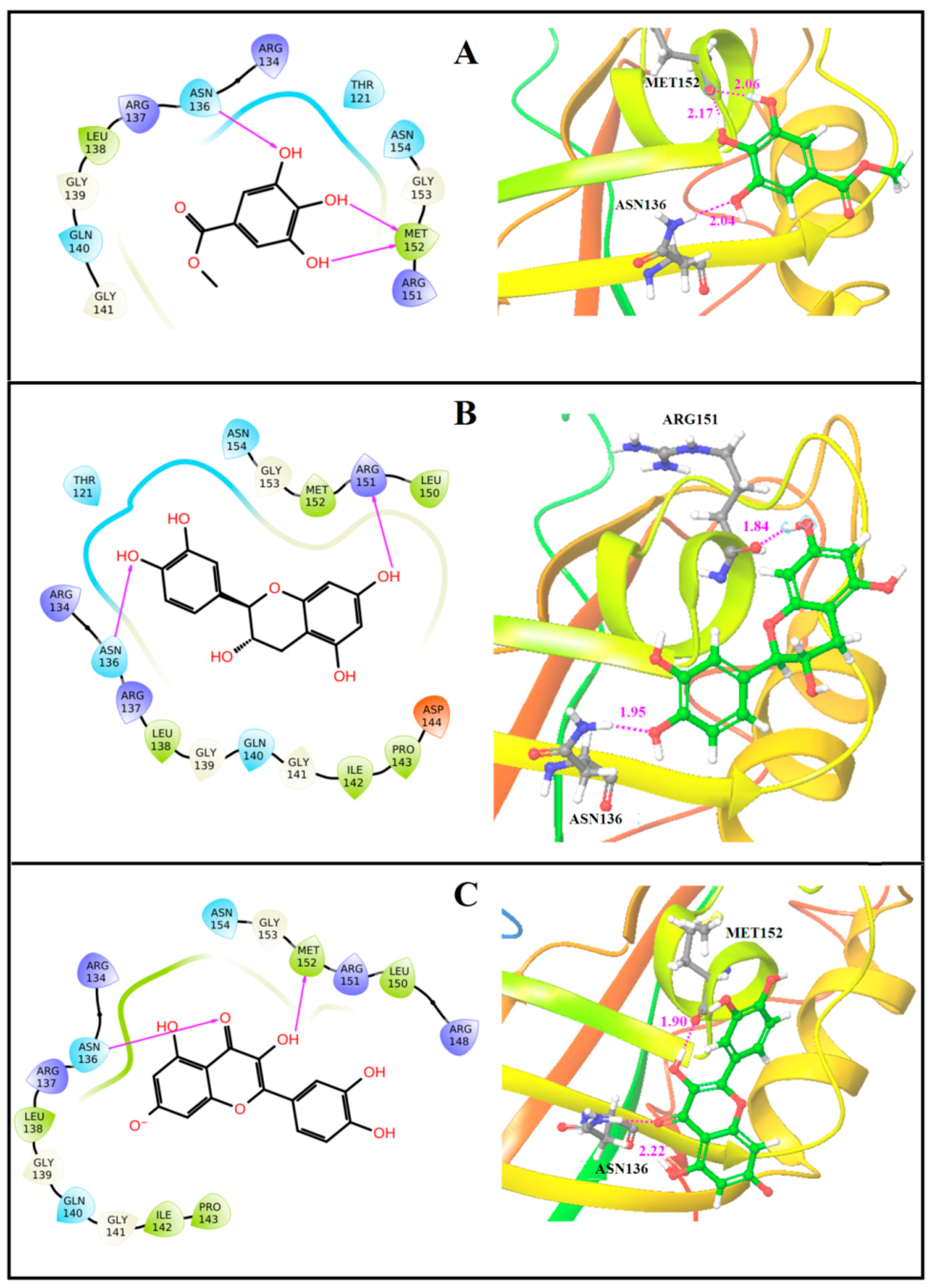
The molecular docking pose of methyl gallate (Figure 1A) revealed that it occupied the RNA binding region of VP40 and yielded three hydrogen bonding interactions with Asn136 (bond distance 2.04 Å) and Met152 (bond distance 2.06 Å and 2.17 Å). Some hydrophobic interactions with Thr121, Arg134, Arg137, Leu138, Gly139, Gln140, Gly141, Arg151, Gly153, and Asn154 amino acid residues were also observed. Similarly, the second bioactive phytocompound, catechin, was bound to the RNA binding region of VP40, exhibiting hydrogen bonding (H-bond) interactions with the residues of Asn136 (bond distance 1.95 Å) and Arg151 (bond distance 1.84 Å), correspondingly. The binding site amino acid residues of Thr121, Arg134, Arg137, Leu138, Gly139, Gln140, Gly141, Ile142, Pro143, Asp144, Leu150, Met152, Gly153, and Asn154 were involved in the hydrophobic interaction with catechin (Figure 1B). On the other hand, quercetin (Figure 1C) also interacted with the RNA binding region of VP40, generating two characteristic hydrogen bonding interactions with Asn136 (bond distance 2.22 Å) and Met152 (bond distance 1.90 Å). Moreover, hydrophobic interactions with residues Arg134, Arg137, Leu138, Gly139, Gln140, Gly141, Ile142, Pro143, Arg148, Leu150, Arg151, Gly153 and Asn154 in the RNA binding region of the VP40 were observed in the quercetin–VP40 protein complex.

3.2. Binding Free Energy

The MM-GBSA post-scoring approach was used to estimate the relative stability of the docked protein–ligand complexes and to evaluate binding orientation and interaction consistency of the bioactive compounds [63]. As shown in Table 1, the MM-GBSA (ΔG_bind) values of the docked complexes were −51.16 kcal mol−1, −49.20 kcal mol−1, and −47.92 kcal mol−1 for methyl gallate, catechin, and quercetin, respectively. Since Prime MM-GBSA provided endpoint free energy estimates and did not include explicit entropic contributions in this study, the reported ΔG_bind values were interpreted comparatively to evaluate relative binding preference, interaction stability, and complex consistency among the compounds rather than as absolute quantitative binding affinities. The results therefore indicate comparatively favorable binding stability and relative interaction strength trends, supporting their potential as lead candidates for further experimental and functional validation rather than confirming definitive inhibitory activity.

3.3. Molecular Dynamics Simulation

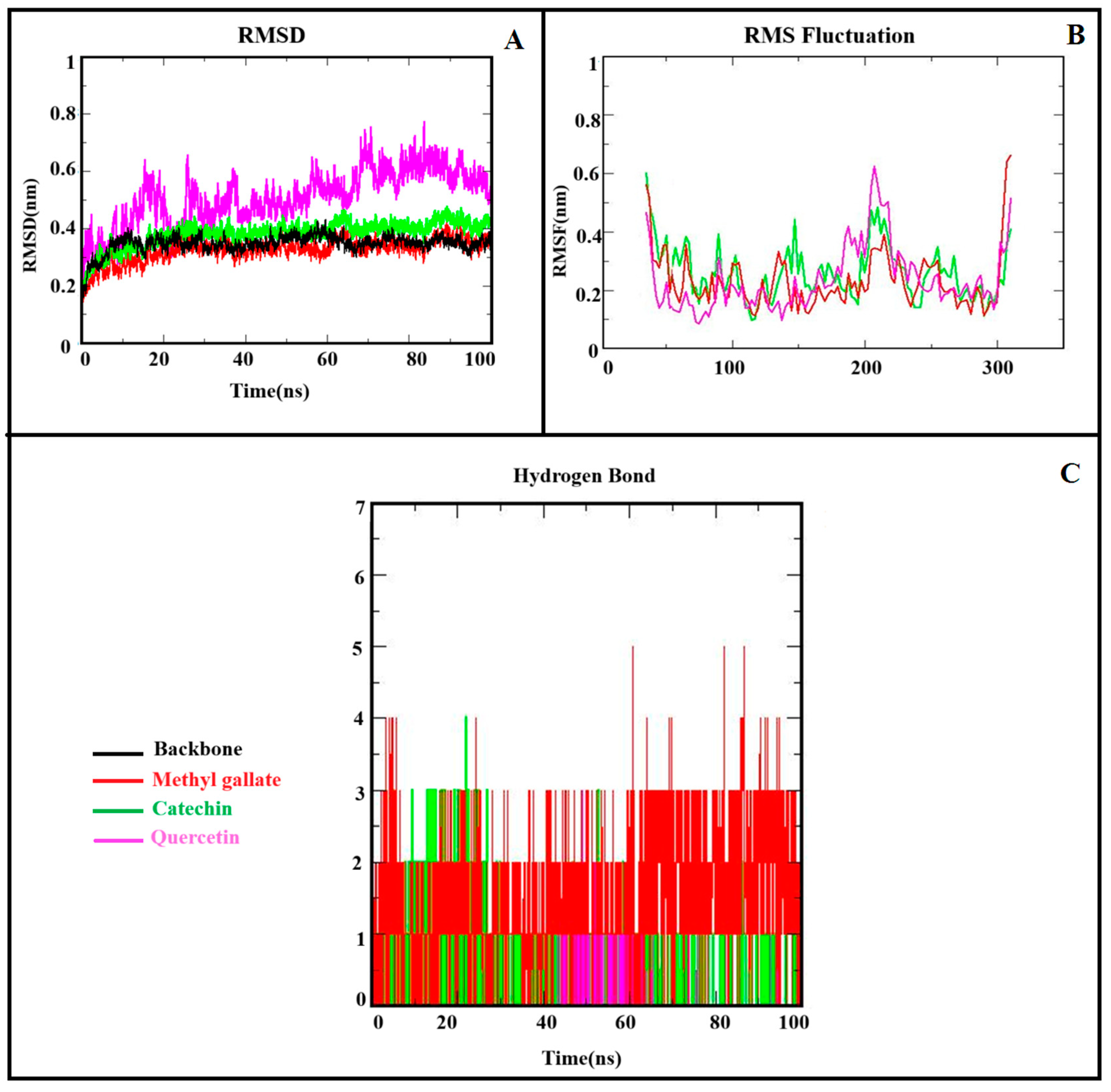
To analyze the conformational changes, structural stability, and dynamic behavior of the Apo and complexes, dynamics were performed for 100 ns MDS. The stability of the Apo protein and complexes was analyzed using RMSD, RMSF, and H-bonds. The RMSD is an important parameter to examine the equilibration of MD trajectories. The RMSD plot is used to check the stability of each system during the simulation [64]. The RMSD plot of the VP40 backbone and its complexes was computed using the simulation time scale, and the results are shown in Figure 2A. The RMSD value for the free VP40 (indicated in black) system was 0.28 nm, and it maintained stability until the end of the simulation. In the case of complexes, the average RMSD values of VP40–methyl gallate and VP40–catechin complexes were 0.13 and 0.17 nm, respectively. In the VP40–quercetin complex, RMSD values gradually increased until ~60–70 ns, after which the system attained stability, with an RMSD value of 0.22. The RMSD plots reveal that the complexes were constantly stable during the simulation time except for quercetin, which demanded a longer equilibration period. To ensure reproducibility and robustness, all simulations were performed in triplicate. The RMSD values (mean ± standard deviation) across independent runs were backbone (0.332 ± 0.026 nm), methyl gallate (0.357 ± 0.041 nm), quercetin (0.307 ± 0.065 nm), and catechin (0.326 ± 0.029 nm). The relatively low standard deviation values indicated consistent structural behavior across replicates, confirming the reliability of the simulations (Figure S1). The ligand RMSD analysis further confirmed the stable ligand retention within the VP40 binding cavity, with only minor positional fluctuations during the simulation (Figure S2).
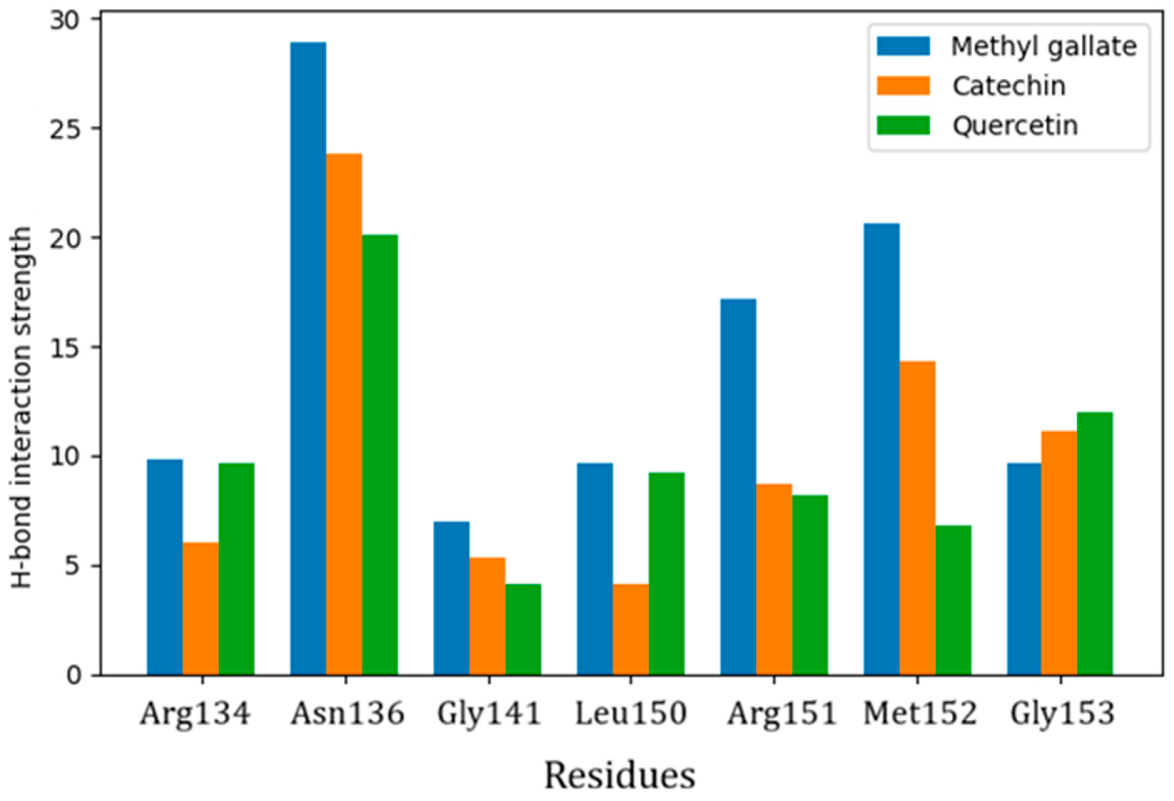
The RMSF plot can be used to describe structural flexibility and changes in protein conformation due to ligand binding during MDS [65]. The RMSF plots of all the complexes are shown in Figure 2B. The average RMSF values for VP40–methyl gallate, VP40–catechin, and VP40–quercetin complexes were found to be 0.07 nm, 0.08 nm, and 0.08 nm, respectively. Although higher fluctuations were observed between residues 210 and 235, these fluctuated regions were referred to as the loop. As a result, the fluctuation did not affect the ligand binding to the active site of the protein, and the VP40 protein remained more rigid and stable during simulation. The intermolecular hydrogen bonding between the protein and ligands plays a vital role in binding strength, molecular recognition and the overall stability of the protein structure [66]. The stability of the hydrogen bond (H-Bond) interaction between docked complexes was analyzed throughout the simulation period, and the result is shown in Figure 2C. Methyl gallate–VP40 showed five H-bonds, catechin–VP40 revealed four H-bonds, and quercetin–Vp20 exhibited two H-bonds throughout the simulation period. These results indicate that methyl gallate–VP40 and catechin–VP40 show stable and strong H-bonds. Hydrogen bond interaction profiling revealed persistent interactions of methyl gallate, catechin, and quercetin with key VP40 residues, particularly Arg134, Asn136, Gly141, Leu150, Arg151, Met152, and Gly153, indicating stable hydrogen bonding networks within the RNA binding region. These interactions contributed to ligand retention and binding stability within the VP40 cavity (Figure 3).

3.4. ADME Pharmacokinetics Analysis

Qikprop was used to analyze the pharmacokinetic properties and forecast drug-likeness for the bioactive compounds [67]. The classification of a specific molecule as a potential therapeutic candidate must agree with a variety of criteria, and Lipinski’s rule of five (RO5) is one of the most frequently accepted criteria in new drug discovery and development [68]. RO5 specifies that bioactive components with good oral absorption and bioavailability must have molecular weights of 500 or less, Log P of 5, less than five hydrogen bond donor (HBD) groups, and less than 10 hydrogen bond acceptor (HBA) groups. If any candidate does not satisfy more than one of these criteria, it is less probable that it will be developed as a possible orally administered drug. In our analysis, all three of the bioactive compounds complied with RO5, and the pharmacokinetic properties were within the acceptable ranges defined for human use. The results for pharmacokinetic properties and drug-likeness of the bioactive compounds are collected in Table 2.

4. Discussion

The discovery of novel and effective therapeutic agents is not only essential to treat EVD, but also to prevent and mitigate the devastating impact on global health [69]. Hence, there is an urgent need for rapid and cost-effective strategies. The requirement for biosafety level four (BSL-4) facilities for preclinical research on EBOV makes experimental drug discovery more challenging and expensive [70]. Thus, computational drug design helps to overcome such challenges and holds promise for developing novel and effective anti-Ebola therapeutics [71].
The reports in the literature suggested that VP40–RNA binding activity plays a key role during the life cycle of the Ebola virus [72]. Arg134 and Phe125 were identified as essential amino acid residues involved in RNA binding activity [73]. The RNA binding mechanism is affected by mutations on these residues, with Phe125 to Ala125, decreasing RNA binding activity and Arg134 to Ala134, abolishing RNA binding activity as well as the formation of the VP40 octamer [74]. This RNA binding activity is important for viral transcription regulation because VP40 in ring form can replicate the transcription control function and so plays an important role in the early stages of the Ebola virus’s life cycle [75]. In our study, the top three bioactive compounds were involved in the formation of hydrophobic interactions with the crucial amino acid Arg134 in the VP40 matrix protein of the Ebola virus, since they bind within the RNA binding region and may potentially interfere with VP40 function. Thus, three bioactive phytocompounds—methyl gallate, catechin, and quercetin—formed strong hydrogen bonds with residues Asn136, Arg151, and Met152, along with hydrophobic interactions with Arg134 of VP40, leading to their identification as promising lead candidates for further experimental investigation against EBOV.
MM-GBSA can offer higher enrichment factors for the ligands than scoring functions alone [76]. Despite the modular nature of MM/GBSA [77], it does not demand calculations on a training set and can be used effectively to rationalize experimental outcomes in enhancing the results of docking and virtual screening. In our MM-GBSA analysis, the compounds methyl gallate, catechin, and quercetin exhibited higher binding free energy values, indicating the possibility of higher affinity with VP40 protein, besides better stability of the complexes. Thus, binding affinities ranged from −51.16 to −47.92 kcal/mol, indicating stable interactions and consistent orientations across multiple conformations. The 100 ns MDS further validated the stability of these complexes. RMSD ranged from 0.13 nm to 0.22 nm, with methyl gallate being the most stable. RMSF values averaged 0.08 nm, with fluctuations to distal loops (210–235). Two to five hydrogen bonds were maintained throughout the simulation.
The medicinal compounds may be active and selective, yet they might not be good candidates for further development due to a lack of desired pharmacokinetic properties [78]. It is possible to have insufficient absorption or permeability if two parameters are out of range. The bioactive compounds may very well be absorbed in the digestive system if any one of the criteria is not met [79]. ADME pharmacokinetic properties determine the efficiency of a drug’s movement throughout the body [80]. Compounds need to undergo ADME screening to ensure that they meet the criteria required for a promising drug candidate [81]. High molecular weight can reduce a compound’s permeability across biological barriers [82]. Lipophilicity, expressed as the log of the partition coefficient (log P) between n-octanol and water, influences the absorption of the drug, with higher log P values often associated with lower bioavailability [83]. Solubility, indicated by the log S value, also affects absorption and distribution [84]. A drug’s ability to cross cell membranes depends on the number of hydrogen bond donors and acceptors [85]. Additionally, the number of rotatable bonds influences bioavailability, with an optimal range of up to 10 promoting effective absorption and distribution [86].
These computational findings provide a roadmap for translating in silico predictions into experimental studies. Methyl gallate, catechin, and quercetin demonstrate strong binding affinities, stable molecular interactions, favorable pharmacokinetic profiles, and potential antiviral effects. Our study was limited to computational calculations based on molecular docking and MD simulations. The binding affinities and interaction stabilities reported here characterize in silico/theoretical predictions that will definitely require in vitro and in vivo experiments for further validation. MD simulations primarily corroborate the stability of the ligand–protein engagement in a solvated state and do not apprehend the real-time physiological context of its binding characteristics. To transition our in silico predictions into a viable therapeutic pipeline, future research must focus on experimental validation within BSL-4 facilities. Hence, studies integrating membrane models would afford more biologically relevant insights. With an initial focus on characterizing the biophysical interaction between the identified candidates and the EBOV VP40 matrix protein specifically, surface plasmon resonance (SPR) or isothermal titration calorimetry (ITC) should be employed to target a dissociation constant to confirm the stability of the complex. Functional efficacy must be assessed through VP40-mediated viral-like particle (VLP) budding assays quantified by qPCR, alongside synergy testing with viral entry inhibitors against contemporary circulating EBOV strains, including the DRC variants.
Future studies should include in vitro binding assays (SPR, ITC) and cell-based functional assays (cell viability, RT-PCR, CPE reduction assay) to confirm our in silico findings. Additionally, in vivo studies will be indispensable to evaluate the bioavailability, pharmacokinetics, and therapeutic efficacy. Since computational insights alone cannot confirm biological efficacy or selectivity, the interactions reported here should therefore be considered predictive and will require further biochemical, cellular, and biophysical validation to determine their therapeutic relevance.

5. Conclusions

The present study demonstrates that methyl gallate, catechin, and quercetin are the major bioactive compounds derived from the medicinal plant Cistus incanus, through docking scores, binding affinities, and interactions with the VP40 protein, a critical target in EBOV’s life cycle. Molecular docking results revealed excellent docking scores of −9.8, −8.8, and −7.7 kcal/mol for methyl gallate, catechin, and quercetin, respectively, with significant molecular interactions at the active site of the protein. This has been further corroborated with the post-docking procedure of MM-GBSA, which demonstrated higher binding energies. Molecular dynamics over 100 ns confirmed the stability of these complexes, while ADME analysis verified that these phytocompounds have favorable pharmacokinetics and drug-like properties. This research work suggests further experimental validation to develop promising natural compound-based antiviral agents targeting the VP40 protein of the Ebola virus. These computational findings serve as a foundation for future experiments to determine whether these predicted interactions could translate to pharmacological effectiveness. Hence, in vitro and in vivo evaluations are warranted to confirm the antiviral effects of these phytocompounds.

Supplementary Materials

The following supporting information can be downloaded at: https://www.mdpi.com/article/10.3390/biophysica6020029/s1, Table S1: The 2D structures, docking scores, Glide energy, and Glide Emodel of phytocompounds of Cistus incanus; Figure S1: Backbone RMSD of VP40 (A) and ligand RMSD of methyl gallate (B), quercetin (C), and catechin (D) over 100 ns molecular dynamics simulations. Triplicate runs indicate stable systems with low fluctuations (mean ± SD): backbone (0.332 ± 0.026 nm), methyl gallate (0.357 ± 0.041 nm), quercetin (0.307 ± 0.065 nm), and catechin (0.326 ± 0.029 nm); Figure S2: Ligand RMSD within the VP40 binding cavity over the 100 ns MD simulation, illustrating stable ligand positioning and consistent binding orientation.

Author Contributions

Conceptualization, W.H. and B.C.; Data curation, W.H. and S.M.; Formal analysis, S.M. and P.B.; Funding acquisition, B.C.; Investigation, S.M. and P.B.; Methodology, W.H. and S.M.; Project administration, W.H. and B.C.; Resources, W.H. and P.B.; Software, S.M.; Supervision, P.B.; Validation. B.C.; Visualization, S.M.; Writing—original draft, W.H. and B.C.; Writing—review and editing, S.M. and P.B. All authors have read and agreed to the published version of the manuscript.

Funding

The author WH gratefully acknowledges the Deanship of Scientific Research and Graduate Studies, Philadelphia University, Jordan for providing the research fund (Grant Number 100/34/541/2022).

Institutional Review Board Statement

Not applicable.

Informed Consent Statement

Not applicable.

Data Availability Statement

All of the data generated or analyzed during this study are included within this article and Supplementary Materials. Further inquiries can be directed to the corresponding author.

Acknowledgments

The Author BC wishes to thank Zarqa University, Jordan for the research facilities and partial funding of this project.

Conflicts of Interest

The authors declare no conflicts of interest.

Abbreviations

The following abbreviations are used in this manuscript:
ADMEAbsorption, distribution, metabolism, and excretion
BEBOV Bundibugyo ebolavirus
CADD Computer-aided drug design
CSF Cerebrospinal fluid
Ci Cistus incanus
CTDs C-terminal domains
DRC Democratic Republic of the Congo
EBOVEbola virus
EVD Ebola virus disease
ΔG-bind Binding free energy (change in Gibbs free energy)
GROMACSGROningen MAchine for Chemical Simulations
H-bondHydrogen bonding
HBA Hydrogen bond acceptor
HBD Hydrogen bond donor
HIV Human immunodeficiency viruses
ICEBOV Côte d’Ivoire ebolavirus
ITC Isothermal titration calorimetry
MDSMolecular dynamics simulation
MM-GBSA Molecular mechanics generalized born surface area
MTK Martyna–Tobias–Klein
NTDs N-terminal domains
PDBProtein data bank
REBOV Reston ebolavirus
RNA Ribonucleic acid
RMSD Root-mean-square deviation
RMSF Root-mean-square fluctuation
RO5Rule of five
RT-PCR Reverse transcription polymerase chain reaction
SEBOV Sudan ebolavirus
SPC Simple point charge
SPRSurface plasmon resonance
vdW Van der Waals
VLP Viral-like particle
WHO World health organization
XP Extra precision
ZBOVZaire ebolavirus

References

  1. Kadanali, A. An Overview of Ebola Virus Disease. North. Clin. Istanbul 2016, 2, 81–86. [Google Scholar] [CrossRef]
  2. Chavez, S.; Koyfman, A.; Gottlieb, M.; Brady, W.J.; Carius, B.M.; Liang, S.Y.; Long, B. Ebola Virus Disease: A Review for the Emergency Medicine Clinician. Am. J. Emerg. Med. 2023, 70, 30–40. [Google Scholar] [CrossRef] [PubMed]
  3. World Health Organization. Democratic Republic of the Congo Declares End of 16th Ebola Outbreak. 2025. Available online: https://www.afro.who.int/countries/democratic-republic-of-congo/news/democratic-republic-congo-declares-end-of16thebola-outbreak (accessed on 10 January 2026).
  4. Taylor, L. Ebola: 15 Dead in New DRC Outbreak, WHO Confirms. BMJ 2025, 390, r1873. [Google Scholar] [CrossRef]
  5. Matson, M.J.; Chertow, D.S.; Munster, V.J. Ebola Virus Disease: Uniquely Challenging among the Viral Hemorrhagic Fevers. Open Forum Infect. Dis. 2025, 12, ofaf464. [Google Scholar] [CrossRef] [PubMed]
  6. Languon, S.; Quaye, O. Filovirus Disease Outbreaks: A Chronological Overview. Virol. Res. Treat. 2019, 10, 1178122X19849927. [Google Scholar] [CrossRef]
  7. Woolsey, C.; Borisevich, V.; Agans, K.N.; Fenton, K.A.; Cross, R.W.; Geisbert, T.W. Bundibugyo Ebolavirus Survival Is Associated with Early Activation of Adaptive Immunity and Reduced Myeloid-Derived Suppressor Cell Signaling. mBio 2021, 12, e0151721. [Google Scholar] [CrossRef]
  8. Bettini, A.; Lapa, D.; Garbuglia, A.R. Diagnostics of Ebola Virus. Front. Public Health 2023, 11, 1123024. [Google Scholar] [CrossRef] [PubMed]
  9. Chughtai, A.A.; Barnes, M.; Macintyre, C.R. Persistence of Ebola Virus in Various Body Fluids during Convalescence: Evidence and Implications for Disease Transmission and Control. Epidemiol. Infect. 2016, 144, 1652–1660. [Google Scholar] [CrossRef] [PubMed]
  10. Jacob, S.T.; Crozier, I.; Fischer, W.A.; Hewlett, A.; Kraft, C.S.; de La Vega, M.A.; Soka, M.J.; Wahl, V.; Griffiths, A.; Bollinger, L.; et al. Ebola Virus Disease. Nat. Rev. Dis. Primers 2020, 6, 13. [Google Scholar] [CrossRef]
  11. Ndayambaje, M.; Yadufashije, C.; Habyarimana, T.; Niyonsaba, T.; Wahnou, H.; Iradukunda, P.G.; Izere, C.; Uwishema, O.; Ndishimye, P.; Oudghiri, M. Molecular Characterization of Ebola Virus, Immune Response, and Therapeutic Challenges: A Narrative Review. Egypt. J. Med. Hum. Genet. 2024, 25, 133. [Google Scholar] [CrossRef]
  12. Ahmad, A.; Tigabu, B.; Ivanov, A.; Jerebtsova, M.; Ammosova, T.; Ramanathan, P.; Kumari, N.; Brantner, C.A.; Pietzsch, C.A.; Simhadri, J.; et al. Ebola Virus Nucleoprotein Interaction with Host Protein Phosphatase-1 Regulates Its Dimerization and Capsid Formation. J. Biol. Chem. 2025, 301, 108541. [Google Scholar] [CrossRef] [PubMed]
  13. Johnson, R.F.; McCarthy, S.E.; Godlewski, P.J.; Harty, R.N. Ebola Virus VP35-VP40 Interaction Is Sufficient for Packaging 3E-5E Minigenome RNA into Virus-like Particles. J. Virol. 2006, 80, 5135–5144. [Google Scholar] [CrossRef]
  14. Jain, S.; Martynova, E.; Rizvanov, A.; Khaiboullina, S.; Baranwal, M. Structural and Functional Aspects of Ebola Virus Proteins. Pathogens 2021, 10, 1330. [Google Scholar] [CrossRef]
  15. Madara, J.J.; Han, Z.; Ruthel, G.; Freedman, B.D.; Harty, R.N. The Multifunctional Ebola Virus VP40 Matrix Protein Is a Promising Therapeutic Target. Future Virol. 2015, 10, 537–546. [Google Scholar] [CrossRef] [PubMed]
  16. Motsa, B.B.; Bhowal, B.; Narkhede, Y.B.; Karki, U.; Ramirez, V.T.; Eger, S.W.; Wiest, O.; Chapagain, P.P.; Stahelin, R.V. Variants of the Ebola Virus Matrix Protein VP40 Have Differential Effects on Oligomerization and Plasma Membrane Interactions. J. Biol. Chem. 2025, 301, 110489. [Google Scholar] [CrossRef]
  17. Bornholdt, Z.A.; Noda, T.; Abelson, D.M.; Halfmann, P.; Wood, M.R.; Kawaoka, Y.; Saphire, E.O. Structural Rearrangement of Ebola Virus VP40 Begets Multiple Functions in the Virus Life Cycle. Cell 2013, 154, 763–774. [Google Scholar] [CrossRef]
  18. Johnson, K.A.; Taghon, G.J.F.; Scott, J.L.; Stahelin, R.V. The Ebola Virus Matrix Protein, VP40, Requires Phosphatidylinositol 4,5-Bisphosphate (PI(4,5)P2) for Extensive Oligomerization at the Plasma Membrane and Viral Egress. Sci. Rep. 2016, 6, 19125. [Google Scholar] [CrossRef]
  19. Bennett, R.P.; Finch, C.L.; Postnikova, E.N.; Stewart, R.A.; Cai, Y.; Yu, S.; Liang, J.; Dyall, J.; Salter, J.D.; Smith, H.C.; et al. A Novel Ebola Virus Vp40 Matrix Protein-Based Screening for Identification of Novel Candidate Medical Countermeasures. Viruses 2021, 13, 52. [Google Scholar] [CrossRef] [PubMed]
  20. Hoenen, T.; Volchkov, V.; Kolesnikova, L.; Mittler, E.; Timmins, J.; Ottmann, M.; Reynard, O.; Becker, S.; Weissenhorn, W. VP40 Octamers Are Essential for Ebola Virus Replication. J. Virol. 2005, 79, 1898–1905. [Google Scholar] [CrossRef] [PubMed]
  21. Stahelin, R.V. Could the Ebola Virus Matrix Protein VP40 Be a Drug Target? Expert Opin. Ther. Targets 2014, 18, 115–120. [Google Scholar] [CrossRef] [PubMed]
  22. Khan, S.; Fakhar, Z.; Ahmad, A. Targeting Ebola Virus VP40 Protein through Novel Inhibitors: Exploring the Structural and Dynamic Perspectives on Molecular Landscapes. J. Mol. Model. 2021, 27, 49. [Google Scholar] [CrossRef] [PubMed]
  23. Sullivan, N.; Yang, Z.-Y.; Nabel, G.J. Ebola Virus Pathogenesis: Implications for Vaccines and Therapies. J. Virol. 2003, 77, 9733–9737. [Google Scholar] [CrossRef]
  24. Adeosun, W.B.; Loots, D.T. Medicinal Plants against Viral Infections: A Review of Metabolomics Evidence for the Antiviral Properties and Potentials in Plant Sources. Viruses 2024, 16, 218. [Google Scholar] [CrossRef] [PubMed]
  25. Atampugbire, G.; Adomako, E.E.A.; Quaye, O. Medicinal Plants as Effective Antiviral Agents and Their Potential Benefits. Nat. Prod. Commun. 2024, 19, 1–13. [Google Scholar] [CrossRef]
  26. Bandara, H.M.S.K.H.; Alakolanga, A.G.A.W.; Amarasinghe, N.R.; Adikaram, N.K.B.; Jayasinghe, L.; Fujimoto, Y. Antiviral Activities of Some Traditional Medicinal Plants of Sri Lanka. Curr. Tradit. Med. 2022, 9, e291222212268. [Google Scholar] [CrossRef]
  27. Raekiansyah, M.; Ngwe Tun, M.M.; Ang, A.; Lee, A.; Macalino, S.J.; Billones, J.; Takamatsu, Y.; Urano, T.; Murao, L.A.E.; Quiming, N.; et al. Antiviral Activity of Medicinal Plant Extracts Vitex Negundo and Macaranga Tanarius Against SARS-CoV-2. Pathogens 2025, 14, 820. [Google Scholar] [CrossRef] [PubMed]
  28. El-Saadony, M.T.; Saad, A.M.; Mohammed, D.M.; Korma, S.A.; Alshahrani, M.Y.; Ahmed, A.E.; Ibrahim, E.H.; Salem, H.M.; Alkafaas, S.S.; Saif, A.M.; et al. Medicinal Plants: Bioactive Compounds, Biological Activities, Combating Multidrug-Resistant Microorganisms, and Human Health Benefits—A Comprehensive Review. Front. Immunol. 2025, 16, 1491777. [Google Scholar] [CrossRef]
  29. Angelova, P.; Hinkov, A.; Gerasimova, V.; Staleva, P.; Kamenova-Nacheva, M.; Alipieva, K.; Shivachev, D.; Shishkov, S.; Shishkova, K. Antiviral Activity of Water–Alcoholic Extract of Cistus incanus L. Int. J. Mol. Sci. 2025, 26, 947. [Google Scholar] [CrossRef]
  30. Rebensburg, S.; Helfer, M.; Schneider, M.; Koppensteiner, H.; Eberle, J.; Schindler, M.; Gürtler, L.; Brack-Werner, R. Potent in Vitro Antiviral Activity of Cistus Incanus Extract against HIV and Filoviruses Targets Viral Envelope Proteins. Sci. Rep. 2016, 6, 20394. [Google Scholar] [CrossRef] [PubMed]
  31. Čarni, A.; Matevski, V.; Šilc, U. Morphological, Chorological and Ecological Plasticity of Cistus incanus in the Southern Balkans. Plant Biosyst. 2010, 144, 602–617. [Google Scholar] [CrossRef]
  32. Martinelli, G.; Maranta, N.; Nicotra, G.; Vicentini, S.F.; Bruno, B.; Baron, G.; Di Lorenzo, C.; El Haddad, S.M.; Fumagalli, M.; Pozzoli, C.; et al. Cistus incanus L. Extract as a Complex Polyphenolic Blend with Retained Anti-Adhesive and Anti-Inflammatory Properties in a Model of E. Coli-Induced UTI Following Simulated Digestion. J. Ethnopharmacol. 2026, 356, 120786. [Google Scholar] [CrossRef] [PubMed]
  33. Moreira, H.; Ślȩzak, A.; Szyjka, A.; Oszmiański, J.; Gasiorowski, K. Antioxidant and Cancer Chemopreventive Activities of Cistus and Pomegranate Polyphenols. Acta Pol. Pharm.—Drug Res. 2017, 74, 688–698. [Google Scholar]
  34. Vitali, F.; Pennisi, G.; Attaguile, G.; Savoca, F.; Tita, B. Antiproliferative and Cytotoxic Activity of Extracts from Cistus incanus L. and Cistus monspeliensis L. on Human Prostate Cell Lines. Nat. Prod. Res. 2011, 25, 188–202. [Google Scholar] [CrossRef] [PubMed]
  35. Attaguile, G.; Caruso, A.; Pennisi, G.; Savoca, F. Gastroprotective Effect of Aqueous Extract of Cistus Incanus L. in Rats. Pharmacol. Res. 1995, 31, 29–32. [Google Scholar] [CrossRef] [PubMed]
  36. Zalegh, I.; Akssira, M.; Bourhia, M.; Mellouki, F.; Rhallabi, N.; Salamatullah, A.M.; Alkaltham, M.S.; Khalil Alyahya, H.; Mhand, R.A. A Review on Cistus Sp.: Phytochemical and Antimicrobial Activities. Plants 2021, 10, 1214. [Google Scholar] [CrossRef] [PubMed]
  37. Bouamama, H.; Villard, J.; Benharref, A.; Jana, M. Antibacterial and Antifungal Activities of Cistus incanus and C. monspeliensis Leaf Extracts. Therapie 1999, 54, 731–733. [Google Scholar] [PubMed]
  38. De Filippis, A.; D’Amelia, V.; Folliero, V.; Zannella, C.; Franci, G.; Galdiero, M.; Di Loria, A.; Laezza, C.; Monti, S.M.; Piccinelli, A.L.; et al. Cistus incanus: A Natural Source of Antimicrobial Metabolites. Nat. Prod. Res. 2025, 39, 3396–3409. [Google Scholar] [CrossRef] [PubMed]
  39. Droebner, K.; Ehrhardt, C.; Poetter, A.; Ludwig, S.; Planz, O. CYSTUS052, a Polyphenol-Rich Plant Extract, Exerts Anti-Influenza Virus Activity in Mice. Antivir. Res. 2007, 76, 1–10. [Google Scholar] [CrossRef]
  40. Kalus, U.; Grigorov, A.; Kadecki, O.; Jansen, J.P.; Kiesewetter, H.; Radtke, H. Cistus incanus (CYSTUS052) for Treating Patients with Infection of the Upper Respiratory Tract. A Prospective, Randomised, Placebo-Controlled Clinical Study. Antivir. Res. 2009, 84, 267–271. [Google Scholar] [CrossRef]
  41. Kielar, P.; Kobylińska, Z.; Biesiadecki, M.; Mołoń, M.; Galiniak, S. Biology, Antioxidant Activity, and Therapeutic Potential of Cistus Sp.—A Comprehensive Review. Int. J. Mol. Sci. 2025, 26, 6400. [Google Scholar] [CrossRef]
  42. Albratty, M.; Thangavel, N.; Chandrasekaran, B.; Meraya, A.M.; Alhazmi, H.A.; Muthumanickam, S.; Boomi, P.; Bhagavan, N.B.; Saleh, S.F. Benchmarking Docking, Density Functional Theory and Molecular Dynamics Studies to Assess the Aldose Reductase Inhibitory Potential of Trigonella foenum-graecum Compounds for Managing Diabetes-Associated Complications. Pharmacia 2024, 71, 1–10. [Google Scholar] [CrossRef]
  43. Zeng, T.; Li, J.; Wu, R. Natural Product Databases for Drug Discovery: Features and Applications. Pharm. Sci. Adv. 2024, 2, 100050. [Google Scholar] [CrossRef]
  44. Butler, M.S.; Capon, R.J.; Blaskovich, M.A.T.; Henderson, I.R. Natural Product-Derived Compounds in Clinical Trials and Drug Approvals. Nat. Prod. Rep. 2025, 43, 20–88. [Google Scholar] [CrossRef]
  45. Niazi, S.K.; Mariam, Z. Computer-Aided Drug Design and Drug Discovery: A Prospective Analysis. Pharmaceuticals 2024, 17, 22. [Google Scholar] [CrossRef] [PubMed]
  46. Oliveira, T.; Silva, M.; Maia, E.; Silva, A.; Taranto, A. Virtual Screening Algorithms in Drug Discovery: A Review Focused on Machine and Deep Learning Methods. Drugs Drug Candidates 2023, 2, 311–334. [Google Scholar] [CrossRef]
  47. Paggi, J.M.; Pandit, A.; Dror, R.O. The Art and Science of Molecular Docking. Annu. Rev. Biochem. 2024, 93, 389–410. [Google Scholar] [CrossRef] [PubMed]
  48. Agu, P.C.; Afiukwa, C.A.; Orji, O.U.; Ezeh, E.M.; Ofoke, I.H.; Ogbu, C.O.; Ugwuja, E.I.; Aja, P.M. Molecular Docking as a Tool for the Discovery of Molecular Targets of Nutraceuticals in Diseases Management. Sci. Rep. 2023, 13, 13398. [Google Scholar] [CrossRef]
  49. Choudhary, D.; Kaur, R.; Jangid, K.; Kumar, V.; Kumar, B.; Singh, T.; Kaur, A.; Singh, S.; Singh, M.; Singh, T.G.; et al. Synthesis, Antidepressant Evaluation and Computational Insights on Substituted Pyrazoles as Selective MAO-A Inhibitors. Eur. J. Med. Chem. 2026, 305, 118556. [Google Scholar] [CrossRef] [PubMed]
  50. Chandrasekaran, B.; Bayan, M.F.; Hmedat, A.; Al-Jaidi, B.A.; Al-Tawalbeh, D.M.; Abuarqoub, D.; Rasras, A.J.; Jaradat, D.M.M.; Dakkah, A.N.; Hourani, W.; et al. Synthesis, Anticancer Screening, and In Silico Evaluations of Thieno[2,3-c]Pyridine Derivatives as Hsp90 Inhibitors. Pharmaceuticals 2025, 18, 153. [Google Scholar] [CrossRef] [PubMed]
  51. Clifton, M.C.; Bruhn, J.F.; Atkins, K.; Webb, T.L.; Baydo, R.O.; Raymond, A.; Lorimer, D.D.; Edwards, T.E.; Myler, P.J.; Saphire, E.O. High-Resolution Crystal Structure of Dimeric VP40 from Sudan Ebolavirus. J. Infect. Dis. 2015, 212, S167–S171. [Google Scholar] [CrossRef] [PubMed]
  52. Bashirova, A.; Pramanik, S.; Volkov, P.; Rozhkova, A.; Nemashkalov, V.; Zorov, I.; Gusakov, A.; Sinitsyn, A.; Schwaneberg, U.; Davari, M.D. Disulfide Bond Engineering of an Endoglucanase from Penicillium verruculosum to Improve Its Thermostability. Int. J. Mol. Sci. 2019, 20, 1602. [Google Scholar] [CrossRef]
  53. Lu, C.; Wu, C.; Ghoreishi, D.; Chen, W.; Wang, L.; Damm, W.; Ross, G.A.; Dahlgren, M.K.; Russell, E.; Von Bargen, C.D.; et al. OPLS4: Improving Force Field Accuracy on Challenging Regimes of Chemical Space. J. Chem. Theory Comput. 2021, 17, 4291–4300. [Google Scholar] [CrossRef] [PubMed]
  54. Halgren, T.A.; Murphy, R.B.; Friesner, R.A.; Beard, H.S.; Frye, L.L.; Pollard, W.T.; Banks, J.L. Glide: A New Approach for Rapid, Accurate Docking and Scoring. 2. Enrichment Factors in Database Screening. J. Med. Chem. 2004, 47, 1750–1759. [Google Scholar] [CrossRef] [PubMed]
  55. Chen, I.J.; Foloppe, N. Drug-like Bioactive Structures and Conformational Coverage with the Ligprep/Confgen Suite: Comparison to Programs MOE and Catalyst. J. Chem. Inf. Model. 2010, 50, 822–839. [Google Scholar] [CrossRef] [PubMed]
  56. Friesner, R.A.; Murphy, R.B.; Repasky, M.P.; Frye, L.L.; Greenwood, J.R.; Halgren, T.A.; Sanschagrin, P.C.; Mainz, D.T. Extra Precision Glide: Docking and Scoring Incorporating a Model of Hydrophobic Enclosure for Protein-Ligand Complexes. J. Med. Chem. 2006, 49, 6177–6196. [Google Scholar] [CrossRef] [PubMed]
  57. Mulakala, C.; Viswanadhan, V.N. Could MM-GBSA Be Accurate Enough for Calculation of Absolute Protein/Ligand Binding Free Energies? J. Mol. Graph. Model. 2013, 46, 41–51. [Google Scholar] [CrossRef] [PubMed]
  58. Chandrasekaran, B.; Boomi, P.; Bayan, M.F.; Muthumanickam, S.; Alyami, M.H. Computational Design of a Novel Inhibitor against COVID. Lett. Drug Des. Discov. 2024, 21, 3934–3945. [Google Scholar] [CrossRef]
  59. Abraham, M.J.; Murtola, T.; Schulz, R.; Páll, S.; Smith, J.C.; Hess, B.; Lindah, E. Gromacs: High Performance Molecular Simulations through Multi-Level Parallelism from Laptops to Supercomputers. SoftwareX 2015, 1–2, 19–25. [Google Scholar] [CrossRef]
  60. Abu Nuwar, M.; Jaradat, D.M.M.; Chandrasekaran, B.; Natsheh, I.; Rasras, A.J.; Alzubi, M.S.H.; Karpoormath, R.; Khalili, F.; Hamadneh, L.; Dahadha, A.A.; et al. Design, Synthesis, Anticancer Screening and Molecular Modelling Studies of Novel Thiazoles. ChemistrySelect 2023, 8, e202302319. [Google Scholar] [CrossRef]
  61. QikProp, Schrödinger Release 2025-4; Schrödinger, LLC: New York, NY, USA, 2025.
  62. Rasras, A.J.; Jaradat, D.M.M.; Chandrasekaran, B.; Hamadneh, L.; Younes, E.A.; Nuwar, M.A.; Khazaleh, N.; Mahli, A.; Al Zubi, M.S. Evaluation of Anticancer Activity of Some New Hybrids of 1,3,4-Oxadiazole Tethered Cinnamamides. J. Mol. Struct. 2025, 1322, 140438. [Google Scholar] [CrossRef]
  63. Sgobba, M.; Caporuscio, F.; Anighoro, A.; Portioli, C.; Rastelli, G. Application of a Post-Docking Procedure Based on MM-PBSA and MM-GBSA on Single and Multiple Protein Conformations. Eur. J. Med. Chem. 2012, 58, 431–440. [Google Scholar] [CrossRef]
  64. Maruyama, Y.; Igarashi, R.; Ushiku, Y.; Mitsutake, A. Analysis of Protein Folding Simulation with Moving Root Mean Square Deviation. J. Chem. Inf. Model. 2023, 63, 1529–1541. [Google Scholar] [CrossRef]
  65. Martínez, L. Automatic Identification of Mobile and Rigid Substructures in Molecular Dynamics Simulations and Fractional Structural Fluctuation Analysis. PLoS ONE 2015, 10, e0119264. [Google Scholar] [CrossRef]
  66. Olivieri, L.; Gardebien, F. Molecular Dynamics Simulations of a Binding Intermediate between FKBP12 and a High-Affinity Ligand. J. Chem. Theory Comput. 2011, 7, 725–741. [Google Scholar] [CrossRef] [PubMed]
  67. Kushwaha, B.; Kushwaha, N.D.; Priya, M.; Chandrasekaran, B.; Obakachi, V.A.; Chauhan, R.; Kidwai, S.; Singh, R.; Ganai, A.M.; Karpoormath, R. Novel Fluorophenyl Tethered Thiazole and Chalcone Analogues as Potential Anti-Tubercular Agents: Design, Synthesis, Biological and in Silico Evaluations. J. Mol. Struct. 2023, 1276, 134791. [Google Scholar] [CrossRef]
  68. Lipinski, C.A.; Lombardo, F.; Dominy, B.W.; Feeney, P.J. Experimental and Computational Approaches to Estimate Solubility and Permeability in Drug Discovery and Development Settings. Adv. Drug Deliv. Rev. 1997, 23, 3–25. [Google Scholar] [CrossRef]
  69. Shcherbakov, D.N.; Isaeva, A.A.; Mustaev, E.A. Treatment of Ebola Virus Disease: From Serotherapy to the Use of Monoclonal Antibodies. Antibodies 2025, 14, 22. [Google Scholar] [CrossRef] [PubMed]
  70. Yang, W.; Li, W.; Zhou, W.; Wang, S.; Wang, W.; Wang, Z.; Feng, N.; Wang, T.; Xie, Y.; Zhao, Y.; et al. Establishment and Application of a Surrogate Model for Human Ebola Virus Disease in BSL-2 Laboratory. Virol. Sin. 2024, 39, 434–446. [Google Scholar] [CrossRef] [PubMed]
  71. Skariyachan, S.; Niranjan, V.; Setlur, A.S.; Vijayan, S.; Sebastian, D. Integrative Computational Approaches in the Quest for Novel Ebolavirus Therapeutics. Expert Opin. Drug Discov. 2025, 21, 117–131. [Google Scholar] [CrossRef]
  72. Noda, T.; Watanabe, S.; Sagara, H.; Kawaoka, Y. Mapping of the VP40-Binding Regions of the Nucleoprotein of Ebola Virus. J. Virol. 2007, 81, 3554–3562. [Google Scholar] [CrossRef] [PubMed]
  73. Broni, E.; Ashley, C.; Adams, J.; Manu, H.; Aikins, E.; Okom, M.; Miller, W.A.; Wilson, M.D.; Kwofie, S.K. Cheminformatics-Based Study Identifies Potential Ebola VP40 Inhibitors. Int. J. Mol. Sci. 2023, 24, 6298. [Google Scholar] [CrossRef]
  74. Gomis-Rüth, F.X.; Dessen, A.; Timmins, J.; Bracher, A.; Kolesnikowa, L.; Becker, S.; Klenk, H.D.; Weissenhorn, W. The Matrix Protein VP40 from Ebola Virus Octamerizes into Pore-like Structures with Specific RNA Binding Properties. Structure 2003, 11, 423–433. [Google Scholar] [CrossRef] [PubMed]
  75. Hoenen, T.; Biedenkopf, N.; Zielecki, F.; Jung, S.; Groseth, A.; Feldmann, H.; Becker, S. Oligomerization of Ebola Virus VP40 Is Essential for Particle Morphogenesis and Regulation of Viral Transcription. J. Virol. 2010, 84, 7053–7063. [Google Scholar] [CrossRef]
  76. Zhang, X.; Perez-Sanchez, H.; Lightstone, F.C. A Comprehensive Docking and MM/GBSA Rescoring Study of Ligand Recognition upon Binding Antithrombin. Curr. Top. Med. Chem. 2017, 17, 1631–1639. [Google Scholar] [CrossRef]
  77. Genheden, S.; Ryde, U. The MM/PBSA and MM/GBSA Methods to Estimate Ligand-Binding Affinities. Expert Opin. Drug Discov. 2015, 10, 449–461. [Google Scholar] [CrossRef]
  78. Sun, D.; Gao, W.; Hu, H.; Zhou, S. Why 90% of Clinical Drug Development Fails and How to Improve It? Acta Pharm. Sin. B 2022, 12, 3049–3062. [Google Scholar] [CrossRef] [PubMed]
  79. Azman, M.; Sabri, A.H.; Anjani, Q.K.; Mustaffa, M.F.; Hamid, K.A. Intestinal Absorption Study: Challenges and Absorption Enhancement Strategies in Improving Oral Drug Delivery. Pharmaceuticals 2022, 15, 975. [Google Scholar] [CrossRef] [PubMed]
  80. Jamrozik, E.; Śmieja, M.; Podlewska, S. ADMET-PrInt: Evaluation of ADMET Properties: Prediction and Interpretation. J. Chem. Inf. Model. 2024, 64, 1425–1432. [Google Scholar] [CrossRef] [PubMed]
  81. Guan, L.; Yang, H.; Cai, Y.; Sun, L.; Di, P.; Li, W.; Liu, G.; Tang, Y. ADMET-Score—A Comprehensive Scoring Function for Evaluation of Chemical Drug-Likeness. Medchemcomm 2019, 10, 148–157. [Google Scholar] [CrossRef]
  82. Matsson, P.; Kihlberg, J. How Big Is Too Big for Cell Permeability? J. Med. Chem. 2017, 60, 1662–1664. [Google Scholar] [CrossRef] [PubMed]
  83. Ghose, A.K.; Viswanadhan, V.N.; Wendoloski, J.J. Prediction of Hydrophobic (Lipophilic) Properties of Small Organic Molecules Using Fragmental Methods: An Analysis of ALOGP and CLOGP Methods. J. Phys. Chem. A 1998, 102, 3762–3772. [Google Scholar] [CrossRef]
  84. Jorgensen, W.L.; Duffy, E.M. Prediction of Drug Solubility from Structure. Adv. Drug Deliv. Rev. 2002, 54, 355–366. [Google Scholar] [CrossRef] [PubMed]
  85. Kenny, P.W. Hydrogen-Bond Donors in Drug Design. J. Med. Chem. 2022, 65, 14261–14275. [Google Scholar] [CrossRef]
  86. Caminero Gomes Soares, A.; Marques Sousa, G.H.; Calil, R.L.; Goulart Trossini, G.H. Absorption Matters: A Closer Look at Popular Oral Bioavailability Rules for Drug Approvals. Mol. Inform. 2023, 42, e202300115. [Google Scholar] [CrossRef] [PubMed]
Figure 1. Three-dimensional interaction diagram of the top three bioactive compounds: methyl gallate (A), catechin (B), and quercetin (C) in the active site of VP40 protein protease. The arrow marks indicate the formation of hydrogen bonding between atoms of the phytoligands and amino acids of the protein, while different colors represent different amino acids.
Biophysica 06 00029 g001
Figure 2. MD simulation for the docked complex: RMSD plot (A), RMSF plot (B), and Hydrogen bond (C).
Biophysica 06 00029 g002
Figure 3. Hydrogen bond interactions of the studied ligands with VP40 residues. Bars represent relative interaction contributions of methyl gallate (blue), catechin (orange), and quercetin (green) with key RNA-binding region residues.
Biophysica 06 00029 g003
Table 1. Docking results (kcal/mol) for the top three bioactive compounds.
PhytocompoundDocking Score (kcal/mol)Glide Energy (kcal/mol)Glide Emodel (kcal/mol)∆G_bind (kcal/mol)
Methyl gallate −9.8 −43.59−68.71−51.16
Catechin−8.8−50.02−65.26−49.20
Quercetin−7.7−36.80−44.95−47.92
Table 2. Drug-likeness and ADME profiles of the phytocompounds.
EntryDrug-Likeness (Lipinski’s Rule of Five)ADME
Mol
Wt
QPlogP O/W aH-Bond
Donor
H-Bond
Acceptor
Violation of
Lipinski’s
Rule
QP
logS b
QPlogHERG cQPP
Caco d
QPP
MDCK e
QPlogKhsa f% Human Oral Absorption gViolation of Rule of
Three
Methyl gallate184.150.52.05.280−1.466−4.56−3.82644.441−0.32672.120
Catechin290.270.455.455.00−2.676−4.78−4.80919.632−0.41559.990
Quercetin302.240.3625.07.50−2.909−5.05−5.1096.511−0.34352.900
Gallic acid170.121−0.57844.250−0.714−1.4289.5244.113−0.98241.0771
Epicatechin306.271−0.21966.21−2.45−4.77117.456.222−0.5634.9332
Epigallocatechin306.271−0.18766.21−2.373−4.59119.0676.848−0.41635.8082
Gallo catechin306.271−0.20966.21−2.452−4.77517.7566.34−0.5635.1262
Epigallocatechin gallate458.378−0.2588.752−3.561−5.7061.030.292−0.44802
Gallocatechin gallate458.378−0.22388.752−3.567−5.7921.0810.308−0.4570.3282
Epicatechin gallate442.3780.442781−3.832−5.7912.870.884−0.29224.7672
Punicalagin (isomer b)1084.731−5.4341728.43−2.208−6.03800−1.70702
Terflavin A (isomer a)1086.747−4.9751728.43−4.124−7.55300−1.69102
Terflavin A (isomer b)784.55−4.6431323.33−3.317−6.8210.0010−1.62302
HHDP-Glc (isomer b)934.684−3.0351521.253−4.961−7.0400−0.90602
Monogalloyl glucose332.263−2.506712.751−1.688−4.484.0521.284−1.11910.1912
Ellagic acid302.197−1.295480−1.918−3.8427.9072.645−0.65835.4381
Myricetin318.239−0.299561−2.672−5.0086.5272.149−0.48926.8161
Myricetin-hexoside gallate480.381−1.895814.52−1.992−4.5372.0830.625−0.94102
Isoquercitrin (quercetin-3-O-glucoside)464.382−1.427713.752−2.672−5.3972.5090.765−0.90202
Procyanidin B (dimer)578.5280.1651010.93−3.656−5.3571.1840.34−0.26202
a Predicted octanol/water partition coefficient log p (acceptable range from −2.0 to 6.5). b Predicted aqueous solubility in mol/L (acceptable range: −6.5 to 0.5). c Predicted IC50 value for blockage of HERG K+ channels (concern below −5.0). d Predicted Caco-2 cell permeability in nm/s (acceptable range: <25 is poor, and >500 is good). e Predicted apparent MDCK cell permeability in nm/s (acceptable range: <25 is poor, and >500 is good). f Prediction of binding to human serum albumin (acceptable range: −1.5 to 1.5). g Percentage of human oral absorption (<25% is poor, and >80% is high).
Disclaimer/Publisher’s Note: The statements, opinions and data contained in all publications are solely those of the individual author(s) and contributor(s) and not of MDPI and/or the editor(s). MDPI and/or the editor(s) disclaim responsibility for any injury to people or property resulting from any ideas, methods, instructions or products referred to in the content.