


Abstract
Electron transfer is one of the most essential processes in biological systems. Redox reactions, either directly or indirectly, drive the main ATP-synthesizing pathways, especially those relying on a chemiosmotic mechanism, and as such, they are fundamental to photosynthesis and respiration. During biochemical redox reactions, electrons are transferred from a low-potential donor to a high-potential acceptor, mainly affecting the oxidation state of carbon atoms. The mechanism of electron transfer remains an intriguing enigma because of the wave-particle duality of subatomic particles. According to the biophysical conditions, electrons can be transferred by quantum tunneling or hopping from one redox site to another. While the driving force is always the electrochemical potential, a particularly interesting case is reversible electron bifurcation, where downhill (exergonic) redox reactions are coupled with uphill (endergonic) reactions by splitting the electrons of a two-electron donor. Here, we aim to discuss these different mechanisms in a comprehensive review accessible to students, teachers, and researchers in biological sciences.
1. Introduction
Life is based on the chemistry of carbon, and living organisms are made of organic molecules. It is therefore important to begin with a definition of an organic molecule. To do this, we will use the European directive 1999/13/EC (Article 2, definition 16) of 11 March 1999 [1], which defines an organic molecule as follows: “any compound containing at least the element carbon and one or more of the following: hydrogen, halogens, oxygen, sulfur, phosphorus, silicon or nitrogen, with the exception of carbon oxides and inorganic carbonates and bicarbonates”. According to this definition, CO2 or bicarbonates (HCO3−) are therefore not organic molecules, unlike CN−.
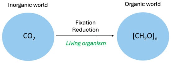
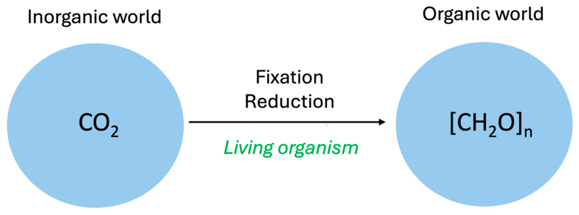
Many organic molecules can easily be formed under reductive prebiotic conditions, such as those that have prevailed in deep ocean thermal vents, for instance [2,3]. Autotrophic organisms have the ability to fix and reduce CO2, thereby setting the first steps towards the formation of organic molecules (Figure 1) [4].
Figure 1.
In living organisms, the passage from an inorganic to an organic world is strictly dependent on the fixation and reduction of CO2.
The carbon atom has the unique ability to form very stable covalent bonds with other carbon atoms or hydrogen, oxygen, and nitrogen atoms. Because of its tetravalent architecture, it can easily form stable three-dimensional assemblies necessary for the building of macromolecules.
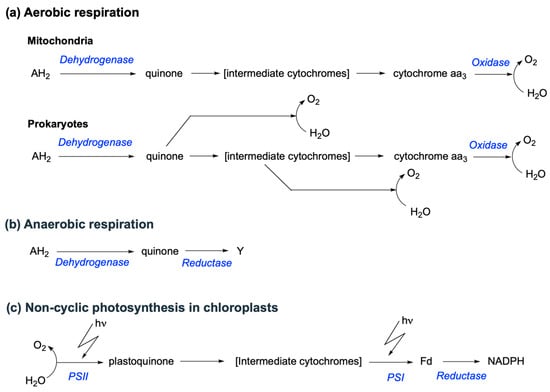
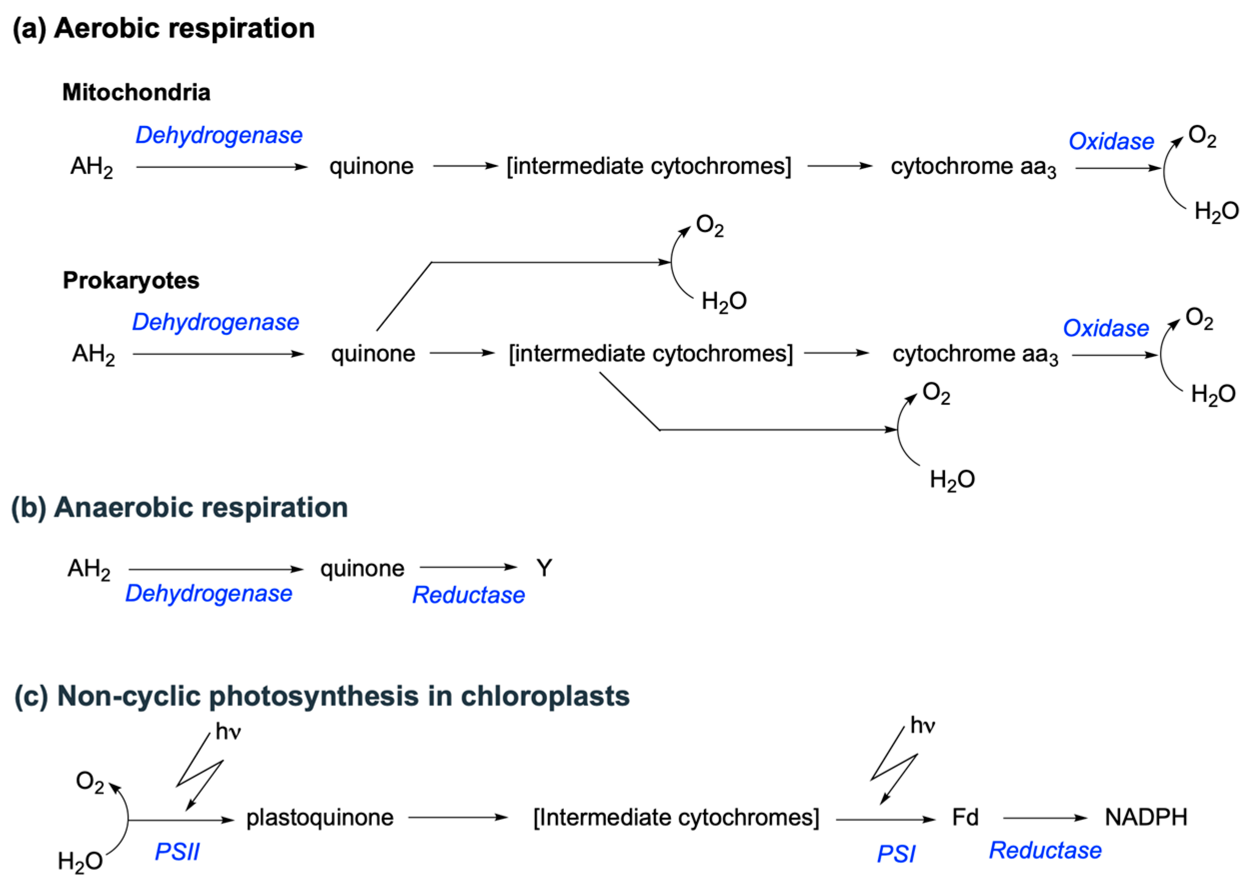
Simple organic molecules (sugars, amino acids, fatty acids…) are the building blocks of living organisms, but many are also the fuels that are consumed for energy production through redox processes. Depending on the nature of the oxidant (electron acceptor), we can distinguish between aerobic (the oxidant is O2) and anaerobic (the oxidant is an inorganic molecule different from O2 or an organic molecule) respiration (Figure 2). The energy released during the electron transfer is partially stored in a proton gradient, which is the driving force for the synthesis of ATP by proton-dependent ATP synthases.
Figure 2.
Generalized electron pathways in aerobic and anaerobic respiration and photosynthesis. In aerobic respiration (a), the electron donor (AH2, specifically a H− donor) is most commonly NADH or FADH2, and the terminal electron acceptor is O2 [5]. In anaerobic respiration (b), the terminal electron acceptor (Y) is a molecule other than O2. This can be either an inorganic (nitrate, ferric iron, sulfate, sulfur) or an organic molecule (fumarate). In these systems, the electron donor may include NADH or FADH2, H2 or organic acids like formate or lactate. (c) Schematic pattern of electron flow in non-cyclic photosynthesis in chloroplasts. The electrons are energized at the level of photosystem II (PSII), facilitating the transfer of two electrons from H2O to plastoquinone. They are energized again at photosystem I (PSI), enabling the reduction of ferredoxin (Fd). Note that when the final electron acceptor is a species other than O2, the final complex is technically referred to as a reductase rather than an oxidase.
While mitochondrial respiratory chains are highly conserved across eukaryotic organisms, prokaryotic respiratory chains exhibit remarkable diversity in their electron donors and acceptors, redox site compositions, and protein subunits. A fundamental distinction is that prokaryotic pathways are often branched toward multiple terminal oxidases. These branch points, typically located at the quinone (Q) or cytochrome levels, provide the metabolic flexibility necessary to thrive under fluctuating environmental conditions [5].
From a phenomenological perspective, photosynthesis can be viewed as the reversal of respiration. In this process, electrons flow from a high (positive) to a low (negative) reduction potential. This thermodynamically “uphill” movement is driven by light energy, which excites electrons to states with highly negative potentials.
In chloroplasts, pigments (chlorophylls) capture photon energy to excite electrons, which are ultimately used to reduce NADP+ to NADH. The resulting oxidized chlorophyll possesses an exceptionally high redox potential (+1.1 V), making it a potent enough oxidant to extract electrons from water (H2O → ½ O2 + 2 H+ + 2 e−). Thus, while respiration consumes O2 to form water, photosynthesis splits water to release O2. Similarly to respiratory chains, photosynthetic transport involves specific plastoquinones, cytochromes, and ferredoxins (Fd) as intermediates. In non-cyclic photophosphorylation, the enzyme ferredoxin-NADP+ reductase (EC 1.18.1.2) catalyzes the final step (Figure 2): 2 Fdred + NADP+ + H+ ⇌ 2 Fdox + NADPH.
Note that some organisms possess divergent respiratory chains. This is, for instance, the case of Cryptosporidium parvum with a highly minimized respiratory chain in a remnant mitochondrion called a mitosome, reflecting an adaptation to its parasitic mode of life [6].
2. Redox Reactions from Organic Chemistry to Biochemistry
Electrons and protons are at the origin of all biochemical energy. In chloroplasts and microbial lithotrophs, the energy of photons is transformed into an electrochemical reduction potential and a proton gradient that provide the energy and the redox equivalents (NADPH) for the synthesis of organic compounds (carbohydrates):
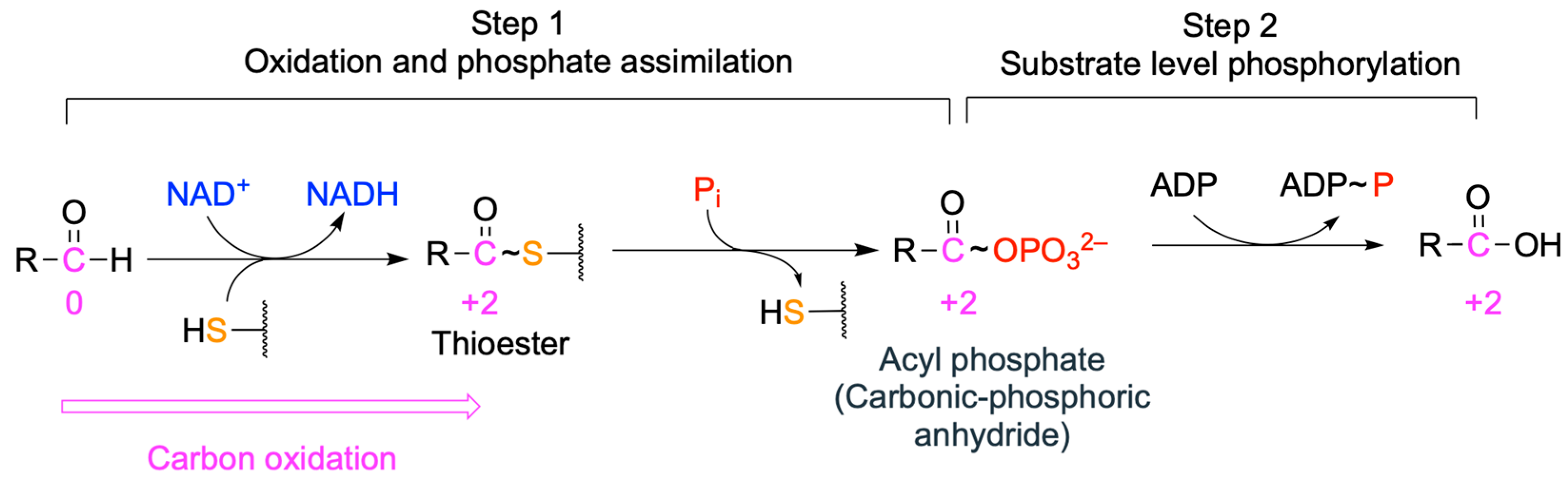
In mitochondrial and bacterial respiratory chains, the redox potential provides the drive for the establishment of a transmembrane proton gradient. Finally, proton gradients drive ATP synthesis by a chemiosmotic mechanism. Moreover, as we previously discussed, substrate-level phosphorylation ultimately relies on the energy of redox reactions too [7] (Figure 3).
Figure 3.
General mechanism for coupling the formation of a thioester to ATP synthesis as occurring in glycolysis. The first step, catalyzed by glyceraldehyde-3-phosphate dehydrogenase (EC 1.2.1.12), corresponds to the redox-dependent formation of a thioester intermediate on the enzyme, followed by the formation of 1,3-bisphosphoglycerate (acyl-phosphate). The second step corresponds to the transfer of the phosphoryl group from 1,3-bisphosphoglycerate to ADP with synthesis of ATP (substrate-level phosphorylation) catalyzed by phosphoglycerate kinase (EC 2.7.2.3). The oxidation numbers of the carbon atom are indicated in magenta. The high-transfer-potential phosphoryl group can then be transferred to ADP to form ATP [7].
Another important point generally ignored (maybe because it is considered too trivial) in all major textbooks is that, in biochemical reactions and even in whole metabolic sequences (see Equations (1) and (2)), the number of electron pair bonds (a double bond counts for two bonds) is identical between substrates and products [7]. The difference in free energy between substrates and products resides solely in their different bonding energies (i.e., different molecular rearrangement or connectivity between atoms) [7].
Electrons in the outer shells of atoms are involved in the formation of molecular bonds, and practically, all chemical reactions involve the transfer of electrons between atoms.
The association of atoms to form molecules considerably reduces the freedom of electrons that form the cement of molecular bonds. Quantum mechanics teaches us that electrons are not free to occupy any energy level but that they are limited to discontinuous transitions, which are also known as quantum leaps. Moving to a higher level requires an input of energy from the external environment, while spontaneously falling back to a lower level releases energy in the form of quanta.
The formation of bonds between elements in their standard state (pure substance in its most stable physical form at a pressure of 1 bar: for instance, pure O2 at a pressure of 1 bar) to form molecules is accompanied by a decrease in energy [7]. Therefore, the enthalpies of bond formation are always negative, and the more negative they are, the stronger the bond. Hence, the driving force for any energy-releasing reaction (∆G < 0) derives from the difference in free energy between the initial and final products: i.e., the change in bond connectivity [7,8,9]. Indeed, the replacement of weak bonds in reaction substrates by stronger bonds in product bonds results in a release of free energy. For instance, in O2, the bonding energy is particularly low, providing an explanation that combustion reactions are always accompanied by an energy release [7,9].
On the other hand, splitting molecular bonds requires an energy input. The breaking of molecular bonds can proceed either by homolytic cleavage (for instance, the dissociation of a gaseous hydrogen molecule into two hydrogen atoms: H—H → H• + •H) or, most frequently in biochemistry, by heterolytic cleavage. In the latter case, the most electronegative atom monopolizes a major part of the bond electrons and bears a partial negative charge (for instance: —Cδ−—Oδ+).
In some cases, the separation is complete: an electron is entirely lost by one atom and gained by another. A classic example, frequently cited in textbooks, is the oxidation of zinc by copper ions: Zn + Cu2+ → Zn2+ + Cu. In this reaction, two electrons are transferred directly from Zn to Cu2+. A more biochemically relevant example is the photolysis that occurs during photosynthesis: H2O → 2H+ + 2 e− + ½ O2. In the respiratory chain, cytochrome c oxidase catalyzes the reverse process: O2 + 4 e− + 4H+ → 2 H2O. This occurs through the sequential transfer of four electrons to O2 [10].
This brings us to the main subject of this review: the transfer of electrons between molecules.
2.1. The Redox State of the Carbon Atom
Redox reactions are very important in biochemistry as they are the source of ATP production. Catabolic and anabolic metabolic reactions are accompanied by a change in the state of oxidation of carbon atoms (Figure 1), though it is understood that redox reactions in biochemistry are not limited to carbon; hence, the reduction of O2 on the respiratory chain to form H2O is of course a redox reaction (Figure 2), and so is H2 production by bacteria.
Whereas, in electrochemistry, redox reactions mainly concern ionic species (e.g., Fe2+, O2−, Na+, etc.) and lead to a modification of their charge, organic redox reactions concern the covalent bonds between a carbon atom and another atom.
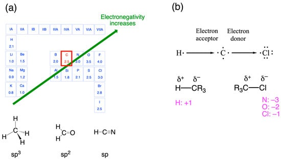
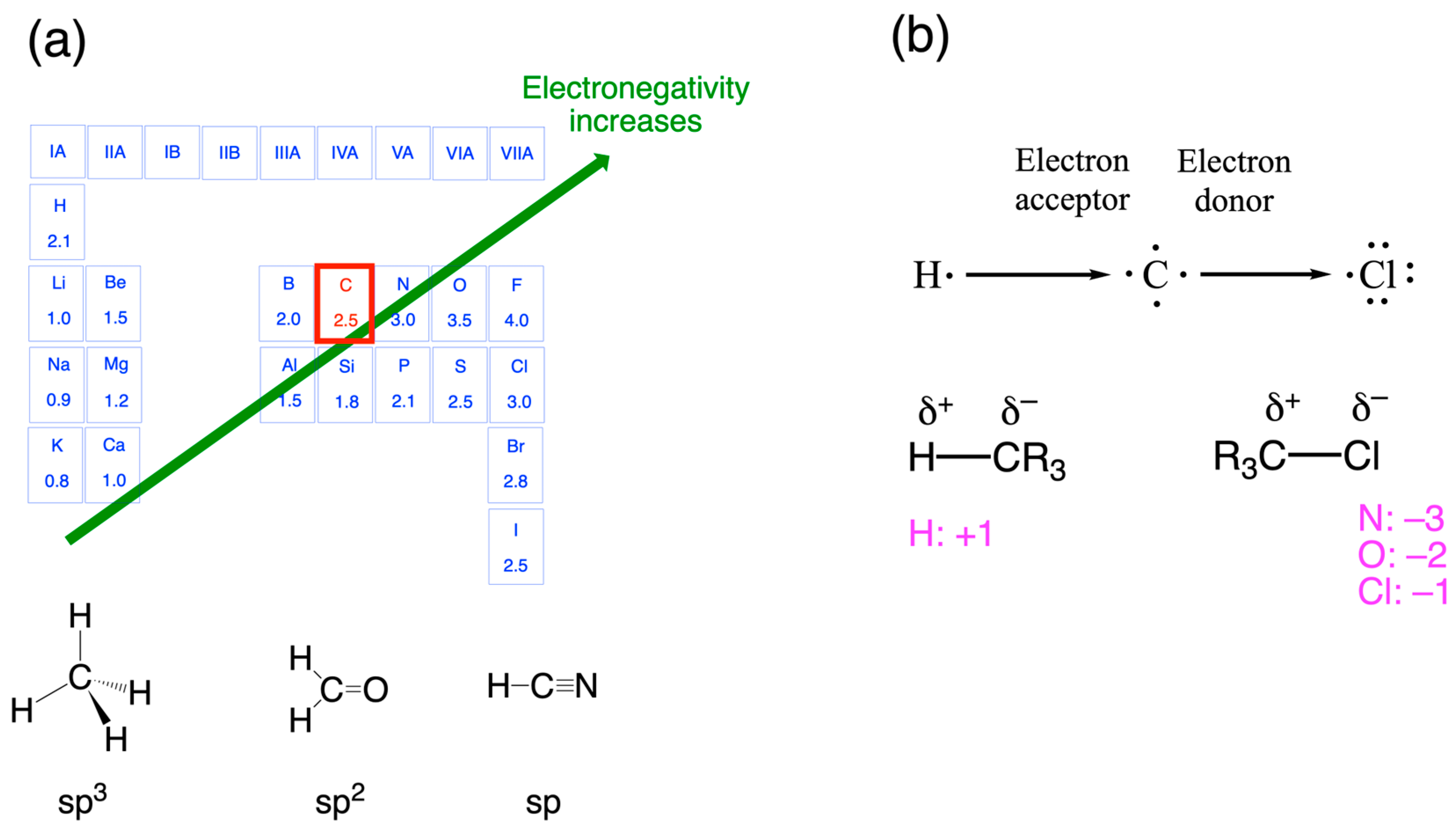
Since the carbon atom is located in the middle of the second period of the periodic table of elements (Figure 4a), it can react with either more electronegative (N, O, F, Cl) or less electronegative (H) elements. We empirically assign a positive charge to the carbon atom in the first case and a negative one in the second case (Figure 4b).
Figure 4.
Covalent bonds formed by carbon atoms. (a) The carbon atom sits in the middle of the 2nd period in the periodic table of the elements. The ground-state electron configuration of carbon is 1s2 2s2 2p2, of which the four outer electrons are valence electrons that may be in different states of hybridization (sp3, sp2 or sp1). (b) The carbon atom can be either an electron donor or an electron acceptor, depending on the substituent. By convention, the oxidation number of H is +1; those of N, O, and Cl are, respectively, −3, −2, and −1 when bound to a carbon atom.
So how can we determine the degree of oxidation of a carbon atom within a neutral molecule such as CH4 or CO2? It is obvious that, in the following irreversible reaction, the carbon atom in CH4 is oxidized to CO2, though none of the reactants is ionic (charged):
CH4 + 2 O2 → CO2 + 2 H2O
The redox state cannot, therefore, be determined by its charge (such as in Fe2+, for instance). Chemists have established a few simple rules that, in many cases, make it possible to determine the oxidation state of a carbon atom within a simple molecule [11]:
- The degree of oxidation of an organic molecule depends on the oxidation state of the carbon (reference atom);
- The sum of the oxidation numbers of a molecule is equal to the total charge of the molecule (often zero in organic chemistry);
- The oxidation number of other atoms is positive when the atom is less electronegative than carbon and negative when the atom is more electronegative than carbon (Figure 4b).
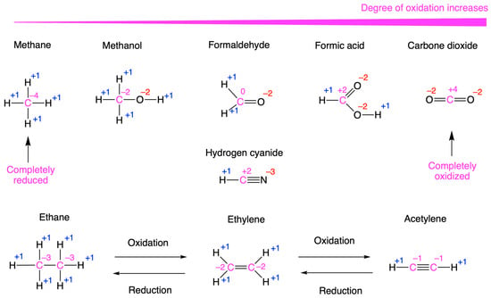
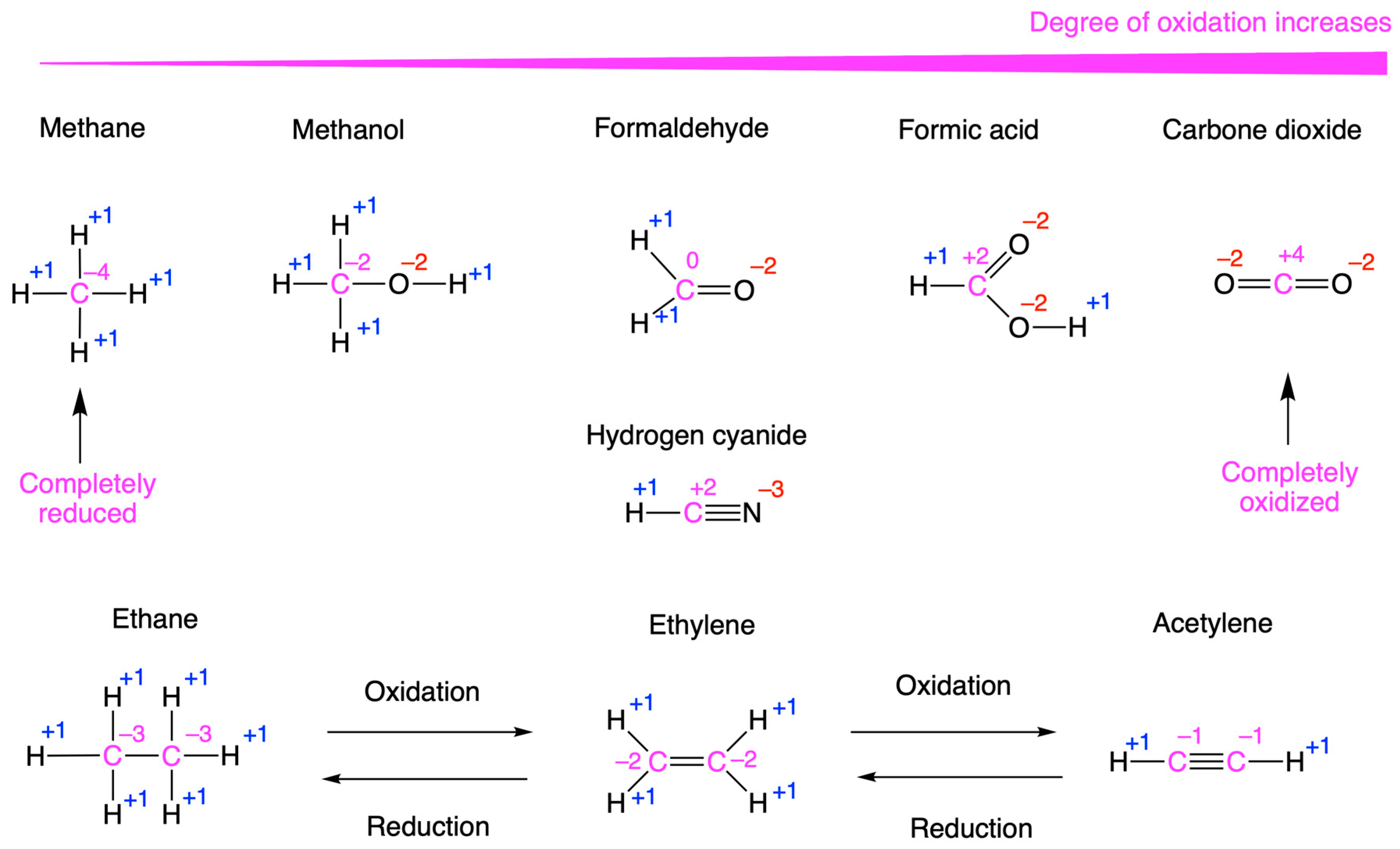
According to this method, the oxidation number of a carbon atom can therefore vary from −4 in methane (the most reduced state) to +4 in CO2 (the most oxidized state) (Figure 5). Another important point, often forgotten by students, is that replacing a single bond with a double bond consists of the oxidation of the two carbon atoms of the bond (sp3 → sp2) and that the opposite reaction corresponds to a reduction. Hence, in biochemistry, the reduction of the carbon atom generally corresponds to the gain of one or two hydrogen atoms [8].
Figure 5.
Oxidation number of the carbon atom in several organic molecules. Carbon atoms and their oxidation number are indicated in magenta. For substituents, the negative oxidation numbers are in red, and the positive oxidation numbers are in blue.
It is understood that attributing an oxidation number to a carbon atom is an empirical stratagem to tackle the stoichiometry of redox reactions in organic chemistry [8]. The carbon oxidation number, as defined here, is a fictitious quantity because there is no complete separation of charges: it is only defined with respect to the differences in electronegativity (relative affinity for electrons) between the carbon atom and its substituents. Therefore, the oxidation state of an atom should not be confused with the actual charge of an atom or molecule.
2.2. Reducing Power and Chemical Transformation in Single Organic Molecules
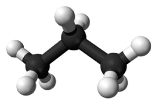
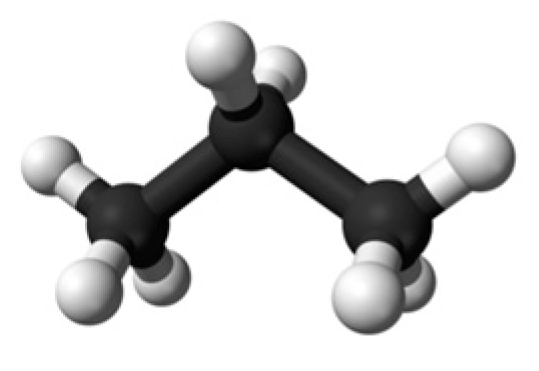
Let us now engage in a simple exercise involving propane, a very simple molecule composed of three saturated carbon atoms (Figure 6).
Figure 6.
Three-dimensional structure of n-propane. All three carbon atoms are in the sp3 hybridization state.
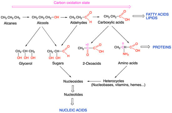
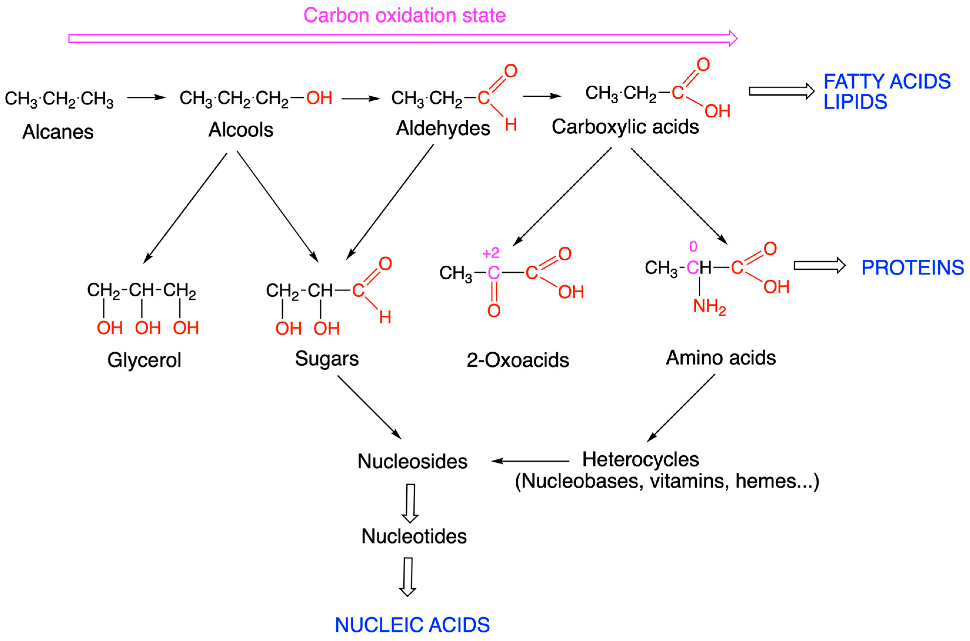
The simplest molecular modification we can make is the replacement of a hydrogen atom by an oxygen or nitrogen atom (Figure 7). In the first case, we obtain an alcohol, namely, 1-propanol. The alcohol function can be oxidized to an aldehyde (propanal) and then to carboxylic acid (propanoic acid). The lengthening of aliphatic chains leads to fatty acids, constituents of lipids such as membrane phospholipids. Note that the passage from a saturated carbon to a carboxyl group consists of a series of oxidations.
Figure 7.
Main categories of metabolites and building blocks of complex macromolecules. The oxidation number of carbon 2 in 2-oxoacids and α-amino acids is indicated in magenta.
The replacement of one hydrogen atom in each carbon atom with an oxygen atom provides glycerol, another fundamental constituent of many lipids such as phosphoglycerates. Starting with propanol, the oxidation of the remaining two carbons to an alcohol provides us with glyceraldehyde, the simplest sugar there is. Note that the middle carbon is asymmetric, and there are therefore two different enantiomers.
Another alternative is to replace a hydrogen atom with a nitrogen atom, resulting in an α-amino acid. The amino acids, in addition to being the constituents of proteins, are precursors for the synthesis of many heterocycles (nucleic bases, vitamins, hemes, etc.). The formation of a glycosidic bond between sugars and bases gives rise to nucleosides, precursors of nucleotides and nucleic acids.
Let us take our reasoning even further. From propanoic acid, we can oxidize carbon 2 into a carbonyl to obtain a 2-oxoacid (pyruvate, oxaloacetate, or 2-oxoglutarate for the most important) that plays a fundamental role in biochemistry. Indeed, the sp2 carbon of the carbonyl group, which is relatively unshielded, carries a partial positive charge, allowing a nucleophilic attack, a strategy widely used in biochemistry.
Note that the oxidation number of carbon 2 is +2 in 2-oxoacids but 0 in α-amino acids. This explains why the transfer of a —NH2 group on a carbonyl is a redox reaction catalyzed by dehydrogenases. These mostly reversible reactions (often called reductive amination or oxidative deamination processes according to the metabolically dominant direction) are encountered in many biochemical pathways (see, for instance, glutamate dehydrogenase, EC 1.4.1.2).
The three-dimensional model of propane, as illustrated (Figure 6), shows that all three carbon atoms are shielded all around by hydrogen or carbon atoms. Note that the difference in electronegativity between the carbon and hydrogen atoms is relatively small (∆Electronegativity = 2.5 − 2.1 = 0.4, Figure 4a) so that the bonds have a low level of polarization. Such a molecule has very high kinetic stability (we transport propane gas in cylinders), but its combustion in the presence of oxygen is very exergonic (∆G ≪ 0): It requires high activation energy that is not compatible with mild biochemical conditions. This same argument applies to any other (at least partially) saturated organic molecules, including the aliphatic chain of fatty acids or carbohydrates.
As we previously suggested [7], one can consider that fuel molecules store potential reducing power. The release of this reducing power under mild biochemical conditions requires molecular bond rearrangements that lead to the oxidation of their carbon atoms, powering an electron flow towards NAD(P)H, which can then drive (O2-dependent) ATP synthesis and biosynthetic processes.
As noted above, propane and other fuel molecules are kinetically stable under. Paradoxically, though highly reduced, these molecules do not have a high electron transfer potential, such as true reductants (NAD(P)H, for instance) [7]. The half-reactions of true redox reactions can be physically separated, each taking place in one compartment, with the electrons flowing from one compartment to the other. There is no such reaction with glucose or fatty acids, at least not one compatible with biological conditions.
Such a transformation from a molecule endowed with potential reducing power to a molecule that can directly be used as a reducing agent is illustrated by a sequence of reactions common to several pathways, such as the TCA cycle (succinate → fumarate → malate → oxaloacetate) and β-oxidation of fatty acids (Figure 8).
Figure 8.
Preparation of a saturated carbon for nucleophilic attack. Such a sequence involving reduction and formation of a double C=C bond, followed by hydration and oxidation of the alcohol to a carbonyl, is a recurring sequence observed during the last part of the TCA cycle or the ß-oxidation of fatty acids. A practically reversed reaction sequence is observed during the synthesis of fatty acids [4].
The reduced tetrahedral sp3 target carbon is transformed to a trigonal planar sp2 carbon in the 2-oxoacid, allowing nucleophilic attack. During this process, reducing equivalents are gradually transferred to FAD and NAD+. This pattern is mechanistically remarkably similar to the steps involved in ß-oxidation of fatty acids. Note that this sequence would correspond to the passage from a carboxylic acid to a 2-oxoacid, as illustrated in Figure 7.
The examples quoted above nicely illustrate how, starting from a very simple propane molecule, a few modifications allow the reconstitution of the main groups of molecules of life, as well as a strategy for extracting reducing power from a precursor that is otherwise inert under mild biochemical conditions.
This example emphasizes the concept that the essence of life lies in the way molecules are transformed into each other, namely through metabolism. It also shows that most of the basic transformations involve a change in the redox state of organic molecules.
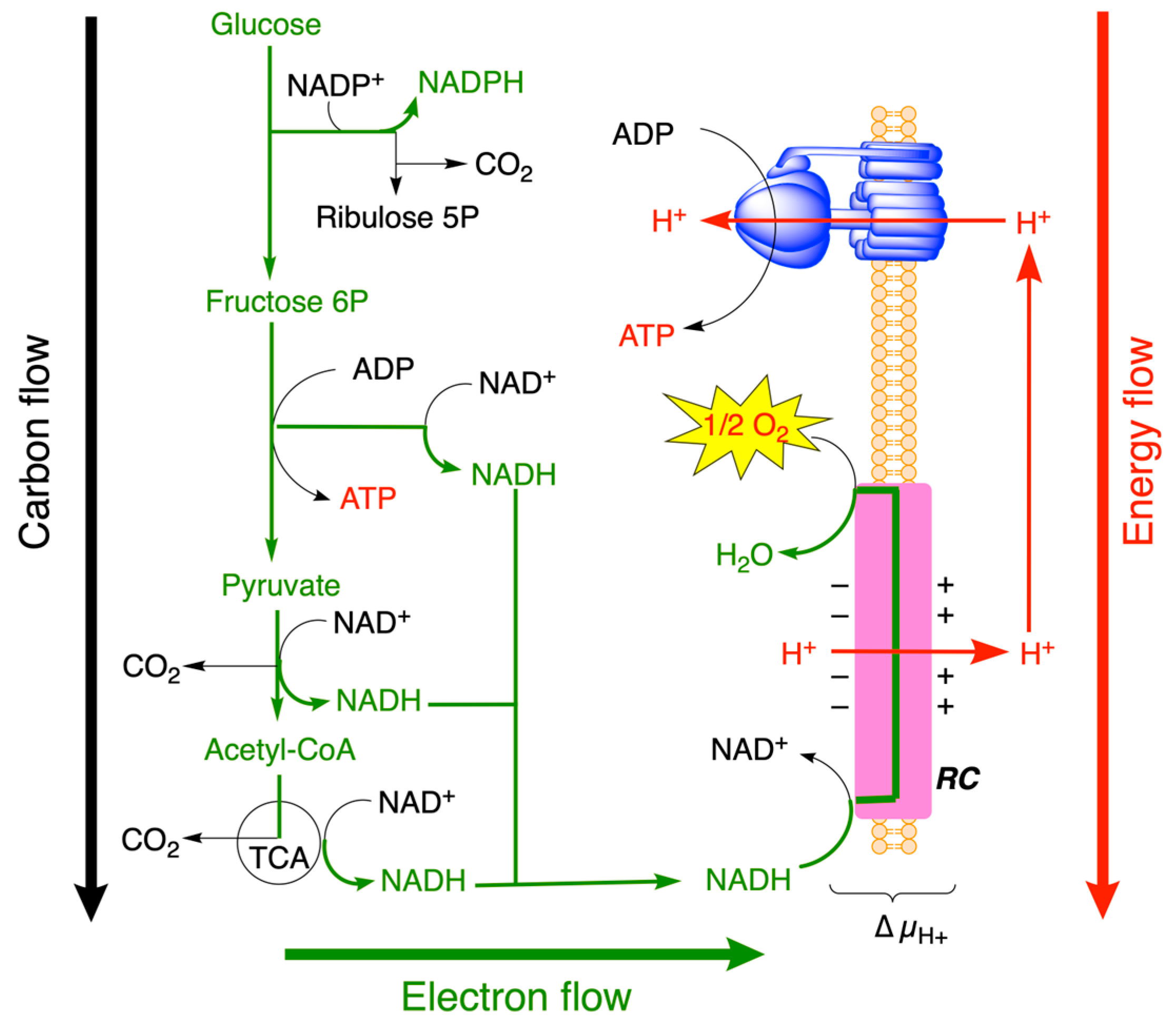
3. Carbon, Electron, and Energy Flows Through Central Catabolic Pathways
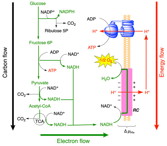
As previously discussed [7,8], the oxidation of partially reduced organic fuel molecules to CO2 gives rise to the following (Figure 9):
Figure 9.
Catabolic flows of matter (carbon), energy, and electrons through the central energy metabolic pathways (glycolysis, tricarboxylic acid (TCA) cycle, and pentose phosphate pathway (PPP)). For simplification, no subcellular compartmentations are indicated, and the scheme may concern any prokaryotic or eukaryotic organism. The driving forces (∆G’, defined at pH 7) depend on differences in concentrations (Q) and the structural rearrangement between initial and final reactants (∆G’°] and redox potential (∆E’) (adapted from [7]).
- (1)
- A (metabolic) flow of matter (actually, the transformation of carbonated molecules, rather than a true vectorial process).
- (2)
- A flow of electrons from fuel molecules to NAD(P)H and FADH2.
- (3)
- A flow of energy.
The final carbon product (CO2) is in a higher oxidation state than the initial reactants: CO2 is the most oxidized form of carbon (oxidation state +4), while organic molecules are hydrogenated and therefore in a lower oxidation state than CO2. During metabolic activity, electrons (usually in the form of hydride anions, H−) are transferred to NADPH and NADH. The latter is oxidized in the respiratory chain by O2, and the electrons are transferred to H2O (Figure 9). Just as some reactions are coupled to ATP synthesis, many redox reactions are coupled to the synthesis of reducing equivalents (NADH and FADH2). It is important to remember that no compound can lose an electron without an acceptor.
O2 has a particularly weak double bond that, when substituted by stronger bonds (H2O or CO2) in a reaction (see Equation (2)), will result in the release of a considerable amount of free energy [7,9,12].
It is intellectually satisfying to observe that nature has associated a specific type of molecule with each of the metabolic flows. Carbohydrates undergo chemical transformations and oxidation of their carbon atoms. NAD(P)H is the main vector of reduction equivalents and supports the “electron flow”, while “energy-rich” molecules, nucleoside triphosphates and O2, store chemical energy to release it on demand.
4. Electron Transfer in Biochemical Reactions
4.1. General Considerations on Electron Transfer in Biochemical Reactions
As we explained above, fuel molecules, such as glucose or fatty acids, are partially reduced molecules that are oxidized during catabolism (Figure 7). Hence, this catabolic activity gives rise to a flow of electrons from the fuel molecules to only a few reduced coupling intermediates: NADPH (oxidative portion of the pentose phosphate shunt), NADH and FADH2 (glycolysis and TCA cycle), and ferredoxins ([Fe-S]: cluster proteins involved in electron transfer reactions in photosynthesis and bacterial metabolism). In the respiratory chain, NADH and FADH2 are oxidized by O2, a process coupled to ATP synthesis through a chemiosmotic mechanism. In contrast, NADPH is involved in the transfer of reducing equivalents from catabolism to power anabolic reactions.
Before pursuing our research further, it is not unnecessary to remind the reader of the origin of the notions of high and low electrostatic or redox potential. Indeed, in physics, the conventional direction of currents corresponds to the flow of positive charges, though the charge carrier is most often electrons. Hence, the current flows from a region of high (+) potential, where positive charges have the highest potential energy, to a region of low potential energy (less positive or more negative).
All redox reactions require a transfer of electrons from a donor (reductant) to an acceptor (oxidant). In metals, which have high electrical conductivity, electrons can move relatively freely from a low (negative) electrostatic potential to a high (positive) electrostatic potential. This is not the case in solutions. There are no free electrons in solutions, and electrons are transferred from a donor molecule to an acceptor molecule, directly or indirectly, through enzymes, coenzymes, or proteins. Indeed, in biology, electrons are transferred between organic molecules and adjacent proteins or through proteins (oxidoreductases) such as the multienzyme membrane complexes of the respiratory chain and photosystems.
Though the notion of electron flow is essentially correct from a phenomenological point of view, in biological systems, however, electron transfer is always local, as electrons are transferred only over very short distances (typically in the nanomolar range) from one complex to another (see below [13,14]). Electron transfer is critical for driving essential processes such as respiration, photosynthesis, and bacterial proton pumping.
Some authors make a distinction between electron transport and electron transfer [14,15]. In both cases, the driving force is the electrochemical potential, but while electron transport is purely electrical (electron flow through a wire or across a protein in the absence of a redox reaction), electron transfer is a chemical reaction with at least two redox sites [14]. Electron transport is generally measured in a solid phase between two electrodes [14,15], and it is not clear to what extent such a phenomenon is useful in biochemistry, as, to our knowledge, electron flow is always accompanied by the oxidation of the donor and the reduction of the acceptor.
However, many redox reactions are more than just the transfer of electrons from one redox center to another. Redox reactions can happen with a change in connectivity, in which case the driving force is the difference in bond energies between products and substrates, and the difference in bond energies between products and substrates could be the main contribution to the overall ∆G of the reaction [9].
While the transfer of electrons from one redox site to another can proceed either straightforwardly (by tunneling or hopping) or in a more complex fashion in a chemical reaction involving bond (connectivity) changes, the reaction always proceeds toward lowering the free energy of the system (∆G < 0).
Electron bifurcation (see Section 4.4) represents a particular type of reaction where an exergonic redox reaction is coupled to an endergonic one. But the overall free energy change is often close to 0, making these reactions reversible.
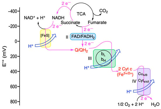
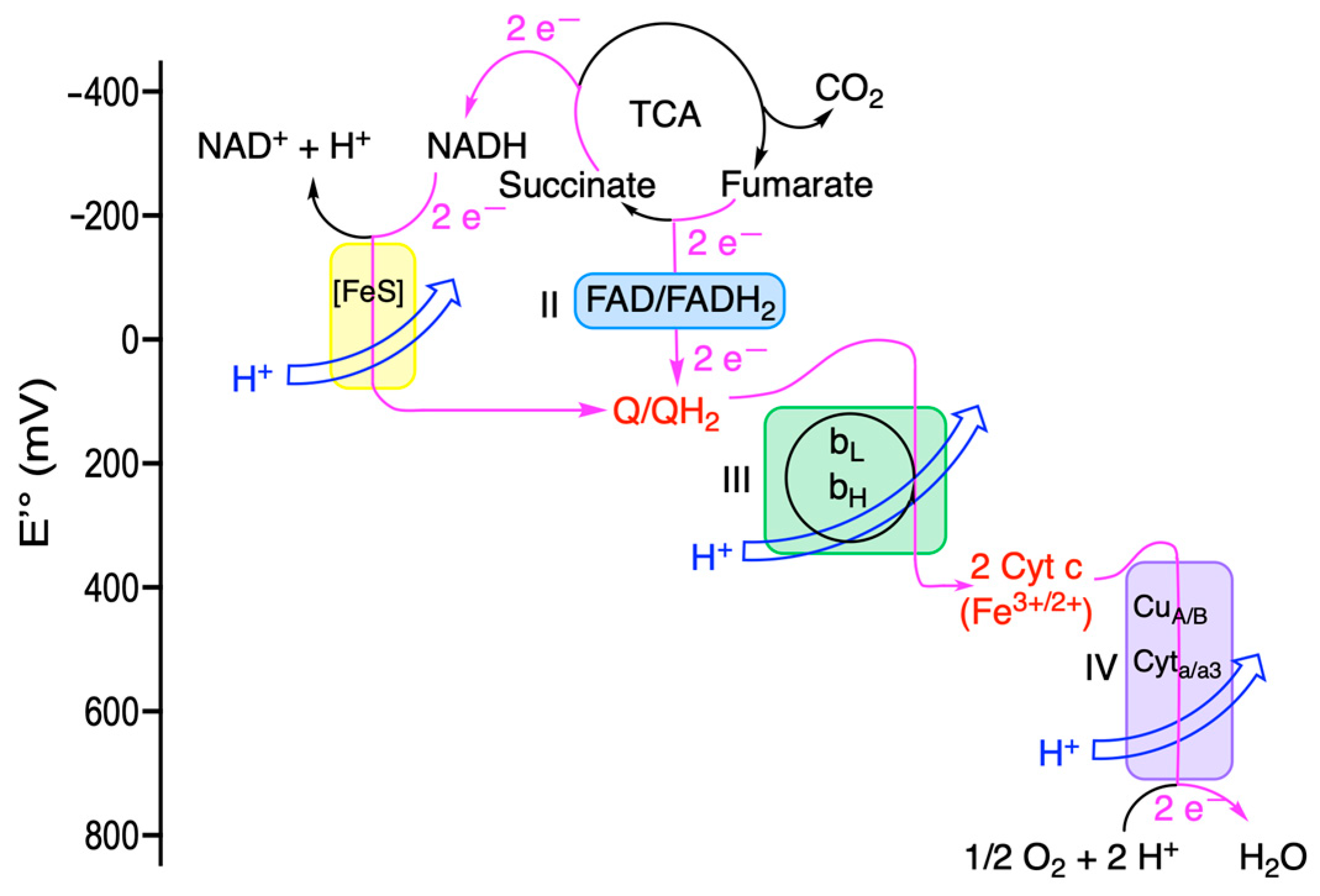
These concepts are clearly illustrated by the mitochondrial respiratory chain (Figure 10).
Figure 10.
Electron flow in the mitochondrial respiratory chain (from [8]). The first reactions involve a transfer of H– + H+ towards the acceptor Q. On the other hand, complexes III and IV act more like wires, conducting electrons coupled to proton pumps. This is, of course, only a phenomenological interpretation, as there is no continuous flow of electrons like in a wire, but the electrons hop from one redox site to the other (see below). Mobile carriers are indicated in red. Electron flow is shown in magenta lines.
Reducing equivalents are captured in the form of NADH or succinate (rather than fumarate) generated during the TCA cycle. Electrons from NADH enter complex I (NADH dehydrogenase), which shuttles them toward the ubiquinone (Q) pool via a series of iron–sulfur (Fe-S) clusters [16]. While the oxidation of NADH or succinate involves changes in molecular connectivity, the subsequent transfer of electrons through protein complexes and their various redox centers occurs via electron hopping or tunneling.
Succinate is oxidized to fumarate by complex II (succinate dehydrogenase); its two electrons are also used to reduce ubiquinone via an intermediate FAD/FADH2 couple. These electrons are then sequentially transmitted to complex III through a mechanism of quinone-based electron bifurcation. This process involves two heme groups of cytochrome (heme bL and heme bH) and ultimately results in the reduction of two mobile cytochrome c carriers (see Section 4.4.1).
The reduced cytochrome then docks at complex IV (cytochrome c oxidase), where its electrons are transferred to the final acceptor, O2, via two copper centers (CuA and CuB) and two cytochromes (a and a3). Quantum mechanical calculations indicate that heme-to-heme electron transfer is consistent with quantum tunneling [17]. The complete reduction of one O2 molecule requires the sequential transfer of four electrons from four reduced cytochrome c molecules [10].
The complexes of the respiratory chain are targets for several clinically significant inhibitors. Complex I is inhibited by rotenone; chronic exposure to this toxin has been linked to an increased risk of Parkinson’s disease [18]. Complex IV is targeted by potent inhibitors such as cyanide (CN−), azide (N3−), and carbon monoxide (CO). Notably, all four mitochondrial complexes are strategic targets in oncology, as cancer cells often rely heavily on the respiratory chain to meet the energy and biosynthetic demands of rapid growth [19].
4.2. Electron Transfer by Tunneling or Hopping
When looking at Figure 10 or at a similar figure in biochemical textbooks, one is left with the impression that the electrons simply “flow” from a low redox potential (the negative electrode) to a higher potential site so as to minimize free energy. But be reminded that there are no free stable electrons in aqueous media and protein complexes. The respiratory chain or the photosynthetic systems are discontinuous media where electrons are transferred from one site to the other over very small distances.
In classical mechanics, an electron is represented as a particle and behaves as such. When it encounters a potential energy barrier with higher energy than its own, it eventually ricochets off the barrier: It can only pass the barrier with sufficient activation energy. In principle, an electron donor (NADH, QH2) is oxidized by a metal center or a heme group, with the electron hopping through the transmembrane protein from one redox site to the next until it reaches another soluble acceptor (Figure 11a). Hopping, which is common in multicenter transmembrane proteins such as cytochrome complexes, flavoprotein, or iron–sulphur clusters [13], allows the transfer of electrons over longer distances (10–30 Å).
However, it was found that, in a number of cases, the transfer of electrons is independent of temperature, an observation thought to be incompatible with a transfer in the form of hopping. The possibility of tunneling transfer was then raised [13,14,20].
In quantum mechanics, a subatomic particle is represented by a wave function, and there is a certain probability that the wave function can cross an energy barrier that is not infinitely high or wide (Figure 11b). This phenomenon is called the tunneling effect. “Tunneling” can only occur with a certain probability when the particle is light enough. In practice, it is therefore limited to electrons and protons. Proton tunneling is probably limited to hydrogen bonding in biology and could be significant in base pairing in DNA [21]. Indeed, in most cases, rather than the proton, it is its charge that is transferred (by permutations of covalent and hydrogen bonds) via a network of hydrogen bonds: the so-called Grotthuss mechanism [22,23].
Electron tunneling is only possible between centers less than 10–14 Å apart [15,20], but it occurs very rapidly with rate constants (kelectron transfer) of the order of 106–109 s−1 so as not to be rate-limiting in enzyme kinetics [17,24].
Figure 11.
Transport of electrons through protein complexes and the tunnel effect. (a) Electrons do not exist in isolation: They can only be located at the donor site or at the acceptor site, never in between. In classical mechanics, they behave as particles and hop from one site to the other. Tunneling implies a residence time of less than 1 femtosecond near any nucleus, which is consistent with a current flowing through the conducting protein over distances of about 2.1 nm [14,24]. (b) The concept of electron tunneling. The red arrow illustrates the pathway of an electron with sufficient energy to pass over the activation barrier (black line). The blue line illustrates the tunneling of an electron (or rather its wave function) through the activation barrier.
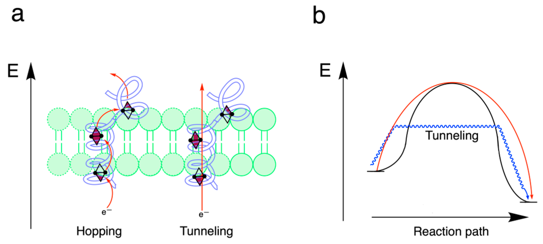
The important point here is not so much to understand the exact mechanism by which an electron passes through a protein (which would exceed the scope of this review) but to realize that this mechanism is, from a phenomenological point of view, comparable to the conduction of an electron along a conducting wire where the driving force is equal to the redox potential difference (∆G = −nF ∆E). This contrasts with redox reactions with connectivity changes, which we will address now.
4.3. Redox Reactions with Connectivity Change
As discussed above, electron flow may be appropriate for a few simple half-reactions involving only a change in the charge of an ion (e.g., from Fe3+ to Fe2+). The motion of electrons is usually limited to the reduction of metals, mainly Cu2+ and Fe3+, the latter very often in iron–sulfur clusters or hemes (cytochromes, for example, Figure 10) within protein complexes. This is because electrons only transfer energy when they move through a “wire”, which, in the case of respiratory chain complexes, powers the transport of H+ to the internal compartment (matrix) by respiratory complexes I, III, and IV [9]. Put differently, electrons are not at the origin of the underlying exergonic redox reaction, but on the contrary, they are driven by the difference in free energy between the reactants and the products.
However, when the chemical connectivity between atoms and molecules changes, for example, in the reaction O2(g) + 4 H+(aq) + 4 e− (or 2 H+ + 2 H−) → 2 H2O(l), the difference in binding energies of reactants and products often makes a dominant contribution to the free energy of reactions and drives the electron flow [9].
Another important point is that in biochemical redox reactions involving organic compounds, electrons are very often not transferred as is but as hydride anions (H−, or 2 e− + H+) (see, e.g., oxidation of NADH and FADH2 or the reduction of glutathione), hence the name “dehydrogenases” for the enzymes catalyzing such reactions. That is why the reduction of a carbon atom is, in many cases, equivalent to hydrogenation. Indeed, from Figure 5, it is evident that the oxidation state of carbon in organic molecules varies by increments of two, justifying the transfer of two reducing equivalents.
Often, reduction potentials are simply considered to be intrinsic properties of molecules or ions, when in fact they derive from the binding, ionization, and hydration energies of conjugated oxidized and reduced species [9].
4.4. Electron Bifurcation
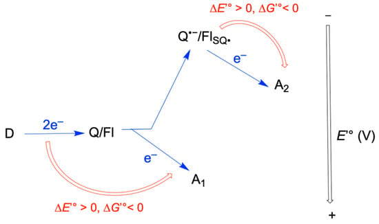
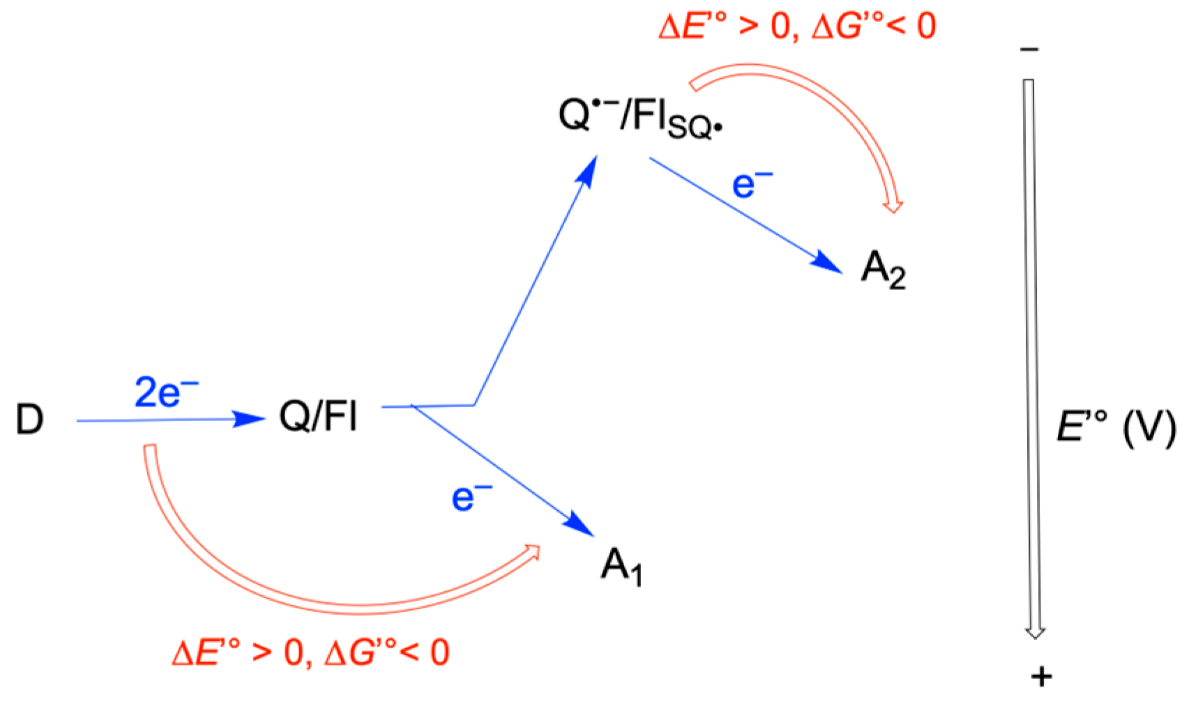
In biochemistry, the term “electron bifurcation” describes the coupling of exergonic and endergonic chemical reactions. In this process, a two-electron donor transfers two electrons to two separate one-electron acceptors with widely divergent redox potentials, minimizing the dissipation of energy in the form of heat (Figure 12).
Figure 12.
General model of electron bifurcation. First, the electron donor (commonly NAD(P)+, FADH2, or H2) transfers 2 electrons to the bifurcating quinone/flavin (Q/Fl). The reduced bifurcation quinone/flavine transfers one electron to the high-potential (more positive) acceptor A1 (∆G < 0). This creates a semi-reduced radical intermediate (Q•−/FlSQ•) containing a second electron that is less tightly bound—meaning that it possesses a redox potential lower (more negative) than that of the initial donor D. In the subsequent step, this radical donates its remaining electron to the low-potential acceptor A2 (∆G < 0).
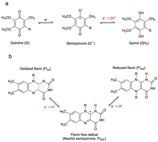
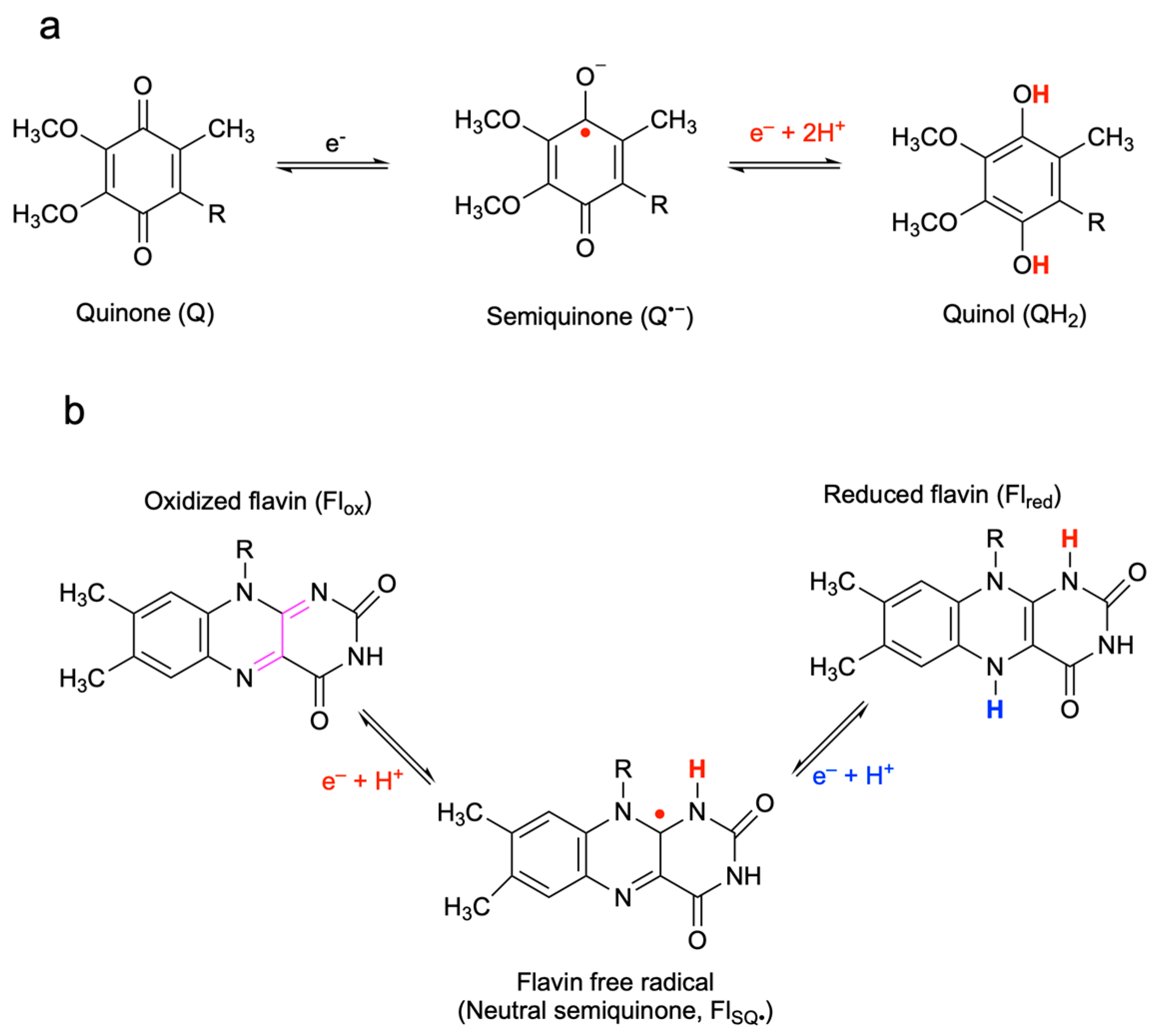
The initial two-electron donor (such as NADH) transfers its electrons to a specialized bifurcating quinone or flavin (Figure 13). This cofactor can exist in three states: fully reduced (two electrons), a semiquinone intermediate (one electron), or fully oxidized (0 electrons). The mechanism operates through a sequence of “crossed” redox potentials. The first electron is transferred to a high-potential acceptor (A1). This is a highly exergonic (downhill) step. This first transfer leaves behind an unstable, low-potential semiquinone radical. Crucially, the redox potential of this semiquinone is much lower (more negative) than that of the original fully reduced species. This extremely “reductive” power allows the second electron to be transferred to a low-potential acceptor (A2), driving an endergonic (uphill) reaction.
Figure 13.
The catalytic cycles of quinones and flavins. (a) The two steps of the reduction of quinone (Q) to quinol (QH2) via a negatively charged semiquinone (Q•−). (b) Structure of the flavin heterocycle and its three states: oxidized (Flox), semi-reduced neutral (FlSQ•, a radical), and reduced (Flred). Electron bifurcation is only possible because of the crossed-over redox potentials of quinones and flavins. This means that the Q/Q•− and Flox/FlSQ• couples have a more negative midpoint potential than the SQ•−/QH2 and FlSQ•−/Flred couples. In other words, the semiquinones have more reducing power than the totally reduced species. The unpaired electrons are indicated as red dots.
The defining feature of electron bifurcation is this concerted flow: The energy “dropped” by the first electron effectively “pushes” the second electron uphill. This tight coupling ensures that the overall reaction remains thermodynamically spontaneous while conserving energy that would otherwise be lost.
First, the electron donor—commonly NAD(P)H, FADH2, or H2—transfers two electrons to the bifurcating quinone or flavin (Q/Fl). The reduced cofactor then transfers one electron to the high-potential (more positive) acceptor A1, a highly exergonic step (ΔG ≪ 0). This creates a highly reactive semi-reduced radical intermediate (Q•−/FlSQ•) containing a second electron that is less tightly bound. Consequently, this second electron possesses a redox potential that is significantly lower (more negative) than that of the initial donor D. In the subsequent step, this “energized” radical donates its remaining electron to the low-potential acceptor A2 (ΔG < 0).
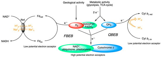
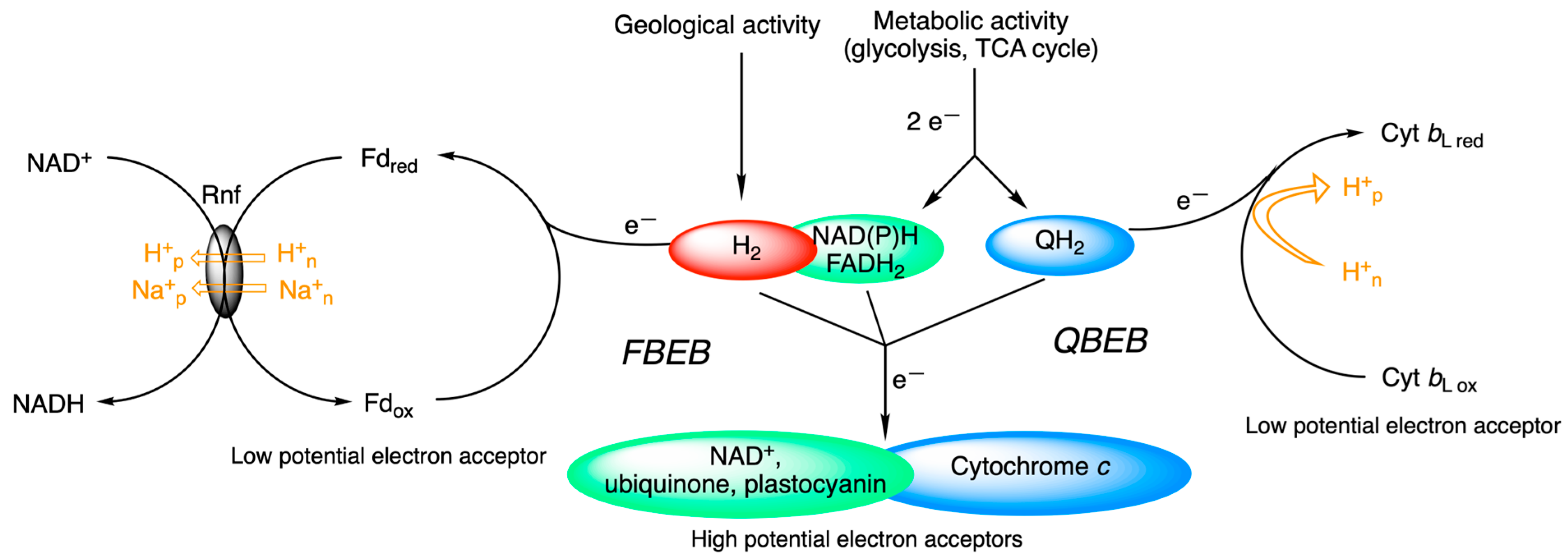
Electron bifurcation is a very fundamental mechanism that probably played a primary role in proto-metabolism and in the development of life [25]. It is a fundamental energy-coupling mechanism in anaerobic microorganisms that likely originated in methanogens near geological, iron-rich, serpentinizing hydrothermal vents. During this process, electron pairs extracted from geological H2 allow thermodynamically uphill reductions (e.g., reduction of ferredoxin) to drive carbon fixation and create a proton gradient [25,26,27] (Figure 14). As already recognized by Peter Mitchell [28], electron bifurcation also plays a fundamental role in photosynthesis and mitochondrial respiration through the Q cycle of b6f and bc1 complexes [29,30,31].
Figure 14.
Electron bifurcation in metabolism. In FBEB, common two-electron donors are NAD(P)H or H2. The low-potential acceptor is often ferredoxin (Fd). In some anaerobic bacteria, reoxidation of Fedred by NAD+ is coupled to H+/Na+ translocation by H+/Na+-translocating ferredoxin: NAD+ reductase (Rnf) [32]. In QBEB (in mitochondrial bc1 complex, for instance), one electron derived from metabolic activity through NADH or FADH2 directly goes to the acceptor cytochrome c, while the second electron is recycled back via cytochrome b for another round. The whole process is coupled to proton pumping. p, Positive side of the membrane; n, negative side of the membrane.
The delocalized π-electron system of flavins favors the stabilization of the semiquinone form, allowing a sequential transfer of the two electrons. This makes flavins ideal intermediates between one-electron reductors (Fe-S complexes, for example) and pyrimidine nucleotides NAD(P)+/NAD(P)H (see below).
While quinone-based electron bifurcation (QBEB) is inherently involved in the establishment of electrochemical proton gradients through the Q-cycle, flavin-based electron bifurcation (FBEB) is mainly used to couple exergonic reactions (NAD+ reduction) to endergonic reactions such as ferredoxin reduction. Establishment of a H+ or Na+ gradient can be a secondary event coupled to the reoxidation of ferredoxin (Figure 14).
4.4.1. Quinone-Based Electron Bifurcation (QBEB)
The Q cycle, first proposed by Peter Mitchell in 1975 [28] for the mitochondrial respiratory chain, is a typical example of QBEB (Figure 15). During a complete cycle, quinones assume the role of H+ transporters, releasing 4 H+ in the intermembrane space [33]:
QH2 + 2 cytochrome c (Fe3+) + 2 H+in → Q + cytochrome c (Fe2+) + 4 H+out
Figure 15.
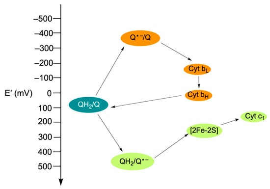
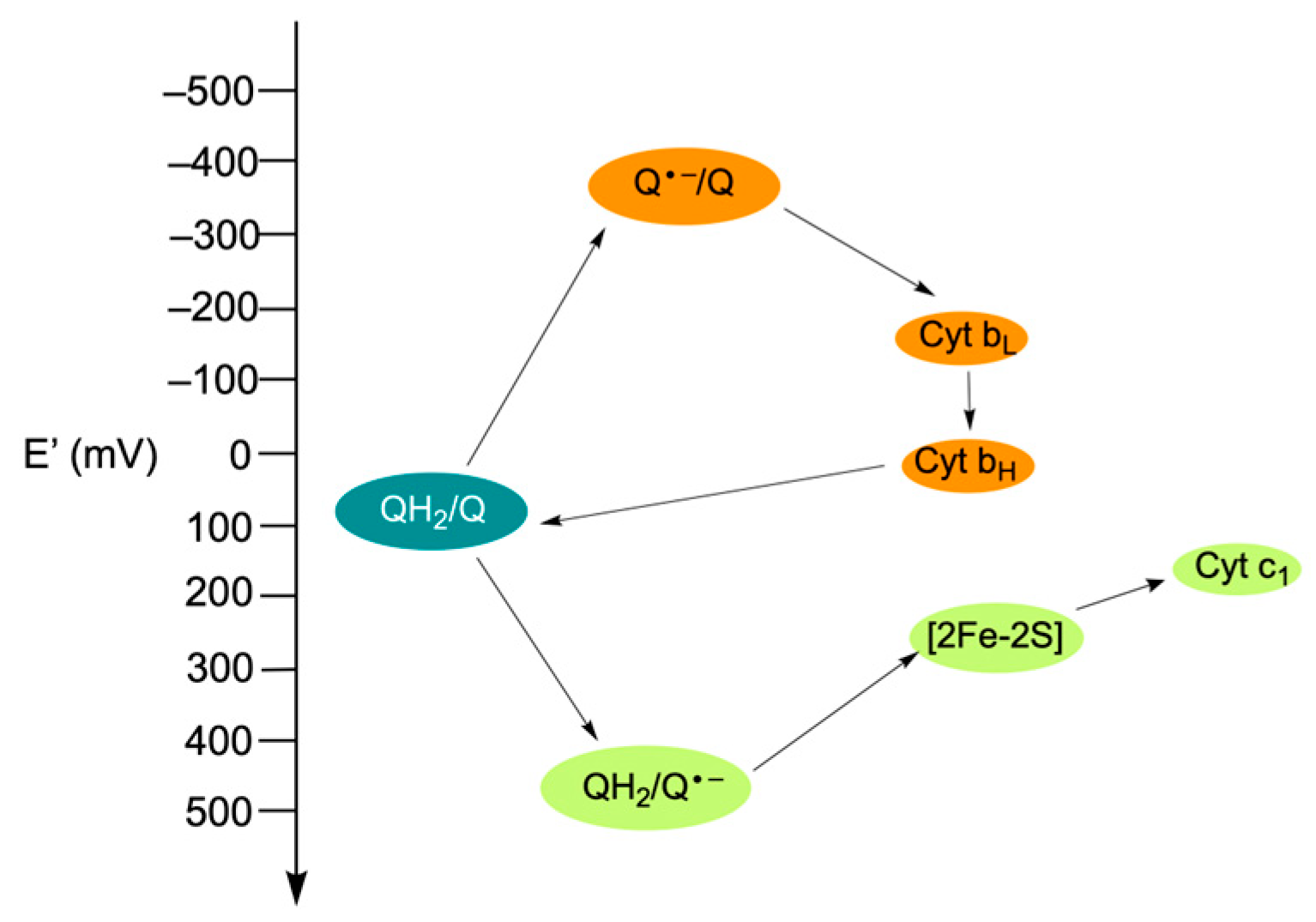
Redox potential (E’) and electron transfer in the Q cycle. The redox potentials E’ are at pH 7, taking into account actual cellular conditions. Modified and redrawn from [34].
The quinone (Q) and quinol (or hydroquinone, QH2) family of molecules form important redox pairs in many biological systems. A semiquinone radical (Q•−) functions as intermediate between the oxidized and reduced forms.
Note that the redox potentials are crossed: The oxidation of the quinone to a semiquinone, Q•−, generates a more potent reductant (E’ = −320 mV) than the initial QH2 (E’ = 90 mV) and is able to reduce cyt bL. This property is important for electron bifurcation, as will be discussed below. An important question to ask is what prevents Q•− from also reducing the [2Fe-2S] center? In order to prevent the two electrons from following the same path to the high-potential acceptor, in this case, [2Fe-2S], unequivocal routing of the electrons is an essential part of this mechanism. This is accomplished, in particular, by a change in the conformation of the complex by moving the Q•– oxidation site away from the [2Fe-2S] center.
This example perfectly illustrates the concept of cross-redox potentials, without which the mechanism of electron bifurcation could not work. In the conventional mode, both electrons reduce a higher potential (less negative) acceptor, making electron bifurcation impossible. Inversion of this order ensures that the first electron can only be removed by a high-potential acceptor, which gives the remaining semiquinone a very low reduction potential able to reduce any nearby low-potential acceptor [35].
We already discussed another point deserving our attention once again (see Section 4.3). Indeed, very often, redox reactions are presented as if the ∆E° of a chemical reaction results only from an electron moving from a lower potential to a higher potential. This is true during the passage of the electron from the center [2Fe-2S] to cyt c1 or from cyt bL to cyt bH, which can be assimilated to the transfer of the electron from one site to another without modification of the molecular bond: only the oxidation state of an iron ion is changed [17,36,37].
However, when we consider the electron transfer reaction of QH2 towards the cyt c1, it is evident that, in addition to the change in the oxidation state of the iron ion, the connectivity of atoms in coenzyme Q is altered. As emphasized above, the difference between the binding energies between the products and the reactants plays at least as important a role as the potential of the electrons [9].
4.4.2. Flavin-Based Electron Bifurcation (FBEB)
FBEB is a recently discovered mechanism in many strict anaerobic bacteria and archaea [35,38]. FBEB depends on a flavin as a cofactor (FAD or FMN). Flavins can exist in three oxidation states: fully oxidized (Flox), partially reduced semiquinones (FlSQ•), and completely reduced hydroquinone (Flred) (Figure 13). This makes flavins ideal candidates for accepting a pair of electrons and then transferring them sequentially.
High-potential terminal one-electron acceptors are multiple and depend on the organism and the metabolic pathway involved (NAD+, crotonyl-CoA, or ubiquinone, for instance). Ferredoxins act as a low-potential terminal acceptor, and the reduced ferredoxin generated can then be used by membrane-bound complexes like Rnf (Fd:NAD reductase) [32] or Ech (Fd:H+ reductase) [35] to generate a proton or sodium motive force (ΔμH+/ΔμNa+) (Figure 14).
Ferredoxin: NADP+ NADH-dependent oxidoreductase (EC 1.6.1.4) is involved in the regulation of the NADH/NADPH balance and represents the prototype of an FBEB enzyme [39,40]. It reversibly catalyzes the simultaneous oxidation of NADH and ferredoxin to produce NADPH:
2 NADP+ + Fd2−red + H+ + NADH ⇌ NAD+ + 2 NADPH + Fd0ox ∆G’° = −20 kJ/mol
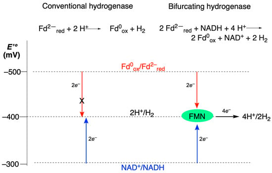
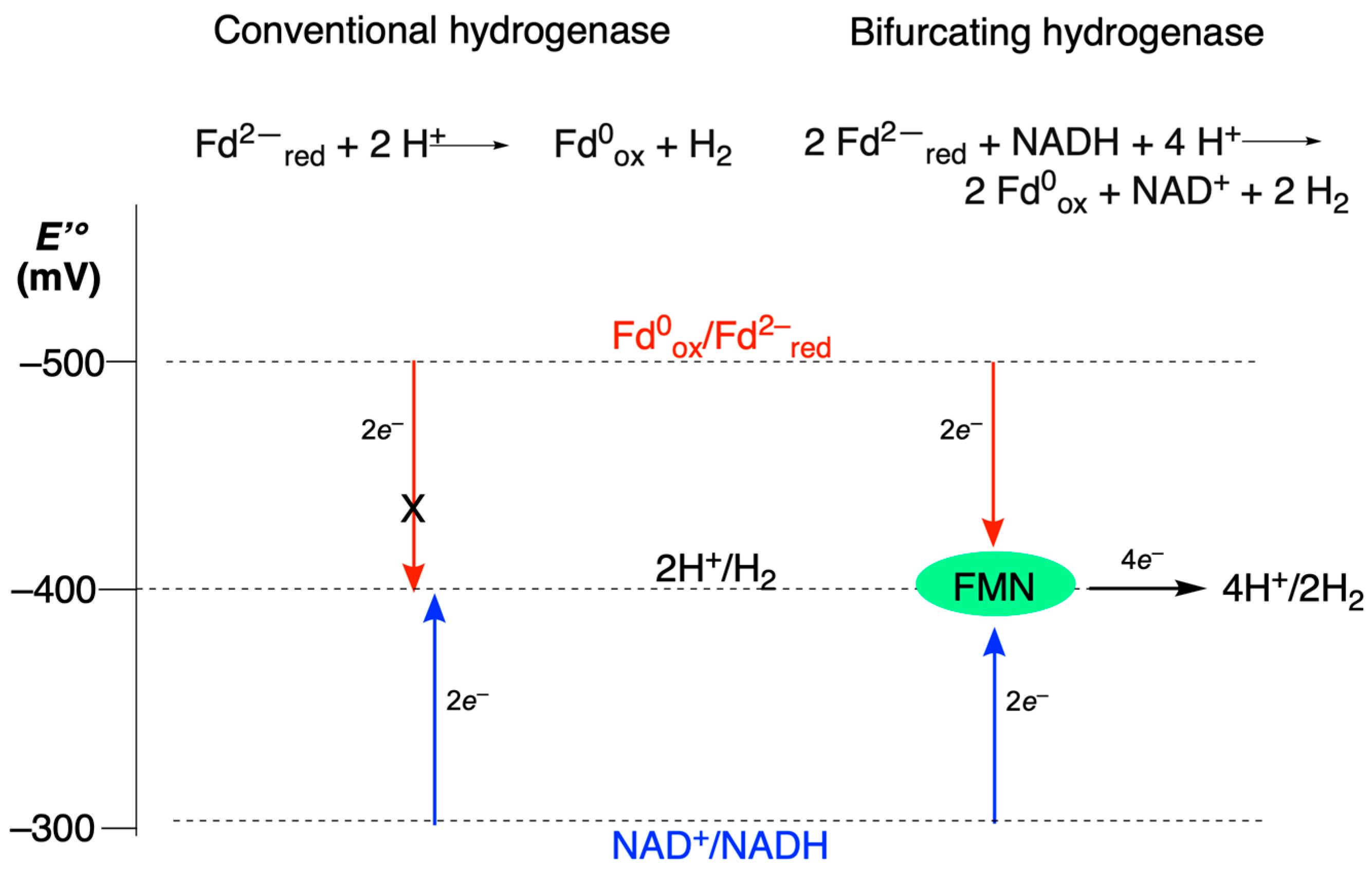
Figure 16 shows a comparison of the reactions catalyzed by a conventional non-bifurcating and a bifurcation hydrogenase [41] (2 Fd2−red + NADH + 4 H+ ⇌ 2 Fd0ox + NAD+ + 2 H2; EC 1.12.1.4).
Figure 16.
Flavin-based electron bifurcation. Comparison of the reactions catalyzed by a conventional non-bifurcating dehydrogenase (left) and a H2-producing bifurcating hydrogenase (right) [27]. In conventional dehydrogenase-catalyzed reactions, the reduced low-potential ferredoxin spontaneously forms H2. Conventional hydrogenases cannot produce H2 from NADH. On the other hand, the bifurcating dehydrogenase is able to couple ferredoxin and NADH oxidation to produce H2. In this reaction, 2 H2 could be replaced by 2 NADPH to illustrate the functioning of ferredoxin: NADP+ NADH-dependent oxidoreductase. The oxidation of the low-potential NADH coupled to H+ reduction is energetically unfavorable (∆G < 0) and cannot occur if not coupled to an exergonic reaction. The bifurcation flavin (FMN) is indicated in a green box.
In addition to an energy-coupling mechanism where an exergonic (favorable) electron transfer drives an endergonic (unfavorable) one within a single enzyme complex, electron bifurcation, whether based on quinone or flavin, solves the problem of switching from two-electron donors (i.e., NADH and FADH2, QH2) generated by metabolic activity (oxidation, alcohol, aldehyde, and carboxyl) to single-electron acceptors of membrane complexes (complexes of the respiratory chain, photosynthesis, and specific complexes of prokaryotes and archaea).
5. Conclusions
Oxidation of organic substrates is at the origin of ATP production in biological systems, either at the level of substrate phosphorylation or oxidative phosphorylation [7]. All these processes rely on redox reactions, with the driving force being the difference in free energy between products and substrates. It is important to realize that when we address the notion of redox reactions, this invariably refers to the carbon atom of a substrate that transits from a reduced state to an oxidized state in catabolism, and the reverse during anabolism. Electron transfer is a cardinal mechanism for the transformation of light into chemical energy during photosynthesis or the combustion of fuel molecules during cellular respiration. However, the mechanism by which electrons are transferred from one redox site to the other within proteins remains a hotly debated subject. Electrons can either quantum mechanically tunnel between redox centers over nanometer distances or hop in multiple steps between intermediate redox sites over longer distances. Electron bifurcation represents a particular case that is fundamental in biological energy conversion processes, coupling exergonic and endergonic redox reactions and driving unfavorable reactions with minimal energy. Understanding the basic mechanisms of electron transfer remains crucial for understanding some basic principles of life.
Funding
L.B. is a Research Director at the Fonds de la Recherche Scientifique—FNRS (Belgium).
Institutional Review Board Statement
Not applicable.
Informed Consent Statement
Not applicable.
Data Availability Statement
No new data were created or analyzed in this study. Data sharing is not applicable to this article.
Conflicts of Interest
The author declares no conflicts of interest.
Abbreviations
The following abbreviations are used in this manuscript:
| FBEB | Flavin-based electron bifurcation; |
| Fd | Ferredoxin; |
| QBEB | Quinone-based electron bifurcation; |
| Q | Quinone; |
| TCA cycle | Tricarboxylic acid cycle. |
References
- Council of the European Union. Council Directive 1999/13/EC of 11 March 1999 on the Limitation of Emissions of Volatile Organic Compounds Due to the Use of Organic Solvents in Certain Activities and Installations; OJ L 85, 29.3.1999; Council of the European Union: Brussels, Belgium; pp. 1–22, Article 2(16).
- Haas, S.; Sinclair, K.P.; Catling, D.C. Biogeochemical Explanations for the World’s Most Phosphate-Rich Lake, an Origin-of-Life Analog. Commun. Earth Environ. 2024, 5, 28. [Google Scholar] [CrossRef] [Scilit]
- Szostak, J.W. Molecular Biology: RNA Gets a Grip on Translation. Nature 2002, 419, 890–891. [Google Scholar] [CrossRef] [Scilit]
- Voet, D.; Voet, J.G.; Pratt, C.W. Principles of Biochemistry; John Wiley & Sons: Singapore, 2013. [Google Scholar]
- White, D.; Drummond, J.; Fuqua, C. Physiology And Biochemistry of Prokaryotes, 4th ed.; Oxford University Press: Oxford, UK, 2012. [Google Scholar]
- Deng, S.; Beatty, W.; Sibley, L.D. Assessing the Function of the Alternative Electron Transport Chain in the Cryptosporidium Parvum Mitosome. mBio 2025, 16, e01120-25. [Google Scholar] [CrossRef] [Scilit]
- Bettendorff, L.; Wins, P. Energy-Rich Molecules and Group Transfer Potentials in Energetic Coupling Reactions. Molecules 2026, 31, 242. [Google Scholar] [CrossRef] [Scilit]
- Bettendorff, L. Reduced Nucleotides, Thiols and O2 in Cellular Redox Balance: A Biochemist’s View. Antioxidants 2022, 11, 1877. [Google Scholar] [CrossRef] [Scilit] [PubMed]
- Schmidt-Rohr, K. Oxygen Is the High-Energy Molecule Powering Complex Multicellular Life: Fundamental Corrections to Traditional Bioenergetics. ACS Omega 2020, 5, 2221–2233. [Google Scholar] [CrossRef] [Scilit] [PubMed]
- Yoshikawa, S.; Shimada, A. Reaction Mechanism of Cytochrome c Oxidase. Chem. Rev. 2015, 115, 1936–1989. [Google Scholar] [CrossRef] [Scilit]
- Hanson, R. Oxidation States of Carbon as Aids to Understanding Oxidative Pathways in Metabolism. Biochem. Educ. 1990, 18, 194–196. [Google Scholar] [CrossRef] [Scilit]
- Schmidt-Rohr, K. Why Combustions Are Always Exothermic, Yielding about 418 kJ per Mole of O2. J. Chem. Edu. 2015, 92, 2094–2099. [Google Scholar] [CrossRef] [Scilit]
- Dalbanjan, N.P.; Korgaonkar, K.; Kadapure, A.J.; Gurav, M.J.; Chachadi, V.B.; Praveen Kumar, S.K. Electrochemical Techniques in Protein Engineering: Innovations in Redox-Active Protein Design and Electron Transfer Mechanisms. Int. J. Biol. Macromol. 2026, 338, 149750. [Google Scholar] [CrossRef] [Scilit] [PubMed]
- Cahen, D.; Pecht, I.; Sheves, M. What Can We Learn from Protein-Based Electron Transport Junctions? J. Phys. Chem. Lett. 2021, 12, 11598–11603. [Google Scholar] [CrossRef] [Scilit]
- Bostick, C.D.; Mukhopadhyay, S.; Pecht, I.; Sheves, M.; Cahen, D.; Lederman, D. Protein Bioelectronics: A Review of What We Do and Do Not Know. Rep. Prog. Phys. 2018, 81, 026601. [Google Scholar] [CrossRef] [Scilit] [PubMed]
- Hirst, J. Mitochondrial Complex I. Annu. Rev. Biochem. 2013, 82, 551–575. [Google Scholar] [CrossRef] [Scilit]
- Kaila, V.R.I.; Johansson, M.P.; Sundholm, D.; Wikström, M. Interheme Electron Tunneling in Cytochrome c Oxidase. Proc. Natl. Acad. Sci. USA 2010, 107, 21470–21475. [Google Scholar] [CrossRef] [Scilit]
- Sharma, L.K.; Lu, J.; Bai, Y. Mitochondrial Respiratory Complex I: Structure, Function and Implication in Human Diseases. Curr. Med. Chem. 2009, 16, 1266–1277. [Google Scholar] [CrossRef] [Scilit]
- Osman, A.M.A.; Arabi, A.A. Targeting Mitochondrial Complexes for Cancer Therapy. Biochem. Pharmacol. 2026, 247, 117695. [Google Scholar] [CrossRef] [Scilit] [PubMed]
- Moser, C.C.; Anderson, J.L.R.; Dutton, P.L. Guidelines for Tunneling in Enzymes. Biochim. Biophys. Acta 2010, 1797, 1573–1586. [Google Scholar] [CrossRef] [Scilit]
- Slocombe, L.; Sacchi, M.; Al-Khalili, J. An Open Quantum Systems Approach to Proton Tunnelling in DNA. Commun. Phys. 2022, 5, 109. [Google Scholar] [CrossRef] [Scilit]
- Agmon, N. The Grotthuss Mechanism. Chem. Phys. Lett. 1995, 244, 456–462. [Google Scholar] [CrossRef] [Scilit]
- Popov, I.; Zhu, Z.; Young-Gonzales, A.R.; Sacci, R.L.; Mamontov, E.; Gainaru, C.; Paddison, S.J.; Sokolov, A.P. Search for a Grotthuss Mechanism through the Observation of Proton Transfer. Commun. Chem. 2023, 6, 77. [Google Scholar] [CrossRef] [Scilit]
- Mostajabi Sarhangi, S.; Matyushov, D.V. Electron Tunneling in Biology: When Does It Matter? ACS Omega 2023, 8, 27355–27365. [Google Scholar] [CrossRef] [Scilit] [PubMed]
- Nitschke, W.; Russell, M.J. Redox Bifurcations: Mechanisms and Importance to Life Now, and at Its Origin. Bioessays 2012, 34, 106–109. [Google Scholar] [CrossRef] [Scilit]
- Huang, H.; Wang, S.; Moll, J.; Thauer, R.K. Electron Bifurcation Involved in the Energy Metabolism of the Acetogenic Bacterium Moorella Thermoacetica Growing on Glucose or H2 plus CO2. J. Bacteriol. 2012, 194, 3689–3699. [Google Scholar] [CrossRef] [Scilit] [PubMed]
- Peters, J.W.; Miller, A.-F.; Jones, A.K.; King, P.W.; Adams, M.W. Electron Bifurcation. Curr. Opin. Chem. Biol. 2016, 31, 146–152. [Google Scholar] [CrossRef] [Scilit] [PubMed]
- Mitchell, P. The Protonmotive Q Cycle: A General Formulation. FEBS Lett. 1975, 59, 137–139. [Google Scholar] [CrossRef] [Scilit]
- Cramer, P.E.; Cirrito, J.R.; Wesson, D.W.; Lee, C.Y.; Karlo, J.C.; Zinn, A.E.; Casali, B.T.; Restivo, J.L.; Goebel, W.D.; James, M.J.; et al. ApoE-Directed Therapeutics Rapidly Clear Beta-Amyloid and Reverse Deficits in AD Mouse Models. Science 2012, 335, 1503–1506. [Google Scholar] [CrossRef] [Scilit]
- Peters, J.W.; Beratan, D.N.; Bothner, B.; Dyer, R.B.; Harwood, C.S.; Heiden, Z.M.; Hille, R.; Jones, A.K.; King, P.W.; Lu, Y.; et al. A New Era for Electron Bifurcation. Curr. Opin. Chem. Biol. 2018, 47, 32–38. [Google Scholar] [CrossRef] [Scilit]
- Yuly, J.L.; Zhang, P.; Lubner, C.E.; Peters, J.W.; Beratan, D.N. Universal Free-Energy Landscape Produces Efficient and Reversible Electron Bifurcation. Proc. Natl. Acad. Sci. USA 2020, 117, 21045–21051. [Google Scholar] [CrossRef] [Scilit]
- Vitt, S.; Prinz, S.; Eisinger, M.; Ermler, U.; Buckel, W. Purification and Structural Characterization of the Na+-Translocating Ferredoxin: NAD+ Reductase (Rnf) Complex of Clostridium Tetanomorphum. Nat. Commun. 2022, 13, 6315. [Google Scholar] [CrossRef] [Scilit]
- Mazat, J.-P.; Ransac, S. The cytochrome bc1 complex in the mitochondrial respiratory chain functions according to the Q cycle hypothesis of Mitchell: The proof using a stochastic approach? Med. Sci. 2010, 26, 1079–1086. [Google Scholar] [CrossRef] [Scilit]
- Yuly, J.L.; Lubner, C.E.; Zhang, P.; Beratan, D.N.; Peters, J.W. Electron Bifurcation: Progress and Grand Challenges. Chem. Commun. 2019, 55, 11823–11832. [Google Scholar] [CrossRef] [Scilit] [PubMed]
- Buckel, W.; Thauer, R.K. Flavin-Based Electron Bifurcation, Ferredoxin, Flavodoxin, and Anaerobic Respiration With Protons (Ech) or NAD+ (Rnf) as Electron Acceptors: A Historical Review. Front. Microbiol. 2018, 9, 401. [Google Scholar] [CrossRef] [Scilit]
- Ransac, S.; Parisey, N.; Mazat, J.-P. The Loneliness of the Electrons in the Bc1 Complex. Biochim. Biophys. Acta (BBA)-Bioenerg. 2008, 1777, 1053–1059. [Google Scholar] [CrossRef] [Scilit] [PubMed]
- Futera, Z.; Wu, X.; Blumberger, J. Tunneling-to-Hopping Transition in Multiheme Cytochrome Bioelectronic Junctions. J. Phys. Chem. Lett. 2023, 14, 445–452. [Google Scholar] [CrossRef] [Scilit]
- Kayastha, K.; Vitt, S.; Buckel, W.; Ermler, U. Flavins in the Electron Bifurcation Process. Arch. Biochem. Biophys. 2021, 701, 108796. [Google Scholar] [CrossRef] [Scilit]
- Imran, S.M.S.; Wiley, S.A.; Lubner, C.E. Electrochemistry of Flavin-Based Electron Bifurcation: ‘Current’ Past and ‘Potential’ Futures. Curr. Opin. Electrochem. 2024, 47, 101536. [Google Scholar] [CrossRef] [Scilit]
- Liang, J.; Huang, H.; Wang, S. Distribution, Evolution, Catalytic Mechanism, and Physiological Functions of the Flavin-Based Electron-Bifurcating NADH-Dependent Reduced Ferredoxin: NADP+ Oxidoreductase. Front. Microbiol. 2019, 10, 373. [Google Scholar] [CrossRef] [Scilit] [PubMed]
- Furlan, C.; Chongdar, N.; Gupta, P.; Lubitz, W.; Ogata, H.; Blaza, J.N.; Birrell, J.A. Structural Insight on the Mechanism of an Electron-Bifurcating [FeFe] Hydrogenase. eLife 2022, 11, e79361. [Google Scholar] [CrossRef] [Scilit]


Disclaimer/Publisher’s Note: The statements, opinions and data contained in all publications are solely those of the individual author(s) and contributor(s) and not of MDPI and/or the editor(s). MDPI and/or the editor(s) disclaim responsibility for any injury to people or property resulting from any ideas, methods, instructions or products referred to in the content. |
© 2026 by the author. Licensee MDPI, Basel, Switzerland. This article is an open access article distributed under the terms and conditions of the Creative Commons Attribution (CC BY) license.