


Abstract
Antimicrobial resistance is a critical global challenge that necessitates the development of durable, material-based strategies to limit pathogen survival and transmission. Conventional cleaning and disinfection methods only provide transient protection due to rapid surface re-contamination. This study investigates the fabrication of polyethylene terephthalate (PET) surfaces designed for antibacterial applications via femtosecond laser-induced micro- and nanostructuring. Surface texturing was performed using a Ti:sapphire femtosecond laser (wavelength λ = 800 nm, pulse duration τ = 70 fs) at peak laser fluences (F) of 2.04 J/cm2 and 4.08 J/cm2, generating hierarchical surface textures with controlled morphology, spacing, and geometry through ultrafast, non-contact laser processing while preserving the bulk properties of PET. The resulting patterns, including parallel and intersecting microchannels decorated with laser-induced nanostructures, enabled tunable surface roughness and wettability, with water contact angles ranging from 33.21° to 118.2°. Comprehensive surface characterization, including morphological, topographical, and wettability analyses, was performed to establish structure–property relationships associated with previously reported antibacterial surface design principles. However, direct antibacterial performance was not evaluated in the present study and will be the subject of future investigations. In addition, the durability of the laser-structured PET was evaluated under simulated real-life conditions, including thermal cycling, ultraviolet exposure, abrasion, chemical resistance, and dust contamination. The structured surfaces demonstrated high structural and functional stability following environmental testing. The results indicate that the laser-induced surface modifications remain stable under conditions representative of prolonged practical use, supporting their potential long-term applicability for antibacterial and self-cleaning PET surfaces.
1. Introduction
Antimicrobial resistance has emerged as a critical global challenge, driving demand for innovative material solutions to limit pathogen survival and transmission [1]. Diseases caused by antibiotic-resistant bacteria are an escalating concern in hospitals, childcare facilities, and public environments, posing a significant threat to global health. According to the World Health Organization (WHO), antimicrobial resistance contributes to prolonged hospital stays, increased healthcare costs, and higher mortality rates [2]. However, conventional approaches for imparting antimicrobial functionality, such as chemical coatings, often provide only short-term effectiveness and may introduce undesirable side effects due to material–coating interactions or degradation [3]. Cleaning and disinfection methods offer only transient protection, as surfaces are rapidly re-contaminated.
Polyethylene terephthalate (PET), along with other polymers, metals, wood, and glass, is widely used in the manufacture of frequently touched surfaces, including packaging, medical devices, and consumer products [4,5]. Its extensive use in high-contact applications—such as beverage bottles, food packaging films, touchscreen protective layers, medical trays, and personal protective equipment—stems from its favorable mechanical strength, chemical stability, transparency, and low cost. However, PET surfaces are prone to microbial contamination, as their typically smooth and adhesive nature can promote initial bacterial attachment under suitable environmental conditions [6]. This is particularly critical in healthcare and public environments, where pathogens can persist on polymeric surfaces and contribute to cross-contamination and infection transmission. For example, PET-based packaging and medical disposables can act as vectors for bacteria such as Escherichia coli and Staphylococcus aureus, especially when surfaces are repeatedly handled [7]. Once adhered, microorganisms may form biofilms, significantly increasing their resistance to cleaning and disinfection procedures. These challenges underscore the importance of surface engineering strategies to mitigate bacterial attachment. In this context, tailoring PET surface topography at the micro- and nanoscale offers a promising route to control wettability and roughness—key determinants of bacterial interaction—without altering bulk properties or relying on chemical antimicrobial agents [8].
To address these limitations, present research explores the design of laser-engineered micro- and nanostructured PET designed to generate surface topographies that have been reported to reduce bacterial adhesion and may contribute to antibacterial performance. The approach is based on creating hierarchical surface topographies inspired by previously reported mechano-bactericidal surfaces, which mechanically compromise bacterial cell membranes, eliminating the need for chemical additives or coatings [8]. Using ultrafast laser processing, diverse micro/nanoscale textures are generated with precise control over geometry, spacing, and energy input [9]. This technique enables direct, non-contact modification of a wide range of engineering materials, including metals and polymers, while preserving their bulk properties. In contrast to conventional methods, ultrashort laser-based surface texturing offers a robust and versatile alternative for functionalizing PET surfaces [10]. This contactless technique enables high precision and tunability of processing parameters, allowing the fabrication of tailored micro- and nanoscale surface features without altering bulk properties [9]. Among the available laser technologies, femtosecond (fs) laser processing offers distinct advantages for polymer surface engineering. Owing to the ultrashort pulse duration (typically below 1 ps), laser energy is deposited within a time shorter than the characteristic thermal diffusion time of the material. Consequently, material removal occurs predominantly through non-equilibrium ablation mechanisms, substantially reducing heat-affected zones, melting, polymer reflow, and thermal degradation. This enables the fabrication of highly reproducible hierarchical micro- and nanostructures with excellent geometrical precision while preserving the bulk mechanical and chemical properties of PET. In contrast, nanosecond laser processing is generally accompanied by stronger thermal effects, including melting, recast material, and carbonization, whereas picosecond lasers provide an intermediate regime with improved precision but still higher thermal accumulation than femtosecond systems. Therefore, femtosecond laser processing represents a particularly suitable approach for generating well-defined functional surface topographies intended for wettability control and future antibacterial applications [9,10]. The relationship between surface roughness, wettability, and bacterial dimensions plays a critical role in microbial adhesion. Reducing initial bacterial attachment is key to preventing biofilm formation and proliferation, which can be effectively addressed through the controlled interplay between surface topography and wettability [11].
Despite significant advances in laser surface engineering, there remains a need for systematic studies that establish clear relationships between femtosecond laser processing parameters, hierarchical surface morphology, wettability, and the long-term stability of laser-engineered PET surfaces under conditions representative of practical use. In particular, the durability of laser-generated surface functionalities following thermal, ultraviolet, mechanical, chemical, and particulate exposure has received considerably less attention than the initial fabrication and characterization of textured polymer surfaces.
The novelty of the present work lies in the comprehensive optimization of femtosecond laser processing parameters for producing hierarchical micro- and nanostructured PET surfaces with controllable morphology and wettability, combined with a systematic evaluation of their durability under simulated real-life environmental conditions. Unlike previous studies that primarily focus on surface fabrication or initial wettability, the present work investigates the structural and functional stability of representative laser-generated topographies after accelerated aging, providing insight into their suitability for long-term applications. Accordingly, the objective of this study is to establish a reproducible femtosecond laser processing methodology for fabricating durable hierarchical PET surface architectures and to investigate the relationships between laser processing parameters, surface morphology, roughness, wettability, and environmental stability. Rather than demonstrating antibacterial activity directly, the study aims to identify laser processing conditions that produce stable hierarchical surface architectures with physicochemical characteristics previously associated with reduced bacterial adhesion and mechano-bactericidal behavior, thereby providing a foundation for future microbiological investigations. The study focuses on physicochemical characterization and durability as essential prerequisites for the future development of antibacterial and self-cleaning PET surfaces. Direct biological validation, including bacterial adhesion, viability, and biofilm formation assays, is beyond the scope of the present work and will be addressed in future studies.
2. Materials and Methods
2.1. Femtosecond Laser Structuring of PET Surfaces
Polyethylene terephthalate (PET-G Copol transparent) starting material with a thickness of 1 mm (Fabtech, Ilfov, Romania) was cut into substrates of 1 × 1 cm2 and subjected to surface modification using a Ti:sapphire femtosecond laser system (Solstice Ace, MKS Spectra-Physics, Santa Clara, CA, USA). The laser operated at a central wavelength of 800 nm with a pulse duration of 70 fs and a repetition rate of 1 kHz. Before systematic processing, a series of preliminary trials was conducted to optimize irradiation conditions, including in-focus and out-of-focus configurations. Preliminary experiments covered laser powers between 20 and 80 mW, scanning speeds between 0.1 and 60 mm/s, hatch distances between 25 and 100 μm and several layer configurations. Parameter combinations were evaluated according to (i) formation of continuous hierarchical structures, (ii) absence of excessive melting or carbonization, (iii) reproducibility over the processed area and (iv) ability to generate distinct wettability regimes. Based on these trials, processing parameters were selected to promote the formation of hierarchical micro/nanostructures, known to significantly influence surface wettability and interfacial properties. Surface patterning was performed using a galvanometric scanning system (IntelliSCAN III 14, SCANLAB, Puchheim, Germany), enabling precise control over beam positioning and pattern geometry. The laser beam was focused onto the sample surface using a 160 mm f-theta lens, resulting in a laser spot diameter of approximately 50 μm (measured at the 1/e2 intensity level). The PET samples were mounted perpendicular to the incident laser beam, and scanning strategies were defined via LaserDesk software (version 1.6.23.0, SCANLAB, Puchheim, Germany). Key parameters included scanning speed (v = 1–32 mm/s), hatch spacing (dx = 45–100 μm), number of overlapped layers (1–3), and relative orientation between successive layers (45°, 60°, 90°). Based on a preliminary parametric optimization study, the laser power was selected and fixed at 20 mW and 40 mW, corresponding to pulse energies of 20 μJ and 40 μJ and peak laser fluences of approximately 2.04 J/cm2 and 4.08 J/cm2, respectively, for a laser spot diameter of approximately 50 μm and a repetition rate of 1 kHz. This approach enabled the fabrication of hierarchical micro- and nanostructures with controlled morphology and spatial organization. Some graded combined “chess board-like” structures were also designed.
2.2. Surface Morphology Characterization
The morphology of the laser-processed PET surfaces was examined using optical (OKM-1, version 1.1, KERN, Balingen, Germany) and scanning electron microscopy (SEM) (Lyra, Tescan Orsay Holding, Brno, Czech Republic). Prior to imaging, samples were coated with a nanometric conductive carbon layer to minimize charging effects. SEM observations were conducted at an accelerating voltage of 20 kV at various magnifications.
In addition, surface topography was quantitatively evaluated using an optical profilometer (Zeta-20, Zeta Instruments, Milpitas, CA, USA). Three-dimensional surface reconstructions were obtained at 20× magnification over an area of 470 × 360 μm. Surface roughness was assessed using the arithmetic mean height (Sa) parameter in accordance with ISO 25178 [12]. Reported values represent the average of five independent measurements collected from both modified and untreated regions of PET samples.
Surface topography of the laser-structured PET samples was further investigated using confocal and atomic force microscopy (AFM). Confocal measurements were performed with a µsurf explorer system (NanoFocus, Oberhausen, Germany), operated via µsoft control software. All images were acquired at a magnification of 20×, allowing for detailed visualization of the micro-scale surface features and overall morphology of the processed areas.
To complement the confocal analysis and provide nanoscale resolution, AFM measurements were carried out using an MFP-3D-BIO system (Asylum Research, Oxford Instruments plc, High Wycombe, UK). The measurements were conducted in tapping mode under ambient air conditions to minimize sample disturbance and obtain high-resolution surface information. A silicon cantilever probe (HQ:NSC18/Al BS, µMasch, Sofia, Bulgaria) with a nominal tip height of 15 µm, tip radius below 8 nm, force constant of 2.8 N/m, and resonance frequency of approximately 75 kHz was employed. The probe was equipped with an aluminum backside coating to enhance reflectivity during detection. The images were taken at a resolution of 512 × 512 points × lines at a rate of 1 Hz. The set point and gain were optimized for each scan to minimize damage and artifacts in the imaging.
2.3. Wettability Measurements
Surface wettability was assessed through static contact angle measurements using a Drop Shape Analyzer (DSA100, KRÜSS GmbH, Hamburg, Germany). A 2 μL droplet of deionized water was deposited onto the sample surface, and the evolution of the contact angle was recorded over a period of 180 s. Data acquisition was performed at 1 s intervals during the first minute, followed by measurements at 60 s intervals. For each processing condition, measurements were conducted at five different locations on three independent samples, and the results are presented as mean value ± standard deviation (SD), calculated from measurements performed at five different locations on three independent samples (n = 15) and compared to untreated PET surfaces.
2.4. Durability Assessment Under Simulated Environmental Conditions
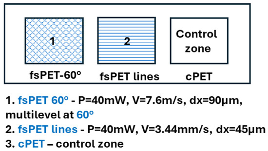
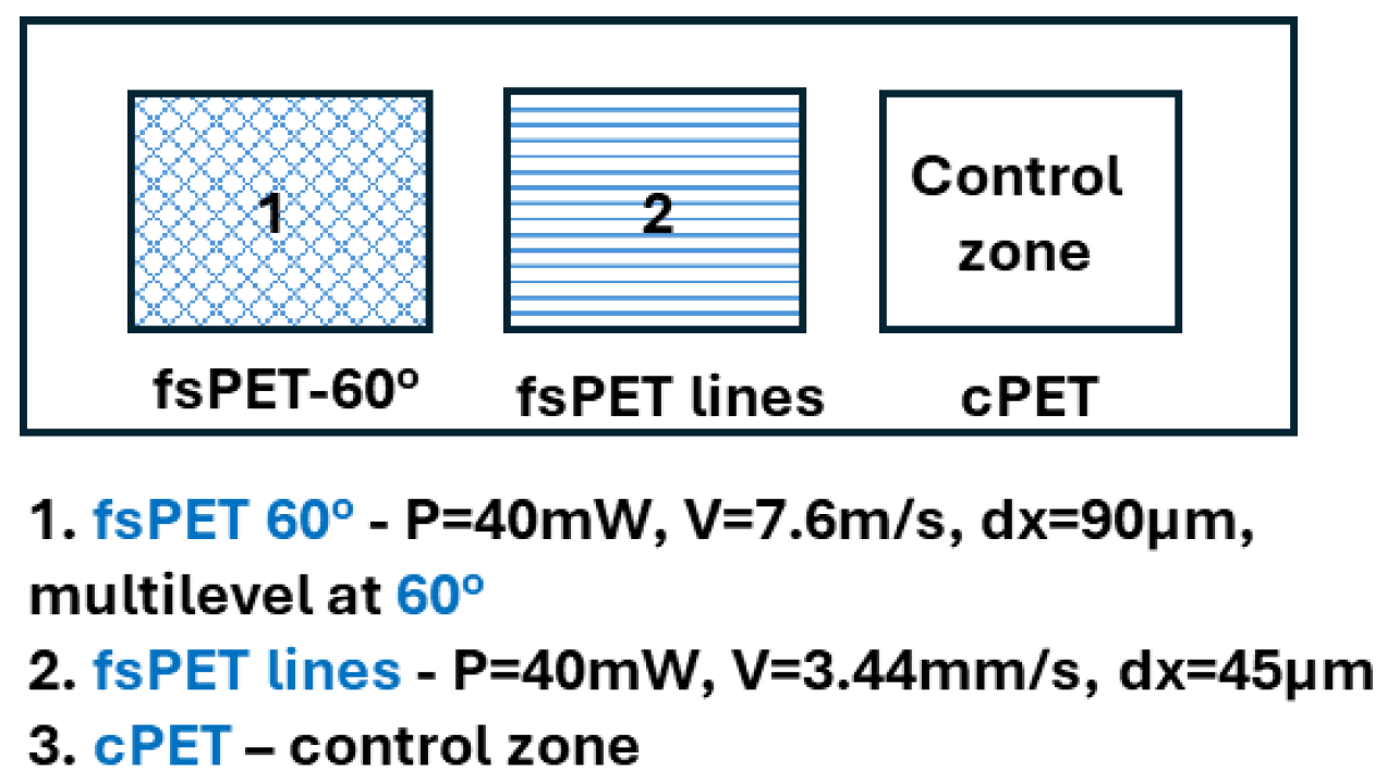
Durability tests were conducted to evaluate the stability and performance of femtosecond (fs) laser-structured polyethylene terephthalate (PET) surfaces under conditions representative of real-life use. Based on preliminary optimization experiments, the following laser processing parameters were selected: laser power of P = 40 mW, scanning speed of v = 7.6 mm/s, hatch spacing of dx = 90 μm (3 layers at 60°) and 45 μm (lines), and pattern configurations consisting of either single-directional lines or three superimposed layers intersecting at 60°. The selected parameters correspond to the two representative structures that exhibited the largest differences in morphology and wettability while maintaining high structural uniformity. For each experimental condition, untreated PET samples were used as controls. Separate sets of laser-structured samples were prepared for each test to avoid cross-interference between degradation mechanisms. After each test, the samples were characterized and compared in terms of morphological features (SEM) and wettability.
2.4.1. Thermal Cycling for Simulating Environmental Temperature Variations
Thermal stability was evaluated by exposing both structured and control PET samples to repeated temperature variations. The test involved cycling temperatures between −40 °C and +85 °C, with each temperature maintained for 10 min over a total of 200 cycles. Between cycles, samples were left to cool down at room temperature (25 °C) for 30 min. Cooling conditions were achieved by means of a laboratory refrigeration system (Electrolux, Stockholm, Sweden), while elevated temperatures were applied using a temperature-controlled laboratory oven (Nahita 631 PLUS, AUXILAB S.L., Navarra, Spain). The lowest temperature was obtained using a controlled freezing spray (Motip freezer, Wolvega, The Netherlands). Surface temperature was monitored at each step by means of a non-contact digital thermometer (TP101, Xuzhou Sanhe Automatic Control Equipment Co., Ltd, Xuzhou, China).
2.4.2. UV Light Exposure
To simulate prolonged daylight exposure, samples were irradiated with ultraviolet (UV) light under controlled environmental conditions inspired by ISO 4892-3 guidelines [13]. A UV source (miniWH, Xuzhou Sanhe Automatic Control Equipment Co., Ltd, Xuzhou, China) emitting in the 390–400 nm range was used. The exposure was conducted at 60 °C and 50% relative humidity for a total duration of 1000 h. This test aimed to assess potential photo- and topography degradation effects on both untreated and laser-modified PET surfaces.
2.4.3. Abrasion Resistance of Laser-Generated Designs
The mechanical durability of frequently touched PET surfaces was investigated using a standardized abrasion test performed with a Taber-type abrasion system (Teledyne Taber, model 503, patent N 2287148, Erichsen, Hemer, Germany). The test was conducted using a CS-10 abrasive wheel (ASTM D1044) under a load of 50 g for 200 cycles. Changes in surface morphology and wettability were evaluated after testing to determine the resistance of the laser-induced structures to mechanical wear.
2.4.4. Chemical Stability
Chemical resistance of the PET surfaces was assessed by immersion in both acidic and saline solutions. Samples were first immersed in 0.5 M hydrochloric acid (HCl) for 48 h at 40 ± 2 °C, followed by immersion in a 5% sodium chloride (NaCl) solution for 72 h at 35 ± 2 °C. After exposure, samples were rinsed thoroughly with distilled water and visually inspected for any signs of degradation, including discoloration, surface morphology deformation, or defect formation.
2.4.5. Dust Contamination and Cleanability
The tendency of the surfaces to retain particulate contamination was evaluated by exposing samples to environmental dust. The dust-covered samples were maintained at 80 °C for 2 h to simulate dry contamination conditions. Subsequently, a standardized cleaning procedure was applied, including air blowing at 3 bar for 30 s, immersion in distilled water under mild agitation for 2 min, and rinsing with running water. Samples were then dried at ambient conditions. Surface cleanliness and changes in wettability were assessed before and after the procedure to determine the effectiveness of particle removal and the influence of surface structuring on dust and particle retention.
3. Results
As a first step in optimizing surface topography and wettability, so that antibacterial self-cleaning surfaces can be obtained, a profound parametric study was performed. As already mentioned, the following parameters of the laser radiation were varied: power, scanning velocity, hatch distance, designs (levels of structuring), in and out of focus irradiation. For better comprehension, the exact parameters applied are given in Table 1.
Table 1.
Different combinations of power, scanning velocity, hatch distances and levels of structuring that were applied during the parametric study performed on PET samples.
As can be clearly seen from the table, a total of 32 different groups of laser structured PET surfaces have been obtained as a result of the detailed parametric study and analyzed with respect to the control, laser-nonprocessed PET substrates. In the current work, a variety of the most representative results are presented.
3.1. Morphological Characterization of PET Samples
3.1.1. Scanning Electron Microscopy
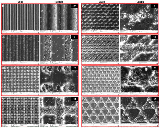
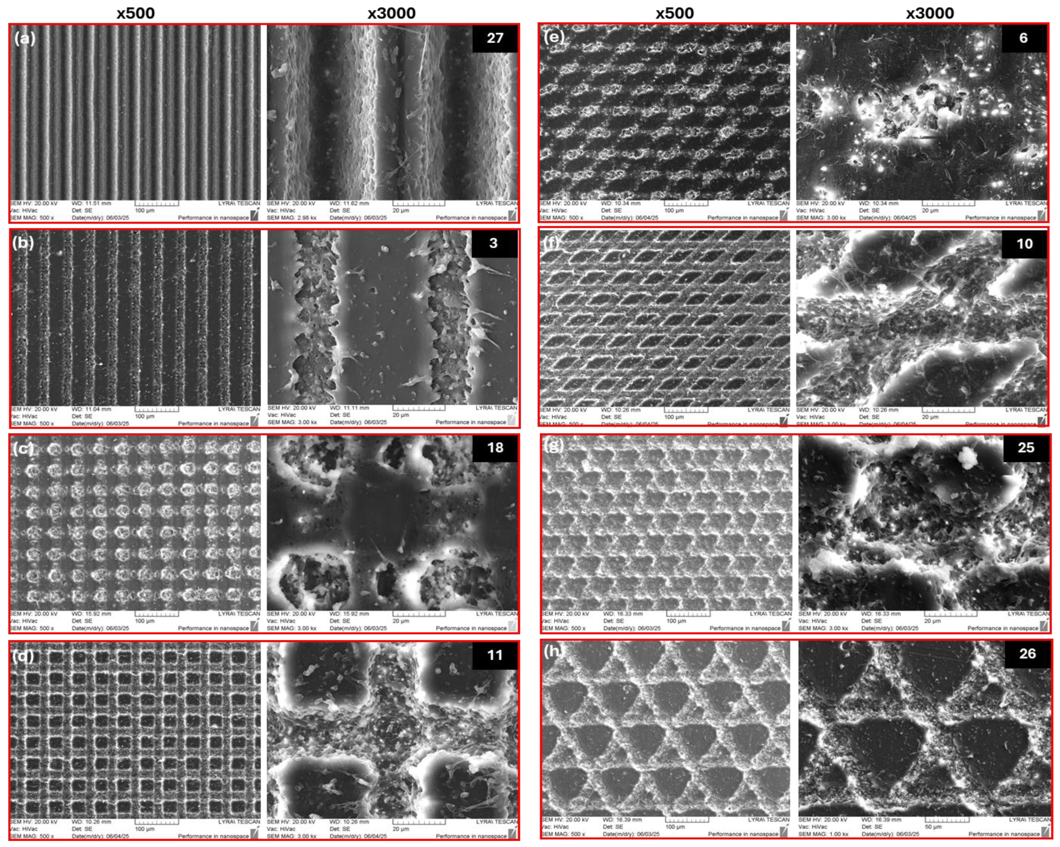
A representative compilation of SEM micrographs of different topography designs of laser-structured PET coupons is presented in Figure 1. As can be seen from the examples presented, hierarchical, multilevel designs with precisely controlled porous grooves of “lines” and “dots” below and above the sample’s baseline can be obtained by finely optimizing the combination of the parameters of the femtosecond laser radiation applied (Table 1). For example, as is presented in panels (a) and (b) of Figure 1, by simply lowering the power of the femtosecond laser radiation applied to the material, the transverse profile of the grooves created can change from “V” to “U” form and from deeper to a shallower view. At the same time, the morphology changes from a more porous to a more granular appearance. Similarly, a “crater” (c) and “dome”-shaped (d) mesh structure is generated by varying the scanning velocity and the power applied-the higher the power and the lower the velocity, the deeper the structures created and vice versa. The same principle is valid for all the designs presented in Figure 1. The designs presented in Figure 1g,h are obtained at the same laser parameters; only the hatch distance is doubled in the lower panel (h), which leads to different morphology.
Figure 1.
Representative SEM images (taken at magnification 500× and 3k×) of fs laser structured PET samples with respect to parameters in Table 1 as follows: (a) PET № 27; (b) PET № 3; (c) PET № 18; (d) PET № 11; (e) PET № 6; (f) PET № 10; (g) PET № 25; (h) PET № 26.
Overall, the results demonstrate that femtosecond laser processing enables a high degree of control over the surface morphology of PET by fine-tuning key irradiation parameters. Variations in laser power, scanning velocity, and hatch spacing lead to distinct and reproducible topographical features, ranging from groove-like structures with tunable cross-sectional profiles to complex multilevel hierarchical patterns. These morphological variations directly influence surface roughness, feature spacing, and aspect ratio—parameters that are critically linked to bacteria–surface interactions. Similar relationships have been reported in the literature, where micro and nanoscale topographies inspired by natural systems such as cicada wings exhibit pronounced bactericidal activity due to their ability to mechanically deform and rupture bacterial cell membranes [8,14]. For instance, Ivanova and co-workers demonstrated that nanopillar arrays on cicada wings can physically penetrate Pseudomonas aeruginosa cells, leading to cell death without the involvement of chemical agents [15]. Subsequent studies have extended this concept to artificially engineered surfaces, showing that parameters such as pillar height, spacing, and sharpness govern the degree of membrane stretching and rupture [14,16]. In particular, high-aspect-ratio and densely packed nanostructures have been shown to enhance bactericidal efficiency, while less pronounced features primarily reduce bacterial adhesion rather than inducing cell lysis.
While nanoscale features are known to induce bactericidal effects via mechanical membrane disruption, microscale structures primarily influence bacterial adhesion by modifying surface wettability and available contact area. For example, the group of Fadeeva demonstrated that laser-fabricated microgrooves on polymer and silicon surfaces significantly decrease bacterial adhesion due to the combined effects of roughness and wettability modulation [17]. In addition, studies on laser-textured titanium and polymers have shown that microscale grooves and ridges can spatially constrain bacterial cells, limiting their ability to spread and form biofilms [18]. These findings suggest that, while microstructures alone may not always induce direct bactericidal action, they play a critical role in preventing initial colonization. Therefore, the hierarchical combination of micro- and nanostructures, as achieved in the present work, is particularly promising, as it has the potential that may enable simultaneous control over bacterial adhesion and viability.
Additionally, modifications in hatch distance significantly influence pattern density and spatial organization without altering the fundamental processing conditions. Therefore, the ability to tailor PET surface morphology via femtosecond laser processing provides a promising route for designing functional surfaces that either inhibit bacterial attachment or actively induce bactericidal effects, depending on the geometrical characteristics of the fabricated structures. These findings confirm that ultrafast laser structuring is a versatile and precise tool for engineering PET surface architectures with tailored geometrical characteristics, providing a solid basis for subsequent investigations into their influence on wettability and antibacterial performance.
3.1.2. 3D-Profilometry, Confocal and Atomic Force Microscopy
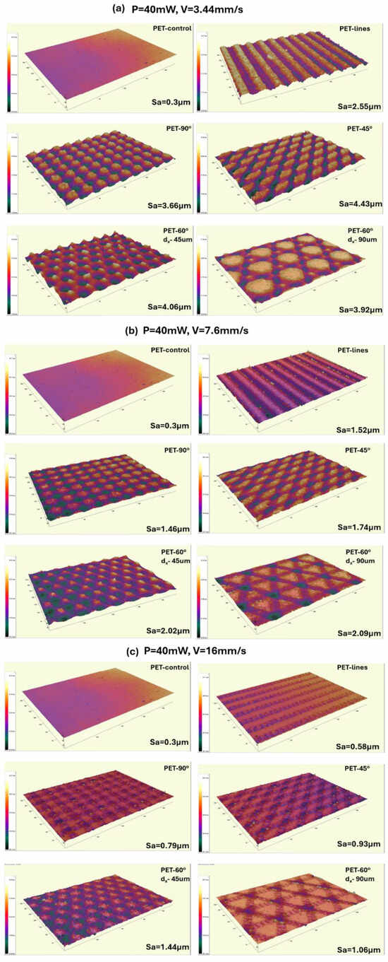
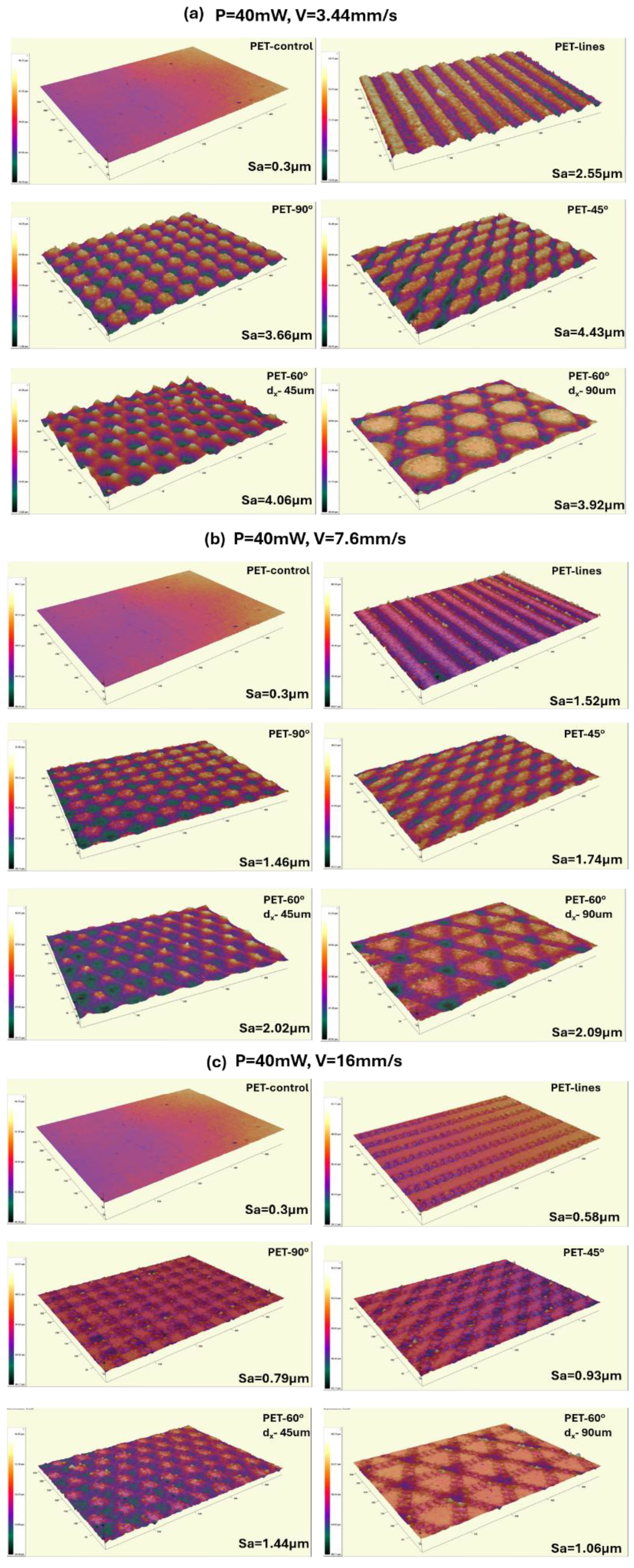
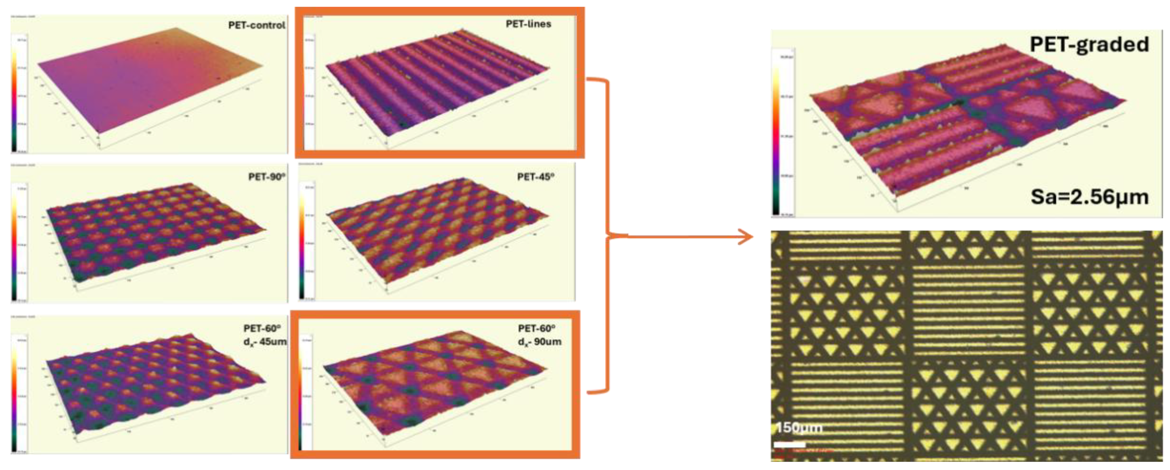
During the parametric study performed, surface roughness was also evaluated. Next, in Figure 2, a representative compilation of 3D-real color images of some of the generated designs (taken at 20× magnification) and the corresponding mean surface area roughness parameter (Sa) are presented. The Sa surface roughness values obtained are summarized in Table 2, providing a quantitative assessment of the topographical modifications induced by the different femtosecond laser processing conditions.
Figure 2.
A representative compilation of 3D-profilometry-real color images (taken at 20× magnification) of different designs of laser-structured PET samples at: (a) P = 40 mW, V = 3.44 mm/s; (b) P = 40 mW, V = 7.6 mm/s; (c) P = 40 mW, V = 16 mm/s.
Table 2.
Summary of areal (Sa) roughness parameters of untreated and femtosecond laser-treated PET surfaces, structured at the following parameters: (a) P = 40 mW, V = 3.44 mm/s; (b) P = 40 mW, V = 7.6 mm/s; (c) P = 40 mW, V = 16 mm/s.
Different designs are obtained in the form of lines or multilevel hierarchical structures, crossed at angles of 45°, 60° and 90° and compared to the control surface. As can be seen from Figure 2, the power of the fs laser radiation applied in the example presented is kept at P = 40 mW, but the velocity is changed as follows: v = 3.44 mm/s (a), v = 7.6 mm/s (b), and v = 16 mm/s (c) panel of Figure 2. The morphology created follows the same trend as monitored during the performed SEM analysis-higher scanning velocity leads to “faster” interaction of the laser radiation with the polymer material, lower energy deposition and subsequently to a lower value of Sa: from the highest presented value of Sa in the current example of 4.43 µm for the lowest scanning speed v = 3.44 mm/s, Figure 2a up to a value of Sa = 58 nm for the highest v = 16 mm/s applied, Figure 2c, which is in the range of the surface roughness of the control PET sample—Sa ≈ 30 nm.
From a functional point of view, these roughness regimes are directly relevant to bacterial interactions. Surfaces with micrometer-scale roughness (Sa in the order of μm) provide increased surface area and sheltering sites that can promote bacterial attachment and colonization, particularly for rod-shaped bacteria with dimensions of ~1–2 μm [19]. In contrast, submicron and nanoscale roughness (Sa < 100 nm) has been associated with reduced bacterial adhesion due to decreased effective contact area and altered surface energy [20]. Moreover, when nanoscale features approach bacterial cell wall dimensions, they can impose mechanical stress on the membrane, potentially leading to deformation or rupture, as demonstrated in mechano-bactericidal surfaces [21]. Therefore, the ability to tune PET surface roughness across micro- to nanoscale regimes enables selective control over bacterial response—ranging from adhesion suppression to potential antibacterial effects—depending on the specific topographical characteristics.
In Figure 3, a combined “chessboard-like” graded structure is presented as an example of another way to finely control surface microdesign, roughness, and, as a result, the wettability (Section 3.2 of the manuscript—Figure 6).
Figure 3.
3D-profilometry-real color (at 20× magnification) and optical image (scale bar = 150 μm) of a design of a combined “chess board-like” graded structure, generated as a combination of two fs laser patterns, obtained at P = 40 mW, V = 7.6 mm/s.
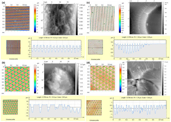
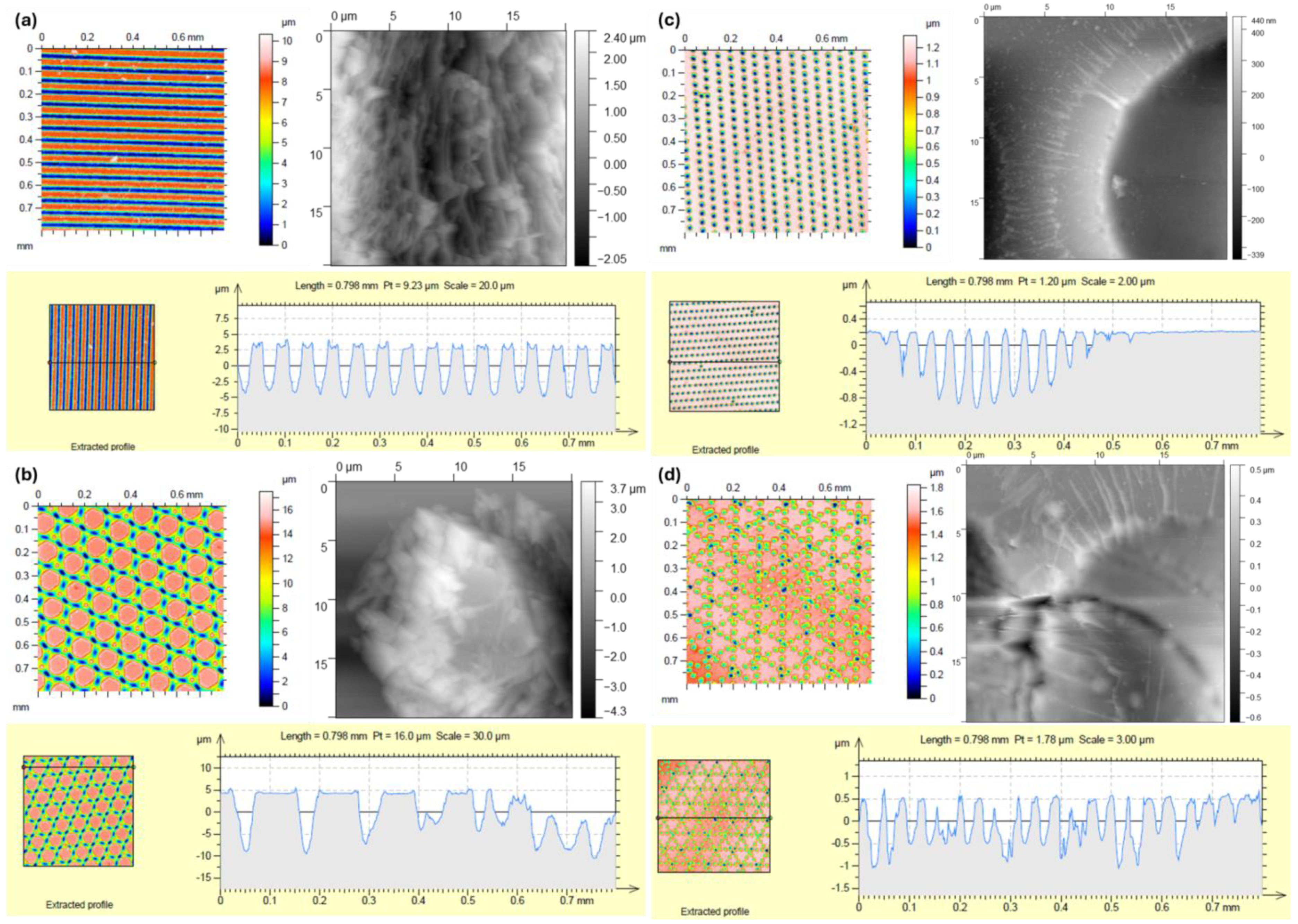
The combination of confocal microscopy and AFM enabled comprehensive multi-scale characterization of the PET surfaces, providing both micrometer-scale surface mapping and nanometer-scale detail essential for evaluating the hierarchical structures generated by femtosecond laser processing—Figure 4. The line roughness of the PET samples presented is also evaluated and shown in Figure 4.
Figure 4.
Confocal (at 20× magnification) and AFM images (20 × 20 μm) of some surface designs on PET, obtained at the following laser patterns: (a) P = 40 mW, V = 3.44 mm/s, dx = 45 μm, lines; (b) P = 40 mW, V = 3.44 mm/s, dx = 90 μm, multilevel lines, crossed at an angle of 60°; (c) P = 40 mW, V = 32 mm/s, dx = 45 μm, lines; (d) P = 40 mW, V = 32 mm/s, dx = 90 μm, multilevel lines, crossed at an angle of 60°.
Panels (a) and (b) of the figure present surface topography designs achieved at P = 40 mW, V = 3.44 mm/s, in the form of lines (a) or multilevel lines, crossed at an angle of 60° (b). The transverse profiles of both types of structures show not only ejected material above the surface baseline (up to 3.5–5 μm), but also the depth of the lines below the PET surface of 5 μm in Figure 4a to almost 10 μm in Figure 4b, when multilevel structuring is applied, giving a total depth of around 15 μm. The AFM images complement the confocal observations by providing higher-resolution characterization of the nanoscale surface morphology within selected regions of the laser-generated structures. In Figure 4a, the AFM image depicts the interior of an individual laser-generated groove, revealing the fine nanoroughness superimposed on the micrometer-scale channel that cannot be resolved by confocal microscopy alone. In Figure 4b, the AFM image shows the characteristic microconical resolidified features located between the intersecting microchannels. These protrusions exhibit additional nanoscale texture on their surfaces, illustrating the hierarchical nature of the laser-generated topography. Such hierarchical morphologies are characteristic of high local energy deposition during ultrafast laser–polymer interaction and have previously been associated with repeated melting, recoil pressure effects, and rapid resolidification of polymer material [9,10].
Panels (c) and (d) of Figure 4 present the same surface morphology of laser-structured PET, but were obtained at an almost 10-fold higher scanning velocity (P = 40 mW, V = 32 mm/s). The faster interaction of laser radiation with the material results in shallower lines with discontinuous “dot-like” structure of separate craters, with mean depth around 400 nm (c) and not more than 970 nm (d); Likewise, the amount of material ejected above the PET baseline decreases considerably to approximately 200–500 nm, corresponding to nearly an order of magnitude reduction compared to the low-velocity structures shown in Figure 4a,b. The corresponding AFM images further reveal the nanoscale morphology of the discontinuous crater-like structures formed at the higher scanning speed. The images show accumulations of resolidified material around the crater edges, appearing as splashed nanomaterial that forms nanofiber-like concentric features. Compared with the structures produced at low scanning speed, these surfaces exhibit substantially shallower relief and a less developed hierarchical morphology, reflecting the lower energy deposited per unit area. This behavior can be explained by the reduced overlap between successive laser pulses at higher scanning speeds, which limits localized heat accumulation and suppresses extensive polymer melting and re-solidification [22]. Similar trends have been reported for femtosecond laser structuring of polymers and metallic substrates, where lower scanning velocities promote the formation of deeper hierarchical structures; in contrast, higher velocities favor nanoscale periodic or discontinuous morphologies due to reduced fluence accumulation [17,18]. Overall, decreasing scanning speed increased deposited fluence, producing deeper hierarchical structures, higher Sa values, and larger modifications in wettability. These observations indicate that scanning speed is the dominant processing parameter controlling PET surface functionality under the investigated conditions.
As already mentioned, these topographic differences could strongly influence bacterial interactions with the PET surfaces. Deep hierarchical microstructures combined with nanoscale roughness may promote anti-adhesive or antibacterial effects by reducing the effective bacteria–surface contact area and generating localized mechanical stress on bacterial membranes [8]. Conversely, shallower discontinuous structures may primarily affect wettability and initial adhesion behavior rather than directly inducing membrane disruption. Therefore, the demonstrated ability to precisely tailor the morphology and roughness of PET surfaces through laser processing provides an important basis for engineering antibacterial surface functionalities.
3.2. Wettability Evaluation of Laser-Structured PET Surfaces
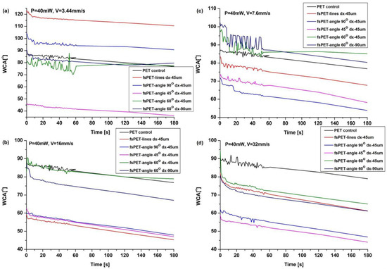
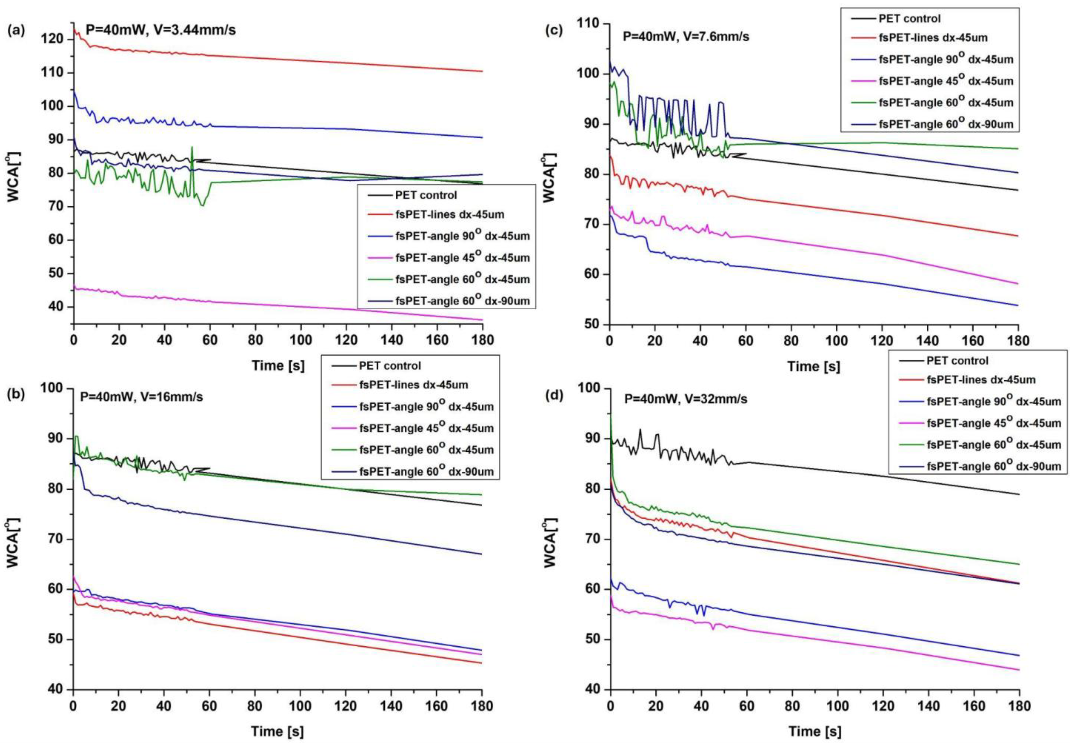
A comprehensive wettability study was also performed on all fs PET designs with respect to control surfaces. Figure 5 shows a compilation of hydrophilic and hydrophobic surfaces obtained after femtosecond laser processing at the same laser power applied (P = 40 mW), with only the scanning speed (V), design, and hatch distance (dx) between the lines being varied. As can be seen from the figure below, at lower velocities (Figure 5a,b), some hydrophobic surfaces are obtained, with mean WCA [°] of above 100° and even 118.2° for one case (P = 40 mW, V = 3.44 mm/s, dx = 45 μm, lines), with respect to the control PET surface (mean WCA [°]- 84.98°). Nevertheless, especially at higher velocities (Figure 5c,d), most of the structures lead to more hydrophilic surfaces with a mean WCA [°] ranging from 78.42° down to 33.21°, depending on the specific combination of laser parameters, design and hatch distance applied for structuring.
Figure 5.
WCA evaluation on control and fs laser-structured PET surfaces, processed at P = 40 mW and scanning velocity as follows: (a) V = 3.44 mm/s; (b) V = 7.6 mm/s; (c) V = 16 mm/s; (d) V = 32 mm/s. The angles (90°, 45°, 60°) indicate the angle of intersecting patterns.
All four panels (a–d) with graphs of Figure 5 of laser-structured PET samples exhibit more or less pronounced heterogeneous wetting after water droplet deposition, reflected by the temporal fluctuations of the measured contact angle before reaching a stable value. This transient wetting behaviour can be interpreted based on the hierarchical surface morphology generated by femtosecond laser processing, whose roughness and topographical characteristics were confirmed by SEM, AFM, confocal microscopy, and optical profilometry (Section 3.1 and Figure 1, Figure 2, Figure 3 and Figure 4). According to the wetting model proposed by Anthony Cassie and Sidney Baxter, the interaction of liquids with such rough or porous surfaces is governed by the formation of a composite solid–air interface [23]. According to this model, when a liquid droplet is placed on a microstructured surface, it does not fully penetrate the surface features but instead rests on a composite interface consisting of solid material and entrapped air [23]. This so-called Cassie–Baxter state results in the characteristic “ups and downs” in the graphs. From the perspective of bacterial adhesion, such a wetting regime reduces the effective solid–liquid contact area, thereby limiting the number of available attachment sites for microorganisms [17,24]. In addition, the presence of air pockets within the surface topography creates an energetically unfavorable interface for stable bacterial anchoring [25]. As a consequence, bacterial cells exhibit reduced adhesion strength and are more easily removed under external forces. In the case of laser-structured PET surfaces, microscale features such as grooves or multilevel periodic patterns can promote the formation of this composite interface, particularly when combined with appropriate surface roughness [26]. Therefore, a reduction in bacterial attachment on microstructured surfaces is expected and may be associated with Cassie–Baxter-type wetting behavior, which primarily contributes to anti-adhesive rather than bactericidal effects [17,25,26].
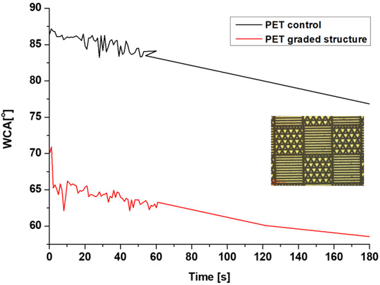
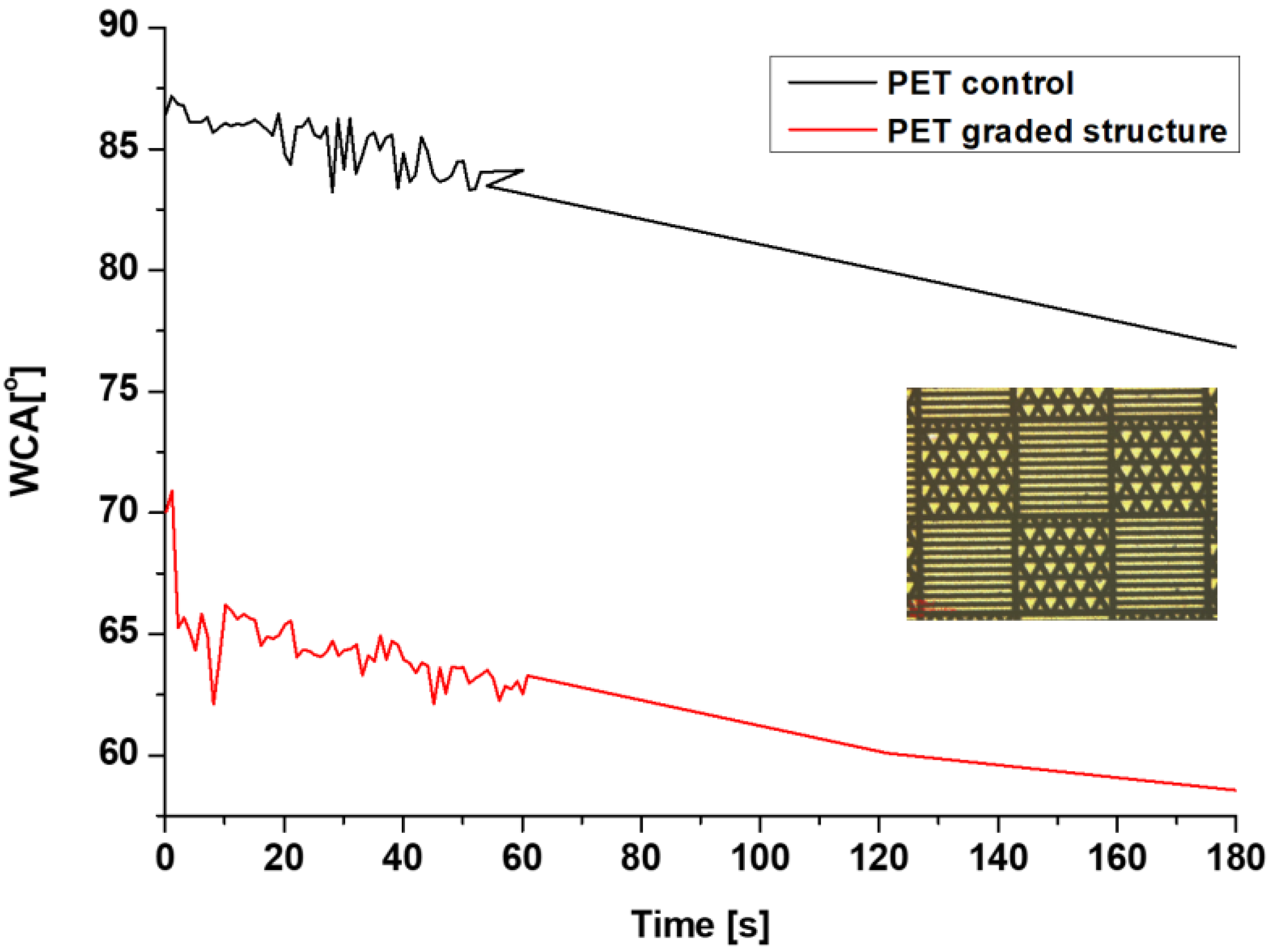
Furthermore, the combined “chessboard-like” graded structure is presented as an example of another way to finely control and locally distribute surface microroughness and, as a result, to tune the wettability—Figure 6, where a mean WCA of 61.12° is measured with respect to the control PET surface (mean WCA [°]-84.98°).
Figure 6.
WCA Evaluation on control and fs laser-structured “chessboard-like” graded PET surface.
A purely micrometer-scale roughness (Sa in the µm range) is generally not sufficient on its own to produce strong bactericidal effects. However, there are well-documented cases where micro-scale roughness combined with hydrophobicity or hierarchical micro/nano features results in both anti-adhesive and bactericidal performance. Surfaces exhibiting micrometer-scale roughness (Sa ≈ 1–10 µm), when combined with appropriate surface chemistry or hierarchical structuring, have been shown to display both anti-adhesive and bactericidal behavior. For example, Jansen et al. and co-workers demonstrated that microstructured polymeric and metallic surfaces with micrometer-scale roughness can significantly reduce bacterial adhesion due to limited contact points and altered surface energy [27]. In a related study, L. B. Boinovich et al. [24] reported that laser-textured surfaces with roughness in the micrometer range, combined with superhydrophobicity, exhibit strong anti-biofouling properties by promoting air entrapment and minimizing liquid-mediated bacterial attachment [28]. Furthermore, W. Barthlott and colleagues showed that hierarchical microstructured surfaces inspired by the lotus leaf can significantly suppress microbial contamination through self-cleaning mechanisms, indirectly contributing to a potential antibacterial performance [29]. These studies confirm that while microroughness alone primarily affects adhesion, its combination with wettability effects and hierarchical structuring can result in multifunctional antibacterial behavior.
3.3. Durability Tests of PET Laser-Optimized Designs for Future Creation of Antibacterial Frequently Touched Surfaces
Based on the results obtained during the profound parametric study of the morphology and wettability presented in the first part of the current manuscript, two designs (PET designs № 22 and PET № 31 from Table 1) were selected for performing a series of simulated real-life conditions durability tests (thermal cycling, ultraviolet exposure, abrasion, chemical resistance, and dust contamination) as a first step towards developing antibacterial frequently touched surfaces. PET design No. 22 was selected because it exhibited the highest hydrophobicity together with well-defined line structures and good structural reproducibility. PET design No. 31 was selected because it combined hierarchical morphology, relatively high roughness and stable hydrophilic behavior, thus representing a complementary topography. Together, these two structures span the range of wetting characteristics obtained in the parametric study and therefore constitute representative candidates for durability evaluation.
Figure 7 is a scheme of samples on which durability tests were performed. Each test was done on 10 replicas of the PET surfaces; surface morphology and wettability were evaluated and compared with the control zones.
Figure 7.
Scheme of the PET samples, subjected to durability tests.
3.3.1. Scanning Electron Microscopy After Durability Tests
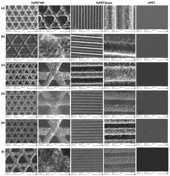
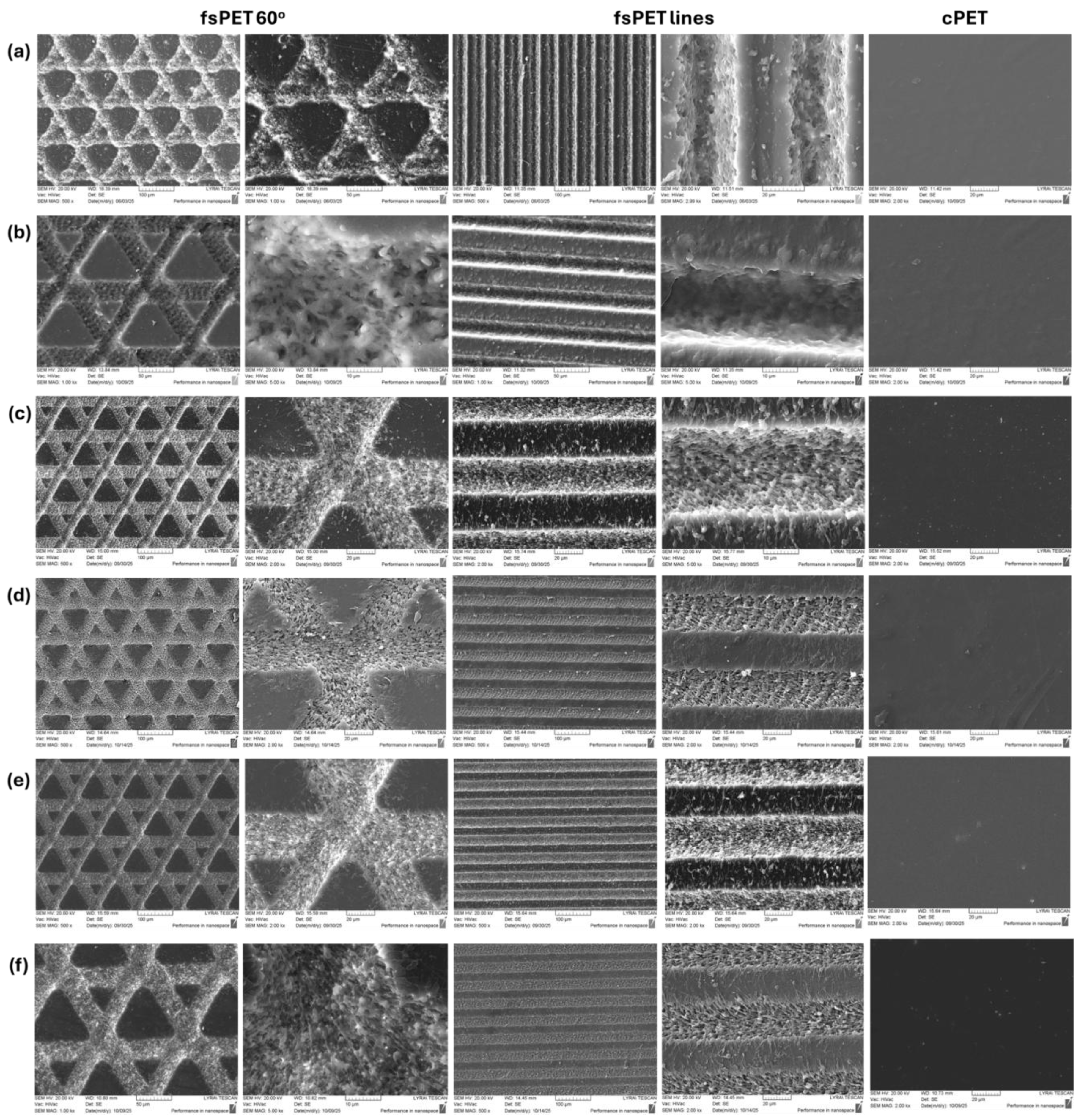
Next, Figure 8 represents a comparison of SEM micrographs of the surface morphology of fs laser-patterned and control PET samples before and after performing the durability tests.
Figure 8.
SEM images (taken at magnification 500×–5k×) of control and fs laser-structured PET samples before (a) and after performing (b) thermal, (c) UV, (d) abrasion, (e) chemical, and (f) dust durability tests.
Qualitative comparison of the SEM micrographs presented in the figure above indicates that the overall micro- and nanoscale morphology of the laser-structured PET surfaces was largely preserved after the durability tests, demonstrating the high stability of the fabricated patterns and their suitability for future long-term application on frequently touched surfaces. In particular, the principal structural features, including laser-generated microchannels, groove geometry, and the superimposed nanoscale textures, remained clearly distinguishable after exposure to thermal cycling Figure 8b, ultraviolet irradiation Figure 8c, chemical resistance testing Figure 8d, abrasion Figure 8e, and dust contamination Figure 8f. SEM analysis revealed that the hierarchical organization of the laser-induced structures was retained under all investigated conditions. The nanoscale features located within the parallel grooves and on the sidewalls of the intersecting channels remained largely intact, confirming the robustness of the femtosecond laser-induced modifications. Minor alterations were detected after the thermal cycling test Figure 8b, where localized smoothing, partial fusion, and a slight reduction in the ordering of the structures inside the grooves were observed. These changes were limited and did not affect the overall hierarchical architecture of the surface. However, the most pronounced modifications were observed after the abrasion test Figure 8d, where partial smoothing and directionality of the protruding grooves and ridge tops occurred as a result of mechanical wear. Nevertheless, the general micro- and nanoscale morphology was preserved, and the structured PET surfaces maintained their characteristic topographical features. These findings demonstrate that femtosecond laser-engineered PET surfaces possess excellent structural durability and can retain their functional architecture under conditions representative of practical use, supporting their future application as robust and long-lasting materials with antibacterial potential.
3.3.2. WCA Analysis After Durability Tests
Surface wettability was evaluated for PET samples that were subjected to each durability test individually. Table 3 summarizes the mean static water contact angle WCA [°] values measured for the untreated PET control and for two representative laser-generated surface designs: parallel line structures and multilevel microchannels intersecting at 60°.
Table 3.
Mean WCA [°] values * of control and fs laser-structured PET samples before and after performing durability tests.
Overall, the wettability measurements demonstrate that the femtosecond laser-structured PET surfaces retained their characteristic wetting behavior after all durability tests, indicating that the physicochemical functionality induced by laser processing was preserved under thermal, ultraviolet, mechanical, chemical, and particulate exposure conditions. The relatively small standard deviations obtained for all measurements indicate good reproducibility of the laser structuring process and homogeneous wetting behavior across the processed surfaces.
Prior to durability testing, the untreated PET exhibited a mean WCA of 85°, corresponding to moderately hydrophobic behavior. In contrast, the line-based design laser structures showed pronounced hydrophobicity, with a mean WCA of 116.8°, while the multilevel intersecting structures displayed a more hydrophilic response, with a mean WCA of 77.9°. These differences confirm that femtosecond laser processing can be used to tailor the wetting characteristics of PET over a broad range by controlling the resulting surface morphology. Following the durability tests, only minor variations in WCA were observed—in the range of a few degrees. The parallel line structures remained strongly hydrophobic throughout the study, with WCA values ranging from 117.9° after abrasion to 121.1° after thermal cycling. Similarly, the intersecting 60° patterns maintained relatively hydrophilic behavior, with WCA values between 76.1° and 82.1°. The untreated PET control exhibited only small fluctuations, remaining within the range of 82–86°. The highest hydrophobicity of the line structures was measured after the thermal and dust tests, reaching 121.1° and 121°, respectively. One possible explanation is a more stable Cassie–Baxter wetting regime resulting from partial air entrapment within the microstructures. However, this mechanism was not directly investigated in the present work and therefore remains speculative.
Taken together, the qualitative SEM observations and the wettability measurements indicate that the laser-induced PET structures maintain their functional surface properties after exposure to harsh environmental and mechanical conditions. This long-term stability is particularly important for future antimicrobial applications, as both hydrophobic and hydrophilic wetting regimes can contribute to reduced bacterial attachment depending on the specific topography and wetting mechanism [30,31].
4. Discussion
The present study demonstrates that femtosecond laser processing can be used to generate a wide range of stable micro- and nanostructured surface morphologies on PET with precisely tunable roughness and wettability. By varying the scanning speed, hatch distance, and pattern design while maintaining constant laser power, the resulting topographies ranged from deep continuous microchannels with pronounced material re-deposition to shallow crater-like structures decorated with nanoscale features. These modifications produced substantial differences in surface roughness and wetting behavior, highlighting the versatility of ultrafast laser processing for engineering functional polymer surfaces. The choice of femtosecond laser irradiation is particularly important for polymer materials such as PET, where excessive thermal loading may lead to melting, deformation, or chemical degradation. The present results demonstrate that ultrashort pulse irradiation enables precise control over groove geometry, hierarchical morphology, and surface roughness without compromising the integrity of the substrate [9]. Although picosecond and nanosecond laser systems are also capable of producing functional surface textures, they generally involve greater thermal accumulation and reduced control over nanoscale feature formation. The high structural fidelity and reproducibility achieved in the present work therefore arise directly from the ultrafast nature of the femtosecond laser–material interaction.
The morphological analysis revealed that decreasing the scanning velocity increased the energy deposited per unit length, resulting in deeper grooves, greater material ejection, and more pronounced hierarchical structures. At the lowest scanning speed investigated (3.44 mm/s), groove depths reached up to 10 μm below the original PET surface, while resolidified ridges extended 3.5–5 μm above the baseline, yielding a total topographical variation of approximately 15 μm. In contrast, increasing the scanning speed to 32 mm/s reduced the interaction time and pulse overlap, leading to discontinuous crater-like structures with depths below 1 μm. Similar trends have been reported for laser processing of polymers, where lower scanning speeds promote stronger heat accumulation and melt dynamics, whereas higher velocities produce finer and shallower textures. Jörn Bonse and co-workers described analogous behavior in ultrafast laser structuring, emphasizing the critical role of accumulated fluence in determining surface morphology [10]. A. Vorobyev and Ch. Guo also demonstrated that laser-induced micro- and nanostructures can be tailored over several length scales through controlled adjustment of processing parameters [9]. The roughness measurements confirmed that the generated textures covered a broad range, from values close to that of untreated PET (Sa ≈ 30 nm) to several micrometers. For example, line-based structures produced at low scanning speeds exhibited Sa values above 4 μm, whereas high-speed processing generated surfaces with roughness Sa values below 100 nm. This tunability is particularly relevant for antibacterial surface design because bacterial adhesion is highly sensitive to topographical dimensions relative to the size of microbial cells. Yang et al. showed that both microscale and nanoscale roughness can reduce bacterial attachment by limiting contact area, altering local surface energy, and modifying hydration layers [30]. Similarly, Cheng and co-workers emphasized that topographies comparable to bacterial dimensions can disrupt the formation of stable adhesion points and inhibit early biofilm development [31].
The quantitative analysis of the arithmetic mean surface roughness (Sa) reveals clear relationships between the laser processing parameters and the resulting surface topography. Rather than varying randomly, the measured Sa values follow systematic trends governed primarily by the energy delivered per unit area during femtosecond laser processing. Among the investigated parameters, scanning speed had the strongest influence on surface roughness. At constant laser power (P = 40 mW), decreasing the scanning speed from 16 mm/s to 7.6 mm/s and further to 3.44 mm/s progressively increased the interaction time between the laser beam and the PET surface. Consequently, a larger amount of energy was deposited locally, promoting stronger ablation, enhanced melting and resolidification, and the formation of deeper microgrooves accompanied by pronounced material redeposition. As a result, the average surface roughness increased from values close to those of untreated PET (Sa ≈ 30–60 nm) to several micrometres (Sa = 4.43 µm), corresponding to nearly two orders of magnitude variation. This evolution is fully consistent with the SEM and confocal observations. Samples exhibiting low Sa values consisted mainly of shallow crater-like features with limited material displacement, whereas high Sa surfaces displayed well-developed hierarchical morphologies composed of deep grooves, elevated ridges, and superimposed nanoscale structures. Thus, the increase in Sa directly reflects the transition from weak surface modification to pronounced hierarchical micro/nanostructuring. Besides scanning speed, the overlap between adjacent laser tracks also contributed to the final topography. Lower scanning speeds and multiple crossed scanning passes increased pulse overlap, resulting in greater cumulative energy deposition and consequently higher roughness values. In contrast, increasing the hatch distance mainly modified the spatial distribution and density of the laser-generated structures rather than producing proportional changes in Sa. Therefore, hatch spacing primarily controlled the organization of the surface features, while scanning speed and pulse overlap dominated the roughness amplitude. The observed roughness evolution was also reflected in wettability measurements. Surfaces exhibiting the highest Sa values generally produced the largest deviations from the wettability of untreated PET, demonstrating that increasing roughness alone is insufficient to explain the wetting behaviour. Instead, wettability resulted from the combined effects of roughness magnitude, hierarchical morphology, groove geometry, and air entrapment within the textured structures. Consequently, surfaces possessing similar Sa values but different structural organizations could exhibit different water contact angles, indicating that the geometrical arrangement of the micro- and nanostructures is equally important as the roughness magnitude itself. From the perspective of antibacterial surface engineering, these observations are particularly relevant. Roughness values in the nanometre range mainly modify the available contact area and surface energy, potentially reducing bacterial attachment. In contrast, hierarchical micro/nanostructures with micrometre-scale roughness provide more complex topographies capable of simultaneously altering wettability and generating local geometrical constraints for bacterial cells. Although the present work does not include microbiological testing, the demonstrated ability to systematically control Sa over nearly two orders of magnitude, together with the accompanying changes in morphology and wettability, provides an important basis for future investigations of bacterial adhesion and antibacterial performance.
The wettability study demonstrated that femtosecond laser structuring enabled precise control over the water contact angle of PET, with values ranging from strongly hydrophilic (33.21°) to distinctly hydrophobic (118.2°). These results are consistent with recent advances in laser surface engineering, where precisely tailored hierarchical micro- and nanostructures have been shown to provide deterministic control over wettability and, consequently, the functional performance of engineered surfaces [32]. Such studies further highlight that the interplay between surface morphology, roughness, and wetting behaviour is fundamental for designing multifunctional interfaces with applications ranging from self-cleaning and anti-fouling to biomedical materials and antimicrobial surfaces [28,30,32]. Parallel microchannels produced at low scanning speeds generated the highest hydrophobicity, while multilevel intersecting structures generally promoted hydrophilic behavior. The observed time-dependent contact angle evolution, characterized by fluctuations during the first minute of measurement, suggests heterogeneous wetting associated with partial penetration of water into the hierarchical surface features. This behavior is consistent with the wetting model proposed by Cassie and Baxter, in which liquid droplets rest on a composite interface consisting of solid and trapped air [23]. On such surfaces, reduced effective contact area and entrapped air pockets can limit bacterial attachment by creating energetically unfavorable conditions for stable anchoring. A Review by Leonid Boinovich and Alexander Emelyanenko further confirmed that both superhydrophobic and highly hydrophilic surfaces can exhibit anti-biofouling properties depending on the dominant wetting mechanism [24].
The present work focuses on morphological optimization, wettability control and durability. Biological validation through bacterial adhesion, viability and biofilm assays will be the subject of future work. The present study was intentionally focused on optimizing laser processing parameters, understanding the resulting surface morphology and wettability, and evaluating the long-term durability of the fabricated PET surfaces. These physicochemical characteristics represent essential prerequisites for the development of antibacterial materials but do not, by themselves, demonstrate antibacterial performance. Therefore, although the hierarchical structures produced in this work exhibit geometrical characteristics consistent with previously reported antibacterial and anti-adhesive surface designs, direct microbiological evaluation—including bacterial adhesion, viability, proliferation, and biofilm formation assays against representative Gram-positive and Gram-negative bacteria—remains necessary and will constitute the next stage of this research. The durability tests demonstrated that the laser-induced structures retained both their morphology and wettability after thermal cycling, UV exposure, chemical resistance testing, abrasion, and dust contamination. SEM and confocal analyses showed that the overall hierarchical architecture was preserved, with only minor smoothing of protruding peaks and localized fusion of nanostructures after the most aggressive treatments. Likewise, the water contact angles remained within the same hydrophilic or hydrophobic regimes observed before testing. These findings confirm that the laser-induced modifications are integral to the polymer surface rather than dependent on unstable coatings or deposited agents. Comparable long-term stability has been reported for laser-structured titanium, stainless steel, and polymers, where functional properties such as wettability and anti-biofouling performance were maintained after prolonged environmental exposure [17,22]. From a practical perspective, long-term usability is a key requirement for antibacterial and self-cleaning surfaces intended for frequently touched objects. Unlike chemically deposited antimicrobial coatings, whose effectiveness may gradually diminish due to wear, degradation, or leaching, laser-induced surface structures are directly integrated into the substrate. The comprehensive durability assessment performed in the present work—including thermal cycling, ultraviolet irradiation, mechanical abrasion, chemical exposure, and dust contamination—demonstrated that both the hierarchical morphology and the associated wetting characteristics were largely preserved after exposure to these accelerated ageing conditions. Although extended real-life service testing and microbiological ageing studies remain subjects of future work, the present results indicate that the fabricated PET surfaces possess the structural robustness required for long-term functional applications.
From the perspective of antimicrobial applications, the results are highly promising. Hydrophobic surfaces with trapped air pockets can reduce bacterial attachment by minimizing the available contact area, whereas hydrophilic structures may promote rapid spreading and drying, limiting the accumulation of nutrient-rich droplets. Moreover, hierarchical micro- and nanostructures may generate localized mechanical stresses capable of damaging bacterial cell envelopes. Ivanova and colleagues, for example, demonstrated that nanoscale protrusions inspired by natural insect wings can physically rupture bacterial membranes, establishing the concept of mechano-bactericidal surfaces [15]. Subsequent reviews by Linklater et al. highlighted that combining micro- and nanoscale features often produces synergistic anti-adhesive and bactericidal effects [8]. Although direct microbiological testing was beyond the scope of the present work, the structural and wetting characteristics achieved on PET are consistent with design principles known to suppress bacterial colonization. The present study demonstrates structural and physicochemical characteristics that are consistent with previously reported antibacterial surface design principles. However, direct antibacterial or anti-adhesion performance of the fabricated PET surfaces remains to be experimentally verified.
PET is widely used in packaging, consumer products, and medical components, making it a particularly relevant substrate for antimicrobial surface engineering. Unlike chemical coatings, which may degrade or leach over time, femtosecond laser texturing offers a solvent-free, additive-free approach that modifies only the surface topography while preserving the bulk properties of the material. The demonstrated durability of the resulting structures under simulated real-life conditions supports their practical implementation on high-contact surfaces where long-term performance is essential.
5. Conclusions
In this study, femtosecond laser processing was successfully employed to fabricate a broad range of hierarchical micro- and nanostructures on PET surfaces with controllable morphology, roughness, and wettability. By systematically varying the scanning speed, hatch spacing, and pattern geometry, the static water contact angle was tuned from hydrophilic to distinctly hydrophobic, while comprehensive durability testing demonstrated that the laser-induced surface architectures retained their morphology and functional wetting characteristics following thermal, ultraviolet, chemical, mechanical, and dust exposure.
The novelty of this work lies in the systematic correlation established between femtosecond laser processing parameters, hierarchical surface morphology, wettability, and long-term durability of laser-engineered PET surfaces. Unlike previous studies, which have primarily focused on the fabrication or initial wettability of laser-textured polymer surfaces, the current work combines optimization of laser processing conditions with a comprehensive evaluation of the structural and functional stability of representative surface architectures under accelerated environmental aging. These findings demonstrate that laser-induced surface functionalities are intrinsic to the PET substrate rather than dependent on temporary chemical modifications, highlighting the potential of femtosecond laser texturing as a durable and environmentally friendly surface engineering strategy. The present work establishes a reproducible methodology for fabricating durable femtosecond laser-engineered PET surfaces with precisely controlled hierarchical morphology and tunable wettability. Although direct antibacterial experiments were beyond the scope of this study, the systematic relationship established between laser processing parameters, surface topography, wettability, and durability provides a solid physicochemical foundation for future microbiological investigations. Overall, the proposed femtosecond laser processing methodology provides a robust and reproducible route for engineering durable PET surfaces with tailored physicochemical properties, establishing a foundation for the future development of coating-free antibacterial and self-cleaning polymeric materials.
Author Contributions
Conceptualization, A.D. and L.L.E.M.; methodology, A.D., A.Z. and L.A.; software, A.Z., L.A. and D.M.; validation, A.D., A.Z. and L.A.; formal analysis, A.Z., L.A., D.M. and R.v.N.; investigation, A.D., A.Z. and L.A.; resources, A.D.; data curation, A.D., A.Z., L.A. and L.L.E.M.; writing—original draft preparation, L.A.; writing—review and editing, A.D., L.A. and L.L.E.M.; visualization, A.Z., L.A., D.M. and R.v.N.; supervision, A.D. and L.L.E.M.; project administration, A.D. and L.L.E.M.; funding acquisition, A.D. and L.L.E.M. All authors have read and agreed to the published version of the manuscript.
Funding
This research was funded by the Bulgarian National Science Fund (BNSF) under grant agreement number KP-06-Austria/5 (2025–2027) “Advancing the understanding the adhesion of antimicrobial and bactericidal surfaces produced through ultrafast laser micro- and nanopatterning” and under the same name, the bilateral project, number BG 04/2025 was funded by the Austrian Federal Ministry of Education, Science and Research (BMBWF). This research was partly funded by BNSF grant agreement number KP-06-H98/1, 2025–2028.
Data Availability Statement
The original contributions presented in the study are included in the article; further inquiries can be directed to the corresponding authors.
Acknowledgments
The research was carried out with the help of infrastructure purchased under the National Roadmap for Scientific Infrastructure (ELI-ERIC-BG), project D01-102. L.L.E.M., D.M. and R.v.N. thank Markus Valtiner for access to the equipment of the Applied Interface Physics group at the Institute for Applied Physics, TU Wien.
Conflicts of Interest
The authors declare no conflicts of interest.
Abbreviations
The following abbreviations are used in this manuscript:
| PET | Polyethylene Terephthalate |
| fs | femtosecond |
| SEM | scanning electron microscopy |
| WCA | water contact angle |
| UV | Ultraviolet |
| AFM | Atomic force microscopy |
References
- Süle, A. Antimicrobial resistance—A global challenge that deserves more attention! Eur. J. Hosp. Pharm. 2022, 29, 65. [Google Scholar] [CrossRef] [Scilit] [PubMed]
- World Health Organization. Antimicrobial Resistance: Global Report on Surveillance; WHO Press: Geneva, Switzerland, 2014; Available online: https://www.who.int/publications/i/item/9789241564748 (accessed on 25 March 2026).
- Page, K.; Wilson, M.; Parkin, I.P. Antimicrobial surfaces and their potential in reducing the role of the inanimate environment in the incidence of hospital-acquired infections. J. Mater. Chem. 2008, 19, 3819–3831. [Google Scholar] [CrossRef] [Scilit]
- Çaykara, T.; Sande, M.G.; Azoia, N.G.; Rodrigues, L.R.; Silva, C.J. Exploring the potential of polyethylene terephthalate in the design of antibacterial surfaces. Med. Microbiol. Immunol. 2020, 209, 363–372. [Google Scholar] [CrossRef] [Scilit] [PubMed]
- Andrady, A.L.; Neal, M.A. Applications and societal benefits of plastics. Philos. Trans. R. Soc. B 2009, 364, 1977–1984. [Google Scholar] [CrossRef] [Scilit] [PubMed]
- Mitik-Dineva, N.; Wang, J.; Truong, V.K.; Stoddart, P.; Malherbe, F.; Crawford, R.J.; Ivanova, E.P. Escherichia coli, Pseudomonas aeruginosa, and Staphylococcus aureus attachment patterns on glass surfaces with nanoscale roughness. Curr. Microbiol. 2009, 58, 268–273. [Google Scholar] [CrossRef] [Scilit] [PubMed]
- Kramer, A.; Schwebke, I.; Kampf, G. How long do nosocomial pathogens persist on inanimate surfaces? A systematic review. BMC Infect. Dis. 2006, 6, 130. [Google Scholar] [CrossRef] [Scilit] [PubMed]
- Linklater, D.P.; Juodkazis, S.; Ivanova, E.P. Nanofabrication of mechano-bactericidal surfaces. Nanoscale 2017, 9, 16564–16585. [Google Scholar] [CrossRef] [Scilit] [PubMed]
- Vorobyev, A.Y.; Guo, C. Direct femtosecond laser surface nano/microstructuring and its applications. Laser Photonics Rev. 2013, 7, 385–407. [Google Scholar] [CrossRef] [Scilit]
- Bonse, J.; Höhm, S.; Kirner, S.V.; Rosenfeld, A.; Krüger, J. Laser-induced periodic surface structures—A scientific evergreen. IEEE J. Sel. Top. Quantum Electron. 2017, 23, 9000615. [Google Scholar] [CrossRef] [Scilit]
- Epstein, A.K.; Hochbaum, A.I.; Kim, P.; Aizenberg, J. Control of bacterial biofilm growth on surfaces by nanostructural mechanics and geometry. Nanotechnology 2011, 22, 494007. [Google Scholar] [CrossRef] [Scilit] [PubMed]
- Podulka, P. The Effect of Surface Topography Feature Size Density and Distribution on the Results of a Data Processing and Parameters Calculation with a Comparison of Regular Methods. Materials 2021, 14, 4077. [Google Scholar] [CrossRef] [Scilit] [PubMed]
- Tubio, C.R.; Seoane-Rivero, R.; Neira, S.; Benito, V.; Zubieta, K.G.; Lanceros-Mendez, S. Fiber-Reinforced Polyester Composites with Photoluminescence Sensing Capabilities for UV Degradation Monitoring. Polymers 2022, 14, 3666. [Google Scholar] [CrossRef] [Scilit] [PubMed]
- Linklater, D.P.; Baulin, V.A.; Juodkazis, S.; Crawford, R.J.; Stoodley, P.; Ivanova, E.P. Mechano-bactericidal actions of nanostructured surfaces. Nat. Rev. Microbiol. 2021, 19, 8–22. [Google Scholar] [CrossRef] [Scilit] [PubMed]
- Ivanova, E.P.; Hasan, J.; Webb, H.K.; Truong, V.K.; Watson, G.S.; Watson, J.A.; Baulin, V.A.; Pogodin, S.; Wang, J.Y.; Tobin, M.J.; et al. Natural bactericidal surfaces: Mechanical rupture of Pseudomonas aeruginosa cells by cicada wings. Small 2012, 8, 2489–2494. [Google Scholar] [CrossRef] [Scilit] [PubMed]
- Pogodin, S.; Hasan, J.; Baulin, V.A.; Webb, H.K.; Truong, V.K.; Phong Nguyen, T.H.; Boshkovikj, V.; Fluke, C.J.; Watson, G.S.; Watson, J.A.; et al. Biophysical model of bacterial cell interactions with nanopatterned cicada wing surfaces. J. Theor. Biol. 2013, 104, 835–840. [Google Scholar] [CrossRef] [Scilit] [PubMed]
- Fadeeva, E.; Schlie, S.; Koch, J.; Chichkov, B.N.; Crawford, R.J.; Wang, J.; Ivanova, E.P. Bacterial retention on superhydrophobic titanium surfaces fabricated by femtosecond laser ablation. Langmuir 2011, 27, 3012–3019. [Google Scholar] [CrossRef] [Scilit] [PubMed]
- Cunha, A.; Elie, A.M.; Plawinski, L.; Serro, A.P.; Botelho do Rego, A.M.; Almeida, A.; Urdaci, M.C.; Durrieu, M.C.; Vilar, R. Femtosecond laser surface texturing of titanium as a method to reduce the adhesion of Staphylococcus aureus and biofilm formation. Appl. Surf. Sci. 2016, 360, 485–493. [Google Scholar] [CrossRef] [Scilit]
- Anselme, K.; Davidson, P.; Popa, A.M.; Giazzon, M.; Liley, M.; Ploux, L. The interaction of cells and bacteria with surfaces structured at the nanometre scale. Acta Biomater. 2010, 6, 3824–3846. [Google Scholar] [CrossRef] [Scilit] [PubMed]
- Whitehead, K.A.; Verran, J. The effect of surface topography on the retention of microorganisms. Food Bioprod. Process. 2006, 84, 253–259. [Google Scholar] [CrossRef] [Scilit]
- Ivanova, E.P.; Hasan, J.; Webb, H.K.; Gervinskas, G.; Juodkazis, S.; Truong, V.K.; Wu, A.H.F.; Lamb, R.N.; Baulin, V.A.; Watson, G.S.; et al. Bactericidal activity of black silicon. Nat. Commun. 2013, 4, 2838. [Google Scholar] [CrossRef] [Scilit] [PubMed]
- Stoian, R.; Colombier, J.-P. Advances in ultrafast laser structuring of materials at the nanoscale. Nanophotonics 2020, 9, 4665–4688. [Google Scholar] [CrossRef] [Scilit]
- Cassie, A.B.D.; Baxter, S. Wettability of porous surfaces. Trans. Faraday Soc. 1944, 40, 546–551. [Google Scholar] [CrossRef] [Scilit]
- Boinovich, L.B.; Emelyanenko, A.M. Anti-biofouling properties of superhydrophobic surfaces. Mendeleev Commun. 2013, 23, 3–10. [Google Scholar] [CrossRef] [Scilit]
- Epstein, A.K.; Wong, T.S.; Belisle, R.A.; Boggs, E.M.; Aizenberg, J. Liquid-infused structured surfaces with exceptional anti-biofouling performance. Proc. Natl. Acad. Sci. USA 2012, 109, 13182–13187. [Google Scholar] [CrossRef] [Scilit] [PubMed]
- Liu, K.; Yao, X.; Jiang, L. Recent developments in bio-inspired special wettability. Chem. Soc. Rev. 2010, 39, 3240–3255. [Google Scholar] [CrossRef] [Scilit] [PubMed]
- Jansen, B.; Peters, G. Modern strategies in the prevention of polymer-associated infections. J. Hosp. Infect. 1991, 19, 83–88. [Google Scholar] [CrossRef] [Scilit] [PubMed]
- Quan, Y.; Chen, Z.; Lai, Y.; Huang, Z.; Li, H. Recent Advances in Fabricating Durable Superhydrophobic Surfaces: A Review in the Aspects of Structures and Materials. Mater. Chem. Front. 2021, 5, 1655–1682. [Google Scholar] [CrossRef] [Scilit]
- Barthlott, W.; Neinhuis, C. Purity of the sacred lotus, or escape from contamination in biological surfaces. Planta 1997, 202, 1–8. [Google Scholar] [CrossRef] [Scilit]
- Yang, K.; Shi, J.; Wang, L.; Chen, Y.; Liang, C.; Yang, L.; Wang, L.-N. Bacterial anti-adhesion surface design: Surface patterning, roughness and wettability: A review. J. Mater. Sci. Technol. 2022, 99, 82–100. [Google Scholar] [CrossRef] [Scilit]
- Cheng, Y.; Feng, G.; Moraru, C.I. Micro- and nanotopography sensitive bacterial attachment mechanisms: A review. Front. Microbiol. 2019, 10, 191. [Google Scholar] [CrossRef] [Scilit] [PubMed]
- Xiao, J.; Yin, K.; Wang, L.; Pei, J.; Song, X.; Huang, Y.; He, J.; Duan, J.A. Femtosecond laser atomic-nano-micro fabrication of biomimetic perovskite quantum dots films toward durable multicolor display. ACS Nano 2025, 19, 23431–23441. [Google Scholar] [CrossRef] [Scilit] [PubMed]

Disclaimer/Publisher’s Note: The statements, opinions and data contained in all publications are solely those of the individual author(s) and contributor(s) and not of MDPI and/or the editor(s). MDPI and/or the editor(s) disclaim responsibility for any injury to people or property resulting from any ideas, methods, instructions or products referred to in the content. |
© 2026 by the authors. Licensee MDPI, Basel, Switzerland. This article is an open access article distributed under the terms and conditions of the Creative Commons Attribution (CC BY) license.