Article

Picosecond Laser Treatment of Cu-Doped TiO2 Coatings: Effects on Mechanical Resistance, Electrochemical Behaviour, and Antibacterial Activity


1
Department of Material Science and Technology, University of Ruse “A. Kanchev”, 8 Studentska Str., 7017 Ruse, Bulgaria
2
Institute of Electronics, Bulgarian Academy of Sciences, 72 Tzarigradsko Chaussee Blvd., 1784 Sofia, Bulgaria
3
Department of Mathematics, Informatics and Natural Sciences, Technical University of Gabrovo, 4 H. Dimitar Str., 5300 Gabrovo, Bulgaria
4
Department of Chemical, Food and Biotechnologies, Razgrad Branch, University of Ruse “A. Kanchev”, 47 Aprilsko Vastanie Blvd., 7200 Razgrad, Bulgaria
5
Faculty of Physics, Sofia University “St. Kliment Ohridski”, 5 James Bourchier Blvd, 1164 Sofia, Bulgaria
6
Rezekne Academy, Riga Technical University, LV-1658 Riga, Latvia
*
Author to whom correspondence should be addressed.
Surfaces 2026, 9(3), 69; https://doi.org/10.3390/surfaces9030069
Submission received: 17 June 2026 / Revised: 20 July 2026 / Accepted: 24 July 2026 / Published: 26 July 2026
(This article belongs to the Special Issue Surface Engineering for Biomedical Applications)

Abstract

Implant-associated infections remain one of the leading causes of failure in orthopaedic and dental implants, necessitating the development of multifunctional surface coatings capable of simultaneously enhancing corrosion resistance, bioactivity, and antibacterial performance. The aim of this study was to investigate how picosecond laser surface treatment modifies the structural, physicochemical, mechanical, electrochemical, bioactive, and antibacterial properties of magnetron-sputtered TiO2/CuO coatings on Ti6Al4V alloy. Structural characterisation revealed that laser treatment transformed the predominantly amorphous TiO2 matrix into a more crystalline rutile-containing structure while preserving the CuO phase. The laser surface-treated (LST) surface exhibited increased surface hydroxylation, enhanced wettability, and a slightly higher release of Cu ions. In addition to modifying the surface chemistry, laser treatment improved the mechanical characteristics of the coating, contributing to its overall durability and suitability for biomedical implant environments. Electrochemical impedance spectroscopy demonstrated that both coatings significantly improved the corrosion resistance of Ti6Al4V in simulated body fluid, whereas the laser-treated coating showed superior long-term stability and passive layer evolution. Following immersion, both surfaces promoted the formation of Ca–P-rich hydroxyapatite deposits, indicating favourable bioactivity. Antibacterial testing against Staphylococcus aureus revealed reductions in bacterial viability of 67% and 74% for the AD and LST coatings, respectively. The enhanced antibacterial performance of the laser-treated surface was attributed to the combined effects of increased crystallinity, surface hydroxylation, hydrophilicity, and copper ion release. The novelty of this work lies in demonstrating that picosecond laser post-treatment can simultaneously tailor the crystallinity, surface chemistry, morphology, corrosion resistance, bioactivity, and antibacterial performance of magnetron-sputtered TiO2/CuO coatings without compromising coating integrity, thereby providing a promising multifunctional surface modification strategy for biomedical implants.

1. Introduction

Because of their very good biocompatibility, mechanical strength, and resistance to corrosion, titanium (Ti) and its alloys are one of the most popular materials for many implantable medical devices as a substitute for load-bearing skeletal structures such as structural components in replacement hips, bone plates, and dental restorations [1,2]. One problem inherent to the use of Ti in the clinical situation is the fact that it is very naturally ‘bi-inert’ and, as such, cannot provide an antimicrobial action [3]. This makes patients highly prone to infections that often cause them chronic problems, ultimately leading to implant failure [4]. As a way of overcoming these infection problems and ensuring successful implantation, the need arises to modify the Ti surface and imbue it with antibacterial properties [5].
Reviews on Ti implant surfaces state that surface modification with biocompatible titanium dioxide (TiO2) can impart key biofunctions such as osseointegration and overall biocompatibility of titanium implants [6]. Moreover, nanostructured TiO2-containing coatings on Ti dental or orthopaedic alloys can increase corrosion resistance, microhardness, and surface wear resistance compared to uncoated Ti [7,8]. However, pure TiO2 generally exhibits only limited antibacterial activity in the absence of light, with its effect being mainly associated with bacterial adsorption and membrane interactions [9]. As a means of addressing the problem of implant-related infections, considerable attention has been drawn to antimicrobial surface coatings enriched with active substances. Usually, this includes the use of metal additions, antimicrobial polymers, or even common antibiotics [10]. In actuality, the effectiveness of current solutions appears short-lived due to the emergence of multidrug-resistant bacteria and other issues (such as polymer layer failure) that compromise the success of these grafts. Inorganic metal additions except gold, such as copper (Cu), silver (Ag), and zinc (Zn), have broad-spectrum and enduring antimicrobial properties when introduced into biological environments [11]. Among these, Cu may be the preferred element due to its role as an essential trace element involved in many enzymatic activities [12]. Furthermore, controlled Cu exposure has been reported to increase osteoblast activity and promote osteoclast differentiation in both in vitro and in vivo models, thereby favouring bone integration [13].
The capacity of copper(II) for inhibiting bacteria depends not only on the Cu ions’ exposure time but also on the amount administered [14]. While high levels of Cu ions could prevent bacterial growth more effectively than low levels, there is the risk that at these high levels, there may be a harmful effect (cytotoxicity) on or a dysfunction of the cells’ mitochondria (their major energy sources) [15]. It also seems that the antibacterial properties of copper are widely effective toward many different types of bacteria, including some strains that have developed an immunity to agents such as antibiotics [16]. However, since copper depends on exposure time, killing bacteria takes some time [17].
Metal oxide thin film deposition is typically performed using sputtering methods. Magnetron sputtering (MS) is a widely adopted, environmentally conscious physical vapour deposition technique for fabricating metal oxide thin films. Differing from electroplating or chemical vapour deposition at high temperatures, MS functions as a pure physical process without producing any detrimental liquid effluent or poisonous gases [18]. Sputtering applies different energy settings according to the nature of the target and application purposes: DC energy is preferred for conducting metals, while pulsed DC energy is applied to ceramics; AC or RF energy can be employed for insulating ceramics [19]. As defect density and composition can be precisely controlled at the atomic scale, sputtered metal oxides demonstrate improved efficiency. Nevertheless, it is common for deposited sputtered ceramic films to adopt an amorphous structure, thus necessitating post-sputtering annealing [20,21].
In the case of oxide layers, laser processing becomes a highly selective method that does not require specific working conditions. In addition, the temperature of films and substrates remains significantly lower than in conventional annealing techniques. Laser pulse annealing allows fast and localised heat treatment of thin films on different substrates and requires a much smaller amount of energy compared to regular thermal processes. Advanced laser-based surface modification techniques, such as those employing picosecond lasers, offer superior control over critical surface characteristics compared to conventional methods. The application of laser pulses to titanium dioxide has been widely described in the literature, with a primary focus on the effects of degradation, chemical changes, and structural transformations for electronics, photocatalysis, and optics [22,23]. Picosecond laser texturing of TiO2/ZnO coatings was reported to reduce the number of viable E. coli colonies compared with untreated coatings [24]. This improvement was attributed to the increased surface area generated by laser texturing, which enhanced the antibacterial effectiveness of ZnO. Recent studies have demonstrated that combining laser surface structuring with antibacterial metal-ion-loaded TiO2 coatings represents an effective strategy for simultaneously enhancing antibacterial performance and osteogenic response through controlled surface morphology and ion-release behaviour [25]. According to numerical calculations made by Van Overschelde et al., TiO2 can melt at the laser energy fluence of 0.13 J/cm2 [26]. However, according to Zimbone et al. [22], the TiO2 films deposited using ALD onto a polyethylene naphthalate substrate experience maximum crystallisation after 10 shots with a laser energy density of 0.05 J/cm2, with theoretical calculations indicating that such energy fluence could increase the surface temperature up to 700 °C. Although numerous studies have investigated laser surface modification to improve either the antibacterial activity or corrosion resistance of titanium-based biomaterials, studies addressing tailoring the properties of Cu-doped TiO2 coatings by picosecond laser post-treatment remain scarce. In particular, the relationships between laser-induced crystallisation, surface chemistry, mechanical performance, corrosion resistance, bioactivity, copper ion release, wettability, and antibacterial activity have not been comprehensively established. Hence, one of the central objectives of this study is to evaluate the effect of picosecond laser treatment on the structural evolution and multifunctional performance of magnetron co-sputtered Cu-doped TiO2 coatings deposited on Ti6Al4V substrates, while preserving the integrity of the deposited films. The present work is focused on elucidating how such picosecond laser treatment modifies magnetron-sputtered TiO2/CuO coatings and correlating the resulting structural and surface modifications with the multifunctional performance of the coatings for biomedical implant applications.

2. Materials and Methods

2.1. Samples’ Preparation

The Ti6Al4V (Grade 5) alloy was employed as the substrate material in this study. Specimens measuring 14 × 14 × 2 mm were fabricated via laser cutting, followed by grinding and polishing. Before coating, the samples were ultrasonically cleaned sequentially in distilled water and absolute ethanol.
Copper-doped TiO2 coatings were deposited onto polished Ti6Al4V substrates using reactive DC magnetron sputtering in a pure oxygen environment. A mosaic (composite) Ti/Cu sputtering target with an exposed Ti:Cu target surface area ratio of 1008:1 (diameter Ø71 mm) was utilised. The target-to-substrate distance was maintained at 50 mm, while the substrate holder rotated at 10 rpm during deposition. Before film growth, the substrates underwent plasma cleaning in argon for 15 min under a pressure of 2.5 Pa, with an applied bias voltage reaching −1000 V. Subsequently, a thin metallic interlayer was deposited in an argon atmosphere for 10 min at a pressure of 2.5 × 10⁻¹ Pa and a target current of 1.5 A (450 V). The oxide films were then formed in pure oxygen for 240 min under identical pressure conditions and the same target current. All depositions were carried out at a substrate temperature of 250 °C with an applied negative bias of −80 V. A photo of the magnetron sputtering setup within the vacuum chamber is presented in Figure 1.
Laser surface treatment of the AD coatings was carried out using a picosecond Nd: YAG pulsed laser (CNI, Jilin, China) operating at a fundamental wavelength of 1064 nm, with a pulse duration of 10 ps and a repetition rate of 1 kHz at ambient conditions. The sample surface was scanned by the laser radiation using a computer-controlled scanner head. The spot size after focusing was about 60 µm, and the scanning speed was 70 mm/s. The laser energy fluence was set to 0.2 J cm−2. Laser parameters were tuned to avoid ablation or surface melting. It should be noted that both the sputtering and laser parameters were selected based on preliminary experiments. These conditions were adjusted to obtain coatings with an initially partially ordered structure, which could subsequently undergo controlled crystallisation upon laser irradiation without inducing melting or other undesirable surface modifications.

2.2. Characterisation Techniques

The surface morphology, cross-sections, and composition were examined using a scanning electron microscope (SEM, KMAT Evo 10, Zeiss, Oberkochen, Germany), equipped with a LaB6 cathode and imaging in secondary electron mode. No conductive film was deposited on the surface of the samples. The composition was determined by the EDS (SmartEDX, Zeiss, Jena, Germany) detector. The elemental distribution was analysed using EDS elemental mapping. The voltage was kept at 10 kV.
The phase composition was analysed by XRD (URD-6 Seiferd & Co., Ahrensburg, Germany) within a 2θ range of 20–80° in a step of 0.1°, Ni-filtered CuKα radiation (λ = 0.154178 nm) and a symmetrical Bragg–Brentano mode. The scanning rate was 6 s per step. The chemical composition of the TiO2–Cu coatings was analysed using Fourier-transform infrared (FTIR) spectroscopy with a Nicolet Summit FTIR Spectrometer (Thermo Fisher Scientific, Madison, WI, USA). Spectra were recorded in the 4000–400 cm−1 range with a spectral resolution of 4 cm−1. Each IR spectrum was recorded using 16–64 scans.
The surface morphology and topography of the coatings were analysed using an MFP-3D Classic atomic force microscope (AFM, Asylum Research, Oxford Instruments, Abingdon, UK) operated in non-contact mode with a probe tip radius of approximately 10 nm. Measurements were performed over scan areas of 5 μm × 5 μm along both lateral directions. For each specimen, six distinct regions were evaluated to ensure representative roughness characterisation. The average values of the arithmetic mean height (Sa), maximum surface height (Sz), and skewness (Ssk) are presented in Section 3.
The surface hardness of the TiO2-Cu coatings was measured using a DuraScan 20 Lite (ZwickRoell GmbH Co., KG, Ulm, Germany) microhardness tester equipped with a Knoop indenter and a load of 15 gf. All specimens were tested at multiple points on their surface, with at least six measurements taken per sample. The results were then averaged for data analysis. Polished Ti6Al4V served as the control group.
Coating adhesion was assessed using a CSEM Macroscratch tester (CSEM, Neuchâtel, Switzerland) equipped with an optical microscope and a Rockwell C diamond indenter (120° cone angle, 200 μm tip radius). The scratch tests were carried out under a linearly increasing normal load up to 30 N. The critical load (Fc), defined as the point of coating failure, was identified by correlating optical observations of the scratch track with abrupt changes in the friction coefficient.
All the electrochemical investigations in SBF solution with the composition indicated in [27] were conducted using an Interface-1010E potentiostat (Gamry Instruments, Inc., Philadelphia, PA, USA). In an SBF (10 mL) solution at 37 °C, a three-electrode cell was used, with the investigated specimen as the working electrode (area 0.8 cm2), a Pt wire as the counter electrode, and a calomel electrode as the reference electrode. The EIS survey was measured after 1 day and 7 days of immersion at RMS sinusoidal perturbation (versus OCP) and a frequency range of 104 Hz to 10−2 Hz. The electrochemical data gathered from EIS were analysed using Gamry Echem Analyst 2 software (Gamry Instruments, Inc., Warminster, PA, USA). After 1 h of immersion, the potentiodynamic test was carried out using a scan rate of 1 mV s−1 and ±0.5 V vs. OPC polarisation. The corrosion potential (Ecorr) and corrosion current density (Icorr) were determined from the measurements using Tafel extrapolation. Corrosion protection efficiency of the coatings was calculated using Equation (1):
P.E.   ( % )   =   ( j c o r r 0   j c o r r c j c o r r 0 )   ×   100
where Iocorr and Iccorr represent the corrosion current densities of the coated and uncoated (bare) samples, respectively. This equation was used to quantify the ability of the coatings to inhibit corrosion.
The release of copper ions was investigated by immersing the samples in 10 mL of SBF solution maintained at 37 ± 0.5 °C. At selected time intervals, the Cu ion concentration in the solution was determined using a spectrophotometer (HI83300, Hanna Instruments, Sat Nușfalău, Romania). Quantification was carried out via a bicinchoninic acid (BCA) method, where a Cu–bicinchoninate complex is formed, exhibiting a maximum absorbance at 562 nm, based on a modified EPA protocol. The method provides a detection resolution of 0.001 mg/L and an accuracy of ±0.01 mg/L (±5%). For each coating type, three samples were tested simultaneously, and the reported values represent the average of these measurements.
Contact angles were measured using the sessile drop method with a camera-equipped goniometer. After ethanol cleaning, a 5.0 μL SBF droplet was deposited onto the sample surface using a microliter syringe, and contact was established by raising the substrate toward the droplet. Measurements were performed at room temperature and 21% humidity, with five readings per surface recorded 30 s after droplet stabilisation. Angles were analysed from captured images using ImageJ 1.54g software (Bethesda, MD, USA), and results are reported as mean values with standard deviations.

2.3. Antibacterial Assay

To evaluate the antibacterial efficacy of the coatings, a suspension of the test microorganism Staphylococcus aureus (5 × 10⁶ CFU/mL) was used. The samples were sterilised by flame exposure for 3 s. Each specimen was placed in a separate sterile Petri dish, and 100 µL of the prepared bacterial suspension was deposited onto its surface. The samples were incubated for 24 h at 37 °C in a thermostatic chamber. Following incubation, each sample was rinsed with 10 mL of sterile physiological saline solution. Ten-fold serial dilutions were prepared from the resulting suspensions. Subsequently, 100 µL aliquots from each dilution were spread onto Petri dishes containing 20 mL of soybean–casein digest agar (HIMEDIA). The inoculated plates were incubated at 37 °C for 24 h in a thermostatic chamber. After incubation, the bacterial colonies formed on the agar plates were counted. The resulting colonies were imaged and counted to determine the colony-forming units (CFUs). The antibacterial efficiency (R, %) was calculated using Equation (2):
R = (B − A)/B × 100%
where A and B represent the average CFU counts of the test samples and the control Ti6Al4V samples, respectively. All experiments were performed in triplicate, with three independent technical replicates for each condition.
All experiments were performed in triplicate, with values in the tables averaged and represented with their mean and standard deviation (SD). The experimental data were subjected to statistical evaluation using analysis of variance (ANOVA) with Statgraphics Centurion XVI Version 16.2.04 software (StatPoint Technologies, Inc., Warrenton, VA, USA), and the significance was defined at p < 0.05.

3. Results and Discussion

3.1. Structure, Composition, and Roughness

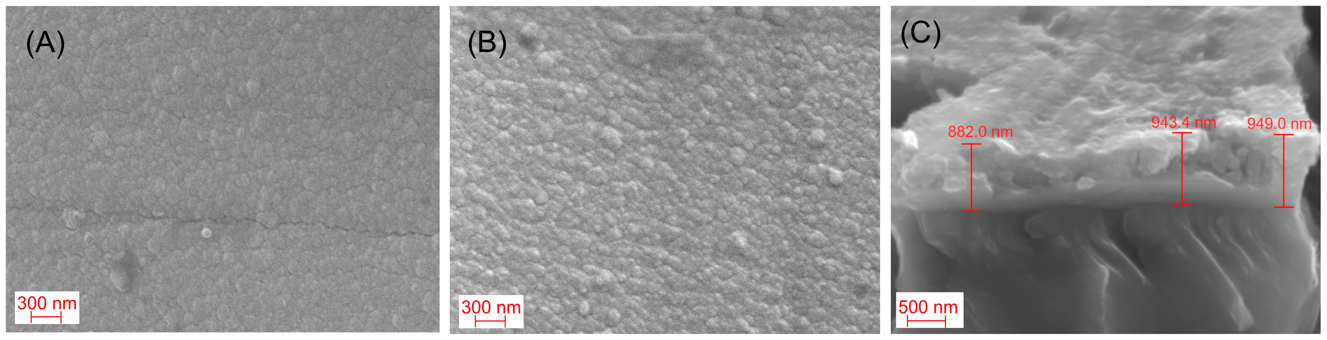
SEM observations of the as-deposited samples (Figure 2A) indicate a slightly rough surface with no second phases. The subsequent laser modification (Figure 2B) with a laser energy fluence of 0.2 J cm−2 ensures preservation of the film morphology. There is no significant qualitative change in the surface morphology of the laser-treated films. Therefore, no significant damage, including decomposition or melting of the material, was observed. Metals are known to tend to break up into droplets or islands more easily when irradiated by pulsed lasers, so dewetting is commonly seen in metal thin films [28]. In contrast, ceramic films are generally more stable and resist this process, so such behaviour is much less often reported [29]. Still, because pulsed lasers can create extremely high temperatures in very small regions, even ceramic films can potentially undergo structural changes if the local heating becomes intense enough. However, the picosecond laser with a 10 ns pulse duration and energy fluence of 0.2 J cm−2 does not substantially change the surface morphology by dewetting. Considering the cross-sections shown in Figure 2C, the average coating thickness before and after the treatment is about 925 nm. Therefore, the deposition (growth) rate of the complex oxide coating is approximately 3.85 nm/min.
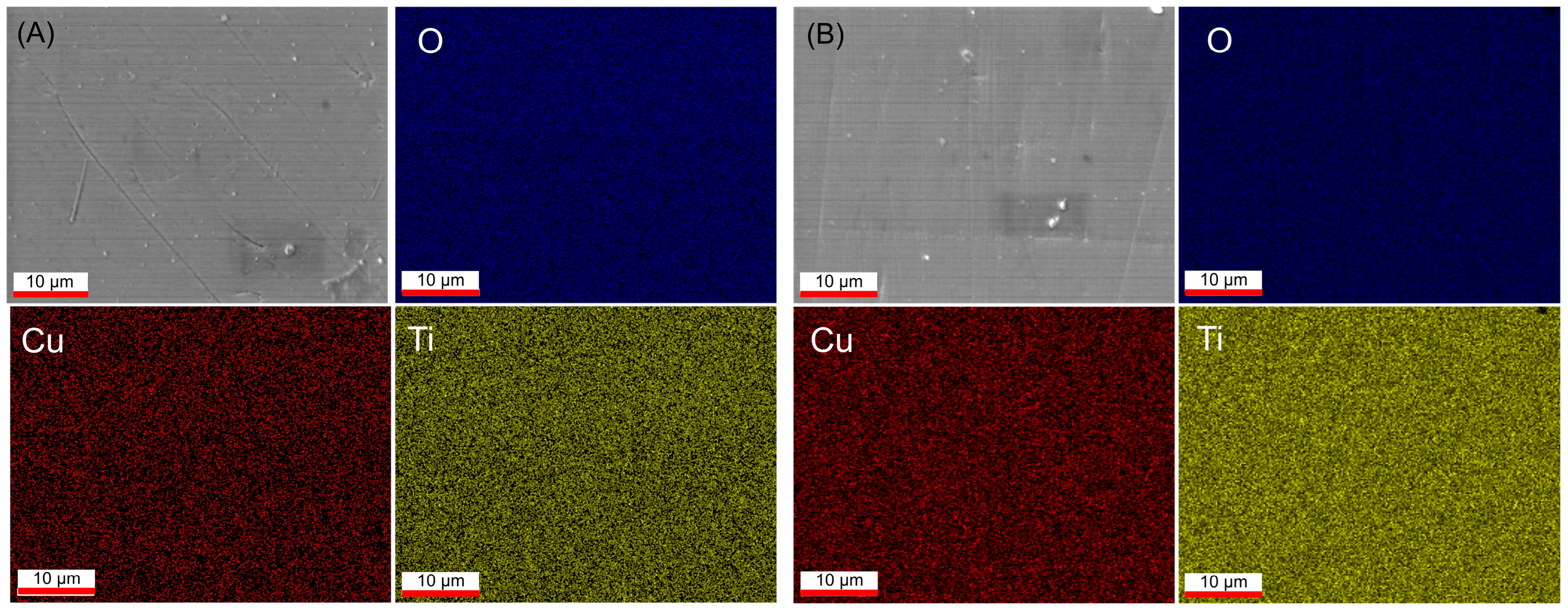
The EDS elemental mapping (Figure 3) of the AD and LST TiO2–Cu films shows that Cu is uniformly distributed across the surface. During laser annealing, Ti and Cu atoms participated in atomic rearrangement and phase transformation. However, both of them exhibit uniform incorporation without inhomogeneous distribution throughout the film. No copper clustering is seen. However, in contrast to both Ti and O content (Table 1), the amount of Cu decreased from about 2 at% for the AD coating down to 1.5 at% after the laser treatment. The decrease in copper content following laser treatment can be attributed to the thermally induced diffusion and sublimation of copper species, driven by the intense heat generated during the pulse.
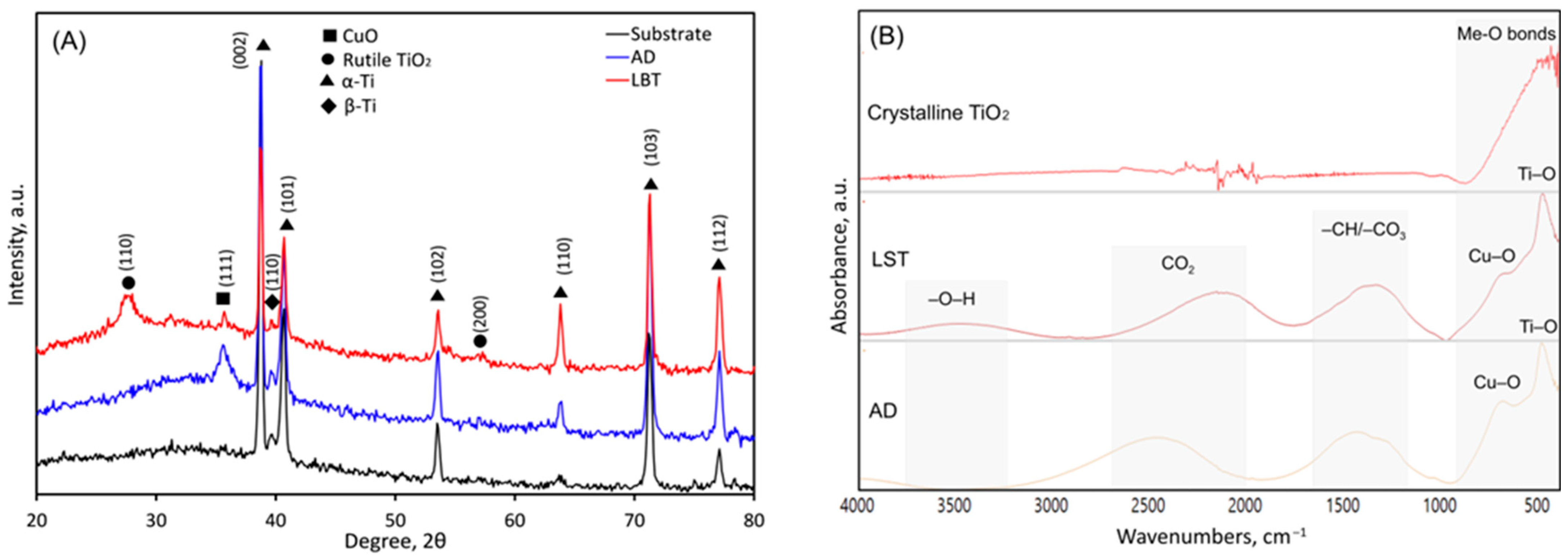
The crystal structure of the substrate, as-deposited and laser-treated samples, was studied by means of XRD analysis. The obtained patterns are displayed in Figure 4A. The peak positions were identified according to the ICDD PDF cards and marked with the corresponding crystallographic planes. The pattern of the as-deposited coating does not reveal any ordered titanium oxide phase. However, a broad CuO peak of (111) orientation can be detected. The peak width suggests the presence of nanosized crystallites with increased microstrain and high defect levels. The results suggest that the coating possesses a partially ordered molecular structure. Following laser post-treatment, rutile-phase TiO2 also crystallises, which may be associated with a laser-induced structural transformation within the film. However, the LST triggers just the early stages of crystallisation. No additional crystalline phases, such as anatase, and no evidence of an anatase-to-rutile phase transformation were observed in the XRD pattern in this sample. The peaks corresponding to copper oxide and rutile are significantly lower in intensity compared to those of the Ti6Al4V substrate. Considering that the oxide layer is approximately 925 nm thick and is formed on a 2 mm titanium alloy, this relative difference in signal intensity is expected and reasonable.
Rutile is the thermodynamically stable phase of titanium dioxide at all temperatures, but its crystallisation from amorphous precursors generally requires high temperatures, typically above 600 to 800 °C for thin films [30]. It is likely that the 0.2 J/cm2 treatment results in a temperature spike slightly above 600–800 °C, which is suitable for rutile crystallisation. According to the XRD analysis, all laser-annealed coatings consisted solely of the rutile phase. At these temperatures, the copper impurity ions within the amorphous matrix gain significant kinetic energy, promoting their migration toward the surface. Slow thermal annealing promotes the formation of crystallites and porous structures, whereas rapid heating suppresses nucleation and enhances densification [31]. Since the laser energy also triggers the structural reorganisation of TiO2 into the more densely packed rutile phase, the lattice effectively “squeezes out” larger copper ions that do not fit easily into the new crystalline structure. Because the environment is hot and the treatment often occurs at the surface, these migrated copper species, particularly from the CuO phases, can undergo thermal evaporation or sublimation, leading to a measurable loss of copper without the formation of metallic clusters. Essentially, the rutile transformation acts as a purifying process that rejects the copper dopants, which then escape the material at the high-temperature surface interface, as shown by the EDS analysis.
To analyse the composition of the materials, Fourier-transform infrared (FTIR) spectroscopy was employed. The FTIR spectra of AD, LST TiO2-Cu, and crystalline TiO2 coating (used as a reference) are shown in Figure 4B. Notably, the wide bands observed between 800 and 400 cm−1 in all coated samples are typically attributed to the vibrations of Ti-O and Ti–O–O bonds. This is further supported by the reference for crystalline TiO2 coating. Aritonang et al. also reported similar findings, indicating that the absorption area between 450 and 600 cm−1 corresponds to Ti-O vibrations [32]. Additionally, a peak at approximately 500–780 cm−1 can be attributed to the Cu-O stretching vibrations of copper oxide in the monoclinic structure [33]. This peak’s intensity diminishes following laser post-treatment, which aligns with the observed decrease in copper content as determined by energy-dispersive spectroscopy (EDS).
In the mid-infrared (IR) region, bands indicative of carbonaceous contaminants, including hydrocarbon species and carbonates interacting with oxygen vacancies and surface hydroxyl groups on hydroxylated TiO2–Cu surfaces, can be found between 1200 and 1800 cm−1 [34]. Additionally, molecularly adsorbed water is expected to contribute at approximately 1620 cm−1 [35]. The band observed around 2200–2400 cm−1 is linked to the asymmetric stretching of CO2 (g) adsorbed on the oxide surface [36]. Wu et al. [36] proposed three types of adsorbed species that can form on the TiO2 surface. In the first pathway, CO2 reacts with surface hydroxyl groups to produce bicarbonate species. The second pathway involves CO2 being captured at oxygen vacancy sites and subsequently converted into carbonate. The third pathway sees CO2 chemisorbed directly onto the TiO2 surface, quickly reaching adsorption equilibrium. After laser-induced structural modifications and partial crystallisation of TiO2 towards the rutile phase, the observed bands shift to lower wavenumbers. This indicates changes in surface hydroxylation, adsorption sites, and the local chemical environment of the oxide matrix. Furthermore, the laser–thermally treated coating likely contains water-related OH groups, as evidenced by the bond vibration observed at 3200–3700 cm−1. This peak may correspond to Ti4+-OH vibrations that were previously reactive defect sites (Ti3+) but have been filled with dissociated water. Together, these findings reveal that the laser treatment not only alters the surface hydroxylation and adsorption characteristics but also rejuvenates reactive defect sites, enhancing the overall chemical reactivity and functionality of the oxide matrix.
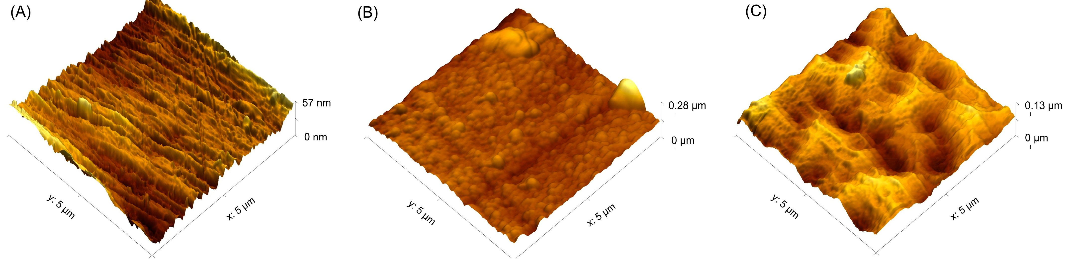
The morphology of the TiO2:Cu sample was further examined using AFM (Figure 5). The roughness values are listed in Table 2. The surface of the polished alloy (Figure 5A) displays a pronounced anisotropic morphology characterised by parallel ridges and valleys extending across the scanned area. The topography appears relatively uniform, with moderate height variations (Sz~57 nm), suggesting a finely textured surface with directional features. The untreated coating region (Figure 5B) exhibited a fine granular structure resembling small spherical particles with several protruding nodular features distributed across the scan area. The presence of micron-scale asperities suggests localised particle agglomeration. The Sz values indicate substantially greater roughness than that observed for the polished surface. After the picosecond laser treatment (Figure 5C), the surface possesses a highly developed cellular or crater-like morphology, consisting of regularly distributed depressions separated by raised rims. The surface exhibits a more complex three-dimensional architecture associated with laser-induced surface restructuring. The periodic arrangement of pits and valleys creates a hierarchical texture that significantly increases surface complexity and the density of surface-active sites. The significant decrease in Ssk from +1.42 for the as-deposited coating to −0.26 after laser treatment (Table 2) indicates a pronounced transformation from a peak-dominated to a valley-dominated surface morphology. This change reflects the removal or remelting of surface asperities and the development of depressions during laser processing, resulting in a more complex hierarchical topography.
Nanoscale surface roughness has been recognised as an important contributor to antibacterial performance, especially when combined with bioactive agents such as Cu. In contrast to smooth surfaces that facilitate bacterial attachment, nanostructured topographies increase the contact area between bacterial cells and the substrate. This enhanced interaction can induce mechanical deformation and disruption of the bacterial membrane, ultimately leading to cell death. Consequently, surfaces with pronounced nanoscale roughness can effectively inhibit bacterial proliferation and biofilm development by altering cell morphology and compromising membrane integrity [37].

3.2. Mechanical Properties

The surface hardness of the TiO2–Cu films, as determined by microhardness testing, is summarised in Table 3. Both TiO2–Cu-coated samples exhibit higher hardness compared to the Ti6Al4V substrate. The as-deposited (AD) coating shows lower hardness than the laser surface-treated (LST) sample and displays only a single discernible diffraction peak corresponding to the CuO phase (Figure 4A), suggesting that a substantial portion of the film is partially ordered. The increased hardness after laser treatment could be attributed to partial rutile formation in the laser-modified oxide layer during rapid heating and cooling, which contributes to an increase in hardness [38,39]. Although the results revealed a slightly lower Cu concentration at the immediate surface after laser treatment, laser-induced changes in the oxide microstructure and surface morphology may facilitate Cu ion release, indicating that the measured surface Cu concentration does not necessarily correlate directly with the amount of Cu ions released. Engelmann et al. reported the deposition of TiO2 films by magnetron sputtering in a reactive oxygen atmosphere, resulting in predominantly amorphous coatings with a thickness of 630 nm and an even lower microhardness of 457 ± 18 HV0.01 (approximately 8.09 GPa) [40]. The hardness of amorphous TiO2 has been reported to lie in the range of 7.4–7.8 GPa [41], which is consistent with the values obtained in the present study. In contrast, the LST coating exhibits higher hardness, which can be attributed to the occurrence of the crystalline rutile phase following laser annealing, as well as to structural densification.
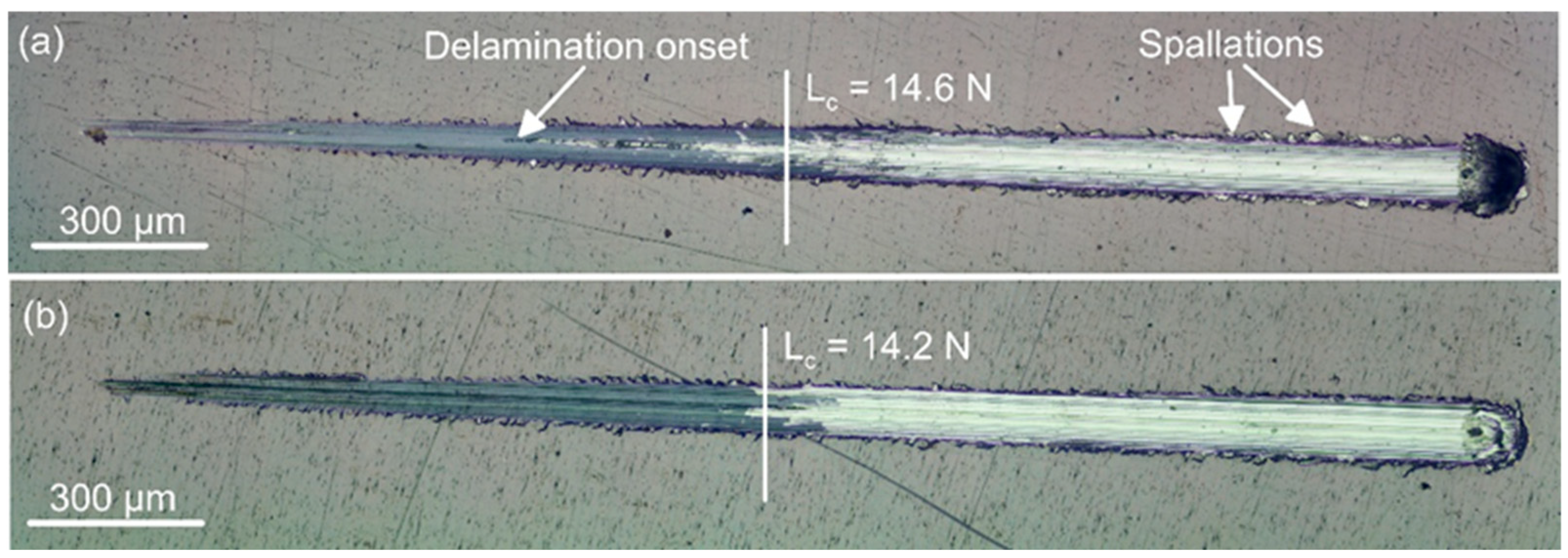
The scratch test results shown in Figure 6 demonstrate the adhesion strength between the as-deposited (AD) and laser surface-treated (LST) coatings. For the as-deposited coating (Figure 6a), the scratch track shows an earlier onset of coating damage. The critical load (Fc) was determined from scratch-track images in combination with a sudden change in the coefficient of friction, both of which indicate coating failure. Some small lateral spallations are also seen. In contrast, the LST coating (Figure 6b) exhibits a more stable scratch response with reduced spallation and a more continuous track morphology. The overall damage morphology appears more controlled. Although the increase in the critical adhesion force is modest, the laser-treated coating exhibits a more uniform scratch damage morphology and reduced local spallation, indicating improved resistance to localised mechanical damage while maintaining comparable coating adhesion. In comparison with similar TiO2–Cu coatings deposited on the Ti6Al4V alloy with approximately twice the thickness, the coatings examined in this study demonstrate intermediate critical load values [42]. Overall, the mechanical behaviour suggests that laser surface treatment modifies the coating structure toward a more compact, harder, and mechanically stable configuration, improving damage tolerance despite a similar critical load threshold. However, dedicated wear and tribocorrosion studies are necessary to evaluate the long-term performance of the coatings under clinically relevant loading conditions.

3.3. Wettability and Ion Release

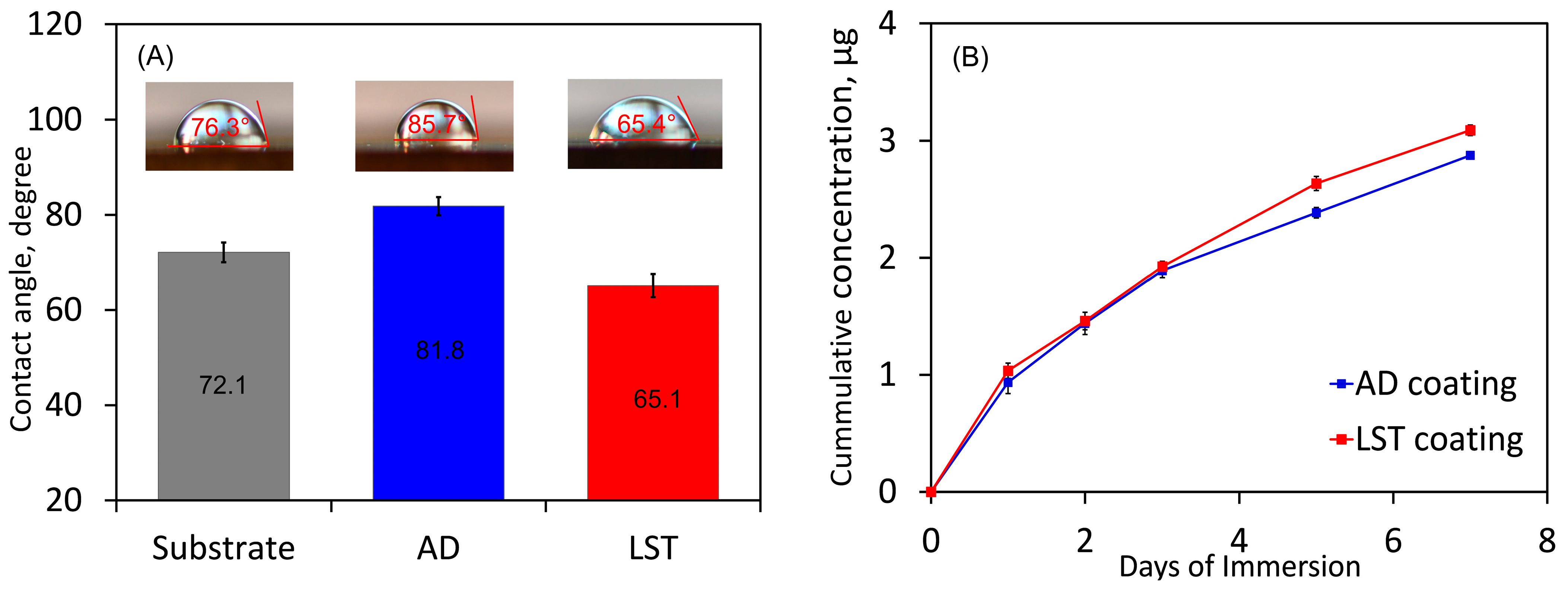
Surface wettability plays a crucial role in cellular interactions with biomaterials, as improved hydrophilicity can facilitate cell adhesion and spreading on the material surface [43]. The wettability of the surfaces was evaluated by static contact angle measurements, as shown in Figure 7A. The pristine substrate exhibited a contact angle of 72.1°, indicating moderate hydrophilicity. After coating, a noticeable change in surface wettability was observed. The AD-coated sample showed an increased contact angle of 81.8°, suggesting a reduction in hydrophilicity and a comparatively more hydrophobic surface. In contrast, the LST-coated sample displayed a lower contact angle of 65.1°, indicating enhanced hydrophilicity relative to both the substrate and the AD coating. This improvement in wettability for the LST coating may be attributed to the presence of more polar functional groups, as determined by the FTIR analysis. It has been reported that micro-arc-oxidised Cu–TiO2 coatings with moderate contact angles of 43–65° support higher viability and proliferation of bone mesenchymal stem cells (BMSCs) than Ti, and at intermediate Cu levels (1Cu-MAO), in particular, they enhance osteogenic marker expression [44]. Except for favourable biomaterial performance, the enhanced wettability of the LST surface may facilitate greater interaction with the surrounding simulated body fluid (SBF), which is consistent with the higher cumulative ion release observed in Figure 7B.
The cumulative copper release from TiO2–Cu coatings under as-deposited (AD) and laser surface-treated (LST) conditions is shown in Figure 7B. Both coatings exhibit a time-dependent increase in Cu release, with an initial rapid release within the first day followed by a slower, sustained release up to 7 days, indicating a transition from surface-controlled to diffusion-controlled behaviour. Despite the lower copper content, the LST coating consistently shows higher Cu release than the AD coating, with the difference becoming slightly more pronounced over time. The slightly increased Cu release after laser treatment may be associated with laser-induced microstructural modifications, including Cu redistribution, grain refinement, increased defect density, and changes in surface morphology, which have been reported to influence ion release behaviour in laser-processed metallic materials [45]. This behaviour can be attributed to laser-induced microstructural changes, such as improved crystallisation, which likely enhance copper redistribution and facilitate ion diffusion. Crystallisation into rutile may force Cu out of the TiO2 lattice and concentrate it at grain boundaries and near-surface regions enhanced by laser treatment, making it far more accessible to the environment than when it is uniformly trapped in a partially ordered matrix. The more hydrophilic laser-treated TiO2-Cu (65.1°) has greater liquid contact and easier electrolyte access to Cu-containing regions, which can favour higher Cu release. Overall, laser surface treatment results in a moderately higher and more sustained copper release, which is advantageous for applications requiring prolonged ion activity. In comparison, Ti–Cu-sintered alloys used in dental and orthopaedic applications have been reported to release up to 0.3 mg·L−1 of Cu within 24 h, a level considered within the safe range while still providing antibacterial effects [46]. Notably, the total Cu ion release from the TiO2–Cu coatings in this study did not exceed 3 µg over 7 days (Figure 7), further supporting the good biosafety of these Cu-containing titanium coatings.

3.4. Electrochemical Tests

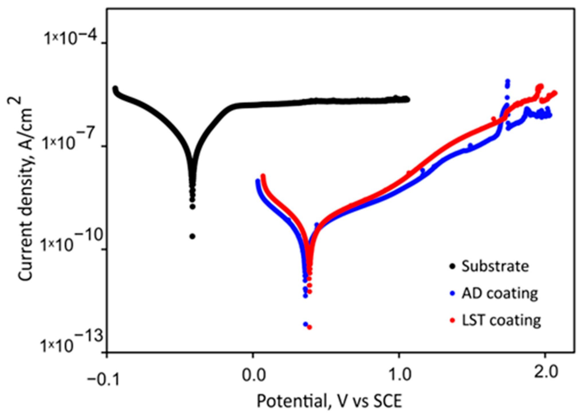
It is well known that many medical devices are prone to corrosion during clinical use. In particular, after implantation into the human body, corrosion at the interface between the implant surface and the surrounding physiological environment can significantly compromise both biocompatibility and service life. To evaluate the corrosion resistance of the TiO2–Cu films, potentiodynamic polarisation tests were conducted in SBF solution (pH = 7.4), and the results are presented in Figure 8. The corresponding anodic and cathodic slopes, Ecorr and jcorr values obtained from the polarisation curves, as well as the protection efficacy (P.E.) values are summarised in Table 4.
Compared with the substrate, the Ecorr values of the AD and LST TiO2–Cu coatings shift toward more positive potentials, while their jcorr values decrease. This behaviour may be attributed to the formation of a compact TiO2/CuO layer on the surface of the coatings, which effectively hinders the penetration of corrosive species through surface defects [47]. Overall, the jcorr values of the TiO2–Cu films show a gradual decreasing trend. This reflects the protection efficacy of both coated samples, which reach 99.4%. Moreover, both βA and βC values increase significantly after applying the TiO2–Cu coatings compared to bare Ti6Al4V, indicating reduced anodic dissolution and cathodic reaction rates. For both parameters, AD shows the highest values, followed by LST, while Ti6Al4V exhibits the lowest values. Therefore, the lower contact angle of the laser-treated TiO2-Cu coatings, combined with an increase in accessible Cu, is consistent with higher copper release and reduced corrosion resistance of the LST compared to the AD sample. Overall, the trend inferred from the PDC curves, namely AD > LST > Ti6Al4V, suggests that AD provides the strongest electrochemical barrier behaviour among the tested samples.
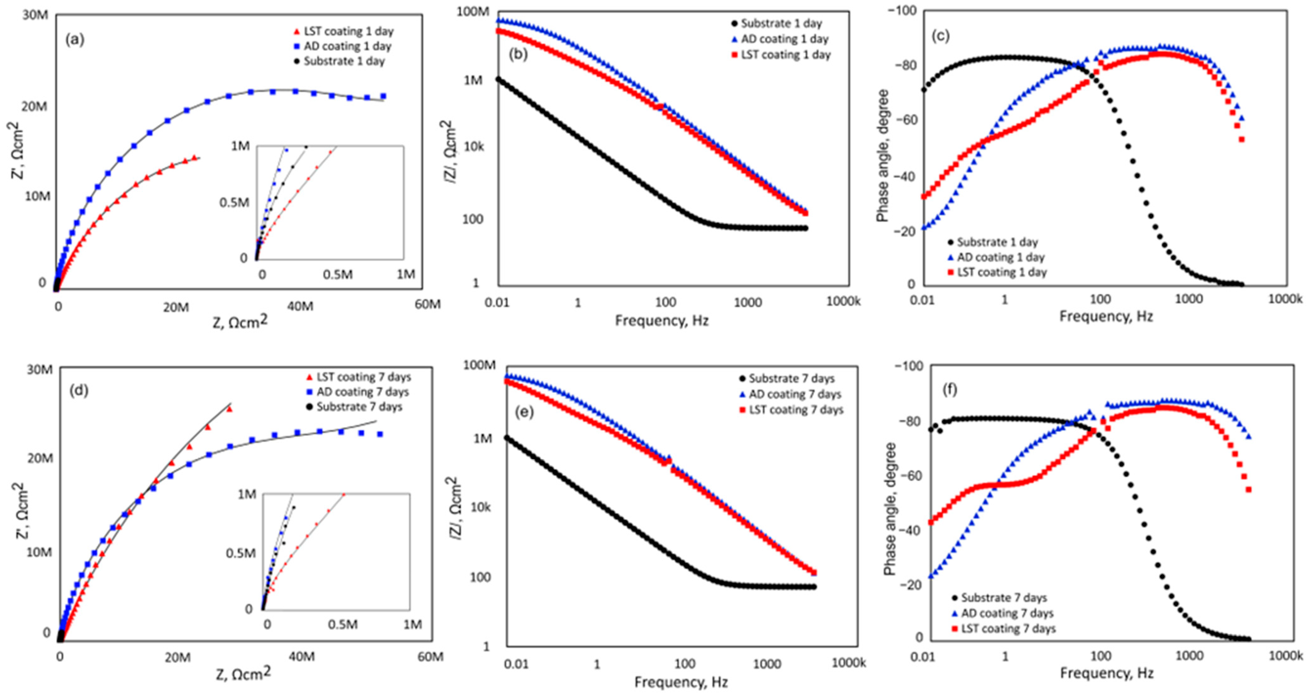
The EIS results shown in Figure 9 demonstrate the evolution of barrier protection and electrochemical stability of the coatings. The Nyquist plots (Figure 9a,d) reveal a significant enlargement of the capacitive semicircle for both coated samples compared with bare Ti6Al4V, confirming the improved corrosion resistance provided by the coatings. After 1 day of immersion, the AD coating exhibits the largest arc diameter, indicating superior initial barrier protection. However, after 7 days, the LST coating shows a more stable and larger impedance response, suggesting enhanced long-term electrochemical stability and lower electrolyte penetration.
The Bode modulus spectra (Figure 9b,e) show that both coatings increase the low-frequency impedance by nearly two orders of magnitude relative to the substrate, demonstrating their strong protective character. The coated samples maintain impedance values close to 108 Ωcm2, whereas the bare alloy remains near 106 Ωcm2. Although the AD coating exhibits high initial impedance, a slight reduction is observed after prolonged immersion, while the LST coating retains a more stable impedance response, indicating better long-term barrier performance.
The phase angle diagrams (Figure 9c,f) indicate clear differences in electrochemical behaviour between the substrate and coated samples. The bare Ti6Al4V alloy exhibits a narrower phase angle region and a rapid decrease at intermediate frequencies, reflecting less stable passive behaviour. In contrast, both coatings show broad phase angle maxima approaching –85°, characteristic of highly capacitive and homogeneous protective layers. The LST coating maintains a wider capacitive region after 7 days, suggesting improved coating integrity and interfacial stability during immersion.
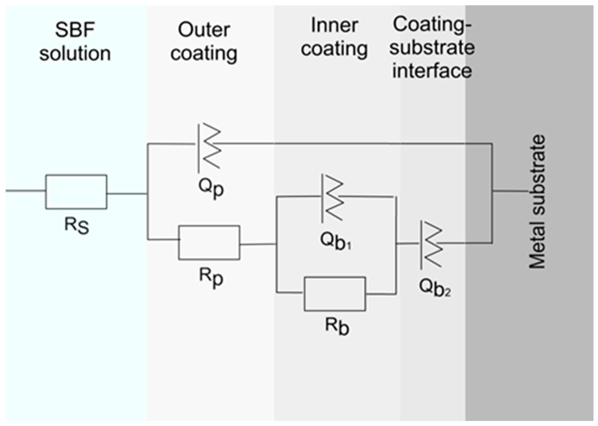
The equivalent circuit scheme used to match the impedance data is presented in Figure 10. In this circuit, RS represents the electrolyte resistance. The Qp–Rp pair corresponds to the porous outer coating, while Qb1–Rb1 represents the dense barrier layer of the coating. The element Qb2 describes the capacitive behaviour of the coating–substrate interface or passive oxide layer on Ti6Al4V. The absence of Rb2 indicates that no distinct charge transfer resistance is detected at this interface, suggesting predominantly capacitive behaviour and limited corrosion activity.
The fitted equivalent circuit parameters (Table 5) further confirm the superior protective behaviour of the coatings. Both coated samples exhibit very low Qp values and high n1 values (0.91–0.97), indicating compact and nearly ideal capacitive behaviour. The AD coating shows a marked decrease in polarisation resistance (Rp) after 7 days, suggesting coating degradation and electrolyte penetration. In contrast, the LST coating maintains or slightly increases its Rp value with immersion time, indicating progressive stabilisation of the protective layer and superior long-term corrosion resistance. The increase of nb1 and nb2 toward unity, particularly for the LST coating, suggests a transition toward more ideal capacitive behaviour and a more homogeneous electrochemical interface. This behaviour may be associated with pore sealing or stabilisation of the passive layer during immersion, contributing to the improved corrosion resistance of the LST coating over prolonged exposure.
The EIS results demonstrate that both coatings significantly improve the corrosion resistance of Ti6Al4V by increasing the impedance modulus by nearly two orders of magnitude. However, their long-term electrochemical evolution differs substantially. The AD coating exhibits high initial resistance but undergoes rapid degradation upon prolonged immersion, evidenced by the sharp decrease in Rp and Rb. In contrast, the LST coating maintains high impedance and even shows an increase in Rp after 7 days, suggesting progressive stabilisation of the passive interface. Compared with previously reported multilayer TiO2-CuO magnetron-sputtered coatings, which exhibited polarisation resistance values on the order of 108 Ω cm2 after immersion in SBF [27], the present coatings demonstrate similarly outstanding corrosion protection.
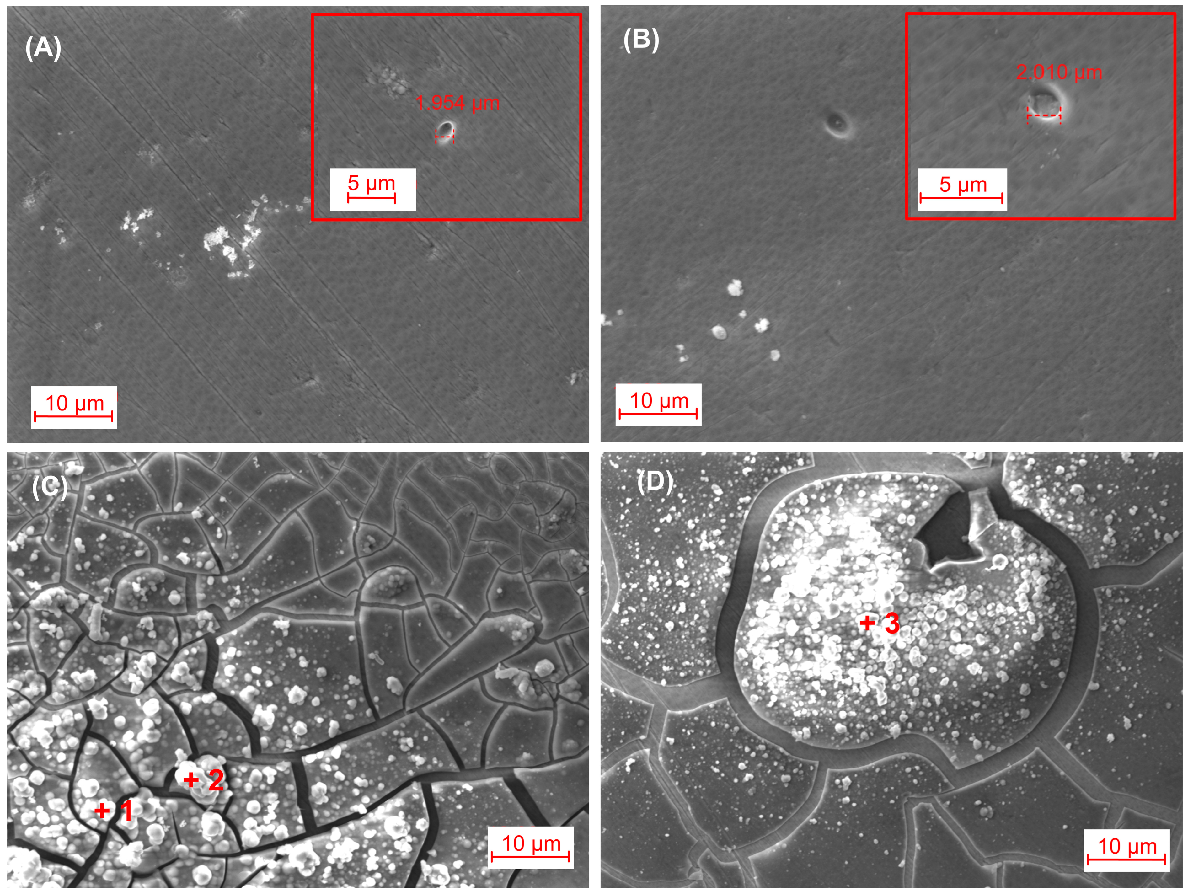
The SEM micrographs after 7 days of EIS immersion reveal differences in the degradation behaviour and surface reactivity of the AD and LST coatings. Both coatings remained generally intact after immersion; however, localised defects and shallow pits with diameters of approximately 1.95–2.01 μm were observed on the surfaces (Figure 11A,B). The presence of these isolated pits suggests limited electrolyte penetration through coating defects, although no severe coating delamination or extensive corrosion attack was detected.
Figure 11C,D reveal abundant particulate deposits distributed across the immersed surfaces. The EDS analyses performed at points 1–3 confirmed the enrichment of Ca, P, and O in the deposited regions. Point 1 contained approximately P (8.8 at%), Ca (2.7 at%), K (0.7 at%), and O (77.4 at%), while point 2 showed P (9.6 at%), Ca (3.1 at%), K (1.1 at%), and O (76 at%). Point 3 exhibited the highest Ca and P contents, with P (10 at%), Ca (3.9 at%), K (1 at%), and O (76.1 at%). The simultaneous presence of Ca and P strongly indicates the formation of hydroxyapatite (HA)-like deposits on the coating surfaces after immersion. All these findings suggest that the coatings not only provide effective corrosion protection but also promote bioactive surface mineralisation beneficial for biomedical applications.

3.5. Antibacterial Properties

The antibacterial activity was assessed against S. aureus, a clinically relevant Gram-positive pathogen and a leading cause of implant-associated infections [48]. Table 6 presents the antibacterial efficacy of the AD and LST surfaces relative to the polished control, quantified through colony-forming unit enumeration. Both modified surfaces significantly suppressed bacterial viability, as evidenced by the reduced number of recoverable colonies. The AD and LST treatments achieved reductions of 67% and 74%, respectively, in the final bacterial population, confirming their pronounced bactericidal activity. The CFU assay employed in this study evaluates the overall antibacterial response after incubation and does not distinguish between reduced initial bacterial adhesion and decreased bacterial viability. Therefore, the observed antibacterial activity likely reflects the combined influence of laser-induced surface modifications on both bacterial attachment and subsequent survival.
The slightly greater reduction in bacterial colonies observed on the LST surface indicates superior bactericidal performance, likely attributable to a combination of its enhanced physicochemical properties. Compared with the AD surface, the LST treatment produced a more crystalline coating containing rutile TiO2 and CuO phases, a higher density of surface hydroxyl (–OH) groups, increased hydrophilicity, and a slightly greater release of Cu ions in SBF. These characteristics can enhance surface reactivity, promote closer interactions between bacteria and the coating, and facilitate the release of bioactive copper species. Previous studies have demonstrated that copper-containing materials can exhibit significant antibacterial activity under dark conditions due to the cytotoxic effects of released Cu ions and the presence of solid-state CuxO nanoclusters and Cu species [49,50]. Furthermore, direct contact between copper-containing particles and bacterial cells can disrupt membrane integrity and cellular metabolism, ultimately reducing bacterial viability [51]. Therefore, the improved antibacterial efficacy of the LST surface is likely a result of the synergistic effects of enhanced crystallinity, surface hydroxylation, morphology, wettability, and copper ion release. The antibacterial effect of copper is multifaceted, involving membrane disruption, oxidative stress, and inhibition of biofilm development [52,53]. These mechanisms can act synergistically, resulting in irreversible damage to essential cellular components, including proteins, lipids, and nucleic acids, ultimately leading to bacterial death [53]. Consequently, surfaces capable of sustaining the release of bioactive copper species while maintaining direct contact with bacterial cells are expected to exhibit enhanced and long-lasting antimicrobial performance.
In addition to the chemical modifications induced by laser treatment, the altered surface nanotopography may also contribute to the antibacterial response. AFM observations revealed the development of a more complex hierarchical surface architecture composed of regularly distributed depressions and valleys, which may influence the initial bacteria–surface interaction by modifying the available contact area and local mechanical interactions. Although the present study does not permit the individual contributions of surface chemistry and nanotopography to be separated, both factors are likely to act synergistically. Recent work by Villanueva et al. [54] demonstrated that nanoscale roughness can alter membrane–surface interactions by changing the energetic barriers associated with membrane deformation and reorganisation. In the case of S. aureus, the presence of a thick peptidoglycan cell wall and the absence of an outer membrane suggest that these effects should be interpreted cautiously and in conjunction with copper-mediated antibacterial mechanisms.
Overall, the present results suggest that the enhanced antibacterial activity of the laser-treated coatings originates from the synergistic effects of laser-induced structural, chemical, and morphological modifications rather than from any single parameter. Further studies specifically designed to decouple the individual contributions of surface chemistry, nanotopography, and copper ion release will be required to establish their relative importance.
The antibacterial performance obtained in this study is comparable to previously reported Cu-containing TiO2 coatings, which exhibited antibacterial efficiencies ranging from 70% for TiO2–Cu films containing ~1.5 at.% Cu [55] to >99% for TiO2/CuO coatings with 3.56 at.% Cu against S. aureus, which still demonstrated improved biocompatibility with MC3T3-E1 osteoblasts [13]. While the present study indicates significant antibacterial activity under dark conditions, the crystalline TiO2/CuO architecture offers additional opportunities for NIR-triggered antimicrobial therapy. This concept is supported by previous studies demonstrating that combining laser irradiation with antimicrobial metal nanostructures, such as Ag nanoparticles, can yield significantly stronger bactericidal effects than either treatment alone against both Gram-positive and Gram-negative bacteria [56]. Enhanced ROS generation and photothermal conversion may amplify the bactericidal response beyond that achieved through copper-mediated mechanisms alone. Evaluating these stimuli-responsive effects constitutes an important direction for future research. Moreover, future work will focus on evaluating bacterial adhesion and viability of the developed coatings against Gram-positive and Gram-negative bacteria, as well as multi-species biofilms, to provide a more comprehensive assessment of their antimicrobial efficacy and clinical potential.

4. Conclusions

TiO2/CuO coatings were successfully fabricated on Ti6Al4V substrates via magnetron sputtering and subsequently modified by selected picosecond laser surface treatment. The novelty of this work lies in demonstrating that picosecond laser post-treatment provides an effective strategy for improving the multifunctional performance of magnetron co-sputtered Cu-doped TiO2 coatings for biomedical applications. The principal findings of this study can be summarised as follows:
  • Picosecond laser treatment promoted the crystallisation of the partially ordered surface structure into a rutile-containing one while preserving the CuO phase, leading to increased surface hydroxylation and improved wettability.
  • The laser-treated coating exhibited improved mechanical properties, including higher hardness and enhanced resistance to surface scratch damage, which can contribute to greater durability and wear resistance under physiological loading conditions.
  • Both AD and LST coatings substantially enhanced the corrosion resistance of Ti6Al4V in simulated body fluid, increasing the impedance modulus by nearly two orders of magnitude compared with the uncoated substrate. However, the LST coating exhibited superior long-term electrochemical stability and maintained a more protective passive interface during prolonged immersion.
  • Immersion studies revealed the formation of Ca–P-rich deposits consistent with hydroxyapatite on both coated surfaces, confirming their ability to promote bioactive mineralisation while maintaining coating integrity.
  • Significant antibacterial activity against Staphylococcus aureus was achieved under dark conditions, with antibacterial efficiencies of 67% and 74% for the AD and LST coatings, respectively. The superior performance of the LST surface was attributed to the synergistic effects of enhanced crystallinity, increased density of surface hydroxyl groups, greater hydrophilicity, and slightly higher copper ion release.
  • The combined corrosion protection, bioactivity, and antibacterial properties demonstrate that laser-modified TiO2/CuO coatings represent a promising surface engineering strategy for reducing implant-associated infections while supporting osseointegration.
Unlike conventional laser surface modification studies that typically focus on a single functional aspect, the present work demonstrates that picosecond laser post-treatment can simultaneously improve the structural, mechanical, electrochemical, bioactive, and antibacterial performance of magnetron-sputtered Cu-doped TiO2 coatings without compromising coating integrity. This integrated approach provides new insight into the design of multifunctional implant surfaces. Future work will focus on investigating their near-infrared (NIR)-activated photothermal and photocatalytic antibacterial performance, which may further enhance bacterial eradication through the generation of reactive oxygen species and localised heating. Although the present study evaluated antibacterial activity only against Staphylococcus aureus, future investigations will include Gram-negative bacteria and polymicrobial biofilm models to provide a more comprehensive assessment of the antimicrobial performance of the laser-treated coatings.

Author Contributions

Conceptualisation, M.P.N. and S.V.; methodology, M.P.N., M.O., E.Z., N.N., I.T., I.K. and S.V.; software, M.P.N.; validation, M.P.N., S.V. and N.N.; formal analysis, E.Z.; investigation, M.P.N., M.O., R.N. (Rosen Nikov), R.N. (Rumen Nikov), E.L., E.Z., A.A., I.T. and I.K.; resources, M.P.N.; data curation, E.Z. and I.A.; writing—original draft preparation, M.P.N.; writing—review and editing, M.P.N., S.D., N.N. and S.V.; visualisation, M.P.N.; supervision, M.P.N.; project administration, M.P.N.; funding acquisition, M.P.N. All authors have read and agreed to the published version of the manuscript.

Funding

This research was funded by the Bulgarian National Science Fund, grant number KII-06-H67-5 (2022).

Institutional Review Board Statement

Not applicable.

Informed Consent Statement

Not applicable.

Data Availability Statement

The original contributions presented in this study are included in the article. Further inquiries can be directed to the corresponding author.

Conflicts of Interest

The authors declare no conflicts of interest. The funders had no role in the design of the study; in the collection, analyses, or interpretation of data; in the writing of the manuscript; or in the decision to publish the results.

References

  1. Davis, R.; Singh, A.; Jackson, M.J.; Coelho, R.T.; Prakash, D.; Charalambous, C.P.; Ahmed, W.; da Silva, L.R.R.; Lawrence, A.A. A comprehensive review on metallic implant biomaterials and their subtractive manufacturing. Int. J. Adv. Manuf. Technol. 2022, 120, 1473–1530. [Google Scholar] [CrossRef] [Scilit] [PubMed]
  2. Yuan, Z.; He, Y.; Lin, C.; Liu, P.; Cai, K. Antibacterial surface design of biomedical titanium materials for orthopedic applications. J. Mater. Sci. Technol. 2021, 78, 51–67. [Google Scholar] [CrossRef] [Scilit]
  3. Xia, C.; Ma, X.; Zhang, X.; Li, K.; Tan, J.; Qiao, Y.; Liu, X. Enhanced physicochemical and biological properties of C/Cu dual ions implanted medical titanium. Bioact. Mater. 2020, 5, 377–386. [Google Scholar] [CrossRef] [Scilit] [PubMed]
  4. Silva Souza, J.G.; Bertolini, M.M.; Costa, R.C.; Nagay, B.E.; Dongari-Bagtzoglou, A.; Barao, V.A.R. Targeting implant-associated infections: Titanium surface loaded with antimicrobial. iScience 2021, 24, 102008. [Google Scholar] [CrossRef] [Scilit] [PubMed]
  5. Sun, X.D.; Liu, T.T.; Wang, Q.Q.; Zhang, J.; Cao, M.S. Surface modification and functionalities for titanium dental implants. ACS Biomater. Sci. Eng. 2023, 9, 4442–4461. [Google Scholar] [CrossRef] [Scilit] [PubMed]
  6. Mishchenko, O.; Volchykhina, K.; Maksymov, D.; Manukhina, O.; Pogorielov, M.; Pavlenko, M.; Iatsunskyi, I. Advanced Strategies for Enhancing the Biocompatibility and Antibacterial Properties of Implantable Structures. Materials 2025, 18, 822. [Google Scholar] [CrossRef] [Scilit] [PubMed]
  7. Ghodrati, H.; Goodarzi, A.; Golrokhian, M.; Fattahi, F.; Anzabi, R.M.; Mohammadikhah, M.; Sadeghi, S.; Mirhadi, S. A narrative review of recent developments in osseointegration and anti-corrosion of titanium dental implants with nano surface. Bone Rep. 2025, 25, 101846. [Google Scholar] [CrossRef] [Scilit] [PubMed]
  8. Bayandinova, M.; Kenesbekov, A.; Serikbaykyzy, A.; Askhatov, A.; Batanov, Y.; Bazarov, N. Development of biocompatible coatings for orthopedic joint implants. Phys. Sci. Technol. 2025, 12, 84–94. [Google Scholar] [CrossRef] [Scilit]
  9. Foster, H.; Ditta, I.B.; Varghese, S.; Steele, A. Photocatalytic disinfection using titanium dioxide: Spectrum and mechanism of antimicrobial activity. Appl. Microbiol. Biotechnol. 2011, 90, 1847–1868. [Google Scholar] [CrossRef] [Scilit] [PubMed]
  10. Akshaya, S.; Rowlo, P.K.; Dukle, A.; Nathanael, A.J. Antibacterial coatings for titanium implants: Recent trends and future perspectives. Antibiotics 2022, 11, 1719. [Google Scholar] [CrossRef] [Scilit] [PubMed]
  11. Zheleva, E.; Nikolova, M.P. Perspective on copper-enhanced titanium implants: Functions, fabrication, and safety. Mater. Des. 2026, 265, 115861. [Google Scholar] [CrossRef] [Scilit]
  12. Yang, H.L.; Zhu, M.Z.; Wang, J.Y.; Ma, C.X.; Zhou, X.W.; Xing, H.X.; Zhang, E.L.; Ji, S.X. Optimization of mechanical and antibacterial properties of Ti-3wt%Cu alloy through cold rolling and annealing. Rare Met. 2022, 41, 610–620. [Google Scholar] [CrossRef] [Scilit]
  13. He, X.J.; Zhang, G.N.; Wang, X.; Hang, R.Q.; Huang, X.B.; Qin, L.; Tang, B.; Zhang, X.Y. Biocompatibility, corrosion resistance and antibacterial activity of TiO2/CuO coating on titanium. Ceram. Int. 2017, 43, 16185–16195. [Google Scholar] [CrossRef] [Scilit]
  14. Romero, L.; Araya, N.; Palacio, D.; Sánchez-Sanhueza, G.; Pérez, E.; Solís, F.; Meléndrez, M.; Medina, C. Study of the antibacterial capacity of a biomaterial of zeolites saturated with copper ions (Cu2+) and supported with copper oxide nanoparticles. Nanomaterials 2023, 13, 2140. [Google Scholar] [CrossRef] [Scilit] [PubMed]
  15. Wang, X.; Wang, W.-X. Cu(I)/Cu(II) released by Cu nanoparticles revealed differential cellular toxicity related to mitochondrial dysfunction. Environ. Sci. Technol. 2023, 57, 9548–9558. [Google Scholar] [CrossRef] [Scilit] [PubMed]
  16. Cao, S.; Zhang, Z.M.; Zhang, J.Q.; Wang, R.X.; Wang, X.Y.; Yang, L.; Chen, D.F.; Qin, G.W.; Zhang, E.L. Improvement in antibacterial ability and cell cytotoxicity of Ti–Cu alloy by anodic oxidation. Rare Met. 2022, 41, 594–609. [Google Scholar] [CrossRef] [Scilit]
  17. Milenkovic, J.; Hrenovic, J.; Matijasevic, D.; Niksic, M.; Rajic, N. Bactericidal activity of Cu-, Zn-, and Ag-containing zeolites toward Escherichia coli isolates. Environ. Sci. Pollut. Res. 2017, 24, 20273–20281. [Google Scholar] [CrossRef] [Scilit] [PubMed]
  18. Wang, F.; Wu, J. Plasma-enhanced chemical vapor deposition. In Modern Ion Plating Technology; Wang, F., Wu, J., Eds.; Elsevier: Amsterdam, The Netherlands, 2023; pp. 247–285. [Google Scholar] [CrossRef] [Scilit]
  19. Wang, F.; Wu, J. Glow discharge ion plating technology. In Modern Ion Plating Technology; Wang, F., Wu, J., Eds.; Elsevier: Amsterdam, The Netherlands, 2023; pp. 115–135. [Google Scholar] [CrossRef] [Scilit]
  20. Asad, J.; Afzal, N.; Rafique, M.; Rizwan, M.; Yasin, M. Annealing effect on DC magnetron sputtered TiO2 film: Theoretical and experimental investigations. Arab. J. Sci. Eng. 2024, 50, 571–581. [Google Scholar] [CrossRef] [Scilit]
  21. Ağırseven, O.; Rivella, D.; Haggerty, J.; Berry, P.; Diffendaffer, K.; Patterson, A.; Kreb, J.; Mangum, J.; Gorman, B.; Perkins, J.; et al. Crystallization of TiO2 polymorphs from RF-sputtered, amorphous thin-film precursors. AIP Adv. 2020, 10, 015036. [Google Scholar] [CrossRef] [Scilit]
  22. Zimbone, M.; Cantarella, M.; Sfuncia, G.; Nicotra, G.; Privitera, V.; Napolitani, E.; Impellizzeri, G. Low-temperature atomic layer deposition of TiO2 activated by laser annealing: Applications in photocatalysis. Appl. Surf. Sci. 2022, 596, 153641. [Google Scholar] [CrossRef] [Scilit]
  23. Wilkes, G.C.; Deng, X.; Choi, J.J.; Gupta, M.C. Laser annealing of TiO2 electron-transporting layer in perovskite solar cells. ACS Appl. Mater. Interfaces 2018, 10, 41312–41317. [Google Scholar] [CrossRef] [Scilit] [PubMed]
  24. Yusuf, Y.; Ghazali, M.; Otsuka, Y.; Ohnuma, K.; Morakul, S.; Nakamura, S.; Abdollah, M. Antibacterial properties of laser surface-textured TiO2/ZnO ceramic coatings. Ceram. Int. 2020, 46, 3949–3959. [Google Scholar] [CrossRef] [Scilit]
  25. Xiong, J.; Tang, X.; Yu, L.; Xiao, D. Antimicrobial and osteogenic performance comparison of titanium implants between the conventional array structure surface and post-etched surface. J. Mater. Eng. Perform. 2025, 34, 20420–20435. [Google Scholar] [CrossRef] [Scilit]
  26. Van Overschelde, O.; Guisbiers, G.; Hamadi, F.; Hemberg, A.; Snyders, R.; Wautelet, M. Alternative to classic annealing treatments for fractally patterned TiO2 thin films. J. Appl. Phys. 2008, 104, 103106. [Google Scholar] [CrossRef] [Scilit]
  27. Valkov, S.; Nikolova, M.P.; Dimitrova, T.V.; Stancheva, M.E.; Dechev, D.; Ivanov, N.; Handzhiyski, Y.; Andreeva, A.; Ormanova, M.; Anchev, A.; et al. Multilayer Ti–Cu oxide coatings on Ti6Al4V: Balancing antibacterial activity, mechanical strength, corrosion resistance, and cytocompatibility. J. Funct. Biomater. 2026, 17, 16. [Google Scholar] [CrossRef] [Scilit] [PubMed]
  28. Kondic, L.; González, A.G.; Diez, J.A.; Fowlkes, J.D.; Rack, P. Liquid-state dewetting of pulsed-laser-heated nanoscale metal films and other geometries. Annu. Rev. Fluid Mech. 2020, 52, 235–262. [Google Scholar] [CrossRef] [Scilit]
  29. Kiisk, V.; Kodu, M.; Pikker, S.; Avarmaa, T.; Jaaniso, R. Oxygen-sensitive luminescence of ultrathin CdWO4:Sm3+ films. Opt. Mater. 2022, 128, 112383. [Google Scholar] [CrossRef] [Scilit]
  30. Saari, J.; Ali-Löytty, H.; Lahtonen, K.; Hannula, M.; Palmolahti, L.; Tukiainen, A.; Valden, M. Low-temperature route to direct amorphous to rutile crystallization of TiO2 thin films grown by atomic layer deposition. J. Phys. Chem. C 2022, 126, 15357–15366. [Google Scholar] [CrossRef] [Scilit]
  31. Keddie, J.L.; Giannelis, E.P. Effect of heating rate on the sintering of titanium dioxide thin films: Competition between densification and crystallization. J. Am. Ceram. Soc. 1991, 74, 2669–2671. [Google Scholar] [CrossRef] [Scilit]
  32. Aritonang, A.B.; Asma, A.; Sapar, A. Synthesis of the Cu(II)-doped TiO2/Bi2O3 as a photocatalyst for Rhodamin B degradation under visible light illumination. Berk. Sainstek 2023, 11, 216. [Google Scholar] [CrossRef] [Scilit]
  33. Varughese, A.; Kaur, R.; Singh, P. Green synthesis and characterization of copper oxide nanoparticles using Psidium guajava leaf extract. In IOP Conference Series: Materials Science and Engineering; IOP Publishing: Bristol, UK, 2020; Volume 961, p. 012011. [Google Scholar] [CrossRef] [Scilit]
  34. Yang, C.; Yu, Y.; Van der Linden, B.; Wu, J.; Mul, G. Artificial photosynthesis over crystalline TiO2-based catalysts: Fact or fiction? J. Am. Chem. Soc. 2010, 132, 8398–8406. [Google Scholar] [CrossRef] [Scilit] [PubMed]
  35. Shaaban, E.; Li, G. Probing active sites for carbon oxides hydrogenation on Cu/TiO2 using infrared spectroscopy. Commun. Chem. 2022, 5, 50. [Google Scholar] [CrossRef] [Scilit] [PubMed]
  36. Wu, J.C.S.; Huang, C.W. In situ DRIFTS study of photocatalytic CO2 reduction under UV irradiation. Front. Chem. Eng. China 2010, 4, 120–126. [Google Scholar] [CrossRef] [Scilit]
  37. Wu, S.; Altenried, S.; Zogg, A.; Zuber, F.; Maniura-Weber, K.; Ren, Q. Role of the surface nanoscale roughness of stainless steel on bacterial adhesion and microcolony formation. ACS Omega 2018, 3, 6456–6464. [Google Scholar] [CrossRef] [Scilit] [PubMed]
  38. Lehmann, J.S.; Schwaiger, R.; Rinke, M.; Greiner, C. How Tribo-Oxidation Alters the Tribological Properties of Copper and Its Oxides. Adv. Mater. Int. 2021, 8, 2001673. [Google Scholar] [CrossRef] [Scilit]
  39. Janus, F.; Kinoshita, R.; Li, R.; Higuchi, K.; Tochigi, E.; Nakamura, A.; Li, Y. A nanoindentation study on the room-temperature plasticity in titanium dioxide bicrystals. J. Am. Ceram. Soc. 2025, 108, 20593. [Google Scholar] [CrossRef] [Scilit]
  40. Engelmann, J.; Stryhalski, J.; Dematte, E.; Fontana, L.; Costa, C.; Milan, J. Tribological behavior of SAE 4140 steel coated with titanium dioxide film. Mater. Res. 2025; in press. [CrossRef] [Scilit]
  41. Zywitzki, O.; Modes, T.; Sahm, H.; Frach, P.; Goedicke, K.; Glöß, D. Structure and properties of crystalline titanium oxide layers deposited by reactive pulse magnetron sputtering. Surf. Coat. Technol. 2004, 180–181, 538–543. [Google Scholar] [CrossRef]
  42. Nikolova, M.; Yousefi, S.; Handzhiyski, Y.; Apostolova, M. One-step magnetron sputtering of crystalline Cu-doped TiO2 coatings: Characterization and antibacterial activity. Appl. Sci. 2024, 14, 9578. [Google Scholar] [CrossRef] [Scilit]
  43. Sousa, S.R.; Lamghari, M.; Sampaio, P.; Moradas-Ferreira, P.; Barbosa, M.A. Osteoblast adhesion and morphology on TiO2 depends on the competitive preadsorption of albumin and fibronectin. J. Biomed. Mater. Res. A 2008, 84A, 281–290. [Google Scholar] [CrossRef] [Scilit] [PubMed]
  44. Kang, B.; Lan, D.; Yao, C.; Liu, P.; Chen, X.; Qi, S. Evaluation of antibacterial property and biocompatibility of Cu-doped TiO2-coated implant prepared by micro-arc oxidation. Front. Bioeng. Biotechnol. 2022, 10, 941109. [Google Scholar] [CrossRef] [Scilit] [PubMed]
  45. Dutta Majumdar, J.; Manna, I. Laser Processing of Materials. Sadhana 2003, 28, 495–562. [Google Scholar] [CrossRef] [Scilit]
  46. Li, K.; Xia, C.; Qiao, Y.; Liu, X. Dose–response relationships between copper and its biocompatibility/antibacterial activities. J. Trace Elem. Med. Biol. 2019, 55, 127–135. [Google Scholar] [CrossRef] [Scilit] [PubMed]
  47. Ait-Djafer, A.Z.; Saoula, N.; Aknouche, H.; Guedouar, B.; Madaoui, N. Deposition and characterization of titanium aluminum nitride coatings prepared by RF magnetron sputtering. Appl. Surf. Sci. 2015, 350, 6–9. [Google Scholar] [CrossRef] [Scilit]
  48. Campoccia, D.; Montanaro, L.; Arciola, C.R. The Significance of Infection Related to Orthopedic Devices and Issues of Antibiotic Resistance. Biomaterials 2006, 27, 2331–2339. [Google Scholar] [CrossRef] [Scilit] [PubMed]
  49. Alotaibi, A.M.; Williamson, B.A.D.; Sathasivam, S.; Kafizas, A.; Alqahtani, M.; Sotelo-Vazquez, C.; Buckeridge, J.; Wu, J.; Nair, S.P.; Scanlon, D.O.; et al. Enhanced Photocatalytic and Antibacterial Ability of Cu-Doped Anatase TiO2 Thin Films: Theory and Experiment. ACS Appl. Mater. Interfaces 2020, 12, 15348–15361. [Google Scholar] [CrossRef] [Scilit] [PubMed]
  50. Qiu, X.; Miyauchi, M.; Sunada, K.; Minoshima, M.; Liu, M.; Lu, Y.; Li, D.; Shimodaira, Y.; Hosogi, Y.; Kuroda, Y.; et al. Hybrid CuxO/TiO2 Nanocomposites as Risk-Reduction Materials in Indoor Environments. ACS Nano 2012, 6, 1609–1618. [Google Scholar] [CrossRef] [Scilit] [PubMed]
  51. Evgenidou, E.; Chatzisalata, Z.; Tsevis, A.; Bourikas, K.; Torounidou, P.; Sergelidis, D.; Koltsakidou, A.; Lambropoulou, D.A. Photocatalytic Degradation of a Mixture of Eight Antibiotics Using Cu-Modified TiO2 Photocatalysts: Kinetics, Mineralization, Antimicrobial Activity Elimination and Disinfection. J. Environ. Chem. Eng. 2021, 9, 105295. [Google Scholar] [CrossRef] [Scilit]
  52. Liu, X.; Tang, J.; Wang, L.; Liu, R. Mechanism of CuO Nanoparticles on Stimulating Production of Actinorhodin in Streptomyces coelicolor by Transcriptional Analysis. Sci. Rep. 2019, 9, 11253. [Google Scholar] [CrossRef] [Scilit] [PubMed]
  53. Zare-Bakheir, E.; Ahghari, M.R.; Maleki, A.; Ghafuri, H. Synthesis of Cu(OH)2 Nanowires Modified by Fe3O4@SiO2 Nanocomposite via a Green and Innovative Method with Antibacterial Activity and Investigation of Magnetic Behaviours. R. Soc. Open Sci. 2022, 9, 212025. [Google Scholar] [CrossRef] [Scilit] [PubMed]
  54. Villanueva, M.E.; Bar, L.; Losada-Pérez, P. Surface nanoroughness impacts the formation and stability of supported lipid bilayers. Colloids Surf. A Physicochem. Eng. Asp. 2024, 682, 132943. [Google Scholar] [CrossRef] [Scilit]
  55. Mungkalasiri, J.; Bedel, L.; Emieux, F.; Doré, J.; Renaud, F.N.R.; Maury, F. DLI-CVD of TiO2–Cu Antibacterial Thin Films: Growth and Characterization. Surf. Coat. Technol. 2009, 204, 887–892. [Google Scholar] [CrossRef] [Scilit]
  56. El-Gendy, A.O.; Samir, A.; Ahmed, E.; Enwemeka, C.S.; Mohamed, T. The antimicrobial effect of 400 nm femtosecond laser and silver nanoparticles on Gram-positive and Gram-negative bacteria. J. Photochem. Photobiol. B Biol. 2021, 223, 112300. [Google Scholar] [CrossRef] [Scilit] [PubMed]
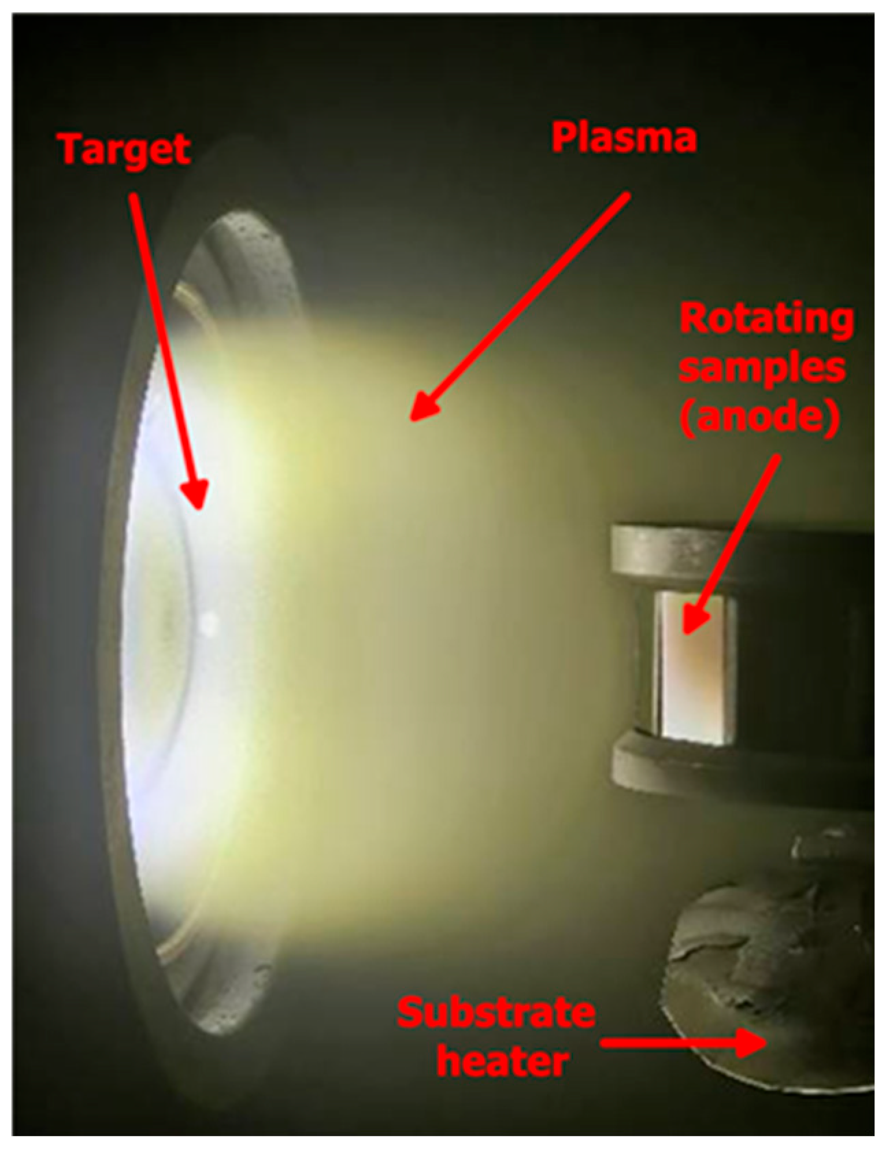
Figure 1. Photograph of a magnetron sputtering process inside the vacuum chamber. The circular target (cathode) is shown on the left, where the plasma is ignited and sustained. A visible glow discharge plasma extends from the target toward the substrate region. On the right, rotating samples (anode/substrate holder) are positioned to ensure uniform thin-film deposition. The system is equipped with a substrate heater located beneath the sample holder, enabling controlled heating during deposition. The rotation, combined with heating, improves film uniformity, adhesion, and microstructure.
Surfaces 09 00069 g001
Figure 2. Representative SEM images of the surface of the as-deposited (AD) coating (A), the laser surface-treated (LST) coating (B), and a cross-section image of the AD coating (C).
Surfaces 09 00069 g002
Figure 3. Chemical element distribution presented via mapping of the top surface areas of the TiO2-Cu coatings without subsequent LST (A) and TiO2-Cu coating after LST (B).
Surfaces 09 00069 g003
Figure 4. XRD spectra of the bare substrate, as-deposited, and laser-treated specimens (A) and FTIR spectra of as-deposited (AD) and laser surface-treated (LST) Cu-doped titania coatings compared with crystalline pure TiO2 (B).
Surfaces 09 00069 g004
Figure 5. Three-dimensional AFM micrographs of the surface architecture of the specimens: (A) bare substrate, (B) AD, and (C) LST-coated specimens.
Surfaces 09 00069 g005
Figure 6. Representative scratch track images of the complex oxide films on the Ti6Al4V alloy: (a) AD specimen; (b) LST-coated sample.
Surfaces 09 00069 g006
Figure 7. (A) Static water contact angle measurements with droplets of 5 µL SBF of the substrate, AD-coated, and LST-coated samples, with representative droplet images shown above each bar. (B) Seven-day cumulative ion release of copper as a function of immersion time for AD and LST coatings in simulated body fluid (SBF) at 37 °C. Error bars represent standard deviation.
Surfaces 09 00069 g007
Figure 8. Representative potentiodynamic polarisation curves measured for the bare and TiO2-Cu coatings in SBF after 1 h of immersion.
Surfaces 09 00069 g008
Figure 9. EIS analysis of both coated and uncoated samples immersed in SBF solution at 37 °C for 1 and 7 days: (a,d) Nyquist diagram; (b,e) Bode plot; (c,f) phase angle curve.
Surfaces 09 00069 g009
Figure 10. An equivalent circuit scheme used to match the impedance data of the substrate and coatings for different immersion periods of the samples in SBF at 37 °C.
Surfaces 09 00069 g010
Figure 11. Representative SEM micrographs of the surface morphology after EIS immersion testing: (A) AD coating after 7 days, (B) LST coating after 7 days, (C) AD-coated surface after immersion showing localised deposition products, and (D) LST coating with hydroxyapatite deposits. Insets in (A,B) show pit morphology with diameters of approximately 1.95–2.01 μm. EDS analysis at points 1–3 revealed Ca-, P-, and O-rich deposits, indicating the formation of hydroxyapatite (HA) layers on the immersed surfaces.
Surfaces 09 00069 g011
Table 1. Atomic % of the chemical elements on the surface of the coated samples.
SampleTi ± Error, %Cu ± Error, %O ± Error, %
AD coating24.0 ± 3.82.0 ± 8.374.0 ± 9.5
LBT coating23.6 ± 3.71.5 ± 7.974.9 ± 9.5
Table 2. Surface roughness values for AD and LST coatings and substrates.
SampleSa, nmSz, nmSsk
Substrate5.80 ± 6.0657.13 ± 0.070.23 ± 0.06
AD17.22 ± 14.34327.26 ± 0.141.42 ± 0.15
LST13.76 ± 16.04127.46 ± 0.15−0.26 ± 0.07
Table 3. Microhardness and critical adhesion forces of the substrate and coated samples.
SampleHK0.015, (kgf mm−2)FC, (N)
Substrate342.8 ± 6.7-
AD845.8 ± 45.514.2 ± 0.2
LST1009 ± 51.214.7 ± 0.4
Table 4. Anodic and cathodic Tafel slope, corrosion potential (Ecorr), corrosion current density (jcorr), and protection efficacy (P.E.) values obtained for the substrate and coated samples after 1 h of immersion.
SampleβA (10−3 V/dec)βC (10−3 V/dec)Ecorr (mV vs. SCE)jcorr (10−9 A cm−2)P.E. (%)
Ti6Al4V223.5254.8−414105-
AD866.9487.13600.6599.4
LST626371.43860.6899.4
Table 5. Electrochemical parameters derived via numerical fitting for uncoated Ti6Al4V and oxide coatings.
SampleTi6Al4VAD CoatingLST Coating
Time1 Day7 Days1 Day7 Days1 Day7 Days
Qp, (Ω−1cm−2sn)1.2 × 10−51.3 × 10−51.6 × 10−81.9 × 10−82.8 × 10−82.7 × 10−8
n10.930.910.970.970.950.96
Rp, (Ωcm2)3.8 × 1031.4 × 1033.3 × 1051 × 1034.4 × 1055.1 × 105
Qb1, (Ω−1cm−2sn)1.3 × 10−33.1 × 10−62.3 × 10−85.6 × 10−81.2 × 10−78.3 × 10−7
nb10.0810.530.740.61
Rb, (Ωcm2)4.3 × 1061.4 × 1067.4 × 1072.4 × 1075.1 × 1076.1 × 106
Qb2, (Ω−1cm−2sn)2.1 × 10−79.5 × 10−73.2 × 10−66.6 × 10−81.3 × 10−61.3 × 10−7
nb21 × 10−70.6510.320.320.53
Table 6. Antibacterial activity of the AR and LST samples against Staphylococcus aureus, evaluated by colony-forming unit (CFU) counting. Representative photographs of bacterial colonies recovered from the tested surfaces after incubation are shown alongside the corresponding antibacterial efficacy values.
SamplePolished Ti6Al4VADLST
Surfaces 09 00069 i001Surfaces 09 00069 i002Surfaces 09 00069 i003
Antibacterial efficiency (R), %-67 a ± 0.6574 b ± 0.71
a, b—Indices showing significant differences (p < 0.05) between the mean values in the column.
Disclaimer/Publisher’s Note: The statements, opinions and data contained in all publications are solely those of the individual author(s) and contributor(s) and not of MDPI and/or the editor(s). MDPI and/or the editor(s) disclaim responsibility for any injury to people or property resulting from any ideas, methods, instructions or products referred to in the content.