


Abstract
This study investigates the composition, morphology and cultural significance of red pigment traces identified on bone pointed tools discovered in the Chalcolithic tell settlement of Pietrele–Măgura Gorgana, attributed to the Kodjadermen–Gumelnița–Karanovo VI cultural complex (4600–4250 BC). The observed use-wear patterns are consistent with repeated contact with soft, non-abrasive materials, including hide working, pigment application on leather or other organic surfaces, fiber manipulation, and perforation of soft substrates. Use-wear analysis revealed polished and flattened distal ends, compatible with repeated use on soft, non-abrasive materials, such as hide, leather, fiber, or other organic substrates. The possibility of pigment application directly on skin, in a practice analogous to tattooing, as previously published, cannot be excluded but remains speculative in the absence of experimental reference data or residue evidence specifically linked to such use. An associated ceramic container was tentatively interpreted as a possible vessel for ochre preparation, suggesting local processing of the pigment. The artifacts were investigated using multi-analytical archaeometric methods: SEM-EDS, AFM, TEM, FTIR, Raman, TGA, CLSM and pseudo-color image segmentation and 3D rendering of porosity distribution. The results consistently identified an iron oxide-based pigment, dominated by hematite and/or goethite, specific to ochre. Pigment particles (50–300 nm) form a well-defined superficial layer on the bone substrate, without Fe–Ca reactions at the interface. The simultaneous presence of Ca, P, Si, Mg and K indicates a silicate matrix with an apatite component, compatible with local and poorly purified raw materials. CIELAB colorimetric analyses revealed significant chromatic variability, suggesting the use of hematite-rich pigments and possible thermal transformations of goethite. The results contribute to the understanding of the pigment technologies of the Chalcolithic communities of the Lower Danube.
1. Introduction
In some studies dedicated to hominid evolution, pigments are considered the first archaeological evidence related to the existence of an aesthetic perception, perhaps even symbolic. The oldest evidence of pigment use is c. 500 ka old coming from Kathu Pan (South Africa) at the transition between the Acheulean and the Middle Stone Age (MSA). In India, similar evidence is dated to c. 300 ka [1]. In Europe, before modern humans, Neanderthals used red ochre in many sites in Europe, dated between 60 and 40 kya [2,3,4], but there is also evidence for much earlier use, minimally 200–250 ka old [5]. We do not know whether they were used to decorate some materials or for skin tattooing, possibly in rituals as identified in some populations in Africa [6]. Obviously, their utilitarian function cannot be excluded either, also starting from ethnographic comparisons: insect repellent and medication [7,8], hide tanning [9], and a component of adhesives for the hafting process [10]. Starting with the Upper Paleolithic, the use of pigments clearly also has a symbolic meaning, attested by paintings on pebbles and cave walls [11,12] or by burial in tombs [13,14]. In more recent periods of prehistory, the use of pigments became widespread, being used especially for decorating ceramics, but they are also present on other categories of artifacts (e.g., walls of dwellings, ornaments, bone and stone tools, etc.).
Among the hypotheses of pigment use, tattoos were mentioned. At the archaeological level, there are few situations where such evidence has been preserved. The unique contexts in which tattooed mummies were preserved, such as the Tyrolean Iceman known as Ötzi, dated to 3370–3100 BC, could be mentioned [15,16]. Another particular situation involves indirect evidence of tattooing: anthropomorphic representations bearing various signs interpreted as tattoos and the presence of pointed tools made of bone with consistent traces of pigment on their surface. Such bone pieces were discovered at the Gumelnița tell settlement from Pietrele (southern Romania). For these discoveries, the hypothesis of their use as tools for applying tattoos was previously raised [17], based on their morphological characteristics and the presence of pigment residues. However, this interpretation remains a working hypothesis, as use-wear patterns consistent with soft-material contact are not exclusively diagnostic of tattooing and could equally reflect other pigment-application activities. In this settlement, traces of pigments are present only on a small number of bone pointed tools, generally extremely thin and sharp, which reduces the likelihood of post-depositional contamination. The present investigation aims to identify the composition of the coloring materials present on these pieces and to contribute contextual evidence relevant to this functional hypothesis while acknowledging that definitive attribution would require experimental reference data.
Ochre is recognized for its diverse color palette ranging from brown to purple, is primarily characterized by its iron content, and in time has become a significant focus in archaeological research due to its cultural implications. Ochre possesses distinct cultural relevance across various societies. Its significance often extends beyond mere aesthetics, suggesting associations with ritualistic, functional, or artistic uses in prehistoric contexts. Additionally, the mention of white ochre highlights the complexity and variability within this material category, prompting further investigation into the cultural and practical applications of ochre through time.
Chemically defined ochres are mixtures characterized by the presence and absence of specific elements or minerals [18]. In an art history and conservation context, ochre is identified narrowly as one of a suite of related earth pigments composed of mixtures of minerals that are differentiated by their manganese oxide content: ochre, umber, and sienna [19]. Studies have identified the materials that fall under that definition [20].
Fe-containing materials for cultural heritage generally arise from accessible geological sources at the surface with or without additional layers of cultural meaning or association with the greater landscape [21,22,23].
Primarily, the Fe-bearing geomaterials of interest contain hematite (α-Fe2O3) and/or goethite (α-FeOOH) in various forms and abundances [24]. Additional ferruginous minerals of interest to the ochre analyst may include ferrihydrite (Fe5O8H·H2O), lepidocrocite (ɣ-FeOOH), maghemite (ɣ-Fe2O3) [25], and jarosite (KFe3[SO4]2(OH)6) [26]. Lu defines three subtypes of goethite, including the “yellow ochreous” that contains excess water and chemical impurities, including amorphous material and high levels of Al2O3 and SiO2 [27]. However, it is not always clear if the Al and Si oxides are additional, diluting minerals or substituted into the crystal lattice [28]. Other associated minerals coexist with the iron minerals, including quartz and other silicates, carbonates, sulfides, and other oxides, which will be discussed below.
When hematite is the main iron oxide, a red color is observed, while ochre is yellow when goethite dominates [28,29]. These colors are due to the Fe3+ ion contained in both oxides and, more precisely, to the charge transfer between Fe3+ and its O2− or OH− ligands. The current literature on iron oxides rarely links spectro-colorimetric analysis to physico-chemical analysis [30,31,32] and, to date, never in a quantitative way. They rely on various techniques such as the Mossbauer effect [33,34], thermal analysis [35], FT-IR spectroscopy [36,37,38,39], X-ray diffraction [40,41,42], Raman microscopy [39,42] and magnetic and voltammetric analyses [43].
The objective of this study is to investigate the composition, morphology and spectro-colorimetric characteristics of the ochre pigment used by the Chalcolithic communities of the Lower Danube, with a focus on the relationship between physico-chemical properties and chromatic variability.
The novelty of this research lies in the quantitative integration of the CIELAB colorimetric analysis with archaeometric and structural investigations of ochre, an approach rarely applied in the specialized literature, as follows: for the morphology of the ochre compounds, additional techniques such as confocal laser scanning microscopy (CLSM), scanning electron microscopy (SEM) coupled with energy dispersive spectroscopy (EDS) and transmission electron microscopy (TEM) highlighted the deposits of ochre pigment particles on the bones and were correlated with pseudo-color image segmentation and 3D rendering of porosity distribution.
Overall, the results contribute to a better development of pigment production, processing and application technologies within prehistoric communities.
2. Cultural Framework
The communities of the Kodjadermen–Gumelniţa–Karanovo VI cultural complex covered a considerable geographic area from the Carpathian Mountains to the Aegean Sea between c. 4600 and 3900 cal BC. The northern Danube area saw in this period the rise of the tell settlements. Larger and multiroomed houses were built, sometimes with one story and stone foundations aligned in symmetrical arrangements [44]. Sometimes, tell settlements were surrounded by defensive structures, while the dwellings were arranged in parallel rows [45,46,47,48]. The necropolises were located outside the settlements, and some of the graves yielded rich grave goods [44,49], comprising personal adornments, pots, flint blades etc. [50]. The social, technological and economic transformations involved changes in the funeral inventories that seem to reflect social hierarchies [44,51,52,53]: the use of copper as a raw material [54,55,56,57,58,59,60], Spondylus ornaments from the Mediterranean world [61], etc.
The tell settlement of Pietrele–Măgura Gorgana (Figure 1a,b) is one of the landmark sites of the Kodjadermen–Gumelniţa–Karanovo VI cultural complex, with its evolution covering the interval 4600–4250 cal BC, according to the 45 radiocarbon dates obtained so far [62]. The tell settlement was located on the lower terrace of the Danube, having at the beginning of archaeological research (in 1943) a diameter of 95 m [63]. After two research campaigns led by Dumitru Berciu in 1943 and 1948, the research resumed in 2004 within the framework of a collaborative project between the German Institute of Archaeology, the “Vasile Pârvan” Institute of Archaeology, Romanian Academy and the Institute of Physical Geography of Goethe University Frankfurt. Research has highlighted intense habitation around the tell settlement, the site being much more extensive than previously assumed [64].
Figure 1.
(a) Geographical location of the Pietrele site in the Balkans; (b) geographical location of the Pietrele site in southern Romania; (c) general plan of the Pietrele tell settlement with both areas (B and F areas) of interest for the present study.
The dwellings were built of massive clay walls (40–60 cm), sometimes reinforced with wooden beams. Some dwellings had two floors. Ovens and other clay installations have been identified. The strong connection with the aquatic environment was proven by the economic model: the inhabitants of Pietrele–Măgura Gorgana were known as fishermen and hunters rather than sedentary farmers [65,66]. Another characteristic was the performance of specific activities by individual groups (functional areas) [64,65], an economic feature rarely highlighted in other Chalcolithic sites.
Ceramic remains vary in shape, size and type of decoration: from common vessels (bowls, cups, jugs, lids) to large-sized storage vessels with high and concave necks and a capacity of up to 30 L [64]. In general, slip was applied, rarely the surface being smoothed and painted with graphite or incised. Archaeological inventories are completed by stone artefacts (scrapers, piercers, arrowheads, super blades), stone millstones, axes made of carved or polished stone, copper and gold objects, ornaments made of various materials (exotic shells, bone, stone, clay, tooth, gold, copper, jade, marble), anthropomorphic and zoomorphic clay figurines, clay weights and a rich osseous industry made of bones, antlers, teeth or shells (pointed tools, beveled tools, spatulas, hammers, harpoons, handles, spindle whorls, abraded astragals, anthropomorphic figurines, etc.) [62,64,65,66,67,68,69,70,71,72,73,74]. Within the osseous industry, a significant amount of thin bone pointed tools as inventory of the households can be highlighted, with some items having visible traces of red pigment on their surface.
3. Materials and Methods
3.1. Archaeological Materials
The present study analyzes a total of seven artifacts recovered from the Chalcolithic tell settlement of Pietrele–Măgura Gorgana. The assemblage comprises six bone pointed tools (P1–P6 samples) bearing macroscopically visible red pigment traces on their surfaces and one ceramic object (P7) presenting internal pigment deposits. All artifacts were selected based on the presence of visible colorant residues and their secure stratigraphic association with occupation levels interpreted as domestic contexts. The bone pieces were subjected to physico-chemical investigations, but the last two (P5 and P6 artifacts) did not provide information, given the very small amount of pigment preserved on their surface.
For consistency throughout the manuscript, a unified alphanumeric sample identification system is adopted (Table 1). Each artifact is assigned a code from P1 to P7, used consistently for all figures, tables, and analytical sections. The original excavation inventory numbers are retained in Table 1 for archival reference only.
Table 1.
Archaeological and analytical variables of the bone pointed tools.
The bone pointed tools belong to two typological categories distinguished by raw material selection and technological production sequence. Type I tools (P1–P4 items) were manufactured from the diaphysis of long bones of medium-sized mammals (Figure 2a,f,h). Flat blanks were obtained through multiple longitudinal partitions of the compact cortical wall, yielding long, thin splints. The convergence of the lateral edges towards the distal point was most likely achieved by longitudinal scraping (Figure 2b,i), although this technique is difficult to confirm since the entire surface of the pieces was subsequently regularized by transverse abrasion (Figure 2c,g). The resulting cross-section is rectangular throughout, with an extremely thin and slightly fractured distal tip at most specimens. A particular subset within this group comprises tools recovered from a heavily burned structure (Figure 2e, P2). These specimens acquired a light grey to white coloration (Figure 2d) and present strong deformations and fractures resulting from high-temperature exposure. The absence of preserved technological marks or use-wear traces on these pieces is noted. In some cases, the high-temperature event welded several pointed tools together. Red pigment traces, however, were preserved on the surfaces of these burned specimens.
Figure 2.
Bone pointed tools and technological traces from the Chalcolithic settlement of Pietrele–Măgura Gorgana: (a) bone pointed tool P1; (b) scraping marks on P1 (detail); (c) abrasion marks on P1 (detail); (d) bone pointed tool P2; (e) in situ position of artifacts during excavation; (f) bone pointed tool P3; (g) abrasion marks on P3 (detail); (h) bone pointed tool P4; (i) scraping marks on P4 (detail).
Type II tools (P5–P6 items) were manufactured from metapodial bones of medium-sized mammals (Figure 3a,d). The flat blank was obtained by longitudinal quadri-partition using a double grooving technique (Figure 3c,f), traces of which remain visible on the medullary canal surfaces. The natural morphology of the epiphysis was retained. The distal end was shaped by circumferential scraping (Figure 3e), followed by abrasion (Figure 3b).
Figure 3.
Bone pointed tools, technological traces, and associated ceramic object from the Chalcolithic settlement of Pietrele–Măgura Gorgana: (a) bone pointed tool P5; (b) abrasion marks on P5 (detail); (c) grooving marks on P5 (detail); (d) bone pointed tool P6; (e) scraping marks on P6 (detail); (f) grooving marks on P6 (detail); (g) ceramic object P7.
In the archaeological assemblage of the tell settlement at Pietrele, ceramic pieces have been identified that can be correlated with the preparation of pigments. These findings will be the subject of a subsequent study; however, we have selected one specimen (Figure 3g) for the present analysis. It possesses a conical shape with a flattened base and an oval cross-section. The height of the piece measures 51.5 mm, its maximum width is 44.8 mm, and its maximum thickness is 37.5 mm. The special find identified with the designation P06F3450574 (P7) was found in the same burnt house located in area F (F12) as the pointed tools with the identification number P2, alongside other objects typically associated with domestic contexts (Figure 4). Initially deemed to be an ochre bar, attributed to the category of special finds used as painting material, the new analysis indicates a change in the object’s function (to a receptacle for preparing the ochre). Nevertheless, the primary purpose remains consistent: to produce red colors for various activities.
Figure 4.
Plan of the burned house located in area F (F12) where bone pointed tools (P2 sample; P06F3700963) and a ceramic piece (P7 sample; P06F3450574) were found (graphics by Meda Toderaș).
On the tell settlement, there are two excavated areas, designated area B and area F (Figure 1c). All the artefacts that make the object of this study come mainly from domestic contexts, different but somehow connected (see Table 1). The main characteristic of the dwellings on the tell settlement consists of successive horizons of houses (burnt and unburnt), separated by a thick layer of soil filling.
Area B is situated on the north side of the site, while area F is on the southern side. The P4 artefact comes from the horizon consisting of the filling in between the sequences of the two houses. The pointed tool with the identification number P3 is from the inventory of a house excavated in area B (named BX5). The main characteristic of this feature was the unburnt walls of the house. The other objects, with the identification numbers P1 and P6, come from another layer of filling, exactly underneath the burnt house excavated in the southern part of the B area, more precise underneath an oven.
In the case of the objects from area F, the situation is very similar; they are coming also from a house and from a layer of filling soil under the house. The pointed tools from which sample P2 was selected were found in the debris of a burnt house (F12) excavated in area F, and the piece with the identification number P5 came from the layer of filling underneath the house.
The possibility of post-depositional pigment contamination was systematically addressed through a contextual comparative analysis of the broader archaeological assemblages recovered from the same stratigraphic units as the analyzed bone pointed tools. This assessment was conducted prior to the selection of artifacts for archaeometric investigation and informed the interpretive framework applied throughout this study. Each domestic context yielding pigment-bearing bone tools was examined in its entirety, with particular attention to all associated artifact categories. The associated assemblages include ceramic vessels, flint tools, osseous objects, faunal remains, as well as other categories of finds typical of Chalcolithic domestic contexts, such as figurines, cooper artifacts, spindle whorls or grinding stones (Figure 5). The macroscopic surfaces of all associated artifacts were carefully inspected for the presence of red colorant deposits. In addition, the bulk composition and visual characteristics of the enclosing sediment matrix were documented in order to evaluate the potential for iron-rich sedimentary input as a source of secondary pigment impregnation. This systematic contextual review yielded a consistent and significant result: no traces of red pigment were identified on other categories of artifacts recovered from the same archaeological contexts. Furthermore, the sediment matrix enclosing the analyzed tools does not present the ochre-rich, iron-stained character that would be expected in the case of diffuse post-depositional contamination. The absence of any generalized iron oxide staining across the associated assemblage and the enclosing sediment matrix suggests that diffuse post-depositional contamination is less likely a post-depositional origin for the observed pigment deposits. To provide a transparent and reproducible basis for this taphonomic assessment, Figure 5 presents a representative selection of artifacts associated with the pigment-bearing bone tools, drawn from the same archaeological contexts (Areas B and F, Pietrele–Măgura Gorgana).
Figure 5.
(a–c) Ceramic fragments ((a)— same context as P2 sample, (b)—same context as P3 sample, (c)— same context as P4 sample); (d) zoomorphic figurine coming from the same context as P4 sample; (e) antler harpoon coming from the same context as P4 sample; (f) cooper specimen coming from the same context as P3 sample; (g–i) flint pieces ((g)— same context as P2 sample, (h)—same context as P3 sample, (i)— same context as P4 sample) (scale: 1 cm).
3.2. Equipment
FTIR spectra were recorded using a Bruker Vertex 80 Fourier Transform Infrared spectrometer equipped with a diamond attenuated total reflectance (ATR) accessory (Bruker Optics Inc., Billerica, MA, USA). Measurements were carried out at a spectral resolution of 0.2 cm−1 and an accuracy of 0.1% T. The spectra were collected within the 4000–400 cm−1 range by averaging 32 scans, without prior sample preparation. Data acquisition was performed using OPUS 7.1 software, and the obtained spectra were subsequently processed in Origin 2019b software. To ensure optimal contact between the sample surface and the ATR crystal, constant contact pressure was applied using the built-in pressure clamp system. Multiple spectra (at least 3 measurements) were collected from each sample in order to assess spectral reproducibility and microstructural heterogeneity. The spectra were collected from pigment-rich and pigment-free areas of the investigated samples.
Raman spectra were measured using a portable Xantus-2 Raman analyzer (Rigaku, Analytical Devices, Wilmington, MA, USA) with a 1064 nm stabilized laser and a CCD detector. The instrument covered a spectral range of 200–2000 cm−1, with a resolution between 7 and 18 cm−1. In this study, all measurements were carried out using the 1064 nm excitation source at a laser power of 100 mW (no thermal alteration of the archaeological residues and pigment phases has been observed). The laser beam was focused through a 50× objective, resulting in an estimated spot size of approximately 1–2 µm. The integration time was 10,000 ms, with five accumulated scans. The analyses were conducted directly on the samples without prior preparation, and the resulting data were subsequently processed using Origin 2019b software.
Prior to each analytical session, spectral calibration was performed using the characteristic silicon standard band at 520.7 cm−1. Raman spectra were acquired within the 200–2000 cm−1 spectral range using acquisition times of 10–30 s and multiple accumulations to improve the signal-to-noise ratio.
For each artifact, at least 5–10 representative points were analyzed, selected according to visible chromatic variability, surface morphology, and the presence of pigment-rich areas observed under optical microscopy. To ensure data reproducibility, measurements were repeated on different regions of each sample.
Due to the sensitivity of iron oxide pigments and archaeological residues to local heating effects, high laser powers were intentionally avoided to preserve the original mineralogical and organic signatures of the samples.
The chromatic parameters were measured using a CR-410 colorimeter from Konica Minolta, Tokyo, Japan, following the CIELAB standard under standardized acquisition conditions [46]. These included Lx* for brightness (ranging from 0 for black to 100 for white), ax* for red/green variation (where +ax* denotes red and −ax* denotes green), and b* for yellow/blue variation (+bx* for yellow and −bx* for blue). The measurements were acquired using the CIE standard illuminant D65 and a 10° standard observer angle. An aperture size of 8 mm was selected for all analyses. Prior to measurements, the instrument was calibrated using the manufacturer-provided white calibration standard. The acquisition geometry was configured in diffuse/8° (d/8) geometry with specular component included (SCI). For each analyzed area, five independent measurements were collected at different points in order to minimize local heterogeneity effects, and the reported L*, a*, and b* values represent the mean ± standard deviation of the replicates. The measurements were performed at room temperature under stable laboratory conditions.
The morphology of the active ends, with the specific details of use-wear (e.g., change in initial volume, polish, functional marks), was systematically recorded with a Keyence VHX-600 digital optical microscope (Keyence Corporation, Osaka, Japan) using lenses of variable strength between 30× and 150×, with an embedded camera and with an Olympus BX53M metallographic microscope (Olympus Europa SE&CO. KG, Hamburg, Germany) magnifications ranging from 100× to 500×), equipped with a EOS 1200D Canon camera.
Scanning electron microscopy (SEM) was conducted using environmental scanning electron microscopy (ESEM) with an FEI Quanta 200 microscope (Eindhoven, The Netherlands) to assess the morphology of the samples. The magnification ranged from 50× to 100,000×. SEM observations were carried out at an accelerating voltage of 15–20 kV, with a working distance of approximately 10 mm and a beam current adjusted automatically by the instrument operating conditions. Secondary electron (SE) imaging modes were employed for morphological and compositional contrast analysis, respectively. The samples were mounted on aluminum stubs using conductive carbon tape. To minimize charging effects, non-conductive samples were coated with a thin gold conductive layer (~10 nm thickness) using a sputter coater prior to analysis.
The microscope is equipped with an Oxford Inca Energy Dispersive X-ray (EDS) system, produced by Oxford Instruments, Abingdon, UK—X-MaxN energy dispersive spectrometer model for chemical analysis of the samples. EDS spectra were acquired using an acquisition time of 60–100 s per analyzed area, with spectral processing and elemental quantification performed using the manufacturer’s software based on ZAF correction procedures. Semi-quantitative elemental analysis was obtained using standardless quantification calibrated against certified internal standards supplied by the instrument manufacturer.
For each sample, at least 5–10 representative regions were analyzed, with analyzed areas ranging from approximately 50 × 50 µm2 to 200 × 200 µm2 depending on microstructural heterogeneity. The reported elemental concentrations represent the average values obtained from multiple measurements. Analytical uncertainty was estimated from repeated measurements and counting statistics, with relative standard deviations generally below 5%.
The confocal laser scanning microscopy (CLSM) analysis was performed using a Leica DCM8 3D surface metrology system (Leica Microsystems CMS GmbH, Wetzlar, Germany), which enabled precise control of imaging conditions (high-resolution topographic imaging, pseudo-color segmentation). It operated with laser lines ranging from 405 nm (UV) to 633 nm (far-red) and allowed surface roughness determination. Three-dimensional optical sectioning was performed using Leica Confocal Software v2.61.
Pseudo-color image segmentation and 3D rendering of porosity distribution were analyzed by image segmentation performed by using ImageJ software for Mac V 1.54 Softonic International S.A. © 1997–2025, Barcelona, Catalonia, Spain [75,76]. The hyper stack plugin’s split channel feature was used to open the stack composite in the software. ImageJ is an open-source analysis and processing program. After that, ImageJ was used for quantification and data processing [77]. Pseudo-color image segmentation and 3D rendering of porosity distribution can provide information about voids, cracks, and pores that are dispersed throughout the matrix.
For transmission electron microscope (TEM), a TecnaiTM G2 F20 TWIN Cryo-TEM (FEI CompanyTM, Hillsboro, OR, USA), with a focus on cryo-electron microscopy (cryo-TEM) and tomography, as well as a software for automated data acquisition and high-resolution digital cameras, like the Eagle 4k CCD, were used. It has a tomography-grade stage and software that enable 3D reconstruction of samples, and it can operate at up to 200 kV, enabling high-resolution imaging.
Powdered microfragments collected from representative pigment-rich regions were gently dispersed in ethanol using ultrasonication for several minutes in order to minimize particle agglomeration. A small droplet of the resulting suspension was deposited onto carbon-coated copper grids (200 mesh) and allowed to dry under ambient laboratory conditions prior to imaging.
TEM observations were conducted to evaluate particle morphology, aggregation state, and nanostructural characteristics. For each sample, multiple representative regions were examined, and at least 50–100 particles were evaluated in order to ensure statistical relevance. Particle size measurements were performed using digital image analysis software on representative micrographs acquired at different magnifications.
For atomic force microscopy (AFM) images, an Agilent 5500 SPM system with a PicoSPM unit, MAC Mode module, and PicoScan controller (Agilent Technologies, Tempe, AZ, USA) was used. Additionally, n-doped silicon without coating Point Probe Plus Silicon SPM Sensor cantilevers (PPP-FM type, NANOSENSORS) was utilized. At room temperature, the AFM images (256 samples per line × 256 lines) were taken in tapping mode at a scan rate of 0.8 to 1.2 lines per second. AFM topographic images were acquired from representative areas selected on the basis of surface homogeneity and absence of large macroscopic defects. Scan sizes ranging from 5 × 5 µm2 to 50 × 50 µm2 were collected with a resolution of 256 × 256 pixels. PicoView SPM Software (version 1.6) was used for section analysis and 3D topographical reconstruction.
Silicon PPP-FM cantilevers (PointProbe® Plus) with a nominal resonance frequency of approximately 75 kHz and a spring constant of ~2.8 N/m were employed for imaging.
Thermogravimetric analysis (TGA) was performed using a TA Instruments Q5000 IR Thermogravimetric Analyzer equipped (TA Instruments, New Castle, DE, USA) with Platinum™ software (version 3.13, Build 261). Approximately 5–15 mg of sample was placed in platinum pans and analyzed under a nitrogen atmosphere with a purge flow of 60 mL/min for the furnace and 40 mL/min for the balance. The measurements were carried out in the temperature range from ambient temperature to 800 °C at a constant heating rate of 10 °C/min. Prior to analysis, the instrument was calibrated for temperature using Curie point standards and for mass using certified reference weights. The instrument sensitivity was <0.1 μg, with a weighing accuracy of ±0.1% and a temperature precision of ±1 °C.
4. Results
4.1. Preamble
The analytical investigation was divided into two complementary datasets: (i) bone specimens containing red pigment residues and (ii) residues associated with the crucible interpreted as a potential pigment-processing structure. This allowed for a better understanding of the composition, technological transformation, and potential processing pathways of the ochre-related archaeological materials.
For the bone samples, the distribution, intensity, and variability of the red pigment layers were first recorded using macroscopic observations and colorimetric measurements. FTIR and Raman spectroscopy were used after these investigations to determine the main mineral phases connected to silicates, phosphates, iron oxides, and potentially thermally altered components. While AFM, TEM, and CLSM analyses were utilized to assess surface morphology, nanoscale particle organization, roughness parameters, and pigment penetration within the microstructural features of the bone surface, SEM–EDS investigations yielded information regarding elemental composition and the spatial distribution of Fe-rich particles within the calcium-phosphate matrix.
For the crucibles, the morphological evaluation and three-dimensional rendering made it possible to see residue distribution, surface degradation patterns, and structural elements that might be connected to heating processes. To locate iron oxide phases that might have been produced by heat treatment, FTIR and Raman spectroscopy were utilized. For the crucible dataset, thermal analysis (TGA-DTG) was especially crucial because it made it possible to assess the dehydration, dehydroxylation, and mineral transformation processes typical of iron-bearing materials exposed to high temperatures. The microstructural organization, vitrification characteristics, elemental distribution, and thermal alteration textures preserved within the residue matrix were further documented by complementary SEM–EDS investigations.
The systematization of all these techniques is shown in Scheme 1.
Scheme 1.
The flowchart for both sample types.
4.2. Use-Wear Analysis
Initial observations that primarily include the microscopic examination of the Type I tools (P1–P4 samples) reveal extremely thin ends, slightly fractured on most pieces (Figure 6a,b). The use-wear is evident, being characterized by the disappearance of technological marks and the flattening of the surface, having a regular appearance without depressional losses of raw materials (Figure 6c,d). These features are consistent with repeated contact with soft, non-abrasive organic materials. However, given the broad range of activities that can produce comparable use-wear traces, including hide working, pigment application on leather or other organic surfaces, fiber manipulation, and perforation of soft substrates, the specific function of these tools cannot be determined solely on the basis of use-wear morphology. While harder or more resistant substrates can be excluded on the basis of tool robustness, the range of compatible soft substrates remains broad. The use-wear evidence alone is therefore insufficient to identify a specific worked material or action. The amount of red pigment has significant accumulation only on the distal end of the pieces (Figure 6e–g), and the traces of pigments suggested that they may have been bound together into a single multipoint tool (see [17]). Among the Type I tools, the burned specimens (P2 sample) present a specific taphonomic profile. Although manufacturing marks and use-wear traces were obliterated by the high-temperature event, estimated at approximately 800–900 °C based on the light grey to white coloration of the bone, consistent with the threshold of calcination [78], red pigment traces were preserved (Figure 6h,i) on the surface, suggesting that the tools had been in active use prior to the burning event. The spatial association of multiple welded tools within the same burned structure may indicate that they were stored or deposited as a group.
Figure 6.
(a,b,j) Distal end details; (c,d,k–m) use-wear marks; (e–i,n,o) red pigment spots ((a,c,e–g)—P1 sample; (b,d)—P4 sample; (h,i)—P2 sample; (j–o)—P5 sample).
For Type II tools (P5–P6), use-wear is intense and clearly visible at the distal end. This is strongly smoothed (Figure 6j), and the surface presents a macroscopic flat polish (Figure 6k–m) that has begun to erase technological marks. Accumulation of red pigment is preserved within the medullary canal cavity (Figure 6n,o), suggesting that the hollow morphology of the blank facilitated pigment retention. The association of morphological use-wear and pigment loading supports a similar functional interpretation as for Type I tools.
Equally informative is the highly selective and spatially restricted distribution of the red colorant on the bone tools. Pigment accumulations are consistently confined to the distal working ends, to the lateral edges of the tool shafts, and—in the case of Type II tools—to the interior of the medullary canal, following the microtopography of the functional surfaces. This pattern of localized, morphologically coherent deposition is incompatible with passive, undifferentiated sedimentary contamination, which would be expected to produce more uniform colorant distribution across all exposed surfaces of both worked and unworked materials from the same context. The spatial congruence between pigment deposition and the areas of active use, as identified by use-wear analysis, further reinforces an anthropogenic origin for the observed residues.
The findings about the investigated samples indicated that the red hues in Chalcolithic ceramics were mainly due to Fe-rich minerals, aligning with observations from various studies across the Balkans and Europe that suggest similarities in pigment preparation and decoration practices during that period [79,80]. The present results are consistent with possible heating-related transformations involving iron oxyhydroxides such as goethite.
Only a few techniques were applied to the bone samples due to the extremely small amount of sample.
The colorimetric measurements (CIELAB) performed on ochre-bearing artifacts from Pietrele–Măgura Gorgana reveal a broad chromatic variability, reflecting both technological processing and weathering status. The predominance of high a* values in several samples suggests the use of hematite-rich pigments, potentially obtained through thermal transformation of goethite, visible on TGA experiments (Table 2). The combined colorimetric, spectroscopic, and thermal data are consistent with possible thermal transformation processes involving iron oxyhydroxides such as goethite, potentially associated with pigment preparation activities.
Table 2.
The chromatic parameters measured for the artifacts from Figure 6e–i,n,o.
The colorimetric variability indicates the coexistence of raw, partially processed, and fully transformed ochre materials within the Pietrele–Măgura Gorgana assemblage. This pattern is consistent with local procurement and on-site processing, rather than the use of standardized, externally sourced pigments.
The chromatic parameters show that warm red–yellow tonalities predominate in all examined samples, as evidenced by the consistently high positive a* and b* values. The presence of iron oxide-rich pigments, most likely hematite-dominated ochre materials, is suggested by the positive a* coordinates (35.44–46.94), which show a strong red component. The coexistence of goethite, clay minerals, or thermally altered iron-bearing phases may be indicated by the elevated b* values (42.37–53.93), which also show a significant yellow contribution. Sample i had the highest luminance (L* = 62.36 ± 0.24) and the highest b* coordinate (53.93 ± 0.26), indicating a lighter and more yellowish appearance, while sample o had the highest a* value (46.94 ± 0.20), indicating the most intense red coloration. Sample h, on the other hand, had the lowest L* value (49.24 ± 0.20), suggesting a darker surface that might be related to localized mineralogical variations, firing effects, or higher pigment concentration. For the cup, the chromatic parameters are similar: L* = 53.73; a* = 40.39; and b* = 48.03, indicating the predominance of hematite-rich compositions.
Due to the extremely small amount of the samples, some analytical techniques, such as FTIR and Raman spectroscopy, have been selected and processed in this paper.
Figure 7 shows the characteristic peaks of FTIR spectra registered for different samples collected from the B and F areas.
Figure 7.
FTIR spectra of different samples collected from different parts of B and F areas ((a)—P1 sample; (b)—P2 sample; (c)—P3 sample; (d)—P4 sample).
The FTIR spectra exhibit similar overall profiles, with the main differences observed in the regions associated with hydroxyl/water vibrations (3400 and 1640 cm−1) and in the fingerprint region between 400 and 1400 cm−1, where the characteristic mineral bands are located. The spectra reveal absorption bands assigned to hematite (~530 and ~470 cm−1, Fe–O stretching), goethite (~872, and ~780, OH bending and Fe–O–OH stretching), organic compounds (2920–2850, 1722, and ~1430 cm−1 corresponding to C–H, C=O, and O–H vibrations), and water-related OH groups (3400 cm−1 assigned to O–H stretching and H–O–H bending vibrations). Several bands observed in the fingerprint region may reflect overlapping contributions from iron oxides/oxyhydroxides, phosphates, and silicate-related phases commonly encountered in archaeological residues. Therefore, individual peak assignments should be interpreted cautiously and in correlation with the broader multi-analytical dataset.
The FTIR results for the pigmented areas of the investigated samples revealed additional bands assigned to iron oxides/oxyhydroxides, such as hematite and/or goethite, and support the presence of ochre-based pigment. A pronounced absorption in the carbonyl region may indicate an organic component, possibly related to degraded organic binder residues, soil-derived organic matter, or an intentionally added organic binder. The term “non-pigmented bone sub-strate” refers to areas of the bone surface where no visible red pigment was detected.
The three bands around 948, 1110, and 1165 cm−1 and the shoulder around 1000 cm−1 are clearly related to the phosphate–goethite complex [81].
On the other hand, the observed vibrations in the present study around 1000 cm−1 (range: 990–1010 cm−1) could be assigned as a mono-protonated bidentate complex (Fe2HPO4) and/or non-protonated monodentate complex (FePO4) [82]. Considering the assignment of Tejedor-Tejedor and Anderson [82], the present IR spectrum indicates the presence of a mixture of monodentate and bidentate complexes, with a majority of the mono-protonated bidentate complex. In contrast, Krumina et al. [83] assigned the vibrational peaks at 940–945, 1004–1012, 1042–1050, 1085–1094, and 1110–1128 cm−1 to a series of monodentate phosphate surface complexes in different protonation states. According to this assignment, the present vibrational bands might indicate a mono-protonated monodentate complex (FeHPO4). This discussion clearly shows the yet unsolved general problem of assignment of phosphate–goethite (or haematite) binding motifs to IR features. The FTIR spectrum of this area shows characteristic phosphate vibrations attributable to bone bioapatite, including bands associated with PO43− groups, in agreement with the Ca and P enrichment observed by EDS.
The Raman spectroscopy analysis was revised to focus on the spectral features directly observed in Figure 8. The main Raman bands were assigned to mineral phases associated with iron oxide pigments and the bone substrate. Characteristic bands attributable to hematite were identified in the low-wavenumber region, supporting the presence of red ochre-type pigment, while phosphate-related contributions associated with the bioapatite structure of bone were also observed. The spectral assignments for the Raman spectra are presented in Figure 8.
Figure 8.
Raman spectra of the investigated samples with pigments and without pigments ((a)—P1 sample; (b)—P2 sample; (c)—P3 sample; (d)—P4 sample).
Raman spectroscopy further supported the presence of iron-bearing phases through characteristic low-wavenumber bands compatible with hematite/goethite-type mineral assemblages. Nevertheless, several spectral features, including the bands observed around ~760, ~950–956, and ~1040 cm−1, may also reflect contributions from phosphate groups, silicates, carbonate phases, or substrate-related signals. Consequently, the Raman interpretation was based on the overall spectral behavior rather than on isolated diagnostic bands alone.
SEM images show irregular particles (50–300 nm), often aggregated into larger clusters. Distinct outlines of hematite/goethite particles embedded in or adhering to the bone surface are shown in Figure 9. The surface shows a heterogeneous landscape with nanoscale height variations. The rougher areas likely correspond to pigment deposition, while smoother zones represent the underlying bone substrate (mineralized collagen matrix).
Figure 9.
SEM images of the P1, P2 and P5 investigated samples.
The spherical and semi-spherical nanostructures represent ochre pigment particles, primarily composed of iron oxides (hematite or goethite). Particle size distribution ranges from ~50 nm to 300 nm, which is typical for mechanically ground or naturally weathered pigment. Particles appear aggregated into clusters, indicating possible use of binders or mechanical adhesion to the bone surface, in good agreement with a number of ethnographic accounts about the use of various organic adhesives, such as fish glue, pine or chokecherry pitch, boiled elk as well as elk neck skin, membrane from beaver tails, tips of elk horns and hide scrapings [84]. In the past, glue was also obtained using fish skin and by boiling sturgeons and other fish (e.g., salmon) heads [85,86].
The interface between pigment clusters and the bone seems to be the pigment application as a surface layer rather than being absorbed into the bone. Microcracks or depressions around pigment aggregates may correspond to either surface degradation of bone or preferential pigment retention in surface defects. The well-defined pigment layer morphology supports a possible intentional surface application of ochre, possibly for symbolic or protective purposes. At the same time, post-depositional alteration processes cannot be entirely excluded, particularly for samples preserving only limited amounts of pigment. However, the absence of generalized iron-rich staining on associated artifacts and surrounding sediment matrices argues against diffuse sedimentary contamination.
The size and aggregation of the pigment particles may provide information about the preparation technology (e.g., grinding intensity, possible mixing with organic binders).
SEM could confirm the iron-rich nature of these particles. The SEM image shows a heterogeneous surface texture, with granular zones corresponding to pigment particles, and smoother background representing the mineralized bone matrix. Iron oxide particles (ochre) appear as well-defined agglomerates attached to the bone surface and filling small pores or micro-depressions. Microcracks and porous regions suggest post-burial diagenetic alterations, which also act as retention sites for pigment particles. SEM–EDS details how repeated heating yields porous, vitrified surfaces and infiltrated oxides—microfeatures similar to our fragment [87].
The EDS maps reveal clear elemental separation between pigment and bone, as is shown in Table 3.
Table 3.
Elemental composition of the investigated sample.
The Fe-rich pigment aggregates remain sharply defined against the Ca–P matrix of bone. No intermediate Fe–Ca reaction layer is observed, which means the pigment was physically deposited, not chemically bound to bone mineral. EDS shows small Fe hotspots in surface pores, indicating pigment mechanically trapped in bone microtopography. The EDS analysis identified high concentrations of Ca and smaller concentrations of Si, P, Fe, and K, which could be assigned to the bone substrate and to ochre applied on a clay binder.
The analyzed material does not correspond to a chemically pure iron ochre but rather to a heterogeneous composite containing an iron-rich pigment fraction associated with a calcium-phosphate phase and a silicate-rich matrix. The Fe content supports the presence of an ochreous chromophore, most likely hematite-dominated, whereas the very high Ca and P contents suggest an apatitic component, potentially related to bone-derived material, phosphate-rich sediment, or substrate contribution. The simultaneous presence of Si, Mg, and K further indicates an earthy silicate matrix, consistent with a locally procured and minimally purified pigment-bearing material or with pigment application onto a complex archaeological substrate.
AFM phase or adhesion mapping would differentiate pigment particles (hard, inorganic) from bone (organic/mineral composite), as observed in Figure 11. AFM confirms the surface roughness and nanoscale pigment deposition. Surface roughness increase (compared to unpainted bone) can be quantitatively measured by AFM and used as an indicator of pigment presence. AFM provides nanoscale topographic information, showing a rough, eroded surface with a distinct central cavity. In the same context, iron oxide particles (hematite/goethite) are investigated by the TEM technique (Figure 12). TEM reveals crystalline particles with sharp edges or rounded shapes (20–200 nm). The morphology suggests surface deposition, but not that post-depositional contamination has been excluded. Pigment particles appear darker (high electron density) compared to bone mineral due to higher atomic number (Fe) and are often aggregated in clusters or attached to the mineralized bone surface (Figure 10 and Figure 11).
Figure 10.
AFM of bone painted with ochre pigment.
Figure 11.
TEM image of the bone covered by ochre pigment.
Similar confirmatory tests (Raman, FTIR, SEM–EDS) were applied to the mini-oven (crucible) identified in the same archaeologic site.
Similar to the investigations on the bone artifacts with red pigments, in the archaeological assemblage of the tell settlement at Pietrele, investigations performed on the ceramic pieces (Figure 12a) can be correlated with the preparation of pigments.
Figure 12.
Combined images of the P7 sample achieved by different techniques. (a) Photo of the mini-oven; (b) SEM of the ceramic crucible sample (20,000×); (c) CLSM image of Fe-oxide aggregate (frontal view of the sample P7); (d) CLSM image of Fe-oxide aggregate (lateral view of the sample P7); (e) CLSM image of Fe-oxide aggregate on substrate; (f) Pseudo-color image segmentation and 3D rendering of porosity distribution.
Due to the very low concentrations of iron, this being inserted into the ceramic mass, some preliminary measurements were performed by EDS (Table 4).
Table 4.
EDS elemental composition.
Future experiments, through new and more performant techniques, will help to detect more exactly the iron form presence and the ceramic type.
The specimen selected for the investigations possesses a conical shape with a flattened base and an oval cross-section. By SEM investigation, the same spherical organization of Fe aggregate is visible (Figure 12b); meanwhile, the Fe aggregate is more visible by CLSM (Figure 12c–e) and pseudo-color image segmentation and 3D rendering of porosity distribution (Figure 12f). The last image is an ImageJ-derived morphology/porosity analysis from SEM/CLSM-like data.
The spatial distribution and morphology of pores within the investigated samples were visualized using pseudo-color image segmentation and 3D rendering. Microscopy-derived grayscale images were used in the analysis, which was processed using threshold segmentation and digital reconstruction algorithms. By assigning pseudo-colors to segmented pore regions based on intensity and depth variations, microstructural heterogeneity and pore connectivity could be better visualized. The 3D renderings that were produced provided qualitative evaluation of structural compactness and porosity evolution.
A heterogeneous porosity distribution with localized compact regions and connected micro-voids was revealed by pseudo-color segmentation. Red–pink areas represent relatively denser or higher microstructural zones, while blue–green domains represent lower-density or recessed areas. Pore sizes were primarily in the micrometric range (5–45 µm), and the estimated porosity fraction varied from 18 to 24 percent. Moderate pore connectivity and uneven surface densification are suggested by the 3D rendering.
The CLSM image maps optical reflectance and depth, emphasizing subtle surface variations in blue–green tones. It helps to visualize the density and the compositional contrasts through a red-to-blue color scale, with denser or iron-rich zones appearing in warmer tones. Together, these complementary views illustrate how combined analytical imaging can reveal both the structural and chemical complexity of ancient mineral grains used in pigments or mortars.
Referring to the central cavity, this feature may represent a gas bubble or shrinkage void, common in ceramic sherds rather than in compact mineral pigments, possibly attributed to an air pocket or micro-channel resulting from repeated heating cycles or degassing during ochre production.
These results are correlated with similar results already reported in the literature: Marketou presents material remains of a pigment-production site, including thermally altered clay fabrics associated with colorant manufacture [88], correlated with other studies for colorant production in the Aegean (sites on Kea, Kos, Rhodes) and emphasizes archaeological, geological, and archaeometric evidence for local pigment/colorant production sites [89]. A paper reviews ochre-processing behavior, including modification by heat [90].
Some works on pigment workshops (e.g., the Ancient Agora of Kos) document raw pigments, processing debris, and thermally altered ceramic fabric associated with colorant manufacture—good context for small-scale firing or heating steps [91]. Experimental and review studies show goethite→hematite conversion from ~250 to 350 °C upward, with progressive color/depth and crystallinity changes up to ~900 °C—fits Fe-enrichment, vitrification patches, and micro-voids that were seen [92].
Studies on pigment manufacture using crucibles show thermally altered, Fe-bearing ceramic fabrics and glassy phases in small vessels—useful analogy for a “mini-oven” scale and the kind of microtextural change repeated firing produces [93].
The ceramic fragment has characteristics, such as a porous, coarse fabric and evidence of oxidizing firing under relatively simple technological conditions, that are consistent with prehistoric utilitarian pottery recorded at the Pietrele–Măgura Gorgana settlement. The chromatic and compositional features point to the presence of iron oxide phases consistent with ochre, most likely originating from the raw clay material.
As applied in archaeology, the term ochre refers to a wide range of materials that exhibit colors ranging from blood red to yellow depending on the presence and proportions of iron oxide chromophore and/iron hydroxides, matrix composition and particle size distribution [94]. A first proof is the FTIR Fe–O bands (~500–550 cm−1) that we identified in the FTIR spectrum (Figure 13).
Figure 13.
FTIR spectra of ceramic crucible sample (P7 sample).
The FTIR spectrum is primarily characteristic of a silicate-rich ceramic matrix, dominated by aluminosilicate vibrations. However, the presence of ochre-related iron oxide phases cannot be excluded (the low-wavenumber features near 512 and 420 cm−1 could include some contribution from iron oxides), particularly if supported by the chromatic appearance and elemental Fe enrichment (~13%). In such composite archaeological materials, the strong silicate bands may mask the weaker spectral contribution of iron oxides. More profound investigations will be possible in our future investigations.
In principle, the FTIR spectra are quite similar for all the collected artifacts but differ in the water area (2900–4000 cm−1) and 400–1400 cm−1, where the main minerals are present, being generated though the firing process. These iron oxides are found in various matrices, which possibly include minerals from ceramic composition: chalk (CaCO3), kaolinite (Al2Si2O5(OH)4), quartz (SiO2), gypsum (CaSO4 2H2O), talc (Mg3Si4O10(OH)2) and related minerals [95]. Yellow ochre consists mainly of goethite/hydrated iron oxides, and red ochres have hematite as the main chromophore [96,97,98].
Thermogravimetric analysis (TGA/DTG) experiments can be used to characterize any material that exhibits a weight change and to detect phase changes due to decomposition, oxidation, or dehydration (Figure 14).
Figure 14.
TGA diagram for P7 sample.
The thermal behavior of the P7 sample is compatible with the possible presence of iron-rich mineral pigments such as ochre. Three minor thermal events were identified. The first mass loss (~0.47%) below 120 °C was attributed to the release of physically adsorbed moisture. A second decomposition stage (~0.65%) centered at approximately 191 °C was associated with dehydration of weakly bound hydrated phases and structurally retained water. The third event (~0.47%) near 416 °C may correspond to partial dehydroxylation or minor decomposition of residual hydroxylated components. The extremely limited mass loss (~1.6%) and the high residual mass (>98%) indicate a predominantly inorganic and thermally stabilized matrix. The absence of significant weight loss at higher temperatures indicates a low content of carbonates and organic compounds, confirming the predominance of thermally stable inorganic mineral phases within the sample, and suggests that most volatile and carbonate phases were previously eliminated during firing. The high residual mass and limited overall weight loss are consistent with thermally stable inorganic constituents typically encountered in ochre-based materials, including hematite and goethite. The thermogravimetric profile of sample P7 is consistent with the behavior of a fired ceramic body, in good agreement with the literature [99,100].
5. Conclusions
The present study integrated archaeometric and use-wear analyses to investigate the nature, composition, and cultural significance of red pigment traces identified on bone pointed tools recovered from the Chalcolithic tell settlement of Pietrele–Măgura Gorgana (southern Romania), dated to 4600–4250 cal BC, within the Kodjadermen–Gumelnița–Karanovo VI cultural complex.
This study provides a multi-analytical archaeometric investigation of red pigment residues identified on Chalcolithic bone pointed tools and associated ceramic residues from the settlement of Pietrele–Măgura Gorgana, contributing to a better understanding of prehistoric pigment technologies and their possible functional implications. The ceramic object P7, bearing internal ochre residues and found in spatial association with the bone tools, was interpreted as a possible pigment-processing vessel. This interpretation, while contextually plausible, remains provisional in the absence of direct functional evidence.
The integrated archaeometric approach, including SEM–EDS, AFM, TEM, CLSM, FTIR, Raman spectroscopy, colorimetry, and 3D rendering analyses, suggests the red residues are iron oxide-based pigments composed predominantly of hematite and/or goethite, corresponding to ochre materials. The pigment particles formed a distinct superficial layer on the osseous substrate without evidence of chemical interaction with the calcium-phosphate matrix, indicating intentional post-manufacturing application rather than post-depositional contamination.
Morphological and use-wear observations suggest that the analyzed bone tools were employed in activities involving soft materials and pigment application. The hypothesis that these implements may have been used for tattooing or body decoration remains plausible and is supported by archaeological and ethnographic parallels. However, the current evidence does not allow definitive functional attribution, as similar wear patterns may also result from other pigment-related activities. Future experimental archaeology and residue analyses are therefore required to validate this interpretation.
The results support the presence of iron-rich ochre materials dominated by hematite- and goethite-related phases and reveal significant chromatic and microstructural variability between the investigated samples. Although the analytical results do not independently demonstrate intentional thermal processing, the combined colorimetric, spectroscopic, and thermal evidence is consistent with possible heating-related transformations of iron-rich ochre materials, potentially associated with pigment preparation activities.
Overall, the multi-analytical investigation contributes to a better understanding of pigment technologies within Chalcolithic communities from the Lower Danube. The mineralogical interpretation results from the correlation of FTIR and Raman data with SEM–EDS, thermal, colorimetric, and microscopic investigations.
Author Contributions
Conceptualization, R.-M.I. and M.M.; methodology, M.M. and R.-M.I.; validation, R.-M.I. and M.M.; investigation, R.-M.I., S.S.-T., M.T., E.A., G.V. and M.M.; writing—original draft preparation, M.M., R.-M.I. and M.T.; writing—review and editing, M.M., R.-M.I. and M.T. All authors have read and agreed to the published version of the manuscript.
Funding
This work was supported by a grant from the Romanian Ministry of Education and Research, National Authority for Scientific Research (ANC), Nucleu project PN 23.06.02.01—InteGral.
Data Availability Statement
Dataset available on request from the authors.
Acknowledgments
During the preparation of this manuscript/study, the authors used ChatGPT, 3.0. for the purposes of generating graphics or for interpretation of data. The authors have reviewed and edited the output and take full responsibility for the content of this publication.
Conflicts of Interest
The authors declare no conflicts of interest.
References
- Watts, I. Pigments. In Encyclopedia of Geoarchaeology; Encyclopedia of Earth Sciences Series; Gilbert, A.S., Ed.; Springer: Dordrecht, The Netherlands, 2017. [Google Scholar] [CrossRef] [Scilit]
- Soressi, M.; d’Errico, F. Pigments, gravures, parures: Les comportements symboliques controversés des Néandertaliens. In Les Néandertaliens. Biologie et Cultures; Vandermeersch, B., Maureille, B., Eds.; Éditions du CTHS: Paris, France, 2007; pp. 297–309. [Google Scholar]
- Zilhão, J.; Angelucci, D.; Argant, J.; Brugal, J.P.; Carrión, J.; Carvalho, R.; Fuentes, N.; Nabais, M. Humans and hyenas in the Middle Paleolithic of Gruta da Oliveira (Almonda karstic system, Torres Novas, Portugal). Zona Arqueol. 2010, 13, 298–308. [Google Scholar]
- Cârciumaru, M.; Nițu, E.C.; Nicolae, A.; Lupu, F.; Dincă, R. Contributions to understanding the Neanderthals symbolism. Examples from the Middle Paleolithic in Romania. Ann. D’universite Valahia Targoviste Sect. D’archeologie D’histoire Tome 2016, XVII, 7–31. [Google Scholar] [CrossRef] [Scilit]
- Roebroeks, W.; Sier, M.J.; Nielsen, T.K.; De Loecker, D.; Parés, J.M.; Arps, C.E.; Mücher, H.J. Use of red ochre by early Neandertals. Proc. Natl. Acad. Sci. USA 2012, 109, 1889–1894. [Google Scholar] [CrossRef] [Scilit] [PubMed]
- Marshall, L. The !Kung of Nyae Nyae; Harvard University Press: Cambridge, MA, USA; London, UK, 1976. [Google Scholar]
- Mandl, I. Collagenases and elastases. Adv. Enzymol. 1961, 23, 164–264. [Google Scholar]
- Velo, J. The problem of ochre. Mank. Q. 1986, 26, 229–237. [Google Scholar] [CrossRef] [Scilit]
- Audouin, F.; Plisson, H. Les ocres et leurs témoins au Paléolithique en France: Enquête et expériences sur leur validité archéologique. Cah. Cent. Rech. Préhist. 1982, 8, 33–80. [Google Scholar]
- Wadley, L.; Williamson, B.; Lombard, M. Ochre hafting in Middle Stone Age southern Africa: A practical role. Antiquity 2004, 78, 661–675. [Google Scholar] [CrossRef] [Scilit]
- Wolf, S.; Dapschauskas, R.; Velliky, E.; Floss, H.; Kande, A.W.; Conard, N.J. The use of ochre and painting during the Upper Paleo-lithic of the Swabian Jura in the context of the development of ochre use in Africa and Europe. Open Archaeol. 2018, 4, 185–205. [Google Scholar] [CrossRef] [Scilit]
- Reiche, I.; Coquinot, Y.; Trosseau, A.; Maigret, A. First discovery of charcoal-based prehistoric cave art in Dordogne. Sci. Rep. 2003, 13, 22235. [Google Scholar] [CrossRef] [Scilit]
- Einwogerer, T.; Handel, M.; Neugebauer-Maresch, C.; Simon, U.; Teschler-Nicola, M. The Gravettian Infant Burials from Krems-Wachtberg, Austria. In Babies Reborn—Infant Child Burials in Pre- and Protohistory. Proceedings of the XV World Congress UISPP (Lisbon, 4–9 September 2006); Bacvarov, K., Ed.; British Archaeological Reports International Series 1835; Archaeopress: Oxford, UK, 2008; pp. 15–22. [Google Scholar]
- Walker, J.W.P.; Clinnick, D.T.G.; Pedersen, J.B.W. Profiled hands in Palaeolithic art: The first universally recognized symbol of the human form. World Art 2016, 8, 1–19. [Google Scholar] [CrossRef] [Scilit]
- Deter-Wolf, A.; Robitaille, B.; Riday, D.; Burlot, A.; Sialuk Jacobsen, M. Chalcolithic Tattooing: Historical and Experimental Evaluation of the Tyrolean Iceman’s Body Markings. Eur. J. Archaeol. 2024, 27, 267–288. [Google Scholar] [CrossRef] [Scilit]
- Samadelli, M.; Melis, M.; Miccoli, M.; Egarter Vigl, E.; Zink, A.R. Complete mapping of the tattoos of the 5300-year-old Tyrolean Iceman. J. Cult. Herit. 2015, 16, 753–758. [Google Scholar] [CrossRef] [Scilit]
- Zidarov, P. The antiquity of Tattooing in Southeastern Europe. In Ancient Ink: The Archaeology of Tattooing; Krutak, L., Deter-Wolf, A., Eds.; A McLellan Book; University of Washington Press: Washington, DC, USA, 2017; pp. 137–149. [Google Scholar]
- Franquelo, M.L.; Perez-Rodriguez, J.L. A new approach to the determination of the synthetic or natural origin of red pigments through spectroscopic analysis. Spectrochim. Acta Part A Mol. Biomol. Spectrosc. 2016, 166, 103–111. [Google Scholar] [CrossRef] [Scilit]
- Faivre, D. Iron Oxides, from Nature to Applications; Wiley-VCH Verlag GmbH & Co. KGaA: Weinheim, Germany, 2016. [Google Scholar]
- Kozowyk, P.R.B.; Langejans, G.H.J.; Poulis, J.A. Lap Shear and Impact Testing of Ochre and Beeswax in Experimental Middle Stone Age Compound Adhesives. PLoS ONE 2016, 11, e0150436. [Google Scholar] [CrossRef] [Scilit]
- Huntley, J.A.; Chalmin, E. Characterizing Rock Art Pigments. In Oxford Handbook of the Archaeology and Anthropology of Rock Art; David, B., McNiven, I., Eds.; Oxford University Press: Oxford, UK, 2017. [Google Scholar] [CrossRef] [Scilit]
- Zipkin, A.M.; Hanchar, J.M.; Brooks, A.S.; Grabowski, M.W.; Thompson, J.C.; Gomani-Chindebvu, E. Ochre fingerprints: Distinguishing among Malawian mineral pigment sources with homogenized ochre chip LA-ICPMS. Archaeometry 2014, 57, 297–317. [Google Scholar] [CrossRef] [Scilit]
- Zipkin, A.M.; Ambrose, S.H.; Lundstrom, C.C.; Bartov, G.; Dwyer, A.; Taylor, A.H. Red Earth, Green Glass, and Compositional Data: A New Procedure for Solid-State Elemental Characterization, Source Discrimination, and Provenience Analysis of Ochres. J. Archaeol. Method Theory 2020, 27, 930–970. [Google Scholar] [CrossRef] [Scilit]
- Cornell, R.; Schwertmann, U. The Iron Oxides. Structure, Properties, Reactions, Occurrences and Uses, 2nd ed.; Wiley-VCH: Weinheim, Germany, 2003. [Google Scholar] [CrossRef] [Scilit]
- Mooney, S.; Geiss, C.; Smith, M. The use of mineral magnetic parameters to characterize archaeological ochres. J. Archaeol. Sci. 2002, 30, 511–523. [Google Scholar] [CrossRef] [Scilit]
- Jercher, M.; Pring, A.; Jones, P.G.; Raven, M.D. Rietveld X-Ray Diffraction and X-Ray Fluorescence Analysis of Australian Aboriginal Ochres. Archaeometry 2007, 40, 383–401. [Google Scholar] [CrossRef] [Scilit]
- Lu, L.; Holmes, R.J.; Manuel, J.R. Effects of Alumina on Sintering Performance of Hematite Iron Ores. ISIJ Int. 2007, 47, 349–358. [Google Scholar] [CrossRef] [Scilit]
- Zuppiroli, L.; Bussac, M.-N. Traité des Couleurs; Presses Polytechniques et Universitaires Romandes: Lausanne, Switzerland, 2001. [Google Scholar]
- Wollschläger, J. Reactive Molecular Beam Encyclopedia of Interfacial Chemistry. In Epitaxy of Iron Oxide Films: Strain, Order, and Interface Properties; Elsevier: Amsterdam, The Netherlands, 2018. [Google Scholar]
- Torrent, J.; Barrón, V. The Visible Diffuse Reflectance Spectrum in Relation to the Color and Crystal Properties of Hematite. Clays Clay Miner. 2003, 51, 309–317. [Google Scholar] [CrossRef] [Scilit]
- Varajao, C.A.; Bruand, R.; Gilkes, E.R. Microporosity of BIF hosted massive hematite ore, Iron Quadrangle, Brazil. An. Acad. Bras. Ciênc. 2002, 74, 113–126. [Google Scholar] [CrossRef] [Scilit]
- Nagano, T.; Nakashima, S.; Nakayama, S.; Senoo, M.; Nagano, T.; Nakashima, S.; Nakayama, S.; Senoo, M. The use of color to quantify the effects of pH and temperature on the crystallization kinetics of ferrihydrite. Clays Miner. Soc. 1994, 42, 226–234. [Google Scholar] [CrossRef] [Scilit]
- Singh, A.K.; Jain, B.K.; Chandra, K. Mossbauer studies of naturally occurring red ochre and yellow ochre. J. Phys. D Appl. Phys. 1978, 11, 55–62. [Google Scholar] [CrossRef] [Scilit]
- Casellato, U.; Vigato, P.A.; Russo, U.; Matteini, M. A Mössbauer approach to the physico-chemical characterization of iron-containing pigments for historical wall paintings. J. Cult. Herit. 2000, 1, 217–232. [Google Scholar] [CrossRef] [Scilit]
- Genestar, J.; Pons Bonafe, C. The use of natural earths in picture: Study and differentiation by thermal analysis. Thermochim. Acta 2004, 413, 85–192. [Google Scholar] [CrossRef] [Scilit]
- Ruan, H.D.; Frost, R.L.; Kloprogge, J.T.; Duong, L. Infrared spectroscopy of goethite dehydroxylation: III. FT-IR microscopy of in situ study of the thermal transformation of goethite to hematite. Spectrochim. Acta Part A 2002, 58, 479–491. [Google Scholar] [CrossRef] [Scilit]
- de Faria, D.L.A.; Lopes, F.N. Heated goethite and natural hematite: Can Raman spectroscopy be used to differentiate them? Vib. Spectrosc. 2007, 45, 117–121. [Google Scholar] [CrossRef] [Scilit]
- Mazzocchin, G.A.; Agnoli, F.; Mazzocchin, S.; Colpo, I. Analysis of pigments from Roman wall paintings found in Vicenza. Talanta 2003, 61, 565–571. [Google Scholar] [CrossRef] [Scilit] [PubMed]
- Bikiaris, D.; Sister, D.; Sotiropoulou, S.; Katsimbiti, O.; Pavlidou, E.; Moutsatsou, A.P.; Chryssoulakis, Y. Ochre-differentiation through micro-Raman and micro-FTIR spectroscopies: Application on wall paintings at Meteora and Mount Athos, Greece. Spectrochim. Acta Part A 1999, 56, 3–18. [Google Scholar] [CrossRef] [Scilit]
- Pomies, M.P.; Morin, G.; Vignaud, C. XRD study of the goethite-hematite transformation: Application to the identification of heated prehistoric pigments. Eur. J. Solid State Inorg. Chem. 1998, 35, 9–25. [Google Scholar] [CrossRef] [Scilit]
- Pomies, M.P.; Menu, M.; Vignaud, C. Red Palaeolithic Pigments: Natural Hematite or Heated GOETHITE? J. Eur. Ceram. Soc. 1999, 19, 1605–1614. [Google Scholar] [CrossRef] [Scilit]
- Clark, R.; Curri, M.L. The identification by Raman microscopy and X-ray diffraction of iron-oxide pigments and of the red pigments found on Italian pottery fragments. J. Mol. Struct. 1998, 440, 105. [Google Scholar] [CrossRef] [Scilit]
- Grygar, T.; Dedecek, J.; Kruiver, P.P.; Dekkers, M.J.; Bezdicka, P.; Schneeweiss, O. Iron oxide mineralogy in late Miocene red beds from La Gloria, Spain: Rock-magnetic, voltammetric and Vis spectroscopy analyses. Catena 2003, 53, 115. [Google Scholar] [CrossRef] [Scilit]
- Lazăr, C.; Mărgărit, M.; Radu, V. Between Dominant Ideologies and Techno-economical Constraints: Spondylus Ornaments from the Balkans in the 5th Millennium BC. In Interchange in Pre- and Protohistory. Case Studies in Iberia, Romania, Turkey and Israel; Cruz, A.R., Gibaja, J.F., Eds.; BAR International Series 2891; Archaeopress: Oxford, UK, 2018; pp. 5–21. [Google Scholar]
- Petrescu-Dîmbovița, M. Eneoliticul dezvoltat. In Istoria Românilor; Petrescu-Dîmbovița, M., Vulpe, A., Eds.; Enciclopedică: București, Romania, 2001; pp. 154–168. [Google Scholar]
- Lazarovici, C.M.; Lazarovici, G. Arhitectura Neoliticului și Epocii Cuprului din România; Trinitas: Iași, Romania, 2007. [Google Scholar]
- Popovici, D. Copper Age traditions north of the Danube River. In The Lost World of Old Europe. The Danube Valley, 5000–3500 BC; Anthony, D.W., Chi, J.Y., Eds.; Princeton University Press: New York, NY, USA, 2010; pp. 112–127. [Google Scholar]
- Ștefan, C.E. Settlement Types and Enclosure in the Gumelnița Culture; Cetatea de Scaun: Târgoviște, Romania, 2010. [Google Scholar]
- Chapman, J.A. Forging Identities in the Prehistory of Old Europe: Dividuals, Individuals and Communities, 7000–3000 BC; Sidestone: Leiden, The Netherlands, 2020. [Google Scholar]
- Lazăr, C. The Eneolithic necropolis from Sultana-Malu Roșu (Romania)—A case study. In Mobility and Transitions in the Holocene; Oosterbeek, L., Fidalgo, C., Eds.; BAR International Ser. 2658; Archaeopress: Oxford, UK, 2014; pp. 67–74. [Google Scholar]
- Chapman, J.A. Social inequality on Bulgarian tells and the Varna problem. In The Social Archaeology of Houses; Samson, R., Ed.; Edinburgh University Press: Edinburgh, UK, 1990; pp. 49–92. [Google Scholar]
- Slavchev, V. The Varna Eneolithic Cemetery in the Context of the Late Copper Age in the East Balkans. In The Lost World of Old Europe. The Danube Valley, 5000–3500 BC; Anthony, D.W., Chi, J.Y., Eds.; Princeton University Press: New York, NY, USA, 2010; pp. 193–210. [Google Scholar]
- Windler, A.; Thiele, R.; Müller, J. Increasing inequality in Chalcolithic Southeast Europe: The case of Durankulak. J. Archaeol. Sci. 2013, 40, 204–210. [Google Scholar] [CrossRef] [Scilit]
- Junghans, S.; Sangmeister, E.; Schröder, M. Kupfer und Bronzein der Frühen Metallzeit Europas: Katalog der Analysen Nr. 985–10 040; Studien zu den Anfängen der Metallurgie 2/3: Berlin, Germany, 1968. [Google Scholar]
- Whittle, A. Europe in Neolithic. The Creation of New Worlds; Cambridge University Press: Cambridge, UK, 1996. [Google Scholar]
- Ryndina, N. The potential of metallography in investigations of early objects made of copper and copper-based alloys. Hist. Metall. 2009, 43, 1–18. [Google Scholar]
- Thomas, J. Understanding the Neolithic; Routledge: London, UK, 1999. [Google Scholar]
- Bailey, D.W. Balkan Prehistory: Exclusion, Incorporation and Identity; Routledge: London, UK, 2000. [Google Scholar]
- Mareș, I. Metalurgia Aramei în Neo-Eneoliticul României; Editura Bucovina Istorică, Seria Arheologie: Suceava, Romania, 2002. [Google Scholar]
- Radivojević, M.; Roberts, B.W.; Marić, M.; Kuzmanović Cvetković, J.; Rehren, T. (Eds.) The Rise of Metallurgy in Eurasia. Evolution, Organisation, and Consumption of Early Metal in the Balkans; Archaeopress: Oxford, UK, 2021. [Google Scholar]
- Mărgărit, M. Personal Adornments in the Prehistory of the Northern Danube Area: From Aesthetic to Socio-Cultural Symbol; Cetatea de Scaun: Târgovişte, Romania, 2019. [Google Scholar]
- Hansen, S. Pietrele—A Lakeside Settlement, 5200–4250 BC. In Neolithic and Copper Age Between the Carpathians and the Aegean Sea. Chronologies and Technologies from the 6th to 4th Millennium BC; Hansen, S., Raczky, P., Anders, A., Reingruber, A., Eds.; Verlag Dr. Rudolf Habelt: Bonn, Germany, 2015; pp. 273–293. [Google Scholar]
- Berciu, D. Săpăturile de la Pietrele, Raionul Giurgiu. 1943 și 1948. Mater. Și Cercet. Arheol. 1956, 2, 503–544. [Google Scholar]
- Toderaş, M.; Hansen, S.; Reingruber, A.; Wunderlich, J. Pietrele-Măgura Gorgana: O așezare eneolitică la Dunărea de Jos între 4500 și 4250 î.e.n. Mater. Și Cercet. Arheol. 2009, 5, 39–90. [Google Scholar]
- Benecke, N.; Hansen, S.; Nowacki, D.; Reingruber, A.; Ritchie, K.; Wunderlich, J. Pietrele in the Lower Danube region: Integrating archaeological, faunal and environmental investigations. Doc. Praehist. 2013, XL, 175–193. [Google Scholar] [CrossRef] [Scilit]
- Ritchie, K. Mixing copper and water: The aquatic focus of Chalcolithic Romania. In From Hunter-Gatherers to Farmers. Human Adaptations at the End of the Pleistocene and the First Part of the Holocene; Mărgărit, M., Boroneanț, A., Eds.; Cetatea de Scaun: Târgoviște, Romania, 2017; pp. 329–338. [Google Scholar]
- Hansen, S.; Dragoman, A.; Reingruber, A.; Benecke, N.; Gatsov, I.; Hoppe, T.; Klimscha, F.; Nedelcheva, P.; Song, B.; Wahl, J.; et al. Der kupferzeitliche Siedlungshügel Pietrele an der Unteren Donau. Bericht über die Ausgrabungen im Sommer 2005. Eurasia Antiq. 2006, 12, 1–62. [Google Scholar]
- Hansen, S.; Toderaș, M.; Reingruber, A.; Gatsov, I.; Georgescu, C.; Görsdorf, J.; Hoppe, T.; Nedelcheva, P.; Prange, M.; Wahl, J.; et al. Pietrele, Măgura Gorgana. Ergebnisse der Ausgrabungen im Sommer 2006. Eurasia Antiq. 2007, 13, 43–112. [Google Scholar]
- Hansen, S.; Toderaş, M.; Reingruber, A.; Gatsov, I.; Kay, M.; Nedelcheva, P.; Nowacki, D.; Röpke, A.; Wahl, J.; Wunderlich, J. Pietrele, “Măgura Gorgana”. Bericht über die Ausgrabungen und geomorphologischen Untersuchungen im Sommer 2009. Eurasia Antiq. 2010, 16, 43–96. [Google Scholar]
- Hansen, S.; Toderaş, M.; Reingruber, A.; Nowacki, D.; Norgaard, H.; Spânu, D.; Wunderlich, J. Die kupferzeitliche Siedlung Pietrele an der Unteren Donau. Bericht über die Ausgrabungen und geomorphologischen Untersuchungen im Sommer 2010. Eurasia Antiq. 2011, 17, 45–120. [Google Scholar]
- Hansen, S.; Toderaş, M.; Reingruber, A.; Wunderlich, J.; Benecke, N.; Gatsov, I.; Marinova, E.; Müller, M.; Nachev, C.; Nedelcheva, P.; et al. Pietrele an der Unteren Donau. Berichtüber die Ausgrabungen und geomorphologischen Untersuchungen im Sommer 2011. Eurasia Antiq. 2012, 18, 1–68. [Google Scholar]
- Hansen, S.; Toderaș, M.; Wunderlich, J.; Beutler, K.; Benecke, N.; Dittus, A.; Karaucak, M.; Müller, M.; Nowacki, D.; Pint, A.; et al. Pietrele “Lacul Gorgana”. Berichtüdie Ausgrabungen in der neolithischen und kupferzeitlichen Siedlung und die geomorphologischen Untersuchungen in den Sommern 2012–2016. Eurasia Antiq. 2017, 20, 1–116. [Google Scholar]
- Hansen, S.; Toderaş, M. Pietrele und die neuen Dimensionen kupferzeitlicher Siedlungen an der Unteren Donau. In Leben auf dem Tell als soziale Praxis; Hansen, S., Ed.; Verlag Dr. Rudolf Habelt: Bonn, Germany, 2010; pp. 85–105. [Google Scholar]
- Gatsov, I.; Nedelcheva, P. Pietrele 2: Lithic Industry. Finds from the Upper Occupation Layers; Habelt-Verlag: Bonn, Germany, 2019. [Google Scholar]
- Abramoff, M.; Magalhaes, P.; Ram, S. Image processing with ImageJ. Biophotonics Int. 2001, 11, 36–42. [Google Scholar]
- Schneider, C.; Rasband, W.; Eliceiri, K. NIH Image to ImageJ: 25 years of image analysis. Nat. Methods 2012, 9, 671–675. [Google Scholar] [CrossRef] [Scilit]
- Bossema, F.G.; Palenstijn, W.J.; Heginbotham, A.; Corona, M.; van Leeuwen, T.; van Liere, R.; Dorscheid, J.; O’Flynn, D.; Dyer, J.; Hermens, E.; et al. Enabling 3D CT-scanning of cultural heritage objects using only in-house 2D X-ray equipment in museums. Nat. Commun. 2024, 15, 3939. [Google Scholar] [CrossRef] [Scilit]
- Walker, P.L.; Miller, K.W.P.; Richman, R. Time, temperature, and oxygen availability: An experimental study of the effects of environmental conditions on the color and organic content of cremated bone. In The Analysis of Burned Human Remains; Schmidt, C.W., Symes, S.A., Eds.; Elsevier Press: London, UK, 2008; pp. 129–136. [Google Scholar]
- Tankova, V.; Atanassova, V.; Mihailov, V.; Pirovska, A. Identification of Mineral Pigments on Red- and Dark-Decorated Prehistoric Pottery from Bulgaria. Minerals 2025, 15, 877. [Google Scholar] [CrossRef] [Scilit]
- Atanassova, V.; Tankova, V.; Mihailov, V.; Pirovska, A. Spectroscopic Study of White Pigments in the Decoration of Neolithic Pottery in the Region of the Thracian Valley, Bulgaria. Minerals 2024, 14, 152. [Google Scholar] [CrossRef] [Scilit]
- Arai, Y.; Sparks, D.L. ATR–FTIR Spectroscopic Investigation on Phosphate Adsorption Mechanisms at the Ferrihydrite–Water Interface. J. Colloid Interface Sci. 2001, 241, 317–326. [Google Scholar] [CrossRef] [Scilit]
- Tejedor-Tejedor, M.I.; Anderson, M.A. The protonation of phosphate on the surface of goethite as studied by CIR-FTIR and electrophoretic mobility. Langmuir 1990, 6, 602–611. [Google Scholar] [CrossRef] [Scilit]
- Krumina, L.; Kenney, J.P.L.; Loring, J.S.; Persson, P. Desorption mechanisms of phosphate from ferrihydrite and goethite surfaces. Chem. Geol. 2016, 427, 54–64. [Google Scholar] [CrossRef] [Scilit]
- Cristiani, E.; Živaljević, I.; Borić, D. Residue analysis and ornament suspension techniques in prehistory: Cyprinid pharyngeal teeth beads from Late Mesolithic burials at Vlasac (Serbia). J. Archaeol. Sci. 2014, 46, 292–310. [Google Scholar] [CrossRef] [Scilit]
- Curtis, E. The North American Indian (Vol. 4): The Apsaroke, or Crows; The Hidatsa; Weston La Barre: Seattle, WA, USA, 1909. [Google Scholar]
- Sturtevant, W. Handbook of North American Indians, Vol. 12: Plains; Smithsonian Institution: Washington, DC, USA, 1978. [Google Scholar]
- Popelka-Filcoff, R.S.; Zipkin, A.M. The archaeometry of ochre sensu lato: A review. J. Archaeol. Sci. 2022, 137, 105530. [Google Scholar] [CrossRef] [Scilit]
- Martinón-Torres, M.; Rehren, T. Post-Medieval Crucible Production And Distribution: A Study Of Materials And Materialities. Archaeometry 2008, 51, 49–74. [Google Scholar] [CrossRef] [Scilit]
- Marketou, A.K.; Kouzeli, K.; Facorellis, Y. Colourful earth: Iron-containing pigments from the Hellenistic pigment production site of the ancient agora of Kos (Greece). J. Archaeol. Sci. Rep. 2019, 26, 101843. [Google Scholar] [CrossRef] [Scilit]
- Kostomitsoupoulou-Marketou, A.; Rodler-Rørbo, A. The Colourant Mapping Project: Approaching the evidence of colourant production in the Aegean during the Hellenistic and Roman Period through Kea, Kos and Rhodes. Archaeol. Anthropol. Sci. 2023, 15, 160. [Google Scholar] [CrossRef] [Scilit]
- Velliky, E.C.; Hodgskiss, T.; Mendoza Straffon, L.; Gustafson, H.; Gollifer, A.; Haaland, M.M. Global Understanding of Ochre Use: Methods, definitions and applications. In Proceedings of the Definitions, and Applications, Society for American Archaeology 81st Annual Meeting, Orlando, FL, USA, 6–10 April 2016. [Google Scholar]
- Marketou, A. The Pigment Production Site of the Ancient Agora of Kos (Greece): Revisiting the material evidence. Thiasos 2019, 8, 61–80. [Google Scholar]
- Wadley, L. Post-depositional heating may cause over-representation of red-coloured ochre in stone age sites. S. Afr. Archaeol. Bull. 2009, 64, 166–171. [Google Scholar]
- Kovalev, I.; Rodler, A.S.; Brøns, C.; Rehren, T. Making and working Egyptian blue—A review of the archaeological evidence. J. Archaeol. Sci. 2023, 153, 105772. [Google Scholar] [CrossRef] [Scilit]
- Mortimore, J.L.; Marshall, L.J.R.; Almond, M.J.; Hollins, P.; Matthews, W. Analysis of Red and Yellow Ochre Samples from Clearwell Caves and Catalhoyuk by Vibrational Spectroscopy and Other Techniques. Spectrochim. Acta Part A Mol. Biomol. Spectrosc. 2004, 60, 1179–1188. [Google Scholar] [CrossRef] [Scilit] [PubMed]
- Montagner, C.; Sanches, D.; Pedroso, J.; Melo, M.J.; Vilarigues, M. Ochres and earths: Matrix and chromophores characterization of 19th and 20th century artist materials. Spectrochim. Acta Part A Mol. Biomol. Spectrosc. 2012, 103C, 409–416. [Google Scholar] [CrossRef] [Scilit]
- Hradil, D.; Grygar, T.; Hradilová, J.; Bezdička, P. Clay and iron oxide pigments in the history of painting. Appl. Clay Sci. 2003, 22, 223–236. [Google Scholar] [CrossRef] [Scilit]
- Elias, M.; Chartier, C.; Prévot, G.; Garay, H.; Vignaud, C. The colour of ochres explained by their composition. Mater. Sci. Eng. B 2003, 127, 70–80. [Google Scholar] [CrossRef] [Scilit]
- Ion, R.-M.; Ion, M.L.; Fierascu, R.C.; Serban, S.; Dumitriu, I.; Radovici, C.; Bauman, I.; Cosulet, S.; Niculescu, V.I.R. Thermal analysis of Romanian ancient ceramics. J. Therm. Anal. Calorim. 2009, 102, 393–398. [Google Scholar] [CrossRef] [Scilit]
- Ion, R.-M.; Dumitriu, I.; Fierascu, R.C.; Ion, M.L.; Pop, S.F.; Radovici, C.; Bunghez, R.I.; Niculescu, V.I.R. Thermal and mineralogical investigations of historical ceramic: A case study. J. Therm. Anal. Calorim. 2011, 104, 487–493. [Google Scholar] [CrossRef] [Scilit]
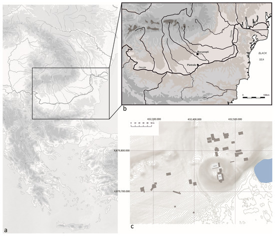
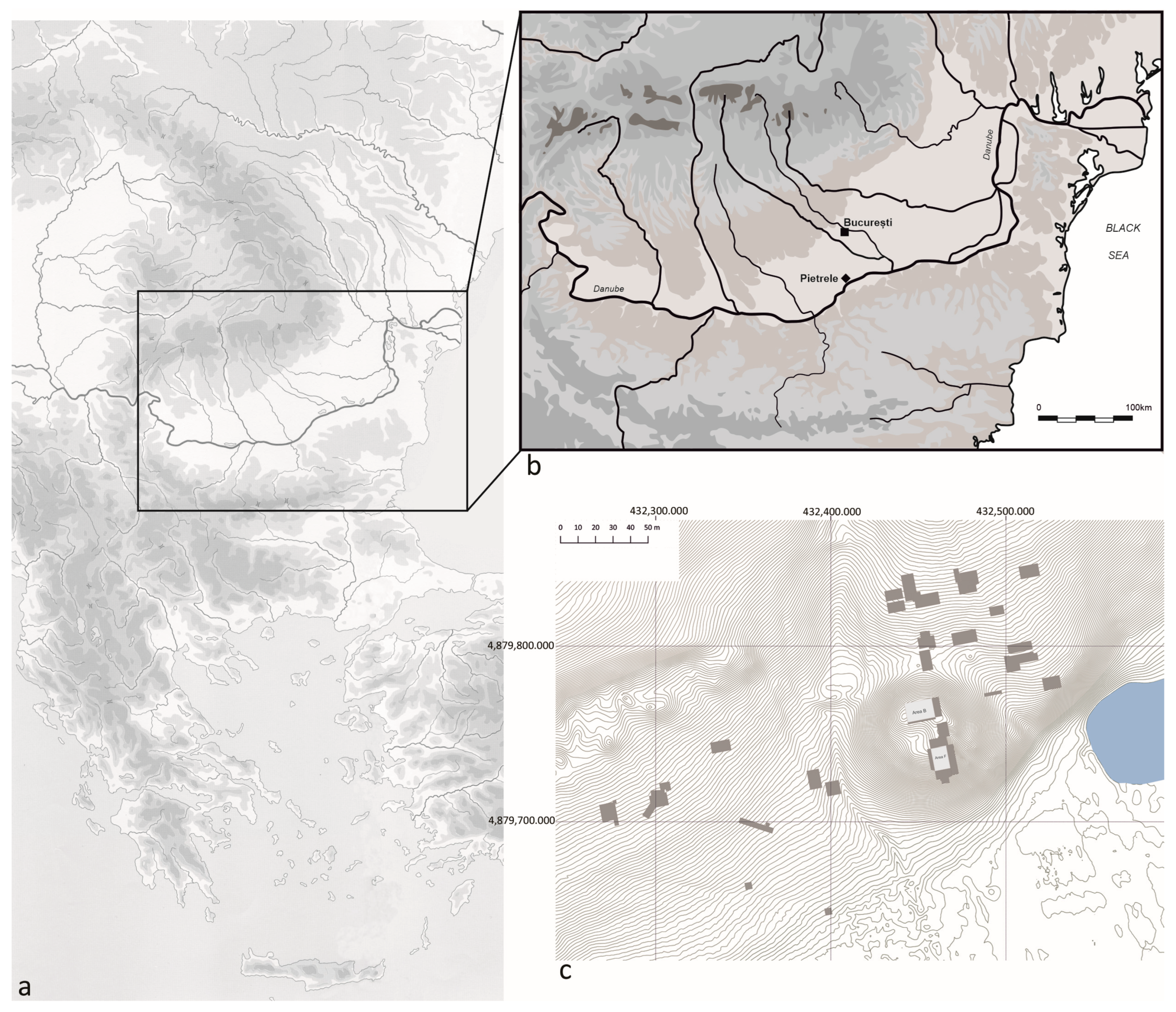
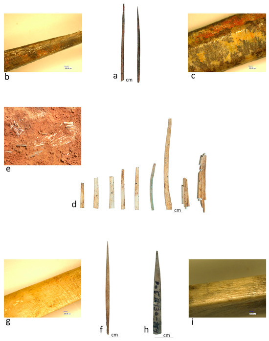
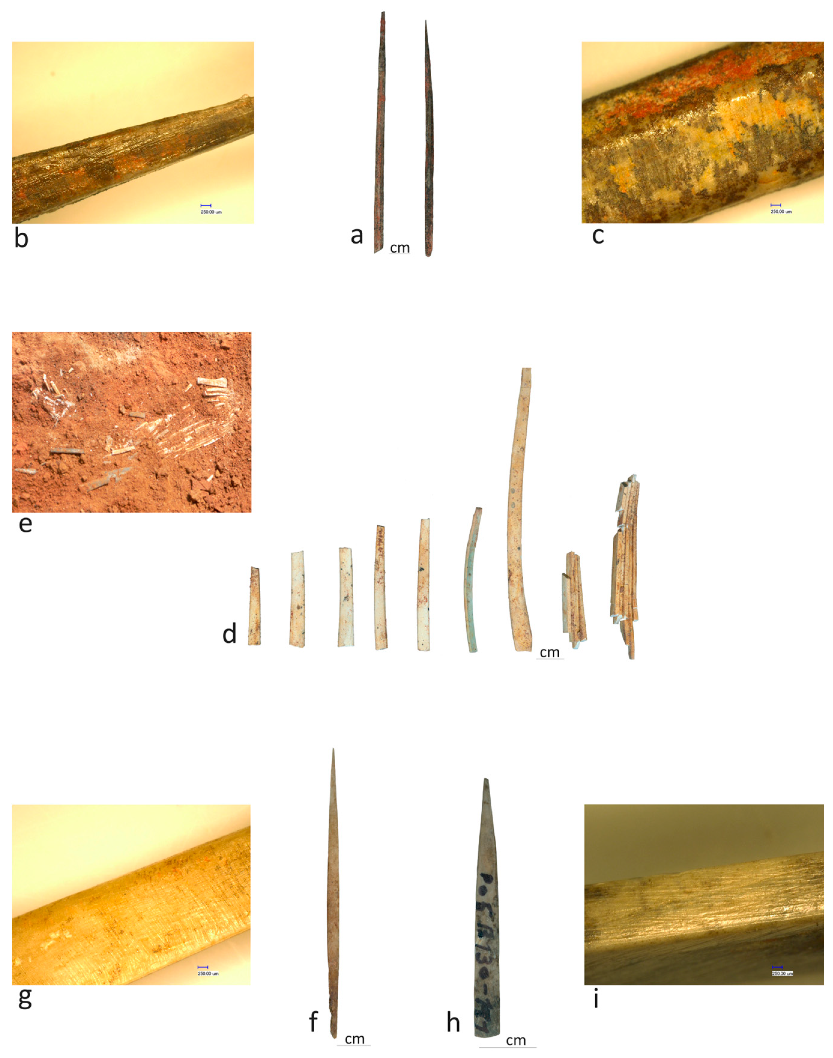
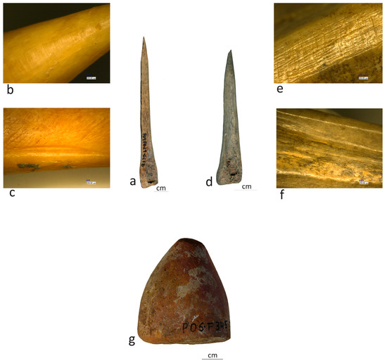
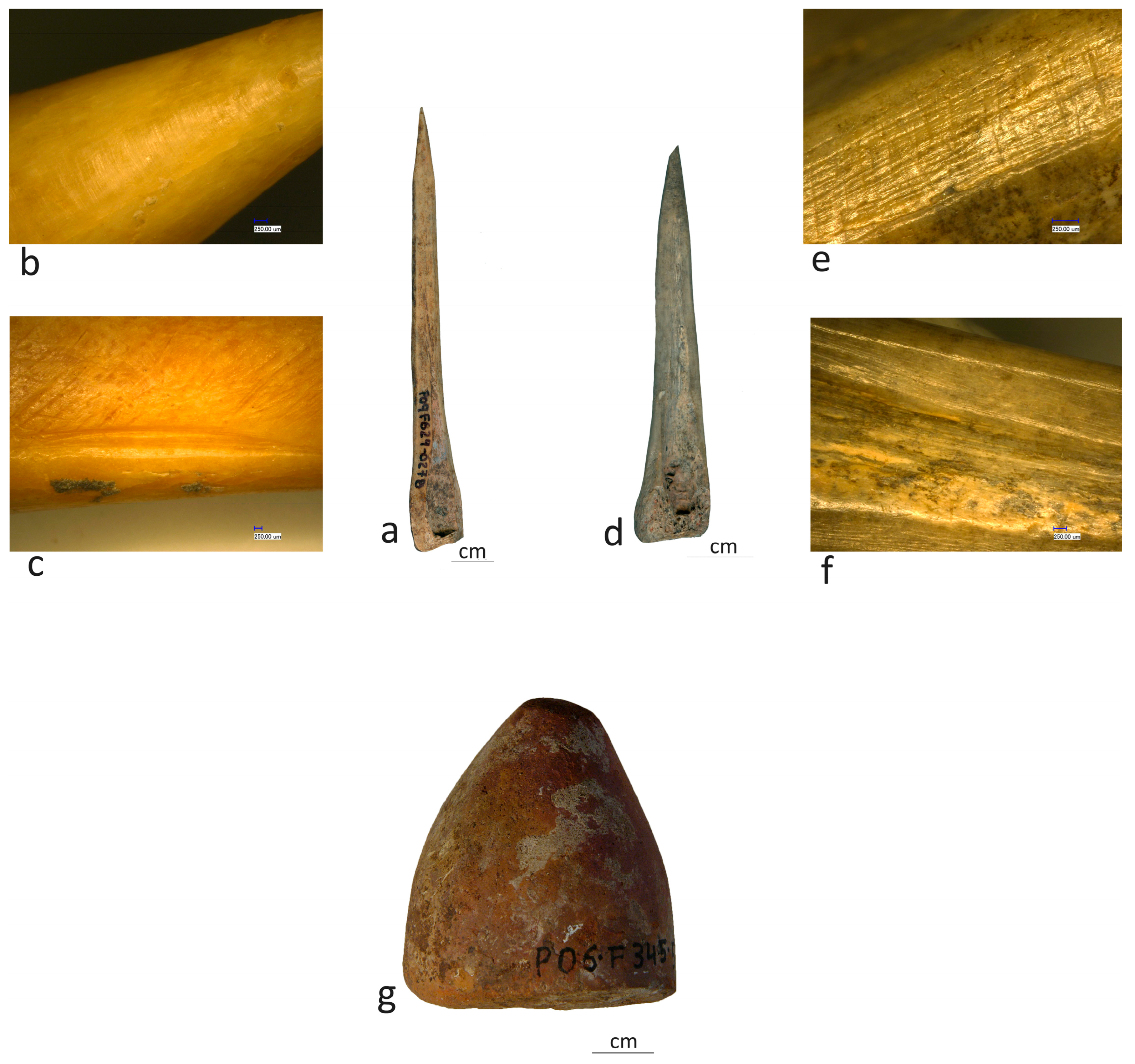
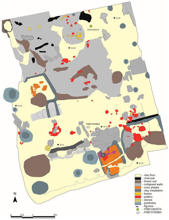
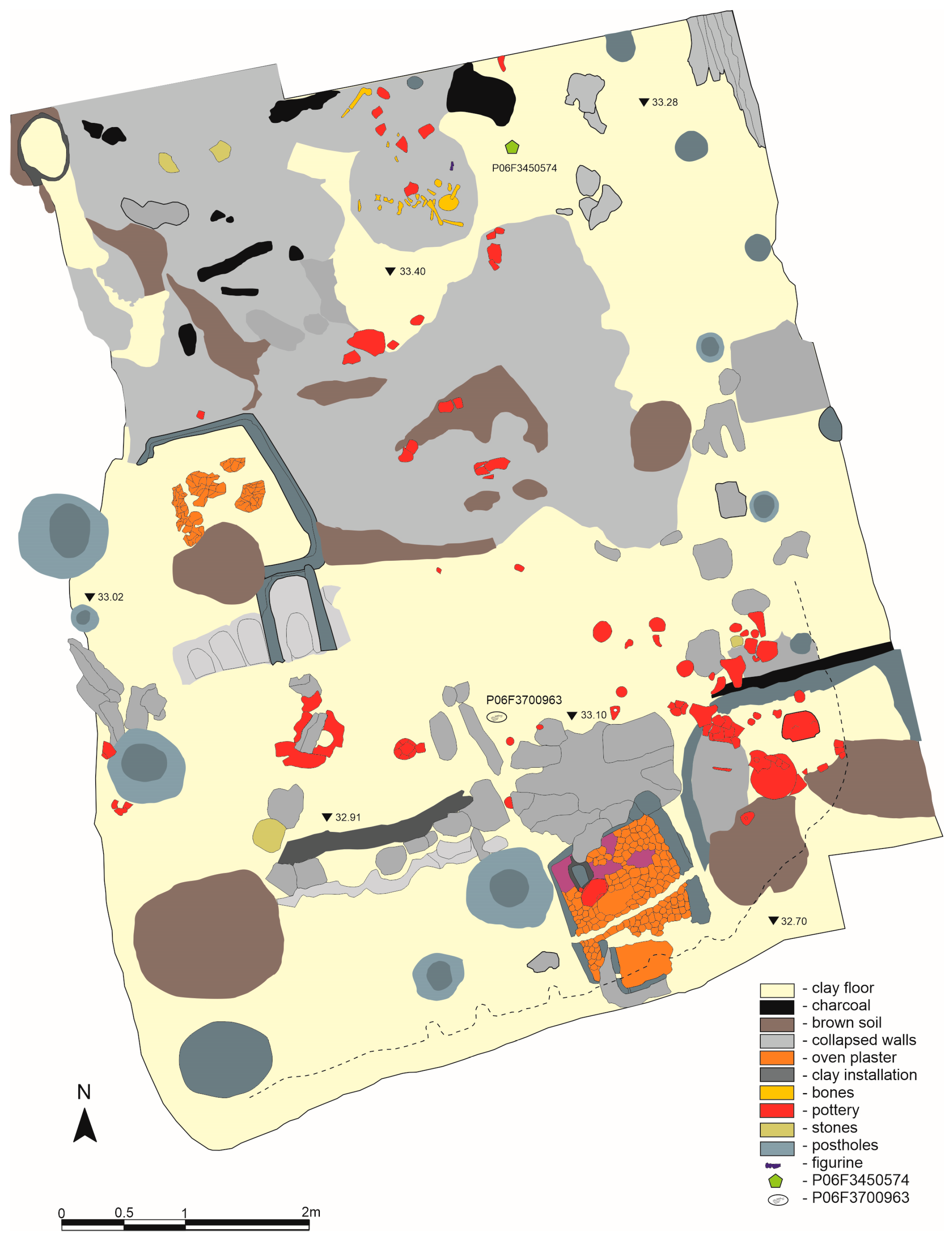
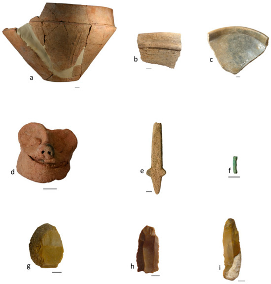
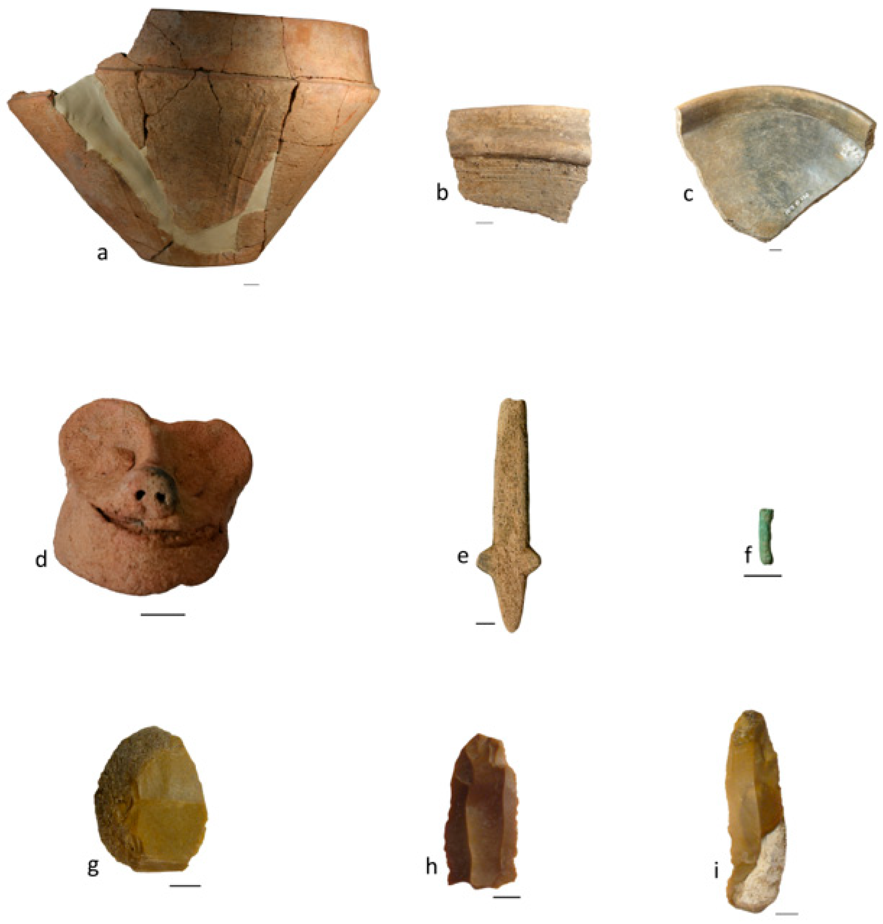
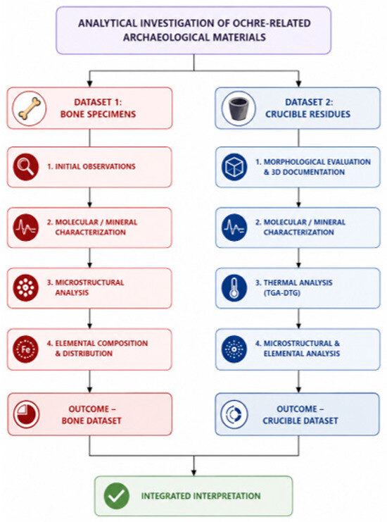
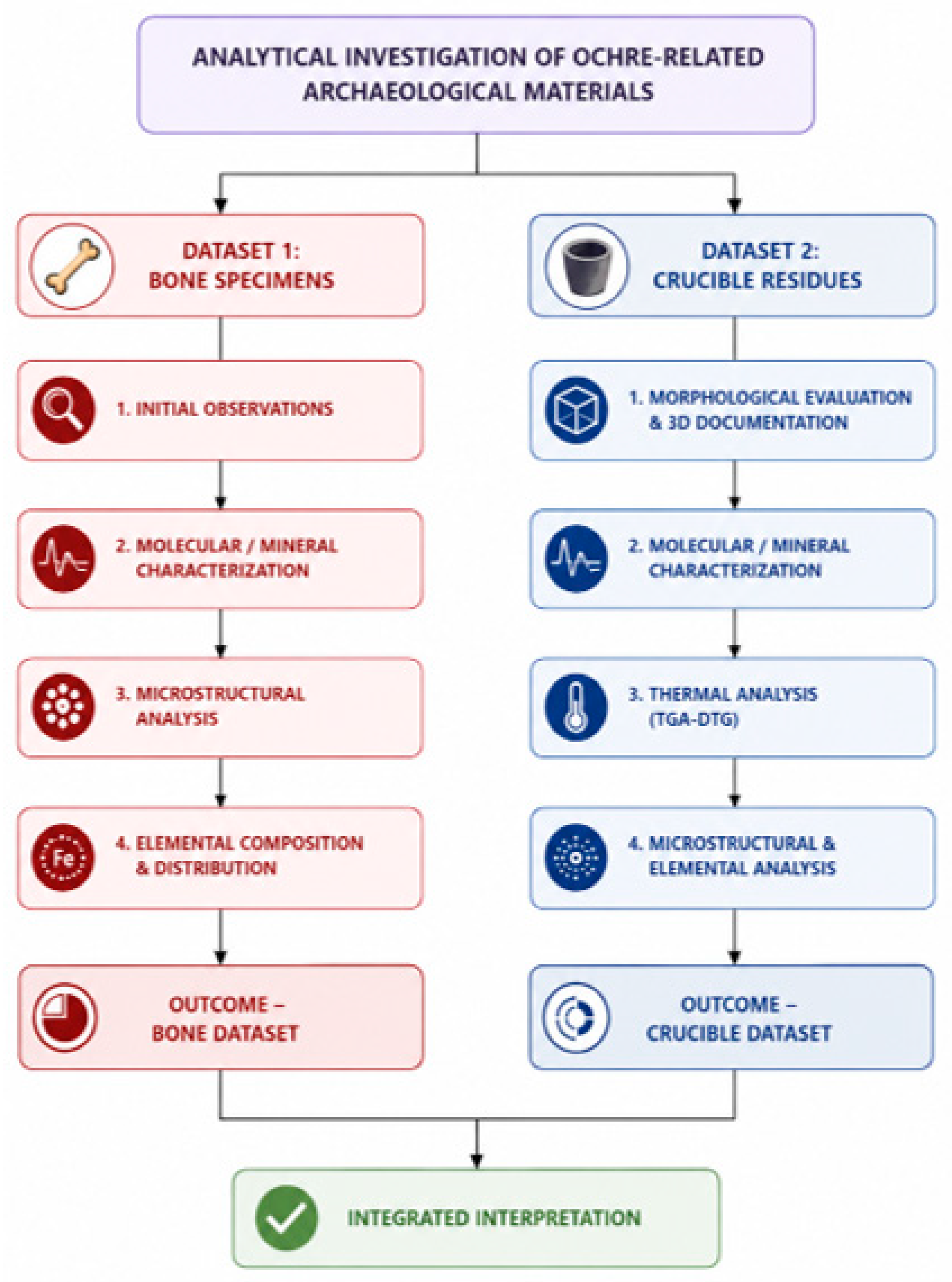
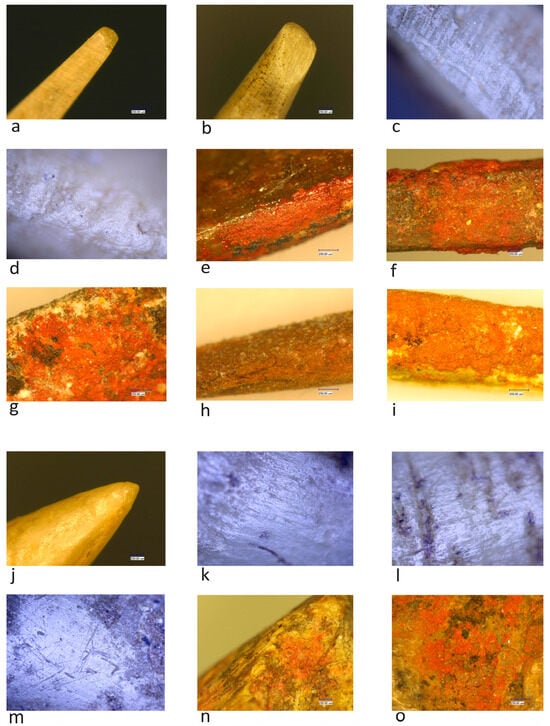
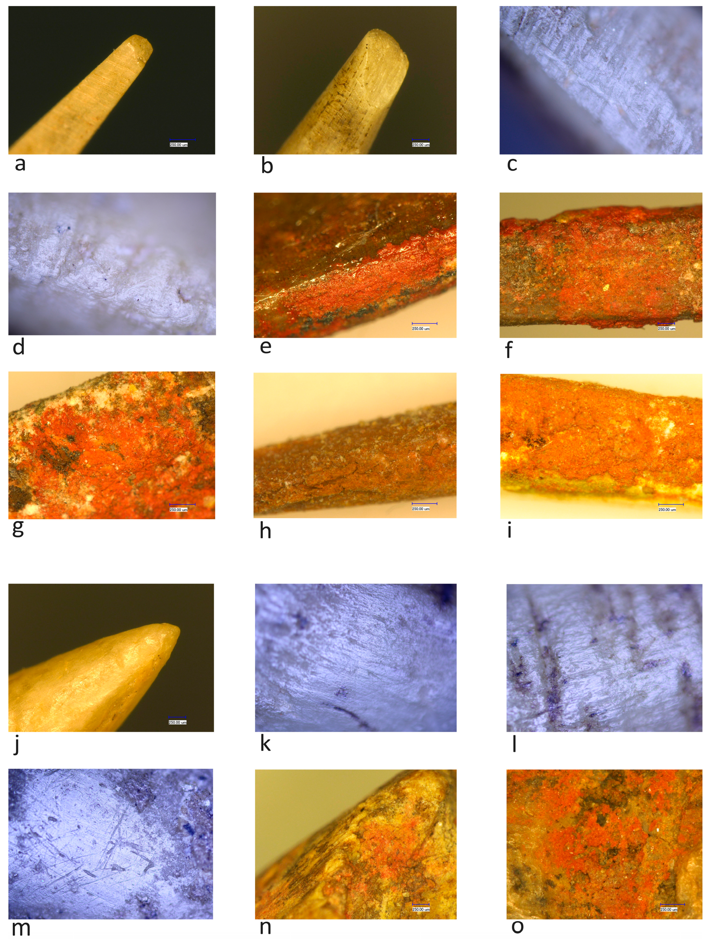
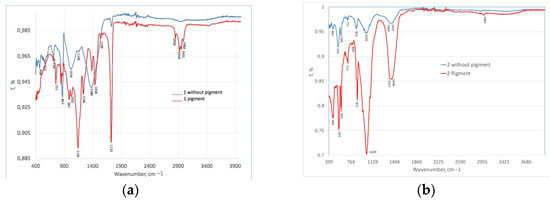
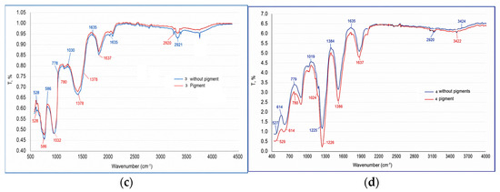
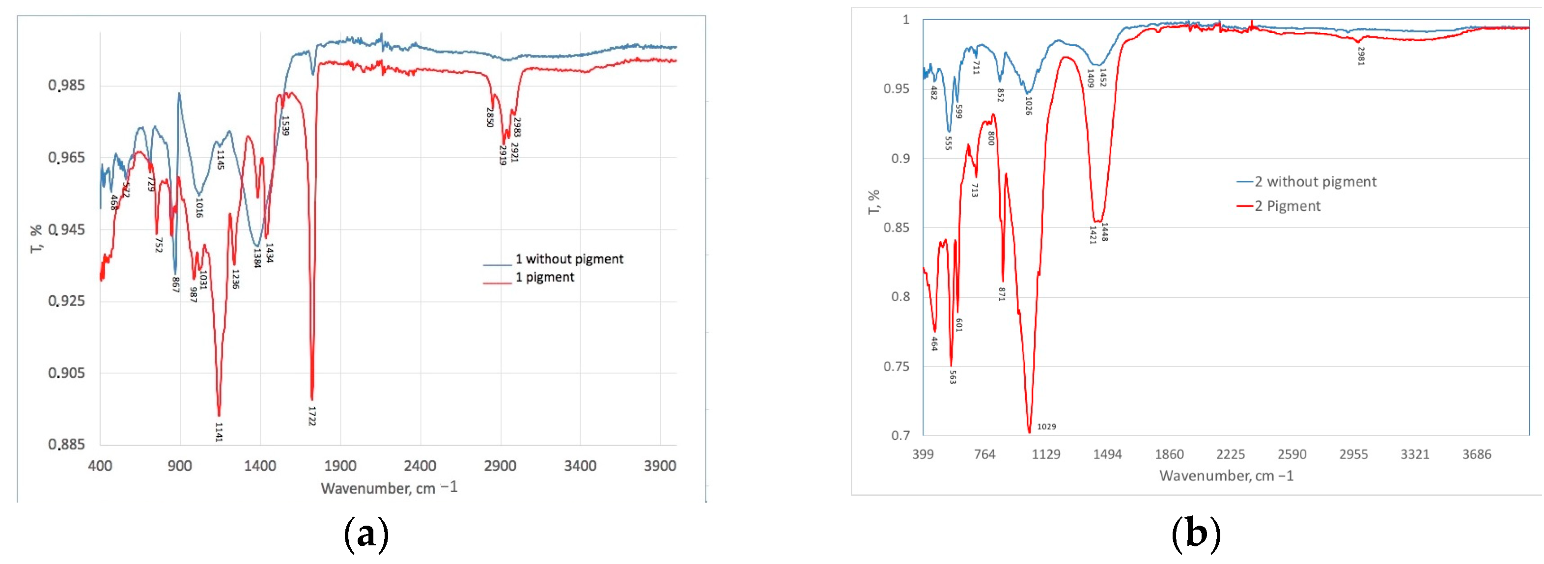
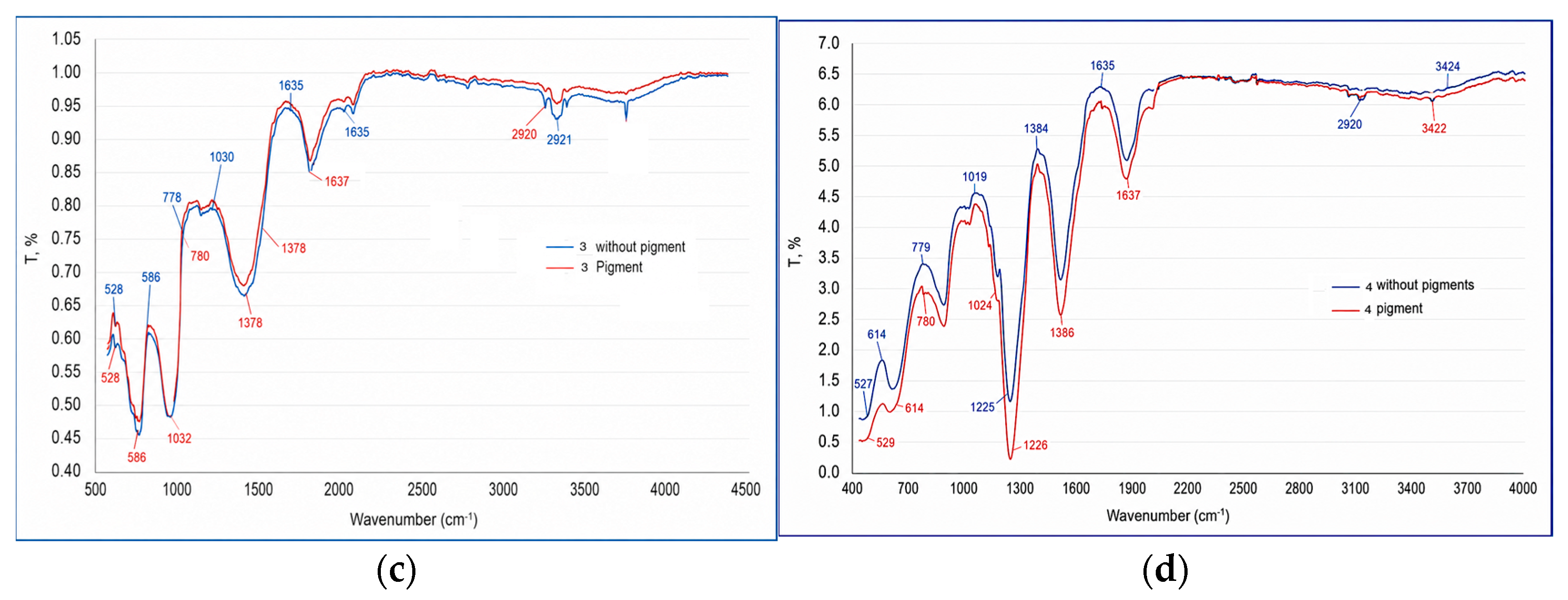
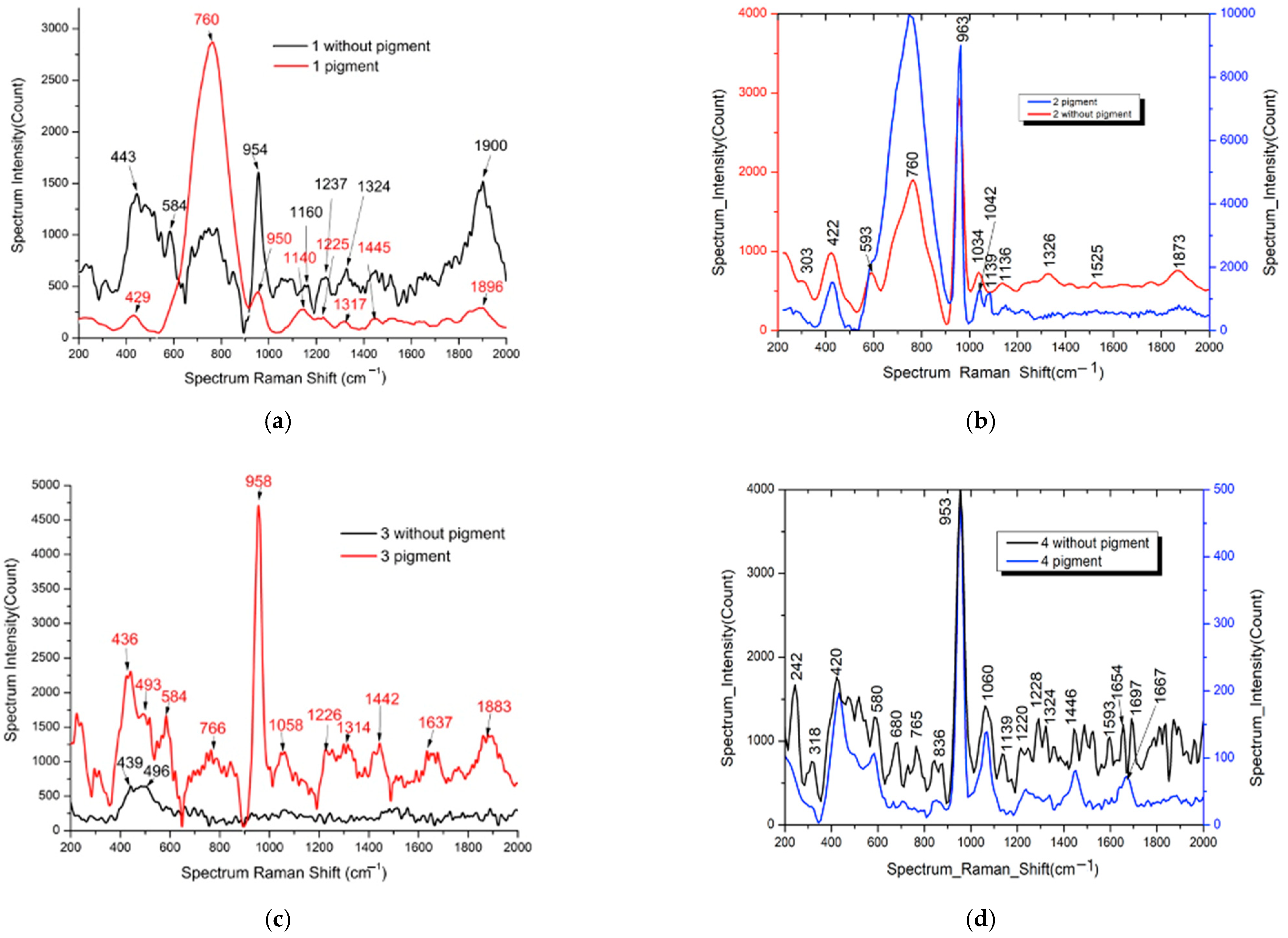
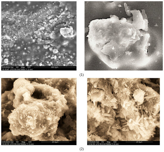
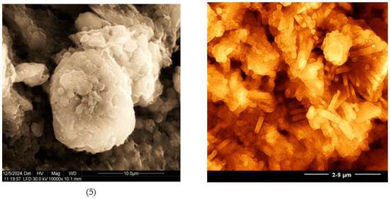
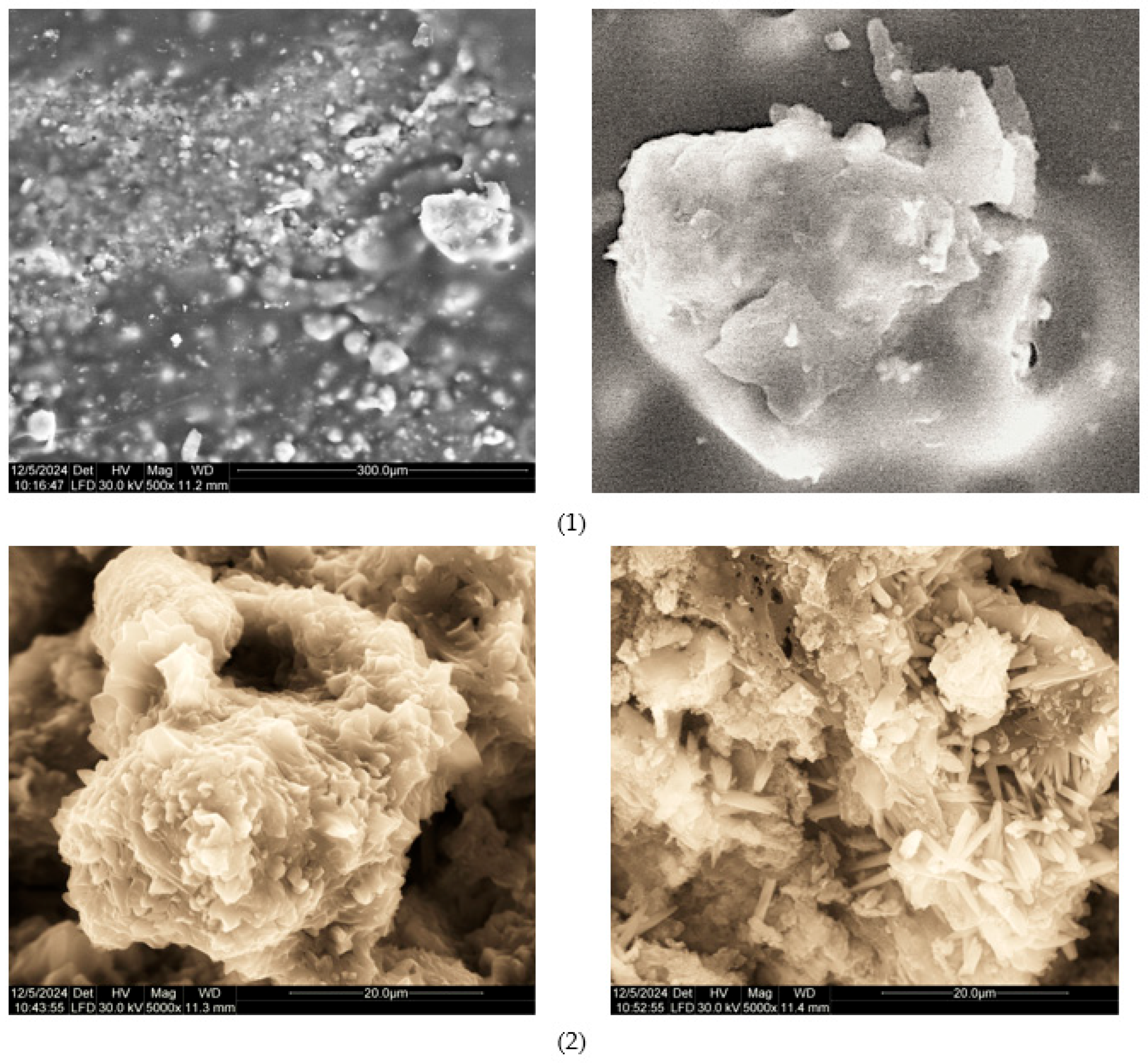
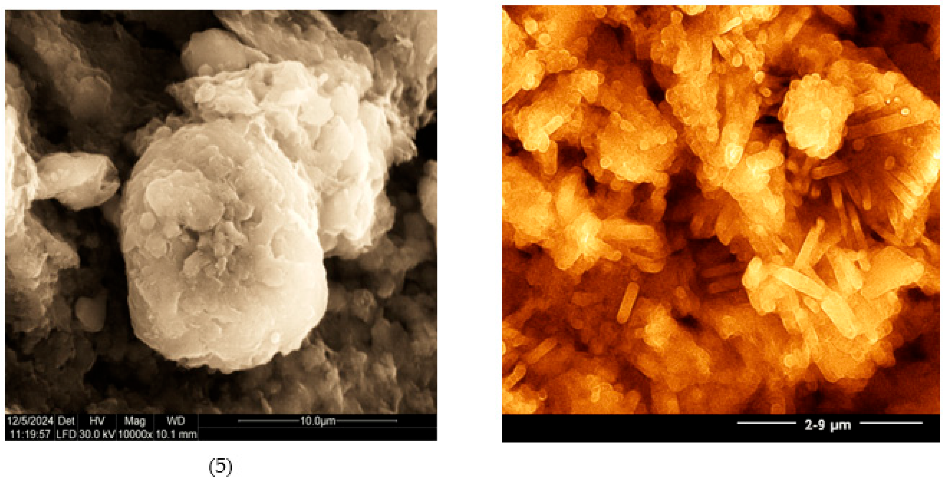
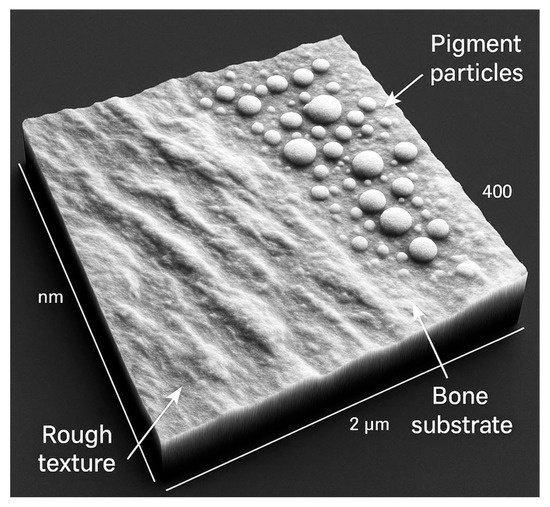
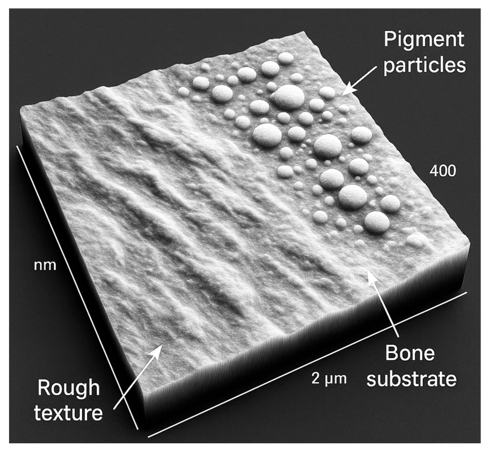
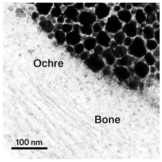
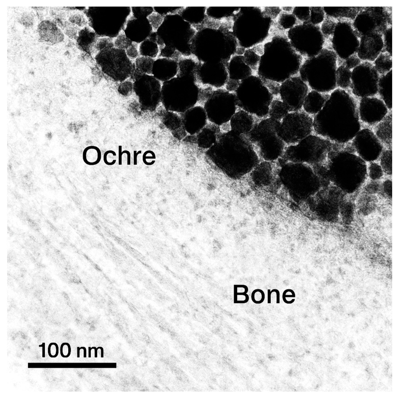
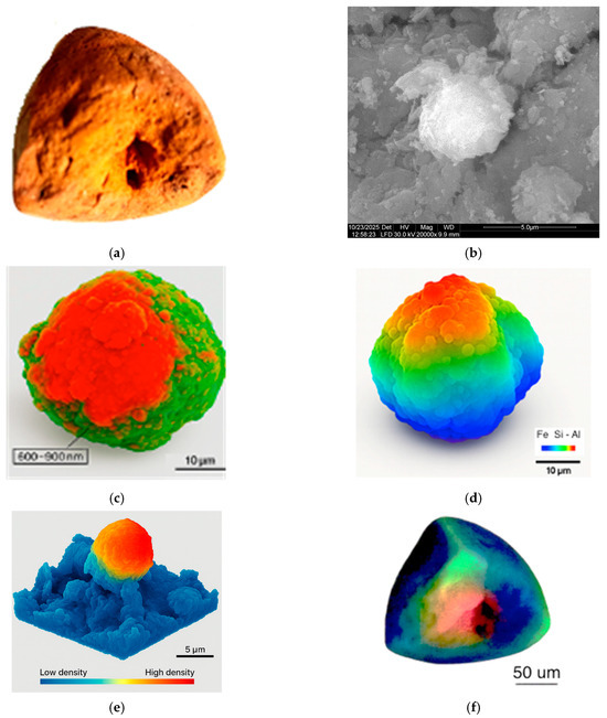
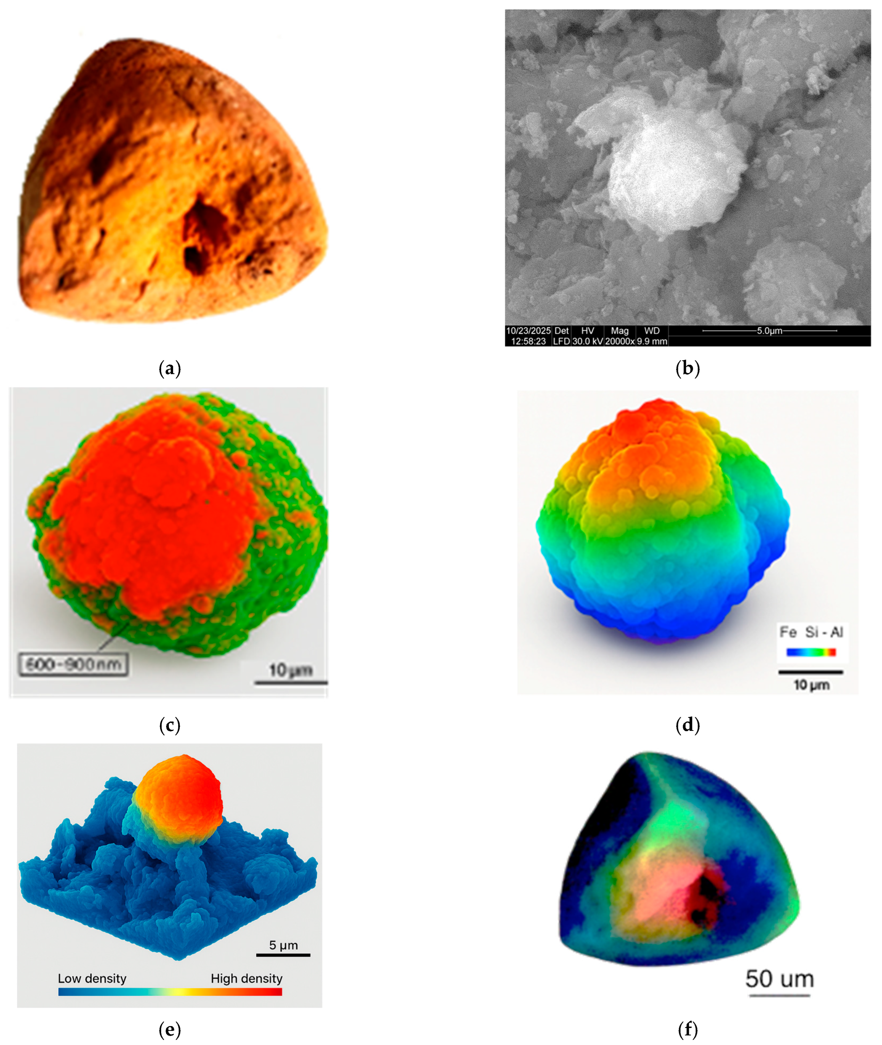
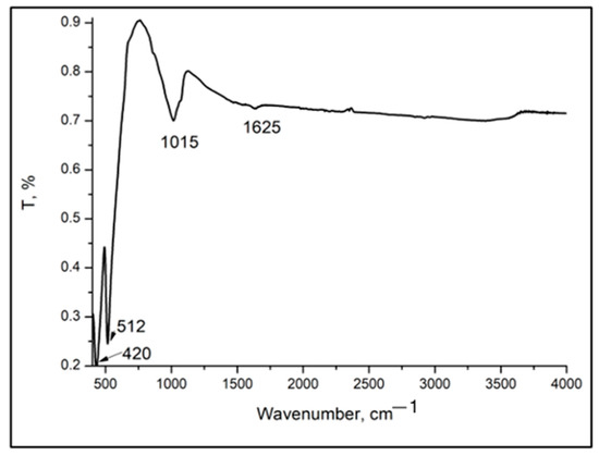
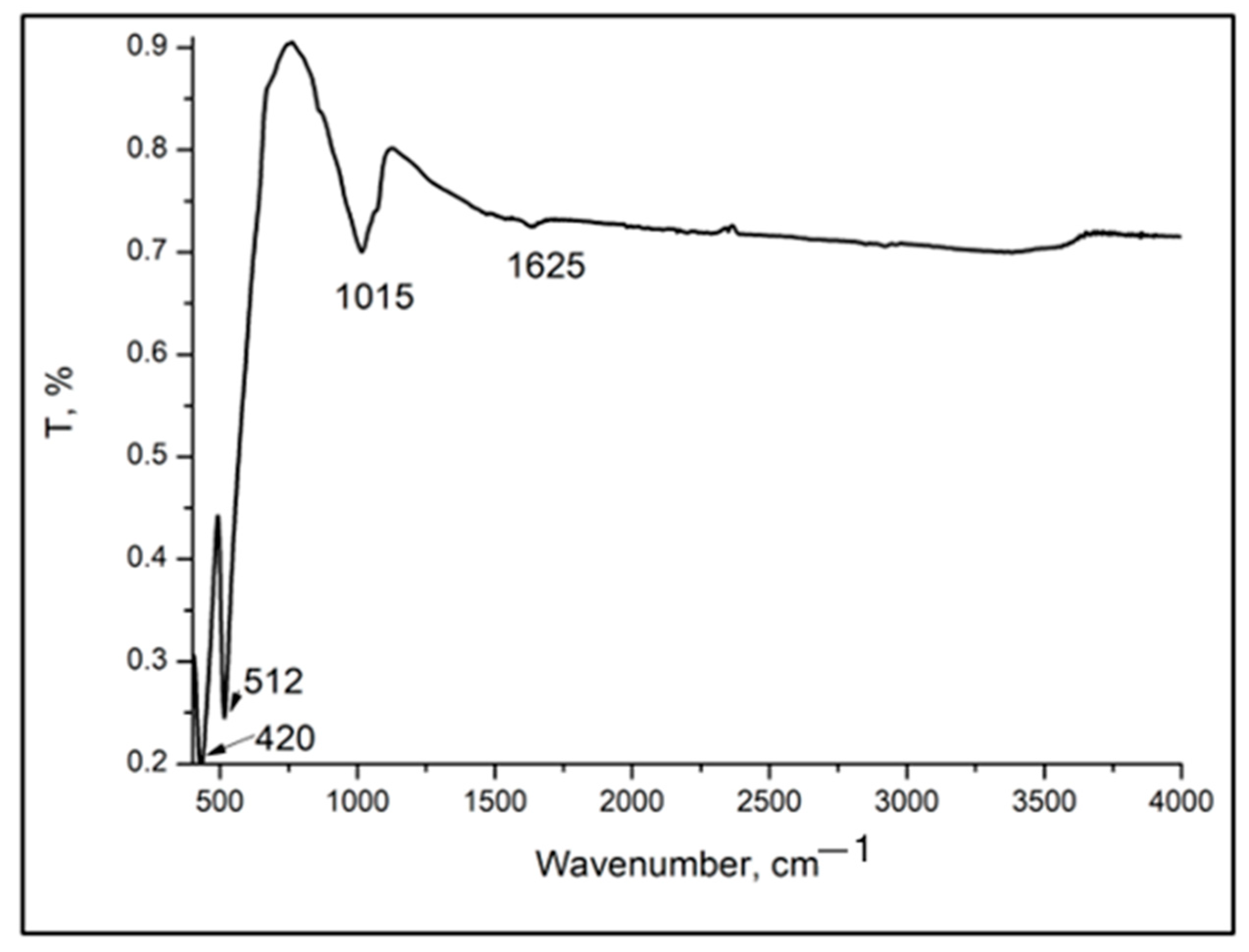
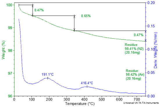
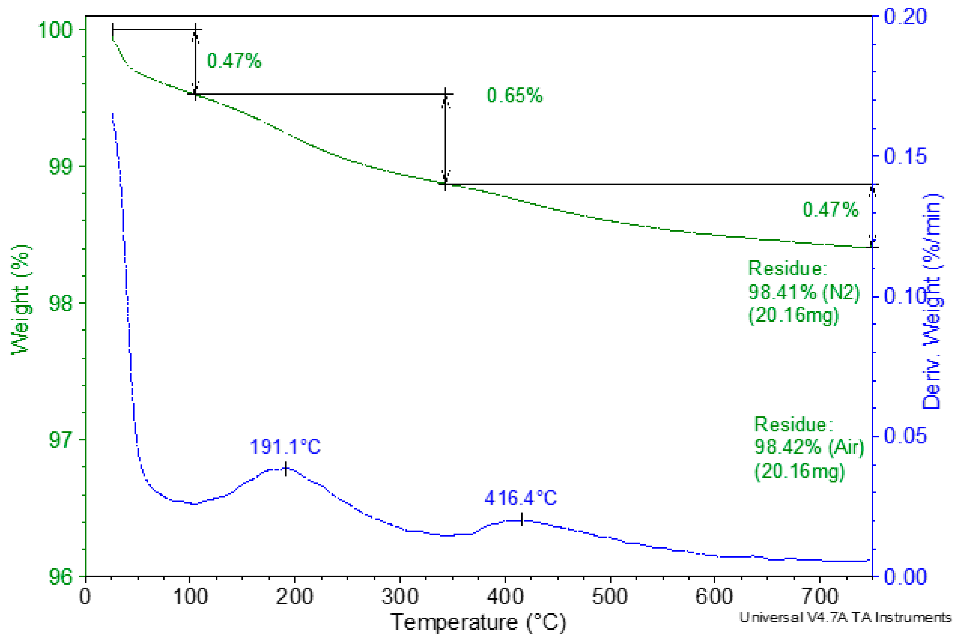

Disclaimer/Publisher’s Note: The statements, opinions and data contained in all publications are solely those of the individual author(s) and contributor(s) and not of MDPI and/or the editor(s). MDPI and/or the editor(s) disclaim responsibility for any injury to people or property resulting from any ideas, methods, instructions or products referred to in the content. |
© 2026 by the authors. Licensee MDPI, Basel, Switzerland. This article is an open access article distributed under the terms and conditions of the Creative Commons Attribution (CC BY) license.