Next Issue
Volume 6, June
Previous Issue
Volume 5, December


Vision, Volume 6, Issue 1 (March 2022) – 19 articles

Cover Story (view full-size image): 
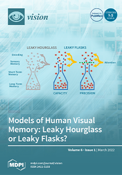
Human memory consists of sensory memory (SM), short-term memory (STM), and long-term memory (LTM). The traditional view of these memory systems resembles a leaky hourglass, the large top and bottom portions representing the large capacities of SM and LTM, whereas the narrow portion in the middle represents the limited capacity of STM. The ‘leak’ in the top part of the hourglass depicts the rapid decay of the contents of SM. In this work, it is shown that major bottlenecks for motion processing exist prior to STM. A model consisting of a pair of ‘leaky flasks’ with narrower top parts capturing distinct bottlenecks prior to STM for capacity and precision of information is proposed to replace the leaky hourglass model. View this paper
  • Issues are regarded as officially published after their release is announced to the table of contents alert mailing list.
  • You may sign up for e-mail alerts to receive table of contents of newly released issues.
  • PDF is the official format for papers published in both, html and pdf forms. To view the papers in pdf format, click on the "PDF Full-text" link, and use the free Adobe Reader to open them.
Order results
Result details
Section
Select all
Export citation of selected articles as:
21 pages, 1635 KiB  
Article
Semantic Expectation Effects on Object Detection: Using Figure Assignment to Elucidate Mechanisms
by Rachel M. Skocypec and Mary A. Peterson
Vision 2022, 6(1), 19; https://doi.org/10.3390/vision6010019 - 21 Mar 2022
Cited by 2 | Viewed by 2593
Abstract
Recent evidence suggesting that object detection is improved following valid rather than invalid labels implies that semantics influence object detection. It is not clear, however, whether the results index object detection or feature detection. Further, because control conditions were absent and labels and [...] Read more.
Recent evidence suggesting that object detection is improved following valid rather than invalid labels implies that semantics influence object detection. It is not clear, however, whether the results index object detection or feature detection. Further, because control conditions were absent and labels and objects were repeated multiple times, the mechanisms are unknown. We assessed object detection via figure assignment, whereby objects are segmented from backgrounds. Masked bipartite displays depicting a portion of a mono-oriented object (a familiar configuration) on one side of a central border were shown once only for 90 or 100 ms. Familiar configuration is a figural prior. Accurate detection was indexed by reports of an object on the familiar configuration side of the border. Compared to control experiments without labels, valid labels improved accuracy and reduced response times (RTs) more for upright than inverted objects (Studies 1 and 2). Invalid labels denoting different superordinate-level objects (DSC; Study 1) or same superordinate-level objects (SSC; Study 2) reduced accuracy for upright displays only. Orientation dependency indicates that effects are mediated by activated object representations rather than features which are invariant over orientation. Following invalid SSC labels (Study 2), accurate detection RTs were longer than control for both orientations, implicating conflict between semantic representations that had to be resolved before object detection. These results demonstrate that object detection is not just affected by semantics, it entails semantics. Full article
Show Figures

Figure 1

9 pages, 990 KiB  
Article
Comparison of Anterior Corneal Aberrometry, Keratometry and Pupil Size with Scheimpflug Tomography and Ray Tracing Aberrometer
by Zahra Ashena, Sean Gallagher, Hasan Naveed, David J. Spalton and Mayank A. Nanavaty
Vision 2022, 6(1), 18; https://doi.org/10.3390/vision6010018 - 18 Mar 2022
Cited by 5 | Viewed by 2321
Abstract
This study aimed to assess the anterior corneal wavefront aberrations, keratometry, astigmatism vectors and pupil size between Pentacam HR® (Oculus Optikgeraete GmbH, Wetzlar, Germany) and iTrace® (Tracey Technologies Corp., Houston, TX, USA). In this observational study, 100 eyes (50 healthy volunteers) [...] Read more.
This study aimed to assess the anterior corneal wavefront aberrations, keratometry, astigmatism vectors and pupil size between Pentacam HR® (Oculus Optikgeraete GmbH, Wetzlar, Germany) and iTrace® (Tracey Technologies Corp., Houston, TX, USA). In this observational study, 100 eyes (50 healthy volunteers) were scanned in mesopic light condition with a Pentacam HR® and iTrace®. Anterior corneal aberrations (spherical aberration (Z40), vertical coma (Z3 − 1), horizontal coma (Z3 + 1)), keratometry in the flattest (K1) and steepest meridian (K2), mean astigmatism, astigmatic vectors (J0 and J45), and pupil size were measured. We found a significant difference in Z40 (Pentacam®: +0.30 ± 0.11 µm and iTrace®: −0.03 µm ± 0.05 µm; p < 0.01) with no correlation between the devices (r = −0.12, p = 0.22). The devices were in complete agreement for Z3 − 1 (p = 0.78) and Z3 + 1 (p = 0.39), with significant correlation between the machines (r = −0.38, p < 0.01 and r = −0.6, p < 0.01). There was no difference in K1, K2 and mean astigmatism. J0 was negative with both devices (against-the-rule astigmatism), but there was no correlation. J45 was negative with the Pentacam HR® (more myopic oblique astigmatism) but significantly correlated between the devices. Pupil size was smaller with Pentacam HR® (p < 0.01). In summary, these devices cannot be used interchangeably. Corneal Z40 was significantly different with more negative Z40 with iTrace® compared to Pentacam HR®. iTrace® operates with lower illumination, giving larger pupil size than Pentacam HR®, which uses intense blue light during measurement. No correlation was found for J0. Pentacam HR® had a trend to record more negative J45 (myopic oblique astigmatism). Full article
Show Figures

Figure 1

16 pages, 2498 KiB  
Review
Pseudomyopia: A Review
by María García-Montero, Gema Felipe-Márquez, Pedro Arriola-Villalobos and Nuria Garzón
Vision 2022, 6(1), 17; https://doi.org/10.3390/vision6010017 - 04 Mar 2022
Cited by 10 | Viewed by 9421
Abstract
This review has identified evidence about pseudomyopia as the result of an increase in ocular refractive power due to an overstimulation of the eye’s accommodative mechanism. It cannot be confused with the term “secondary myopia”, which includes transient myopic shifts caused by lenticular [...] Read more.
This review has identified evidence about pseudomyopia as the result of an increase in ocular refractive power due to an overstimulation of the eye’s accommodative mechanism. It cannot be confused with the term “secondary myopia”, which includes transient myopic shifts caused by lenticular refractive index changes and myopia associated with systemic syndromes. The aim was to synthesize the literature on qualitative evidence about pseudomyopia in terms that clarify its pathophysiology, clinical presentation, assessment and diagnosis and treatment. A comprehensive literature search of PubMed and the Scopus database was carried out for articles published up to November 2021, without a data limit. This review was reported following the preferred reporting items for systematic reviews and meta-analyses (PRISMA) guidelines. Following inclusion and exclusion criteria, a total of 54 studies were included in the qualitative synthesis. The terms pseudomyopia and accommodation spasm have been found in most of the studies reviewed. The review has warned that although there is agreement on the assessment and diagnosis of the condition, there is no consensus on its management, and the literature describes a range of treatment. Full article
Show Figures

Figure 1

9 pages, 3142 KiB  
Article
Choroidal Thickness in Correlation with Axial Length and Myopia Degree
by Habibah Setyawati Muhiddin, Andi Ratna Mayasari, Batari Todja Umar, Junaedi Sirajuddin, Ilhamjaya Patellongi, Itzar Chaidir Islam and Andi Muhammad Ichsan
Vision 2022, 6(1), 16; https://doi.org/10.3390/vision6010016 - 02 Mar 2022
Cited by 10 | Viewed by 3222
Abstract
Background: Myopia is a condition in which the visual images come to a focus in front of the retina of the eye. This disease is a major cause of visual disability, which presents in 108 million persons globally. Purpose: This study aims to [...] Read more.
Background: Myopia is a condition in which the visual images come to a focus in front of the retina of the eye. This disease is a major cause of visual disability, which presents in 108 million persons globally. Purpose: This study aims to determine the relationship between the degree of myopia, the axial length, and the choroidal thickness (CT). Methods: This is an observational analytical study that made use of a cross-sectional design. A total of 59 participants with refractive errors underwent treatment at Hasanuddin University Hospital and 116 eyes were measured and analyzed. The choroidal thickness was measured using the Enhance Depth Imaging OCT (EDI-OCT) tool, which is divided into nine observational areas. Furthermore, all data obtained were compared using statistical analysis, such as the one-way ANOVA and Pearson correlation test (p < 0.05). Results: There was a significant relationship between the choroidal thickness with axial length (p < 0.05) and myopia degrees (p < 0.05). Conclusions: The thickness of the choroid decreases with an increase in the axial length and degree of myopia, which further indicates that the higher the myopia degree, the thinner the choroidal vasculature. Full article
Show Figures

Figure 1

20 pages, 7277 KiB  
Article
Capacity and Allocation across Sensory and Short-Term Memories
by Shaoying Wang, Srimant P. Tripathy and Haluk Öğmen
Vision 2022, 6(1), 15; https://doi.org/10.3390/vision6010015 - 01 Mar 2022
Cited by 1 | Viewed by 2868
Abstract
Human memory consists of sensory memory (SM), short-term memory (STM), and long-term memory (LTM). SM enables a large capacity, but decays rapidly. STM has limited capacity, but lasts longer. The traditional view of these memory systems resembles a leaky hourglass, the large top [...] Read more.
Human memory consists of sensory memory (SM), short-term memory (STM), and long-term memory (LTM). SM enables a large capacity, but decays rapidly. STM has limited capacity, but lasts longer. The traditional view of these memory systems resembles a leaky hourglass, the large top and bottom portions representing the large capacities of SM and LTM, whereas the narrow portion in the middle represents the limited capacity of STM. The “leak” in the top part of the hourglass depicts the rapid decay of the contents of SM. However, recently, it was shown that major bottlenecks for motion processing exist prior to STM, and the “leaky hourglass” model was replaced by a “leaky flask” model with a narrower top part to capture bottlenecks prior to STM. The leaky flask model was based on data from one study, and the first goal of the current paper was to test if the leaky flask model would generalize by using a different set of data. The second goal of the paper was to explore various block diagram models for memory systems and determine the one best supported by the data. We expressed these block diagram models in terms of statistical mixture models and, by using the Bayesian information criterion (BIC), found that a model with four components, viz., SM, attention, STM, and guessing, provided the best fit to our data. In summary, we generalized previous findings about early qualitative and quantitative bottlenecks, as expressed in the leaky flask model and showed that a four-process model can provide a good explanation for how visual information is processed and stored in memory. Full article
(This article belongs to the Special Issue Sensory and Working Memory: Stimulus Encoding, Storage, and Retrieval)
Show Figures

Figure 1

13 pages, 3880 KiB  
Article
Familiarity with an Object’s Size Influences the Perceived Size of Its Image
by Jeroen B. J. Smeets, Pauline E. Weijs and Eli Brenner
Vision 2022, 6(1), 14; https://doi.org/10.3390/vision6010014 - 24 Feb 2022
Viewed by 2713
Abstract
It is known that judgments about objects’ distances are influenced by familiar size: a soccer ball looks farther away than a tennis ball if their images are equally large on the retina. We here investigate whether familiar size also influences judgments about the [...] Read more.
It is known that judgments about objects’ distances are influenced by familiar size: a soccer ball looks farther away than a tennis ball if their images are equally large on the retina. We here investigate whether familiar size also influences judgments about the size of images of objects that are presented side-by-side on a computer screen. Sixty-three participants indicated which of two images appeared larger on the screen in a 2-alternative forced-choice discrimination task. The objects were either two different types of balls, two different types of coins, or a ball and a grey disk. We found that the type of ball biased the comparison between their image sizes: the size of the image of the soccer ball was over-estimated by about 5% (assimilation). The bias in the comparison between the two balls was equal to the sum of the biases in the comparisons with the grey disk. The bias for the coins was smaller and in the opposite direction (contrast). The average precision of the size comparison was 3.5%, irrespective of the type of object. We conclude that knowing a depicted object’s real size can influence the perceived size of its image, but the perceived size is not always attracted towards the familiar size. Full article
(This article belongs to the Special Issue Size Constancy for Perception and Action)
Show Figures

Figure 1

13 pages, 964 KiB  
Review
A Review of Smartphone Apps Used for Toric Intraocular Lens Calculation and Alignment
by Yarrow Scantling-Birch, Hasan Naveed, Ritika Mukhija and Mayank A. Nanavaty
Vision 2022, 6(1), 13; https://doi.org/10.3390/vision6010013 - 18 Feb 2022
Cited by 4 | Viewed by 3042
Abstract
Smartphone apps are becoming increasingly popular in ophthalmology, one specific area of their application being toric intraocular lens (IOL) surgery for astigmatism correction. Our objective was to identify, review and objectively score smartphone apps applicable to toric IOL calculation and/or axis alignment. This [...] Read more.
Smartphone apps are becoming increasingly popular in ophthalmology, one specific area of their application being toric intraocular lens (IOL) surgery for astigmatism correction. Our objective was to identify, review and objectively score smartphone apps applicable to toric IOL calculation and/or axis alignment. This review was divided into three phases. A review was conducted on four major app databases (phase I): National Health Service (NHS) Apps Library, Google Play Store, Apple App Store and Amazon Appstore. A systematic literature review (phase II) was conducted to identify studies for included apps in phase I of our study. Keywords used in both searches included: “toric lens”, “toric IOL”, “refraction”, “astigmatism”, “ophthalmology”, “eye calculator”, “ophthalmology calculator” and “refractive calculator”. Included apps were objectively scored (phase III) by three independent reviewers using the mobile app rating scale (MARS), a validated tool that ranks the quality of mobile health apps using a calculated mean app quality (MAQ) score. Phase I of our study screened 2428 smartphone apps, of which six apps for toric IOL calculation and four apps for axis marking were eligible and were selected for quantitative analysis. Phase II of our study screened 477 studies from PubMed, Medline and Google Scholar. Three studies validating two apps (toriCAM, iToric Patwardhan) in a clinical setting as adjunct tools for preoperative axis marking were identified. Phase III ranked Toric Calculator for iPhone (Apple iOS, MAQ 4.13; average MAQ 3.34 ± 0.54) as the highest-scoring toric IOL calculator, and iToric Patwardhan (Android OS, MAQ 4.13; average MAQ 3.41 ± 0.44) was the highest-scoring axis marker in our study. Our review identified and objectively scored ten smartphone apps available for toric IOL surgery adjuncts. Toric Calculator for iPhone and iToric Patwardhan were the highest-scoring toric IOL calculator and axis marker, respectively. Current literature, though limited, suggests that axis marking smartphone apps can achieve similar levels of misalignment reduction when compared to digital systems. Full article
Show Figures

Figure 1

15 pages, 1453 KiB  
Review
PreserFlo® MicroShunt: An Overview of This Minimally Invasive Device for Open-Angle Glaucoma
by Gloria Gambini, Matteo Mario Carlà, Federico Giannuzzi, Tomaso Caporossi, Umberto De Vico, Alfonso Savastano, Antonio Baldascino, Clara Rizzo, Raphael Kilian, Aldo Caporossi and Stanislao Rizzo
Vision 2022, 6(1), 12; https://doi.org/10.3390/vision6010012 - 09 Feb 2022
Cited by 20 | Viewed by 4486
Abstract
For moderate-to-severe glaucoma, trabeculectomy remains the “gold standard” intraocular pressure (IOP)-lowering treatment; nonetheless, this method requires extensive post-operative maintenance. Microinvasive glaucoma surgery (MIGS) treatments are designed to lessen intra- and post-operative care burden while offering an acceptable IOP decrease for individuals with mild [...] Read more.
For moderate-to-severe glaucoma, trabeculectomy remains the “gold standard” intraocular pressure (IOP)-lowering treatment; nonetheless, this method requires extensive post-operative maintenance. Microinvasive glaucoma surgery (MIGS) treatments are designed to lessen intra- and post-operative care burden while offering an acceptable IOP decrease for individuals with mild to moderate glaucoma. The PreserFlo® MicroShunt (previously InnFocus MicroShunt) is an 8.5 mm glaucoma drainage device manufactured from poly(styrene-block-isobutylene-block-styrene) (SIBS), an extremely biocompatible and bioinert material. The lumen is narrow enough to prevent hypotony, but big enough to avoid being obstructed by sloughed cells or pigment. The device is implanted ab externo, as a stand-alone procedure or in conjunction with cataract surgery, with intraoperative mitomycin C, and a bleb is produced under the conjunctiva and Tenon’s capsule. The MicroShunt was CE-marked in 2012 and designed for primary open-angle glaucoma, the IOP of which remains uncontrolled after maximally tolerated topical treatment. Several clinical trials evaluating the MicroShunt’s long-term safety and effectiveness have been conducted, highlighting the effectiveness of the device over time, along with a tolerable safety profile. The present review aims to gather evidence of PreserFlo’s effectiveness and safety results almost 10 years after its introduction, and furthermore, to compare it with other MIGS and with the gold-standard trabeculectomy for glaucoma management. Full article
Show Figures

Figure 1

10 pages, 235 KiB  
Article
Ocular Surface Disease Signs and Symptoms in Patients with Pseudoexfoliative Glaucoma: A Case—Control Study
by Maria Dermenoudi, Artemis Matsou, Christina Keskini and Eleftherios Anastasopoulos
Vision 2022, 6(1), 11; https://doi.org/10.3390/vision6010011 - 08 Feb 2022
Cited by 2 | Viewed by 2182
Abstract
Purpose: The present study evaluates the differences in the prevalence of the signs and symptoms of ocular surface disease (OSD) in patients with PEX glaucoma (PEXG), compared to other glaucoma types (non-PEXG). Methods: Patients with non-PEXG and PEXG were prospectively examined for the [...] Read more.
Purpose: The present study evaluates the differences in the prevalence of the signs and symptoms of ocular surface disease (OSD) in patients with PEX glaucoma (PEXG), compared to other glaucoma types (non-PEXG). Methods: Patients with non-PEXG and PEXG were prospectively examined for the presence and severity of OSD signs and questioned for symptoms using the OSDI (ocular surface disease index) questionnaire. Results: 116 patients were prospectively enrolled (58 non-PEXG and 58 PEXG). PEXG subjects who were older, had lower central corneal thickness (CCT) values, at a more advanced glaucoma stage and required more IOP lowering drops. OSD signs were prevalent in both groups: conjunctival hyperemia (74.5% non-PEXG vs. 94.8% PEXG), eyelid redness (70.7% vs. 96.6%), conjunctival (74.1% vs. 93.1%) and corneal fluorescein staining (81% vs. 93.1%) and abnormal TFBUT (82.8% vs. 87.9%). When adjusted for potential confounders, (older age, thinner CCT, more advanced glaucoma in PEXG) eyelid redness remained the only parameter significantly associated with PEXG, being 11 times more likely to occur in this group (p = 0.037). Conclusion: Subjects with PEXG presented a higher frequency of signs of OSD compared to other glaucoma types. When accounting for confounding factors, the only difference between the groups was the significantly higher presence (11 times more likely) of eyelid redness in PEXG, suggesting, in addition to glaucoma treatment, the impact of PEX on ocular surface integrity. Full article
13 pages, 285 KiB  
Article
Reading in Children Who Survived Cerebellar Tumors: Evidence from Eye Movements
by Sofia Mironets, Marina Shurupova and Anna Dreneva
Vision 2022, 6(1), 10; https://doi.org/10.3390/vision6010010 - 06 Feb 2022
Cited by 1 | Viewed by 2647
Abstract
Cerebellar tumors often affect the eye movement centers located in vermis, negatively affecting cognitive development and learning abilities in children. Previous research has established that patients who survived cerebellar tumors tend to demonstrate various saccadic impairments (e.g., hypermetria) and poor gaze stability as [...] Read more.
Cerebellar tumors often affect the eye movement centers located in vermis, negatively affecting cognitive development and learning abilities in children. Previous research has established that patients who survived cerebellar tumors tend to demonstrate various saccadic impairments (e.g., hypermetria) and poor gaze stability as compared to healthy controls. The aim of the current study was to evaluate the influence of oculomotor deficits in such patients on reading parameters. A total of 112 children (8–17 y.o.), 65 of whom survived cerebellar tumors, participated in the study. The study design included several oculomotor and reading tasks. Eye movements were recorded every 1/60 s monocularly with an Arrington eye tracker. We observed profound reading impairments in the patients as compared to healthy children, including longer reading time, greater numbers of fixations and regressive saccades, and longer fixation durations. We also found significant correlations between changes in basic oculomotor functions and reading parameters. The patients also demonstrated gaze fixation instability, large number of fixations, and long scanpath reflecting the return of the gaze to the already counted objects. Thus, oculomotor changes caused by cerebellar tumor and its treatment led to disturbances in such neurocognitive activity as reading. Our findings emphasize the necessity of considering these deficits in cerebellar tumor survivors when designing rehabilitation protocols. Full article
2 pages, 225 KiB  
Editorial
Acknowledgment to Reviewers of Vision in 2021
by Vision Editorial Office
Vision 2022, 6(1), 9; https://doi.org/10.3390/vision6010009 - 25 Jan 2022
Viewed by 1627
Abstract
Rigorous peer-reviews are the basis of high-quality academic publishing [...] Full article
13 pages, 292 KiB  
Review
Spotlight on MicroPulse Laser Trabeculoplasty in Open-Angle Glaucoma: What’s on? A Review of the Literature
by Gloria Gambini, Matteo Mario Carlà, Tomaso Caporossi, Umberto De Vico, Alfonso Savastano, Antonio Baldascino, Clara Rizzo, Raphael Kilian and Stanislao Rizzo
Vision 2022, 6(1), 8; https://doi.org/10.3390/vision6010008 - 21 Jan 2022
Cited by 2 | Viewed by 2666
Abstract
Glaucoma is the most common cause of permanent blindness in the world, caused by a progressive optic neuropathy. Patients with glaucoma are often treated with topical medicines therapy in order to reduce intra-ocular pressure (IOP). On the other hand, laser therapies, with the [...] Read more.
Glaucoma is the most common cause of permanent blindness in the world, caused by a progressive optic neuropathy. Patients with glaucoma are often treated with topical medicines therapy in order to reduce intra-ocular pressure (IOP). On the other hand, laser therapies, with the introduction of Argon Laser Trabeculoplasty (ALT) and successively with Selective Laser Trabeculoplasty (SLT), were reported to be effective in IOP control, with low adverse effect rates. In recent years, the micropulse laser, a subthreshold laser technology, was introduced with the goal of reducing side effects while maintaining the effectiveness of the laser treatments. Several studies focused on Micropulse Diode Laser Trabeculoplasty (MDLT) in open-angle glaucoma, to evaluate its effectiveness and possible side effects. Promising results were reported, but irradiation circumstances have not been standardized yet and its role as a substitute for previous laser techniques has yet to be defined. As a result, the goal of this review was to analyze the physical principles at the basis of MDLT and to frame it in the open-angle glaucoma management setting, highlighting the advantages and shortfalls of this technique. Full article
17 pages, 492 KiB  
Systematic Review
Systematic Review of the Longitudinal Sensitivity of Precision Tasks in Visual Working Memory
by James Ades and Jyoti Mishra
Vision 2022, 6(1), 7; https://doi.org/10.3390/vision6010007 - 21 Jan 2022
Cited by 1 | Viewed by 2190
Abstract
Precision of working memory (WM) refers to the objective performance of individuals when trying to recall the features of the encoded WM items. Studies of precision in VWM aim to identify whether differences in WM performance within individuals are sensitive to individual states [...] Read more.
Precision of working memory (WM) refers to the objective performance of individuals when trying to recall the features of the encoded WM items. Studies of precision in VWM aim to identify whether differences in WM performance within individuals are sensitive to individual states or traits. In this systematic review, we study VWM precision and whether it reflects true differences in ability to accurately store information, and thereby possibly a more sensitive measure than discrete VWM span alone. Sifting through 327 abstracts, we identified 34 relevant articles. After assessing these articles with regard to our inclusion criteria to test participants at two separate time points and have a sample size of at least fifteen participants, we found four longitudinal studies regarding VWM precision. One review author and two reviewers independently assessed all studies in the screening and selection process and extracted outcome measures, study characteristics, and, when possible, test–retest reliability metrics. Given the small and heterogeneous sample, this systematic review could not yet provide conclusive evidence on the sensitivity of VWM precision paradigms. Future research of VWM should include longitudinal studies of precision, and address both test–retest reliability in healthy adults and changes in precision during key developmental trajectory periods and in clinical populations. Full article
Show Figures

Figure 1

24 pages, 1467 KiB  
Review
Ocular Autonomic Nervous System: An Update from Anatomy to Physiological Functions
by Feipeng Wu, Yin Zhao and Hong Zhang
Vision 2022, 6(1), 6; https://doi.org/10.3390/vision6010006 - 14 Jan 2022
Cited by 16 | Viewed by 9152
Abstract
The autonomic nervous system (ANS) confers neural control of the entire body, mainly through the sympathetic and parasympathetic nerves. Several studies have observed that the physiological functions of the eye (pupil size, lens accommodation, ocular circulation, and intraocular pressure regulation) are precisely regulated [...] Read more.
The autonomic nervous system (ANS) confers neural control of the entire body, mainly through the sympathetic and parasympathetic nerves. Several studies have observed that the physiological functions of the eye (pupil size, lens accommodation, ocular circulation, and intraocular pressure regulation) are precisely regulated by the ANS. Almost all parts of the eye have autonomic innervation for the regulation of local homeostasis through synergy and antagonism. With the advent of new research methods, novel anatomical characteristics and numerous physiological processes have been elucidated. Herein, we summarize the anatomical and physiological functions of the ANS in the eye within the context of its intrinsic connections. This review provides novel insights into ocular studies. Full article
Show Figures

Figure 1

8 pages, 1055 KiB  
Article
Prediction Error Stabilization and Long-Term Standard Results with a Monofocal Intraocular Lens
by Beatríz Macías-Murelaga, Gonzaga Garay-Aramburu, Roberto Bergado-Mijangos, Daniel Coello-Ojeda, Itziar Ozaeta, Pio Jésus Garcia-Gómez, Jesús Garrido-Fierro, Manuel Rodríguez-Vallejo and Joaquín Fernández
Vision 2022, 6(1), 5; https://doi.org/10.3390/vision6010005 - 13 Jan 2022
Cited by 2 | Viewed by 1973
Abstract
The aim of this study was to assess the stability and differences between objective (O-Rx) and subjective (S-Rx) refraction for the assessment of the prediction error (PE). A secondary aim was to report the results of a monofocal intraocular lens (IOL). 100 subjects [...] Read more.
The aim of this study was to assess the stability and differences between objective (O-Rx) and subjective (S-Rx) refraction for the assessment of the prediction error (PE). A secondary aim was to report the results of a monofocal intraocular lens (IOL). 100 subjects were included for whom S-Rx and O-Rx were obtained for all visits, and for visual performance, posterior capsular opacification incidence and Nd:YAG rates at 12 months. Either S-Rx and O-Rx showed a hyperopic shift from 1 to 6 months (p < 0.05) and stabilization after 6 months. S-Rx was related with the axial length (rho = −0.29, p = 0.007), obtaining a major tendency towards hyperopia in short eyes implanted with high-power IOLs. O-Rx showed a myopic shift in comparison to S-Rx (p < 0.05). This resulted in a decrease of the number of eyes in ±0.50 D and ±1.00 D from 79 to 67% and from 94 to 90%, respectively. The median (interquartile range) uncorrected and corrected visual acuities were 0.1 (0.29) and 0 (0.12) logMAR, respectively, and seven eyes required Nd:YAG capsulotomy at 12 months. Some caution should be taken in PE studies in which O-Rx is used or S-Rx is measured in a 1-month follow-up. Constant optimization should be conducted for this IOL after S-Rx stabilization. Full article
Show Figures

Figure 1

16 pages, 3448 KiB  
Article
Bleb-Independent Glaucoma Surgery to Activate the Uveolymphatic Route of Non-Trabecular Aqueous Humor Outflow: Short-Term Clinical and OCT Results
by Vinod Kumar, Kamal Abdulmuhsen Abu Zaalan, Andrey Igorevich Bezzabotnov, Galina Nikolaevna Dushina, Ahmad Saleh Soliman Shradqa, Zarina Shaykuliyevna Rustamova and Mikhail Aleksandrovich Frolov
Vision 2022, 6(1), 4; https://doi.org/10.3390/vision6010004 - 12 Jan 2022
Cited by 2 | Viewed by 2813
Abstract
The deep sclerectomy technique was modified to enhance aqueous humor (AH) outflow via the non-trabecular pathway. A pilot study was carried out to assess its safety and effectiveness. Thirty-eight patients were under observation. After superficial scleral flap (4 × 4 mm), deep scleral [...] Read more.
The deep sclerectomy technique was modified to enhance aqueous humor (AH) outflow via the non-trabecular pathway. A pilot study was carried out to assess its safety and effectiveness. Thirty-eight patients were under observation. After superficial scleral flap (4 × 4 mm), deep scleral layers were divided into three parts by three parallel-to-limbus incisions. Deep sclerectomy without creating a window in the Descemetes’ membrane was carried out in the distal part. A collagen implant was placed under the sclera of the remaining two parts with one end in the intrascleral pool. The third proximal part was excised to expose the uvea and implant. A Nd:YAG laser trabeculotomy at the surgery site was made on postoperative days 7–10. Outcome measures were IOP change, use of hypotensive medication(s), complications, and the need for a second surgery. At six months, the mean IOP decreased from 29.1 ± 9.2 mm Hg to 14.0 ± 4.3 mm Hg (p = 1.4 × 10⁻⁹); hypotensive medication use reduced from 2.9 ± 0.9 to 0.6 ± 1.0 (p = 1.3 × 10⁻¹⁰); complete success was achieved in 68.4% of cases and partial success was achieved in 31.6% of cases. Intraoperative and postoperative complications were rare and manageable. The OCT of the surgery site revealed the absence of bleb in all cases. Lymphatic vessels with characteristic bicuspid valves in their lumen were detected in conjunctiva near the operation site and over it in 32 patients. IOP decrease in the proposed technique was achieved by activation of the uveolymphatic route of AH outflow. Full article
Show Figures

Figure 1

14 pages, 1496 KiB  
Article
Attentional Orienting in Front and Rear Spaces in a Virtual Reality Discrimination Task
by Rébaï Soret, Pom Charras, Christophe Hurter and Vsevolod Peysakhovich
Vision 2022, 6(1), 3; https://doi.org/10.3390/vision6010003 - 06 Jan 2022
Cited by 1 | Viewed by 2393
Abstract
Recent studies on covert attention suggested that the visual processing of information in front of us is different, depending on whether the information is present in front of us or if it is a reflection of information behind us (mirror information). This difference [...] Read more.
Recent studies on covert attention suggested that the visual processing of information in front of us is different, depending on whether the information is present in front of us or if it is a reflection of information behind us (mirror information). This difference in processing suggests that we have different processes for directing our attention to objects in front of us (front space) or behind us (rear space). In this study, we investigated the effects of attentional orienting in front and rear space consecutive of visual or auditory endogenous cues. Twenty-one participants performed a modified version of the Posner paradigm in virtual reality during a spaceship discrimination task. An eye tracker integrated into the virtual reality headset was used to make sure that the participants did not move their eyes and used their covert attention. The results show that informative cues produced faster response times than non-informative cues but no impact on target identification was observed. In addition, we observed faster response times when the target occurred in front space rather than in rear space. These results are consistent with an orienting cognitive process differentiation in the front and rear spaces. Several explanations are discussed. No effect was found on subjects’ eye movements, suggesting that participants did not use their overt attention to improve task performance. Full article
(This article belongs to the Special Issue Eye Tracking in Human–Computer Interaction)
Show Figures

Figure 1

16 pages, 789 KiB  
Review
A Comparative Analysis of the Camera-like Eyes of Jumping Spiders and Humans
by Irina P. Shepeleva
Vision 2022, 6(1), 2; https://doi.org/10.3390/vision6010002 - 31 Dec 2021
Viewed by 6252
Abstract
Among invertebrates, jumping spiders are one of the few groups whose representatives have camera-like eyes, and the only group whose representatives have fovea. The latter is present in the camera-like eyes of representatives of some groups of vertebrates, including humans. Based on the [...] Read more.
Among invertebrates, jumping spiders are one of the few groups whose representatives have camera-like eyes, and the only group whose representatives have fovea. The latter is present in the camera-like eyes of representatives of some groups of vertebrates, including humans. Based on the literature data, a comparative analysis of the camera-like eyes of jumping spiders and humans was carried out, in the course of which the similarities and differences in the properties and functions of their basic components were identified. The presented data are necessary for the formation of knowledge about jumping spiders as model animals for studying the functioning of the visual system. Full article
Show Figures

Figure 1

16 pages, 1893 KiB  
Article
Pattern-Induced Visual Discomfort and Anxiety in Migraineurs: Their Relationship and the Effect of Colour
by Trevor J. Hine and Yolande B. Z. White
Vision 2022, 6(1), 1; https://doi.org/10.3390/vision6010001 - 24 Dec 2021
Cited by 1 | Viewed by 2733
Abstract
In migraineurs, coloured lenses were found to reduce the visual stress caused by an aversive pattern known to trigger migraines by 70%, but do such patterns also produce a low-level anxiety/fear response? Is this response lessened by colour? We sought to investigate this [...] Read more.
In migraineurs, coloured lenses were found to reduce the visual stress caused by an aversive pattern known to trigger migraines by 70%, but do such patterns also produce a low-level anxiety/fear response? Is this response lessened by colour? We sought to investigate this in a study comprising a broad screening component followed by a dot-probe experiment to elicit attentional biases (AB) to aversive patterns. Undergraduate psychology students completed headache and visual discomfort (VD) questionnaires (N = 358), thereby forming a subject pool from which 13 migraineurs with high visual discomfort and 13 no-headache controls with low visual discomfort, matched on age and sex, completed a dot-probe experiment. Paired stimuli were presented for 500 ms: aversive achromatic 3 cpd square wave gratings vs control, scrambled patterns. These conditions were repeated using the colour that was most comfortable for each participant. VD was greater in the more severe headache groups. On all measures, the migraineurs were more anxious than the controls, and a positive relationship was found between VD and trait anxiety. The 3 cpd gratings elicited an aversive AB in the migraine group which was somewhat reduced by the use of colour, and this was not seen in the controls. The results suggest a new role for colour in reducing visual stress via anxiety/fear reduction. Full article
(This article belongs to the Special Issue The Visual Aura in Migraine)
Show Figures

Figure 1

Previous Issue
Next Issue
Back to TopTop