


Vision, Volume 9, Issue 4 (December 2025) – 21 articles
Cover Story (view full-size image):
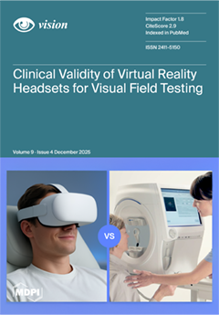
Virtual reality (VR) technology is emerging as a promising alternative to conventional perimetry, offering portable, immersive, and potentially more accessible visual field testing. However, the clinical validity of commercially available VR-based perimetry devices remains uncertain due to heterogeneity in hardware, software, and testing protocols. This systematic review evaluates the clinical validity of VR headsets for visual field assessment by comparing their performance with the Humphrey Field Analyzer, the current gold standard. Nineteen studies were analyzed, highlighting devices with clinically acceptable agreement as well as key limitations related to disease severity, technological features, and regulatory status. The findings provide guidance for clinicians considering VR-based perimetry in clinical and teleophthalmology settings. View this paper
- Issues are regarded as officially published after their release is announced to the table of contents alert mailing list.
- You may sign up for e-mail alerts to receive table of contents of newly released issues.
- PDF is the official format for papers published in both, html and pdf forms. To view the papers in pdf format, click on the "PDF Full-text" link, and use the free Adobe Reader to open them.
Previous Issue
Next Issue