


Vision 2026, 10(2), 27; https://doi.org/10.3390/vision10020027 - 13 May 2026
Abstract
The aim of this study is to determine the optimal functional size of laser peripheral iridotomy (LPI) for anterior chamber parameter improvement in primary angle-closure disease (PACD). This study evaluated 109 eyes from 62 consecutive phakic patients. Baseline and one-week post-LPI anterior segment
[...] Read more.
The aim of this study is to determine the optimal functional size of laser peripheral iridotomy (LPI) for anterior chamber parameter improvement in primary angle-closure disease (PACD). This study evaluated 109 eyes from 62 consecutive phakic patients. Baseline and one-week post-LPI anterior segment OCT were utilized to measure anterior chamber volume (ACV), anterior chamber angle (ACA), and iridotomy dimensions. Data was analyzed using linear mixed-effects models (LMMs), generalized additive models (GAMs), and receiver operating characteristic (ROC) curves. Post-LPI, significant increases occurred in ACA 500 (+7.54°), ACV (+11.09 mm3), and gonioscopic grade. LMMs confirmed a positive association between iridotomy size and anatomical expansion. GAMs demonstrated a saturation effect for ACV improvement, plateauing at 0.1 mm2 (narrow area) and 0.25–0.30 mm2 (superficial area), while the ACA relationship remained predominantly linear. ROC analysis identified preliminary superficial area cutoffs of 0.14 mm2 and 0.12 mm2 as discriminators of above-median volumetric and angular response, respectively. These findings suggest that LPI size is an independent determinant of anatomical response, beyond simple patency. As a preliminary, hypothesis-generating target, a superficial iridotomy area of approximately 0.12–0.14 mm2 was associated with above-median volumetric and angular response in this cohort. Prospective validation is required before these thresholds can be incorporated into clinical practice.
Full article
(This article belongs to the Special Issue Retinal and Optic Nerve Diseases: New Advances and Current Challenges)
►
Show Figures
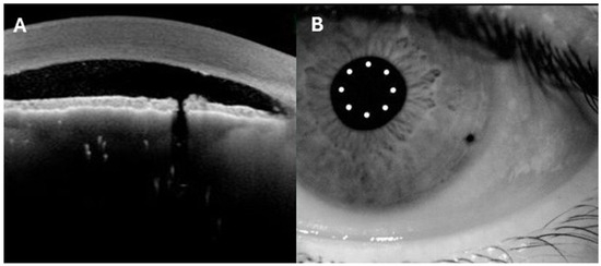
Figure 1


