Next Issue
Volume 7, December
Previous Issue
Volume 7, June


Tomography, Volume 7, Issue 3 (September 2021) – 19 articles

Cover Story (view full-size image): 
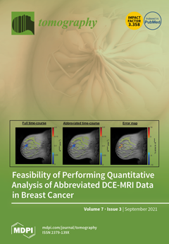
This paper investigates the error that results when quantitatively analyzing abbreviated dynamic contrast-enhanced magnetic resonance imaging (DCE-MRI) data of the breast with the Standard Kety–Tofts model and its Patlak variant. We performed simulations and analyzed patient data to determine the accuracy with which abbreviated time-courses could reproduce the pharmacokinetic parameters, Ktrans (volume transfer constant) and ve (extravascular extracellular volume fraction) when compared to the full time-courses. Most of the analyzed patients had high Ktrans agreement for an abbreviation as short as 4.5 minutes. The results indicate the potential of performing a quantitative analysis of abbreviated breast DCE-MRI without significant loss of information; furthermore, this can be accomplished in the community-based care setting. View this paper
  • Issues are regarded as officially published after their release is announced to the table of contents alert mailing list.
  • You may sign up for e-mail alerts to receive table of contents of newly released issues.
  • PDF is the official format for papers published in both, html and pdf forms. To view the papers in pdf format, click on the "PDF Full-text" link, and use the free Adobe Reader to open them.
Order results
Result details
Section
Select all
Export citation of selected articles as:
11 pages, 1106 KiB  
Article
Radiomics for Everyone: A New Tool Simplifies Creating Parametric Maps for the Visualization and Quantification of Radiomics Features
by Damon Kim, Laura J. Jensen, Thomas Elgeti, Ingo G. Steffen, Bernd Hamm and Sebastian N. Nagel
Tomography 2021, 7(3), 477-487; https://doi.org/10.3390/tomography7030041 - 17 Sep 2021
Cited by 7 | Viewed by 2599
Abstract
Aim was to develop a user-friendly method for creating parametric maps that would provide a comprehensible visualization and allow immediate quantification of radiomics features. For this, a self-explanatory graphical user interface was designed, and for the proof of concept, maps were created for [...] Read more.
Aim was to develop a user-friendly method for creating parametric maps that would provide a comprehensible visualization and allow immediate quantification of radiomics features. For this, a self-explanatory graphical user interface was designed, and for the proof of concept, maps were created for CT and MR images and features were compared to those from conventional extractions. Especially first-order features were concordant between maps and conventional extractions, some even across all examples. Potential clinical applications were tested on CT and MR images for the differentiation of pulmonary lesions. In these sample applications, maps of Skewness enhanced the differentiation of non-malignant lesions and non-small lung carcinoma manifestations on CT images and maps of Variance enhanced the differentiation of pulmonary lymphoma manifestations and fungal infiltrates on MR images. This new and simple method for creating parametric maps makes radiomics features visually perceivable, allows direct feature quantification by placing a region of interest, can improve the assessment of radiological images and, furthermore, can increase the use of radiomics in clinical routine. Full article
Show Figures

Figure 1

12 pages, 3534 KiB  
Article
Metabolite-Specific Echo-Planar Imaging of Hyperpolarized [1-13C]Pyruvate at 4.7 T
by Tyler Blazey, Galen D Reed, Joel R Garbow and Cornelius von Morze
Tomography 2021, 7(3), 466-476; https://doi.org/10.3390/tomography7030040 - 15 Sep 2021
Cited by 1 | Viewed by 1947
Abstract
Although hyperpolarization (HP) greatly increases the sensitivity of 13C MR, the usefulness of HP in vivo is limited by the short lifetime of HP agents. To address this limitation, we developed an echo-planar (EPI) sequence with spectral-spatial radiofrequency (SSRF) pulses for fast [...] Read more.
Although hyperpolarization (HP) greatly increases the sensitivity of 13C MR, the usefulness of HP in vivo is limited by the short lifetime of HP agents. To address this limitation, we developed an echo-planar (EPI) sequence with spectral-spatial radiofrequency (SSRF) pulses for fast and efficient metabolite-specific imaging of HP [1-13C]pyruvate and [1-13C]lactate at 4.7 T. The spatial and spectral selectivity of each SSRF pulse was verified using simulations and phantom testing. EPI and CSI imaging of the rat abdomen were compared in the same rat after injecting HP [1-13C]pyruvate. A procedure was also developed to automatically set the SSRF excitation pulse frequencies based on real-time scanner feedback. The most significant results of this study are the demonstration that a greater spatial and temporal resolution is attainable by metabolite-specific EPI as compared with CSI, and the enhanced lifetime of the HP signal in EPI, which is attributable to the independent flip angle control between metabolites. Real-time center frequency adjustment was also highly effective for minimizing off-resonance effects. To the best of our knowledge, this is the first demonstration of metabolite-specific HP 13C EPI at 4.7 T. In conclusion, metabolite-specific EPI using SSRF pulses is an effective way to image HP [1-13C]pyruvate and [1-13C]lactate at 4.7 T. Full article
Show Figures

Figure 1

15 pages, 4291 KiB  
Article
Evaluation of Regional Lung Function in Pulmonary Fibrosis with Xenon-129 MRI
by Jaime Mata, Steven Guan, Kun Qing, Nicholas Tustison, Yun Shim, John P. Mugler III, Talissa Altes, Jhosep Huaromo and Borna Mehrad
Tomography 2021, 7(3), 452-465; https://doi.org/10.3390/tomography7030039 - 15 Sep 2021
Cited by 12 | Viewed by 2621
Abstract
Idiopathic pulmonary fibrosis, a pattern of interstitial lung disease, is often clinically unpredictable in its progression. This paper presents hyperpolarized Xenon-129 chemical shift imaging as a noninvasive, nonradioactive method of probing lung physiology as well as anatomy to monitor subtle changes in subjects [...] Read more.
Idiopathic pulmonary fibrosis, a pattern of interstitial lung disease, is often clinically unpredictable in its progression. This paper presents hyperpolarized Xenon-129 chemical shift imaging as a noninvasive, nonradioactive method of probing lung physiology as well as anatomy to monitor subtle changes in subjects with IPF. Twenty subjects, nine healthy and eleven IPF, underwent HP Xe-129 ventilation MRI and 3D-SBCSI. Spirometry was performed on all subjects before imaging, and DLCO and hematocrit were measured in IPF subjects after imaging. Images were post-processed in MATLAB and segmented using ANTs. IPF subjects exhibited, on average, higher Tissue/Gas ratios and lower RBC/Gas ratios compared with healthy subjects, and quantitative maps were more heterogeneous in IPF subjects. The higher ratios are likely due to fibrosis and thickening of the pulmonary interstitium. T2* relaxation was longer in IPF subjects and corresponded with hematocrit scores, although the mechanism is not well understood. A lower chemical shift in the red blood cell spectroscopic peak correlated well with a higher Tissue/RBC ratio and may be explained by reduced blood oxygenation. Tissue/RBC also correlated well, spatially, with areas of fibrosis in HRCT images. These results may help us understand the underlying mechanism behind gas exchange impairment and disease progression. Full article
Show Figures

Figure 1

19 pages, 11161 KiB  
Article
Radiofrequency Bias Correction of Magnetization Prepared Rapid Gradient Echo MRI at 7.0 Tesla Using an External Reference in a Sequential Protocol
by Hampus Olsson, Mikael Novén, Jimmy Lätt, Ronnie Wirestam and Gunther Helms
Tomography 2021, 7(3), 434-451; https://doi.org/10.3390/tomography7030038 - 13 Sep 2021
Viewed by 2131
Abstract
At field strengths of 7 T and above, T1-weighted imaging of human brain suffers increasingly from radiofrequency (RF) B1 inhomogeneities. The well-known MP2RAGE (magnetization prepared two rapid acquisition gradient echoes) sequence provides a solution but may not be readily available [...] Read more.
At field strengths of 7 T and above, T1-weighted imaging of human brain suffers increasingly from radiofrequency (RF) B1 inhomogeneities. The well-known MP2RAGE (magnetization prepared two rapid acquisition gradient echoes) sequence provides a solution but may not be readily available for all MR systems. Here, we describe the implementation and evaluation of a sequential protocol to obtain normalized magnetization prepared rapid gradient echo (MPRAGE) images at 0.7, 0.8, or 0.9-mm isotropic spatial resolution. Optimization focused on the reference gradient-recalled echo (GRE) that was used for normalization of the MPRAGE. A good compromise between white-gray matter contrast and the signal-to-noise ratio (SNR) was reached at a flip angle of 3° and total scan time was reduced by increasing the reference voxel size by a factor of 8 relative to the MPRAGE resolution. The average intra-subject coefficient-of-variation (CV) in segmented white matter (WM) was 7.9 ± 3.3% after normalization, compared to 20 ± 8.4% before. The corresponding inter-subject average CV in WM was 7.6 ± 7.6% and 13 ± 7.8%. Maps of T1 derived from forward signal modelling showed no obvious bias after correction by a separately acquired flip angle map. To conclude, a non-interleaved acquisition for normalization of MPRAGE offers a simple alternative to MP2RAGE to obtain semi-quantitative purely T1-weighted images. These images can be converted to T1 maps, analogously to the established MP2RAGE approach. Scan time can be reduced by increasing the reference voxel size which has only a miniscule effect on image quality. Full article
Show Figures

Figure 1

10 pages, 16977 KiB  
Essay
Identification of Traumatic Bone Marrow Oedema: The Pearls and Pitfalls of Dual-Energy CT (DECT)
by Giovanni Foti, Gerardo Serra, Venanzio Iacono and Claudio Zorzi
Tomography 2021, 7(3), 424-433; https://doi.org/10.3390/tomography7030037 - 03 Sep 2021
Cited by 8 | Viewed by 3723
Abstract
Dual-energy computed tomography (DECT) has been reported to successfully identify bone marrow oedema (BME) in various traumatic settings. DECT has multiple strengths, including the availability of both a 3D view of the anatomical area studied and of high-resolution dual energy specific maps super-imposed [...] Read more.
Dual-energy computed tomography (DECT) has been reported to successfully identify bone marrow oedema (BME) in various traumatic settings. DECT has multiple strengths, including the availability of both a 3D view of the anatomical area studied and of high-resolution dual energy specific maps super-imposed onto conventional grayscale morphological images. Windowing can be used to enhance the visualization of BME by increasing the level of the super-imposed images. Conversely, by decreasing the level of the super-imposition of color-coded images, it is possible to progressively enhance the visualization of fine anatomical details, which is useful for diagnosing associated imaging findings. Importantly, bone sclerosis may represent an important pitfall for DECT, potentially generating both false positive and false negative findings by locally altering CT numbers. The aim of this paper was to evaluate the strengths and limitations of DECT in accurately detecting traumatic BME, by considering practical approaches to imaging at several anatomical sites. Full article
Show Figures

Figure 1

12 pages, 3299 KiB  
Article
Branch Pulmonary Artery Regurgitation in Repaired Tetralogy of Fallot: Correlation with Pulmonary Artery Morphology, Distensibility, and Right Ventricular Function
by Suvipaporn Siripornpitak, Duangkanok Lueangwattanapong, Apichaya Sriprachyakul, Suthep Wanitkun and Alisa Limsuwan
Tomography 2021, 7(3), 412-423; https://doi.org/10.3390/tomography7030036 - 01 Sep 2021
Cited by 2 | Viewed by 1709
Abstract
Background: The aim was to determine the effect of pulmonary artery (PA) morphology on the branch pulmonary artery-regurgitation fraction (BPA-RF), the relationship of pulmonary insufficiency (PI) to BPA-RF and PA-distensibility, and factors (BPA-RF and PA-distensibility) associated with right ventricular function (RVF) in repaired [...] Read more.
Background: The aim was to determine the effect of pulmonary artery (PA) morphology on the branch pulmonary artery-regurgitation fraction (BPA-RF), the relationship of pulmonary insufficiency (PI) to BPA-RF and PA-distensibility, and factors (BPA-RF and PA-distensibility) associated with right ventricular function (RVF) in repaired tetralogy of Fallot (rTOF). Methods: A total of 182 rTOF patients (median age 17.1 years) were analyzed for length, angle of PA, BPA-RF, PI, and PA-distensibility, using magnetic resonance imaging. Results: The left PA had a significant greater RF than the right PA (median (interquartile range)): LPA 43.1% (32.6–51.5) and RPA 35.2% (24.7–44.7), p < 0.001. The LPA was shorter with a narrower angle than the RPA (p < 0.001). The anatomy of the branch-PA was not a factor for the greater LPA-RF (odds ratio, 95% confidence interval: CI, p-value): length 0.44 (0.95–2.00), p = 0.28; angle 0.63 (0.13–2.99), p = 0.56. There was a strong positive correlation between PI and BPA-RF-coefficients (95% CI), p-value: LPA 0.78% (0.70–0.86), p < 0.001; RPA 0.78% (0.71–0.84), p < 0.001 and between BPA-RF and distensibility-coefficients (95%CI), p-value: LPA 0.73% (0.37–1.09), p < 0.001; RPA 1.63% (1.22–2.03), p < 0.001, respectively. The adjusted BPA-RF did not predict RVF, RPA (p = 0.434), LPA (p = 0.268). Conclusions: PA morphology is not a significant factor for the differential BPA-RF. The vascular wall in rTOF patients responds to chronic increased intravascular volume by increasing distensibility. BPA-RF is not a determinant of RVF. Full article
Show Figures

Figure 1

15 pages, 4600 KiB  
Review
Idiopathic Interstitial Pneumonias and COVID-19 Pneumonia: Review of the Main Radiological Features and Differential Diagnosis
by Alessia Guarnera, Elena Santini and Pierfrancesco Podda
Tomography 2021, 7(3), 397-411; https://doi.org/10.3390/tomography7030035 - 31 Aug 2021
Cited by 9 | Viewed by 4002
Abstract
COVID-19 pneumonia represents a challenging health emergency, due to the disproportion between the high transmissibility, morbidity, and mortality of the virus and healthcare systems possibilities. Literature has mainly focused on COVID-19 pneumonia clinical-radiological diagnosis and therapy, and on the most common differential diagnoses, [...] Read more.
COVID-19 pneumonia represents a challenging health emergency, due to the disproportion between the high transmissibility, morbidity, and mortality of the virus and healthcare systems possibilities. Literature has mainly focused on COVID-19 pneumonia clinical-radiological diagnosis and therapy, and on the most common differential diagnoses, while few papers investigated rare COVID-19 pneumonia differential diagnoses or the overlapping of COVID-19 pneumonia on pre-existing lung pathologies. This article presents the main radiological characteristics of COVID-19 pneumonia and Idiopathic Interstitial Pneumonias (IIPs) to identify key radiological features for a differential diagnosis among IIPs, and between IIPs and COVID-19 pneumonia. COVID-19 pneumonia differential diagnosis with IIPs is challenging, since these entities may share common radiological findings as ground glass opacities, crazy paving patterns, and consolidations. Multidisciplinary discussion is crucial to reach a final and correct diagnosis. Radiologists have a pivotal role in identifying COVID-19 pneumonia patterns, reporting possible overlapping with long-lasting lung diseases, and suggesting potential differential diagnoses. An optimal evaluation of HRTC may help in containing the disease, in promoting better treatment for patients, and in providing an efficient allocation of human and economic resources. Full article
Show Figures

Figure 1

10 pages, 8788 KiB  
Essay
Identification of Non-Traumatic Bone Marrow Oedema: The Pearls and Pitfalls of Dual-Energy CT (DECT)
by Giovanni Foti, Gerardo Serra, Venanzio Iacono, Stefania Marocco, Giulia Bertoli, Stefania Gori and Claudio Zorzi
Tomography 2021, 7(3), 387-396; https://doi.org/10.3390/tomography7030034 - 26 Aug 2021
Cited by 9 | Viewed by 2940
Abstract
Dual-energy computed tomography (DECT) is an imaging technique widely used in traumatic settings to diagnose bone marrow oedema (BME). This paper describes the role of DECT in diagnosing BME in non-traumatic settings by evaluating its reliability in analyzing some of the most common [...] Read more.
Dual-energy computed tomography (DECT) is an imaging technique widely used in traumatic settings to diagnose bone marrow oedema (BME). This paper describes the role of DECT in diagnosing BME in non-traumatic settings by evaluating its reliability in analyzing some of the most common painful syndromes. In particular, with an illustrative approach, the paper describes the possible use of DECT for the evaluation of osteochondral lesions of the knee and of the ankle, avascular necrosis of the hip, non-traumatic stress fractures, and other inflammatory and infectious disorders of the bones. Full article
Show Figures

Figure 1

14 pages, 4011 KiB  
Article
Retrospective Cohort Study of Frequency and Patterns of Orbital Injuries on Whole-Body CT with Maxillofacial Multi-Slice CT
by Leonie Goelz, Annika Syperek, Stephanie Heske, Sven Mutze, Norbert Hosten and Michael Kirsch
Tomography 2021, 7(3), 373-386; https://doi.org/10.3390/tomography7030033 - 17 Aug 2021
Cited by 4 | Viewed by 3157
Abstract
Background: High-impact trauma frequently leads to injuries of the orbit, but literature focusing on the viscerocranium rather than the neurocranium is underrepresented. Methods: Retrospective cohort study (2006–2014) at an urban level 1 trauma center assessing the frequency and typical patterns of [...] Read more.
Background: High-impact trauma frequently leads to injuries of the orbit, but literature focusing on the viscerocranium rather than the neurocranium is underrepresented. Methods: Retrospective cohort study (2006–2014) at an urban level 1 trauma center assessing the frequency and typical patterns of orbital injuries on whole-body computed tomography (WBCT) with maxillofacial multi-slice CT (MSCT) after severe trauma. (1) Screening of consecutive WBCT cases for dedicated maxillofacial MSCT. (2) Examination by two independent experts’ radiologists for (peri-/)orbital injuries. (3) Case review for trauma mechanisms. Results: 1061 WBCT were included revealing 250 (23.6%) patients with orbital injuries. Less than one-quarter (23.3%) of patients showed osseous and 9.5% showed soft tissue injuries. Combined osseous and soft tissue lesions were present in 39.2% of orbital injuries, isolated soft tissue injuries were rare. Single- or two-wall fractures of the orbit were prevalent, and the orbital floor was affected in 67% of fractures. Dislocated extraocular muscles (44.6%), deformation of the ocular globe (23.8%), and elongation of the optic nerve (12.9%) were the most frequently soft tissue findings. Vascular trauma was suspected in 15.8% of patients. Conclusions: Orbital trauma was confirmed in 23.6% of cases with suspected facial injuries after severe trauma. Concomitant soft tissue injuries should be excluded explicitly in cases with orbital fractures to prevent loss of vision or ocular motility. Full article
Show Figures

Figure 1

15 pages, 3542 KiB  
Article
Detection of Lung Nodules in Micro-CT Imaging Using Deep Learning
by Matthew D. Holbrook, Darin P. Clark, Rutulkumar Patel, Yi Qi, Alex M. Bassil, Yvonne M. Mowery and Cristian T. Badea
Tomography 2021, 7(3), 358-372; https://doi.org/10.3390/tomography7030032 - 07 Aug 2021
Cited by 10 | Viewed by 3785
Abstract
We are developing imaging methods for a co-clinical trial investigating synergy between immunotherapy and radiotherapy. We perform longitudinal micro-computed tomography (micro-CT) of mice to detect lung metastasis after treatment. This work explores deep learning (DL) as a fast approach for automated lung nodule [...] Read more.
We are developing imaging methods for a co-clinical trial investigating synergy between immunotherapy and radiotherapy. We perform longitudinal micro-computed tomography (micro-CT) of mice to detect lung metastasis after treatment. This work explores deep learning (DL) as a fast approach for automated lung nodule detection. We used data from control mice both with and without primary lung tumors. To augment the number of training sets, we have simulated data using real augmented tumors inserted into micro-CT scans. We employed a convolutional neural network (CNN), trained with four competing types of training data: (1) simulated only, (2) real only, (3) simulated and real, and (4) pretraining on simulated followed with real data. We evaluated our model performance using precision and recall curves, as well as receiver operating curves (ROC) and their area under the curve (AUC). The AUC appears to be almost identical (0.76–0.77) for all four cases. However, the combination of real and synthetic data was shown to improve precision by 8%. Smaller tumors have lower rates of detection than larger ones, with networks trained on real data showing better performance. Our work suggests that DL is a promising approach for fast and relatively accurate detection of lung tumors in mice. Full article
Show Figures

Figure 1

14 pages, 2373 KiB  
Article
Combination of Radiomics and Machine Learning with Diffusion-Weighted MR Imaging for Clinical Outcome Prognostication in Cervical Cancer
by Ankush Jajodia, Ayushi Gupta, Helmut Prosch, Marius Mayerhoefer, Swarupa Mitra, Sunil Pasricha, Anurag Mehta, Sunil Puri and Arvind Chaturvedi
Tomography 2021, 7(3), 344-357; https://doi.org/10.3390/tomography7030031 - 05 Aug 2021
Cited by 12 | Viewed by 3415
Abstract
Objectives: To explore the potential of Radiomics alone and in combination with a diffusion-weighted derived quantitative parameter, namely the apparent diffusion co-efficient (ADC), using supervised classification algorithms in the prediction of outcomes and prognosis. Materials and Methods: Retrospective evaluation of the imaging was [...] Read more.
Objectives: To explore the potential of Radiomics alone and in combination with a diffusion-weighted derived quantitative parameter, namely the apparent diffusion co-efficient (ADC), using supervised classification algorithms in the prediction of outcomes and prognosis. Materials and Methods: Retrospective evaluation of the imaging was conducted for a study cohort of uterine cervical cancer, candidates for radical treatment with chemo radiation. ADC values were calculated from the darkest part of the tumor, both before (labeled preADC) and post treatment (labeled postADC) with chemo radiation. Post extraction of 851 Radiomics features and feature selection analysis—by taking the union of the features that had Pearson correlation >0.35 for recurrence, >0.49 for lymph node and >0.40 for metastasis—was performed to predict clinical outcomes. Results: The study enrolled 52 patients who presented with variable FIGO stages in the age range of 28–79 (Median = 53 years) with a median follow-up of 26.5 months (range: 7–76 months). Disease recurrence occurred in 12 patients (23%). Metastasis occurred in 15 patients (28%). A model generated with 24 radiomics features and preADC using a monotone multi-layer perceptron neural network to predict the recurrence yields an AUC of 0.80 and a Kappa value of 0.55 and shows that the addition of radiomics features to ADC values improves the statistical metrics by approximately 40% for AUC and approximately 223% for Kappa. Similarly, the neural network model for prediction of metastasis returns an AUC value of 0.84 and a Kappa value of 0.65, thus exceeding performance expectations by approximately 25% for AUC and approximately 140% for Kappa. There was a significant input of GLSZM features (SALGLE and LGLZE) and GLDM features (SDLGLE and DE) in correlation with clinical outcomes of recurrence and metastasis. Conclusions: The study is an effort to bridge the unmet need of translational predictive biomarkers in the stratification of uterine cervical cancer patients based on prognosis. Full article
Show Figures

Figure 1

11 pages, 7334 KiB  
Article
The Significance of Echo Time in fMRI BOLD Contrast: A Clinical Study during Motor and Visual Activation Tasks at 1.5 T
by Themistoklis Boursianis, Georgios Kalaitzakis, Katerina Nikiforaki, Emmanouela Kosteletou, Despina Antypa, George A. Gourzoulidis, Apostolos Karantanas, Efrosini Papadaki, Panagiotis Simos, Thomas G. Maris and Kostas Marias
Tomography 2021, 7(3), 333-343; https://doi.org/10.3390/tomography7030030 - 05 Aug 2021
Viewed by 1915
Abstract
Blood Oxygen Level Dependent (BOLD) is a commonly-used MR imaging technique in studying brain function. The BOLD signal can be strongly affected by specific sequence parameters, especially in small field strengths. Previous small-scale studies have investigated the effect of TE on BOLD contrast. [...] Read more.
Blood Oxygen Level Dependent (BOLD) is a commonly-used MR imaging technique in studying brain function. The BOLD signal can be strongly affected by specific sequence parameters, especially in small field strengths. Previous small-scale studies have investigated the effect of TE on BOLD contrast. This study evaluates the dependence of fMRI results on echo time (TE) during concurrent activation of the visual and motor cortex at 1.5 T in a larger sample of 21 healthy volunteers. The experiment was repeated using two different TE values (50 and 70 ms) in counterbalanced order. Furthermore, T2* measurements of the gray matter were performed. Results indicated that both peak beta value and number of voxels were significantly higher using TE = 70 than TE = 50 ms in primary motor, primary somatosensory and supplementary motor cortices (p < 0.007). In addition, the amplitude of activation in visual cortices and the dorsal premotor area was also higher using TE = 70 ms (p < 0.001). Gray matter T2* of the corresponding areas did not vary significantly. In conclusion, the optimal TE value (among the two studied) for visual and motor activity is 70 ms affecting both the amplitude and extent of regional hemodynamic activation. Full article
Show Figures

Figure 1

10 pages, 1905 KiB  
Article
7-Tesla Functional Cardiovascular MR Using Vectorcardiographic Triggering—Overcoming the Magnetohydrodynamic Effect
by Christian Hamilton-Craig, Daniel Stäeb, Aiman Al Najjar, Kieran O’Brien, William Crawford, Sabine Fletcher, Markus Barth and Graham Galloway
Tomography 2021, 7(3), 323-332; https://doi.org/10.3390/tomography7030029 - 04 Aug 2021
Cited by 3 | Viewed by 1920
Abstract
Objective: Ultra-high-field B0 ≥ 7 tesla (7T) cardiovascular magnetic resonance (CMR) offers increased resolution. However, electrocardiogram (ECG) gating is impacted by the magneto-hydrodynamic effect distorting the ECG trace. We explored the technical feasibility of a 7T magnetic resonance scanner using an ECG trigger [...] Read more.
Objective: Ultra-high-field B0 ≥ 7 tesla (7T) cardiovascular magnetic resonance (CMR) offers increased resolution. However, electrocardiogram (ECG) gating is impacted by the magneto-hydrodynamic effect distorting the ECG trace. We explored the technical feasibility of a 7T magnetic resonance scanner using an ECG trigger learning algorithm to quantitatively assess cardiac volumes and vascular flow. Methods: 7T scans were performed on 10 healthy volunteers on a whole-body research MRI MR scanner (Siemens Healthineers, Erlangen, Germany) with 8 channel Tx/32 channels Rx cardiac coils (MRI Tools GmbH, Berlin, Germany). Vectorcardiogram ECG was performed using a learning phase outside of the magnetic field, with a trigger algorithm overcoming severe ECG signal distortions. Vectorcardiograms were quantitatively analyzed for false negative and false positive events. Cine CMR was performed after 3rd-order B0 shimming using a high-resolution breath-held ECG-retro-gated segmented spoiled gradient echo, and 2D phase contrast flow imaging. Artefacts were assessed using a semi-quantitative scale. Results: 7T CMR scans were acquired in all patients (100%) using the vectorcardiogram learning method. 3,142 R-waves were quantitatively analyzed, yielding sensitivity of 97.6% and specificity of 98.7%. Mean image quality score was 0.9, sufficient to quantitate both cardiac volumes, ejection fraction, and aortic and pulmonary blood flow. Mean left ventricular ejection fraction was 56.4%, right ventricular ejection fraction was 51.4%. Conclusion: Reliable cardiac ECG triggering is feasible in healthy volunteers at 7T utilizing a state-of-the-art three-lead trigger device despite signal distortion from the magnetohydrodynamic effect. This provides sufficient image quality for quantitative analysis. Other ultra-high-field imaging applications such as human brain functional MRI with physiologic noise correction may benefit from this method of ECG triggering. Full article
Show Figures

Figure 1

10 pages, 5601 KiB  
Article
Influence of Different Arm Positions in the Localizer Radiograph(s) on Patient Dose during Exposure-Controlled CT Examinations of the Neck to Pelvis
by Tony M. Svahn, Lovre Peric and Jennifer C. Ast
Tomography 2021, 7(3), 313-322; https://doi.org/10.3390/tomography7030028 - 29 Jul 2021
Cited by 2 | Viewed by 2662
Abstract
Our aim was to examine the impact of different arm positions during imaging of the localizer radiograph(s) on effective dose for exposure-controlled computed tomography (CT) (Siemens/Canon) scans of the neck to pelvis. An anthropomorphic whole-body phantom was scanned from the neck to pelvis [...] Read more.
Our aim was to examine the impact of different arm positions during imaging of the localizer radiograph(s) on effective dose for exposure-controlled computed tomography (CT) (Siemens/Canon) scans of the neck to pelvis. An anthropomorphic whole-body phantom was scanned from the neck to pelvis with the arms positioned in three different ways during the acquisition of the localizer radiograph: (i) above the head, (ii) alongside the trunk, and (iii) along the trunk with the hands placed on the abdomen. In accordance with clinical routines, the arms were not included in the subsequent helical scans. Effective doses were computed to a standard-sized patient (male/female) using a dedicated system-specific Monte Carlo-based software. Effective doses for the Canon CT scanner for the different alternatives (male/female) were (a) 5.3/6.62 mSv, (b) 5.62/7.15 mSv and (c) 5.92/7.44 mSv. For the Siemens CT scanner, effective doses were (a) 4.47/5.59 mSv, (b) 5.4/6.69 mSv and (c) 5.7/6.99 mSv. Arms placed above the head during localizer radiograph imaging in the current CT procedures substantially reduced the total effective dose to the patient. Full article
Show Figures

Figure 1

12 pages, 995 KiB  
Article
Evaluating a Machine Learning Tool for the Classification of Pathological Uptake in Whole-Body PSMA-PET-CT Scans
by Annette Erle, Sobhan Moazemi, Susanne Lütje, Markus Essler, Thomas Schultz and Ralph A. Bundschuh
Tomography 2021, 7(3), 301-312; https://doi.org/10.3390/tomography7030027 - 29 Jul 2021
Cited by 15 | Viewed by 4865
Abstract
The importance of machine learning (ML) in the clinical environment increases constantly. Differentiation of pathological from physiological tracer-uptake in positron emission tomography/computed tomography (PET/CT) images is considered time-consuming and attention intensive, hence crucial for diagnosis and treatment planning. This study aimed at comparing [...] Read more.
The importance of machine learning (ML) in the clinical environment increases constantly. Differentiation of pathological from physiological tracer-uptake in positron emission tomography/computed tomography (PET/CT) images is considered time-consuming and attention intensive, hence crucial for diagnosis and treatment planning. This study aimed at comparing and validating supervised ML algorithms to classify pathological uptake in prostate cancer (PC) patients based on prostate-specific membrane antigen (PSMA)-PET/CT. Retrospective analysis of 68Ga-PSMA-PET/CTs of 72 PC patients resulted in a total of 77 radiomics features from 2452 manually delineated hotspots for training and labeled pathological (1629) or physiological (823) as ground truth (GT). As the held-out test dataset, 331 hotspots (path.:128, phys.: 203) were delineated in 15 other patients. Three ML classifiers were trained and ranked to assess classification performance. As a result, a high overall average performance (area under the curve (AUC) of 0.98) was achieved, especially to detect pathological uptake (0.97 mean sensitivity). However, there is still room for improvement to detect physiological uptake (0.82 mean specificity), especially for glands. The ML algorithm applied to manually delineated lesions predicts hotspot labels with high accuracy on unseen data and may be an important tool to assist in clinical diagnosis. Full article
(This article belongs to the Special Issue AI Imaging Diagnostic Tools)
Show Figures

Figure 1

15 pages, 2766 KiB  
Article
A Novel Iterative MLEM Image Reconstruction Algorithm Based on Beltrami Filter: Application to ECT Images
by Abdelwahhab Boudjelal, Abderrahim Elmoataz, Bilal Attallah and Zoubeida Messali
Tomography 2021, 7(3), 286-300; https://doi.org/10.3390/tomography7030026 - 28 Jul 2021
Cited by 6 | Viewed by 3068
Abstract
The implementation of emission-computed tomography (ECT), including positron emission tomography and single-photon emission-computed tomography, has been an important research topic in recent years and is of significant and practical importance. However, the slow rate of convergence and the computational complexity have severely impeded [...] Read more.
The implementation of emission-computed tomography (ECT), including positron emission tomography and single-photon emission-computed tomography, has been an important research topic in recent years and is of significant and practical importance. However, the slow rate of convergence and the computational complexity have severely impeded the efficient implementation of iterative reconstruction. By combining the maximum-likelihood expectation maximization (MLEM) iteratively along with the Beltrami filter, this paper proposes a new approach to reformulate the MLEM algorithm. Beltrami filtering is applied to an image obtained using the MLEM algorithm for each iteration. The role of Beltrami filtering is to remove mainly out-of-focus slice blurs, which are artifacts present in most existing images. To improve the quality of an image reconstructed using MLEM, the Beltrami filter employs similar structures, which in turn reduce the number of errors in the reconstructed image. Numerical image reconstruction tomography experiments have demonstrated the performance capability of the proposed algorithm in terms of an increase in signal-to-noise ratio (SNR) and the recovery of fine details that can be hidden in the data. The SNR and visual inspections of the reconstructed images are significantly improved compared to those of a standard MLEM. We conclude that the proposed algorithm provides an edge-preserving image reconstruction and substantially suppress noise and edge artifacts. Full article
Show Figures

Figure 1

8 pages, 754 KiB  
Article
High Rate of False Negative Diagnosis of Silent Patent Ductus Arteriosus on the Chest CT with 3 mm Slice-Thickness, Suggesting the Need for Analysis with Thinner Slice Thickness
by Dongjun Lee, Minji Son, Seungmin Yoo, Sanghoon Jung, Eunju Chun and Charles S. White
Tomography 2021, 7(3), 278-285; https://doi.org/10.3390/tomography7030025 - 27 Jul 2021
Cited by 1 | Viewed by 2092
Abstract
The purpose of this study was to evaluate the diagnostic accuracy of patent with ductus arteriosus (PDA) based on the availability of pretest information on routine chest CT with 3 mm slice-thickness. We retrospectively evaluated CT of 64 patients with PDA. The enrolled [...] Read more.
The purpose of this study was to evaluate the diagnostic accuracy of patent with ductus arteriosus (PDA) based on the availability of pretest information on routine chest CT with 3 mm slice-thickness. We retrospectively evaluated CT of 64 patients with PDA. The enrolled patients were categorized as group 1 (presence of pretest information) and 2 (absence of pretest information, silent PDA). CTs were read by eleven board-certified radiologists, and subsequently by two blind readers. We investigated whether a PDA was mentioned on the initial CT reading. Correct diagnosis of PDA was made in all patients with group 1 (n = 42). In contrast, only 13.7% were correctly diagnosed in group 2. All cases of missed PDA in group 2 were also missed by two blind readers. It is important to realize that the diagnostic accuracy of silent PDA is poor on the chest CT with 3 mm slice-thickness. Thus, use of axial CT images with the thinnest slice-thickness and multi-planar reformatted images (i.e., sagittal and coronal images) may be one way to reduce the number of missed PDA. Full article
Show Figures

Figure 1

10 pages, 7704 KiB  
Commentary
Radiological Reporting of Urgencies Related to Medical Devices: Commentary on a Possible Systematic Approach
by Andrea Contegiacomo, Marco Conti, Massimo Muciaccia, Pietro Trombatore, Michele Dezio, Emilio Lozupone, Agostino Meduri, Riccardo Marano, Luigi Natale and Riccardo Manfredi
Tomography 2021, 7(3), 268-277; https://doi.org/10.3390/tomography7030024 - 29 Jun 2021
Viewed by 2155
Abstract
Most medical devices are routinely recognized on radiological images and described as normal findings in the radiological report, but sometimes they can cause patient access to the emergency department. Multiple possible complications have been described and most of them require prompt recognition by [...] Read more.
Most medical devices are routinely recognized on radiological images and described as normal findings in the radiological report, but sometimes they can cause patient access to the emergency department. Multiple possible complications have been described and most of them require prompt recognition by radiologists for proper clinical management. This commentary proposes a systematic approach to radiological reporting of the most common emergent complications related to medical devices with the intent to avoid the omission of important findings in the final radiological report. Full article
Show Figures

Figure 1

15 pages, 2158 KiB  
Article
Characterizing Errors in Pharmacokinetic Parameters from Analyzing Quantitative Abbreviated DCE-MRI Data in Breast Cancer
by Kalina P. Slavkova, Julie C. DiCarlo, Anum S. Kazerouni, John Virostko, Anna G. Sorace, Debra Patt, Boone Goodgame and Thomas E. Yankeelov
Tomography 2021, 7(3), 253-267; https://doi.org/10.3390/tomography7030023 - 23 Jun 2021
Cited by 2 | Viewed by 3225
Abstract
This study characterizes the error that results when performing quantitative analysis of abbreviated dynamic contrast-enhanced magnetic resonance imaging (DCE-MRI) data of the breast with the Standard Kety–Tofts (SKT) model and its Patlak variant. More specifically, we used simulations and patient data to determine [...] Read more.
This study characterizes the error that results when performing quantitative analysis of abbreviated dynamic contrast-enhanced magnetic resonance imaging (DCE-MRI) data of the breast with the Standard Kety–Tofts (SKT) model and its Patlak variant. More specifically, we used simulations and patient data to determine the accuracy with which abbreviated time course data could reproduce the pharmacokinetic parameters, Ktrans (volume transfer constant) and ve (extravascular/extracellular volume fraction), when compared to the full time course data. SKT analysis of simulated abbreviated time courses (ATCs) based on the imaging parameters from two available datasets (collected with a 3T MRI scanner) at a temporal resolution of 15 s (N = 15) and 7.23 s (N = 15) found a concordance correlation coefficient (CCC) greater than 0.80 for ATCs of length 3.0 and 2.5 min, respectively, for the Ktrans parameter. Analysis of the experimental data found that at least 90% of patients met this CCC cut-off of 0.80 for the ATCs of the aforementioned lengths. Patlak analysis of experimental data found that 80% of patients from the 15 s resolution dataset and 90% of patients from the 7.27 s resolution dataset met the 0.80 CCC cut-off for ATC lengths of 1.25 and 1.09 min, respectively. This study provides evidence for both the feasibility and potential utility of performing a quantitative analysis of abbreviated breast DCE-MRI in conjunction with acquisition of current standard-of-care high resolution scans without significant loss of information in the community setting. Full article
Show Figures

Figure 1

Previous Issue
Next Issue
Back to TopTop