


Highlights
What are the main findings?
- •
- We demonstrated that salinity provides a favorable environment for euhalophytes in terms of growth and cellular homeostasis.
- •
- We have elucidated how salt promotes the growth of euhalophytes.
What are the implications of the main findings?
- •
- They provide new insights into the mechanisms of plant salt tolerance.
- •
- They offer new ideas for the breeding of salt-tolerant crops.
Featured Application
Abstract
Euhalophytes are a group of plants with extremely strong salt tolerance. This study used hydroponically grown Suaeda salsa and Salicornia bigelovii seedlings as model species to probe common features of salt tolerance and photosynthesis among euhalophytes. Exposure to NaCl (100–600 mmol·L−1) for 28 d caused no detectable damage to either biomembranes or photosystem II in the two euhalophytes; moreover, SOD and POD activities were lower than those of the control group. Furthermore, salt treatment increased tissue succulence degree and promoted growth (e.g., more branches and larger photosynthetic organs) in the two euhalophytes. For instance, compared with the control group, plasma membrane permeability (PMP) and malondialdehyde (MDA) contents of S. salsa seedlings grown under 200 mmol·L−1 NaCl decreased by 49.8% and 20.6%, respectively, and the activities of SOD and POD decreased by 75.5% and 87.6%, respectively, whereas the whole-plant fresh weight increased by 94%. These results suggest that 200 mmol·L−1 NaCl is beneficial for the growth of S. salsa. Similar results were observed in S. bigelovii, with an optimal growth salinity of 400 mM NaCl. In saline environments, betaine and Na+ served as key osmotica, promoting water absorption in both euhalophytes. Concurrently, they minimized transpirational water loss through reduced stomatal conductance (Gs) and transpiration rate (Tᵣ), thereby limiting salt influx. Although this response mechanism reduced net photosynthetic rate (Pn), the two euhalophytes compensated by increasing the biomass of photosynthetic organs to enhance carbon fixation, and simultaneously downregulated chlorophyll and protein contents to lower metabolic consumption and respiratory rate (Rr). The above shared mechanism of salt-stimulated growth in the two euhalophytes could offer a breakthrough in breeding vegetable crops with improved salt tolerance and higher yield.
1. Introduction
Soils with NaCl concentrations ≥200 mmol·L−1 are widespread globally, occupying approximately 800 million ha: an environment where only halophytes can complete their life cycles [1,2,3]. Currently, progress in generating highly salt-tolerant crops via cross-breeding or transgenesis remains limited [4]. Cultivating salt-tolerant crops in moderately to highly saline soils remains heavily dependent on high economic costs and freshwater resources [5,6]. Elucidating the salt tolerance mechanisms of halophytes is therefore crucial for breeding salt-tolerant crops [7].
Based on Na+ accumulation and translocation at the whole-plant level, Breckle broadly classified halophytes into three categories: recretohalophytes, euhalophytes, and pseudo-halophytes [8]. Recretohalophytes secrete excess salt via epidermal bladders or salt glands [9]. Euhalophytes, also referred to as succulent euhalophytes or salt-dilution halophytes, accumulate salt in their succulent stems and leaves and dilute it by absorbing additional water [10]. Pseudo-halophytes retain more salt in their roots than in their shoots [11], and were formerly termed salt-excluding halophytes [12]. Collectively, halophytes represent only about 1% of the world’s flora [13]. At the cellular level, the salt-adaptation mechanisms of halophytes can be categorized into three types: osmotic tolerance, Na+/Cl− exclusion and compartmentalization, and tolerance to accumulated Na+ and Cl− [2]. Under high salinity, all halophytes must first achieve osmotic balance. Na+ and Cl− serve as the primary osmotica sequestered in vacuoles, while non-toxic organic solutes (compatible solutes) are mainly used to maintain osmotic equilibrium in the cytoplasm [9]. A range of important compatible solutes, including sugars, sugar alcohols, amino acids, proline-related compounds, betaines, and methylated sulphonium compounds, have been identified as being involved in halophyte salt tolerance [13,14].
As in glycophytes (non-halophytes), high Na+ and Cl− concentrations are cytotoxic to halophytes, and in vitro assays confirm that halophyte enzymes are as NaCl-sensitive as those of glycophytes [2,15]. Nevertheless, halophytes outperform glycophytes in ion compartmentalization and exclusion, thereby maintaining lower cytosolic Na+ and Cl− levels [3,16]. In glycophytes, cytosolic Na+ must be kept below 30 mmol·L−1 [17]. By contrast, halophytes generally accumulate higher Na+ and Cl− concentrations in their shoots than in the external solution [18]. X-ray microanalysis has shown that under high salinity, halophyte cytosolic Na+ can reach 150–200 mmol·L−1, and their chloroplasts and mitochondria can tolerate such elevated ion levels [9]. Many studies have shown that compatible solutes not only contribute to osmotic adjustment but also protect against ion toxicity [5,9,19]. The concentrations of these solutes accumulated in halophyte cells are often higher than those in glycophytes [2]. In addition, halophytes maintain a low cytosolic Na+/K+ ratio through efficient ion homeostasis [9].
Through the above mechanisms, halophytes are not only highly tolerant to salt but can even benefit from moderate salinity in terms of growth. For example, the optimal NaCl concentration for the growth of Limonium bicolour (an obligate recretohalophyte) is approximately 100 mmol·L−1. Under 200 and 400 mmol·L−1 NaCl conditions, although its Na+ secretion rate was enhanced, shoot fresh weight was reduced by 30% and 65%, respectively, compared with the 100 mmol·L−1 treatment [20,21]. Tamarix species (also recretohalophytes) exhibit stronger salt tolerance, yet their growth decreases with increasing salinity, with obvious inhibition even at 100 mmol·L−1 NaCl [22]. Facultative halophytes lacking salt glands (e.g., Artemisia species) grow better in non-saline soils, even though they can tolerate salinity as high as 1000 mmol·L−1 NaCl [23]. The inhibitory effects of high salinity on the growth of these halophytes are closely related to declines in photosynthesis. On the one hand, both stomatal density and stomatal aperture in halophyte leaves decreased under high salinity, which limited CO2 supply [2,18]. On the other hand, the activity of photosynthesis-related enzymes, including chlorophyll (Chl) synthase and Calvin-cycle enzymes, was inhibited by high salt concentrations [19,24]. To date, salt-tolerant crops developed through transgenic or cross-breeding approaches have not yet overcome the growth restrictions imposed by high salinity [4,16].
Under high salinity, a distinctive group of salt-preferring halophytes can thrive and even benefit from saline conditions [10]. These halophytes are therefore termed “euhalophytes”. Treatment with high NaCl concentrations actually promotes their growth, with optimal concentrations generally ≥200 mmol·L−1. Conversely, non-saline soils are unfavorable for their growth, and KCl cannot substitute for NaCl in stimulating their growth [1,3]. Thus, the salt tolerance model of euhalophytes holds great promise for breeding salt-tolerant crops. Moreover, euhalophytes possess considerable economic and ecological value. Among them, Suaeda salsa (L.) Pall. and Salicornia bigelovii Torr. have been well developed and applied in high-saline agriculture as sources of vegetable, edible oil, and forage [3,10]. These two euhalophytes, which can be irrigated directly with seawater, are also known as “seawater vegetables”. Here, we examined and compared their common salt tolerance and photosynthetic traits, with the goal of elucidating the underlying mechanisms in euhalophytes.
2. Materials and Methods
2.1. Plant Materials and Growth Conditions
The seeds of Suaeda salsa (L.) Pall. and Salicornia bigelovii Torr. were purchased from Yancheng Lvyuan Agricultural Technology Co., Ltd. (Yancheng, China). Twenty uniform and plump seeds were sown in each square plastic pot (10 cm × 10 cm × 8 cm) containing clean, moist quartz sand. After cotyledon emergence, half-strength Hoagland’s nutrient solution (containing 5 mmol·L−1 NaCl) was applied. The seedlings were cultivated in a greenhouse with a maximum natural photosynthetic photon flux density (PPFD) of 1200 μmol·m−2·s−1, a photoperiod of 16 h light/8 h dark, relative humidity maintained at 40–50%, and a temperature regime of 20–30 °C.
2.2. Salt Treatment
After gently washing off the fine sand from the roots, five uniform seedlings (4–5 cm in height) were transplanted into each hydroponic pot (13 cm inner diameter × 15 cm depth) and allowed to acclimate for 3 days. Each pot contained 2.0 L of half-strength Hoagland’s nutrient solution. Seedlings were then exposed to NaCl at 100, 200, 400, or 600 mmol·L−1 (dissolved in full-strength Hoagland’s solution), with the salt concentration raised stepwise by 100 mmol·L−1 every 12 h to reach the designated levels on the same day. Ten pots were used per NaCl concentration. For most indices, one biological replicate consisted of one plant from one pot. However, for indices requiring a larger amount of plant material, such as ion content determination and enzyme extraction, plant materials from multiple plants in the same pot were mixed to form one biological replicate. The control group (CK) was maintained in nutrient solution containing 5 mmol·L−1 NaCl. All salt solutions were refreshed three times weekly. After 28 days of salt treatment, plants were harvested in the morning for measurement of physiological and biochemical parameters. Given that the green, fleshy twigs of S. salsa bear semi-cylindrical leaves, whereas the chlorophyll-containing twigs of S. bigelovii have reduced scale-like leaves, the green twigs (including leaves) were selected as the primary material for analysis.
2.3. Biomass and Water Status
After removal from hydroponic pots, roots were blotted dry with absorbent paper, and the fresh weight (FW) of each seedling was recorded promptly using an electronic balance. Seedlings were then oven-dried at 75 °C to constant weight to determine dry weight (DW). The dried tissue was ground and homogenized, and a 100-mg aliquot was incinerated in a muffle furnace at 500 °C to determine ash weight (AW). Organic dry weight (ODW) was calculated by subtracting AW from DW (ODW = DW − AW). The root-to-shoot ratio was calculated based on FW. FW and DW of the green twigs were determined following the same protocol. Water status of the green twigs was expressed as succulence degree = FW/DW.
2.4. Plasma Membrane Permeability (PMP) and Membrane Lipid Peroxidation
PMP of the green twigs was expressed as relative electrical conductivity (%) and measured using a conductivity meter. Before measurement, the twigs were harvested and washed with distilled water [25]. The extent of lipid peroxidation in the green twig tissue was evaluated by measuring malondialdehyde (MDA) content according to the method of Hodges et al. [26].
2.5. Enzyme Extraction and Assay of Antioxidant Enzymes
Fresh green twigs (1 g) were homogenized with 10 mL of extraction buffer [0.05 mol·L−1 NaH2PO4/Na2HPO4, 1 mmol·L−1 EDTA, 1 mmol·L−1 dithiothreitol, and 20% (v/v) glycerol, pH 7.8] on an ice bath. The homogenate was centrifuged at 4 °C and 12,000 g for 10 min. The supernatant was aliquoted into Eppendorf tubes (1 mL each) and stored at –80 °C for subsequent enzyme activity assays.
Antioxidant enzyme activities were measured following the method of Babić et al. [27]. One unit of superoxide dismutase (SOD) activity was defined as the amount of enzyme required to inhibit nitro blue tetrazolium photoreduction to formazan by 50%. Formazan concentration was measured at 560 nm using a spectrophotometer (Evolution 350, Thermo Fisher, Waltham, MA, USA). One unit of peroxidase (POD) activity was defined as the absorbance change of 1.0 per minute at 470 nm, corresponding to the oxidation of guaiacol to tetraguaiacol. One unit of catalase (CAT) activity was defined as the absorbance change of 1.0 per minute at 240 nm, corresponding to the consumption of H2O2.
2.6. Cell Sap Concentration (CSC) and Osmoticum Content
The green twigs were snap-frozen in liquid nitrogen and then thawed in a syringe. The thawed tissue was squeezed with the syringe to collect the cell sap. The CSC was immediately determined using a Vapor Pressure Osmometer (VAPRO 5600, Wescor, Inc., Logan, UT, USA).
Proline (Pro) content in the green twigs was measured according to the method of Bates et al. [28]. Fresh twigs (1.0 g) were homogenized with 10 mL of 3% sulfosalicylic acid. The absorbance of the reaction product of proline with ninhydrin reagent was measured at 520 nm, and proline content was quantified using L-proline as a standard sample. Soluble sugar (SS) content was determined as described by Jermyn [29]. The absorbance of the reaction product of sugars with anthrone reagent was measured at 630 nm, with glucose as a standard sample. Betaine was extracted following the method of Yang and Lu [30], and its content was determined using a betaine assay kit (Solarbio, Inc., Beijing, China) with glycine betaine as a standard sample. Total free amino acids (FAA) were determined following Kowalska et al. [31]. The reaction product of FAA with ninhydrin reagent was measured at 570 nm, and quantified against a glycine standard curve.
Fresh green twigs were rinsed twice with deionized water and then oven-dried at 75 °C. Subsequently, each 0.5 g of dried sample was digested with 20 mL of HNO3:HClO4 (4:1, v/v) at 120 °C for 5 h. The Na+, K+, and Mg2+ concentrations in the diluted digests were determined using an AA-700 atomic absorption spectrometer (Shimadzu Corporation, Kyoto, Japan). The Na+ transfer factor (TF) represents the salt accumulation capacity of plants. It was calculated as the ratio of Na+ content in the twigs to the Na+ concentration in the culture medium.
2.7. Protein and Chlorophyl (Chl) Content
Soluble protein concentration in the water extract from the green twigs was quantified using the Coomassie Brilliant Blue method (G250), with absorbance measured at 595 nm and bovine serum albumin (BSA) used as the standard sample. The green twigs (0.2 g) were cut into 1 mm slices and placed into a test tube containing 20 mL of 80% acetone. The tubes were sealed with stoppers and kept at 0 °C for chlorophyll extraction until the tissues turned white. Chlorophyll concentrations in the well-mixed extract were determined at 663.2 and 646.8 nm following Yue et al. [32].
2.8. Chl a Fluorescence and Gas-Exchange Parameters
For chlorophyll fluorescence measurements, green twigs from fully dark-adapted seedlings were used to determine the maximum photochemical efficiency of photosystem II (Fv/Fm) and the photosynthetic performance index (PI) using a plant efficiency analyser (Handy PEA, Hansatech Instrument Ltd., King’s Lynn, UK) in a dark room at room temperature. The leaves (from S. salsa) or branches (from S. bigelovii) were selected for each measurement.
Gas-exchange parameters were measured with a TARGAS-1 portable photosynthetic system (Hansatech Instrument Ltd., King’s Lynn, UK) under a natural condition of 430 μmol·mol−1 CO2, 50% relative humidity, and saturating light intensity of 1000 μmol·m−2·s−1. Fully light-adapted green twigs were harvested and quickly placed into a small modified open assimilation chamber for measurement. Respiratory rate (Rr) was recorded under dark conditions (covered with black cloth). Net photosynthetic rate (Pn), transpiration rate (Tr), intercellular CO2 concentration (Ci), stomatal conductance (Gs), and Rr were calculated according to Qiu et al. [33]. The total photosynthetic capacity of whole-plant (Pt) was calculated by multiplying the total fresh weight of twigs per plant by Pn.
2.9. Statistical Analyses
All data were expressed as means ± SD. Statistical analyses among different NaCl treatments were performed using one-way ANOVA and Duncan’s LSR (least significant range) test with SPSS 17.0 software. Different lowercase letters above the means indicated significant differences (p < 0.05). The data of the two euhalophytes were not compared with each other.
3. Results
3.1. The Effects of Salinity on the Growth of Two Euhalophytes
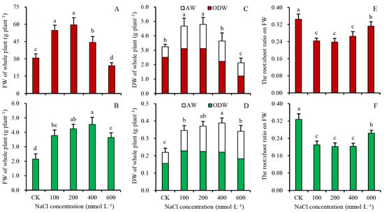
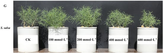
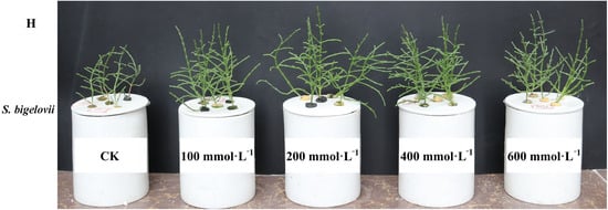
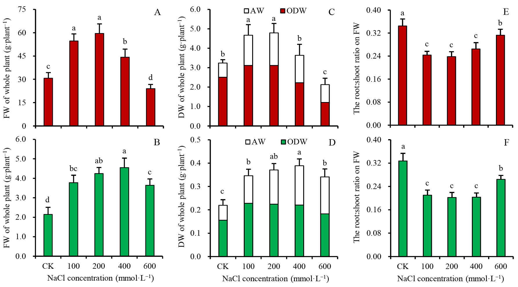
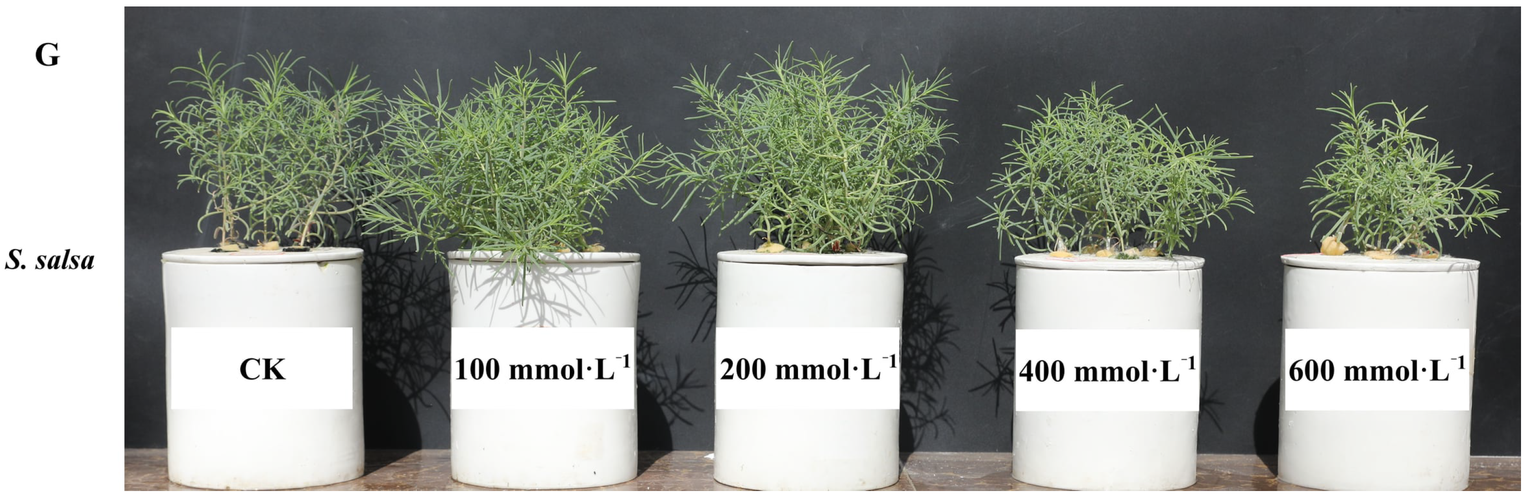
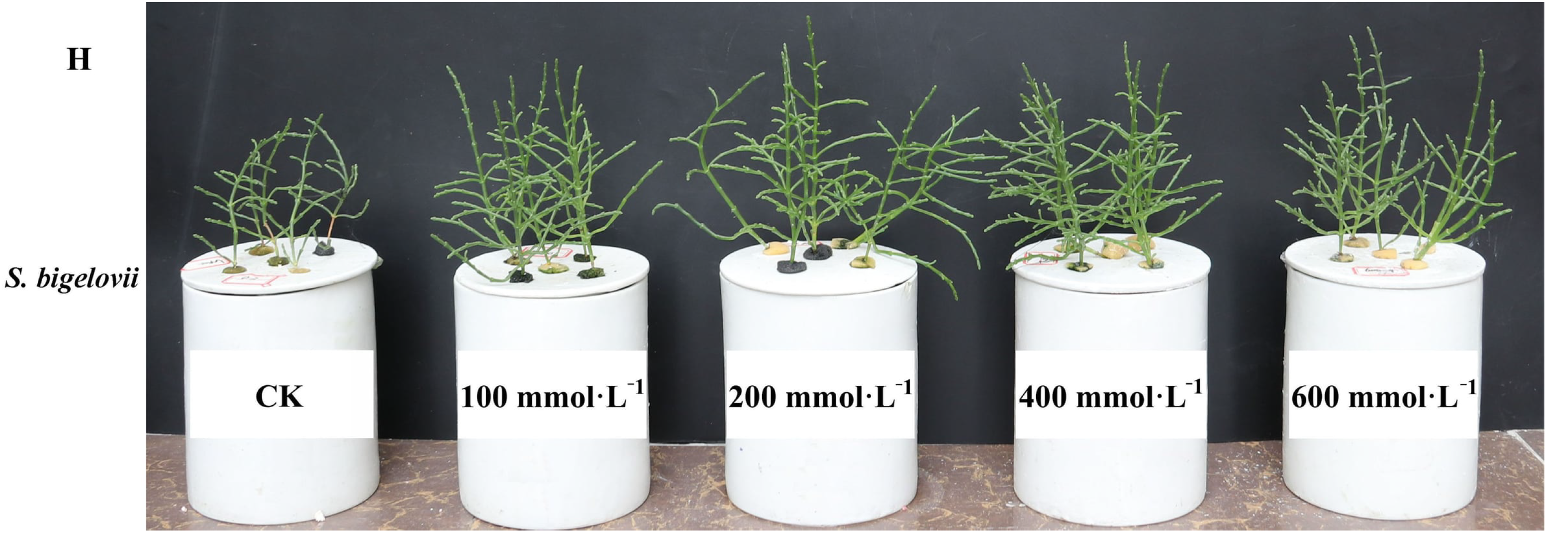
A certain range of salt concentration can promote the growth of both euhalophytes. After a 28-d NaCl treatment, the whole-plant fresh weight (FW) of S. salsa and S. bigelovii seedlings exposed to 100, 200, 400, and 600 mmol·L−1 NaCl was 1.78, 1.94, 1.44, 0.78 and 1.76, 1.97, 2.12, 1.69 times that of their respective control groups, respectively. (Figure 1A,B). The whole-plant dry weight (DW) was 1.44, 1.48, 1.12, 0.66 and 1.58, 1.69, 1.78, 1.55 times that of their respective control groups, respectively (Figure 1C,D). The FW and DW data indicated that the optimal NaCl concentration for growth was 100–200 mmol·L−1 for S. salsa and 200–400 mmol·L−1 for S. bigelovii. Based on organic dry weight (ODW), 400–600 mmol·L−1 NaCl significantly inhibited the growth of S. salsa, whereas even 600 mmol·L−1 NaCl promoted the growth of S. bigelovii (Figure 1C,D). Thus, S. bigelovii exhibited stronger salt tolerance than S. salsa. The root-to-shoot ratio of both species was significantly lower under saline conditions than that of the control (Figure 1E,F), indicating that salt treatment promoted shoot growth more than root growth. Salt-treated plants exhibited increased branch numbers and greater twig succulence, reflecting a vigorous growth phenotype (Figure 1G,H).
Figure 1.
The growth phenotype and biomass of whole-plant of S. salsa and S. bigelovii seedlings treated with 5 (CK), 100, 200, 400 and 600 mmol·L−1 NaCl for 28 d. (A) The whole-plant fresh weight (FW) of S. salsa. (B) The whole-plant FW of S. bigelovii. (C) The whole-plant dry weight (DW) of S. salsa. (D) The whole-plant DW of S. bigelovii. (E) The root-to-shoot ratio of S. salsa. (F) The root-to-shoot ratio of S. bigelovii. (G) The growth phenotype of S. salsa. (H) The growth phenotype of S. bigelovii. The data are means ± SD (n = 10). The means denoted by different lowercase letters are significantly different at p < 0.05.
3.2. The Effects of Salinity on Biomembranes Stability of Two Euhalophytes
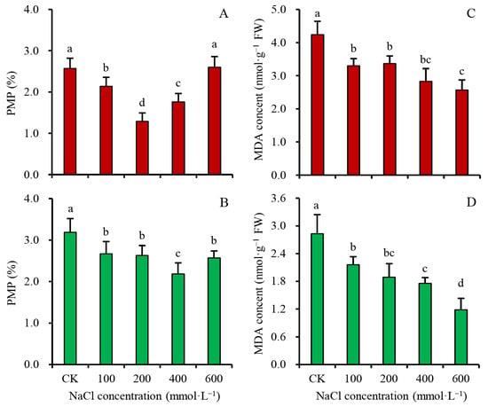
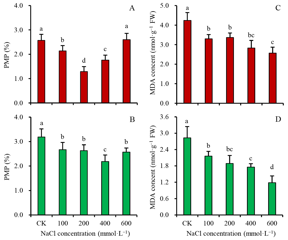
Plasma membrane is a primary site of cellular injury under environmental stress. The results of plasma membrane permeability (PMP) indicated that both euhalophytes could maintain plasma membrane stability under high salinity (100–600 mmol·L−1 NaCl). The PMP of salt-treated twigs was generally lower than that of the control (Figure 2A,B), except for S. salsa at 600 mmol·L−1 NaCl. The malondialdehyde (MDA) content, an indicator of membrane lipid peroxidation, was also significantly lower in salt-treated twigs than in the control (Figure 2C,D). For instance, compared with the control group, the PMP and MDA contents of S. salsa seedlings grown under 200 mmol·L−1 NaCl decreased by 49.8% and 20.6%, respectively. These results indicate that 100–600 mmol·L−1 NaCl does not induce membrane damage in the two euhalophytes.
Figure 2.
The biomembrane stability of twigs of S. salsa and S. bigelovii seedlings treated with 5 (CK), 100, 200, 400 and 600 mmol·L−1 NaCl for 28 d. (A) The plasma membrane permeability (PMP) of twigs of S. salsa. (B) The PMP of twigs of S. bigelovii. (C) The malondialdehyde (MDA) contents in twigs of S. salsa. (D) The MDA contents in twigs of S. bigelovii. The data are means ± SD (n= 6). The means denoted by different lowercase letters are significantly different at p < 0.05.
3.3. Antioxidant Response of Two Euhalophytes Under Saline Conditions
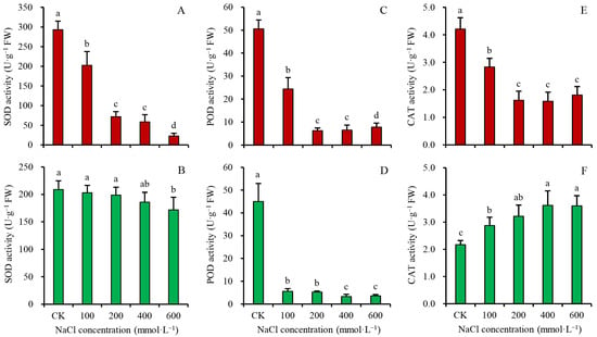
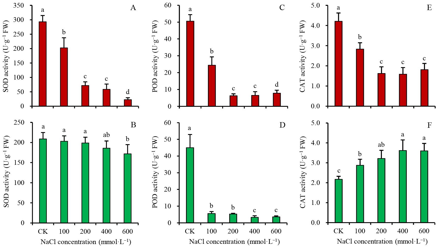
High salinity stress often causes secondary oxidative damage in plants, which in turn induces an increase in antioxidant enzyme activity. As salinity increased, the activities of SOD, POD, and CAT in S. salsa twigs decreased significantly (Figure 3A,C,E). At NaCl concentrations of 100, 200, 400, and 600 mmol·L−1, SOD activity decreased by 30.9%, 75.5%, 80.0%, and 92.3%, respectively, relative to the control. POD activity decreased by 51.8%, 87.6%, 87.3%, and 84.6%, and CAT activity decreased by 32.7%, 61.4%, 62.3%, and 57.0%, respectively.
Figure 3.
The antioxidant enzyme activities in twigs of S. salsa and S. bigelovii seedlings treated with 5 (CK), 100, 200, 400 and 600 mmol·L−1 NaCl for 28 d. (A) The superoxide dismutase (SOD) activities of S. salsa. (B) The SOD activities of S. bigelovii. (C) The peroxidase (POD) activities of S. salsa. (D) The POD activities of S. bigelovii. (E) The catalase (CAT) activities of S. salsa. (F) The CAT activities of S. bigelovii. The data are means ± SD (n = 6). The means labeled with different letters denote significant differences (p < 0.05).
At NaCl concentrations of 100, 200, 400, and 600 mmol·L−1, the POD activity in S. bigelovii showed a similar decreasing trend to that in S. salsa, with reductions of 87.6%, 88.3%, 92.7% and 92.0% relative to the control, respectively (Figure 3D). SOD activity changed little after salt treatment and was only significantly reduced at 600 mmol·L−1 NaCl (Figure 3B). In S. bigelovii, CAT activity showed a significant increase after salt treatment, unlike in S. salsa (Figure 3F).
3.4. Water Status and Osmotic Regulation of Two Euhalophytes Under Saline Conditions
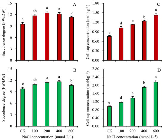
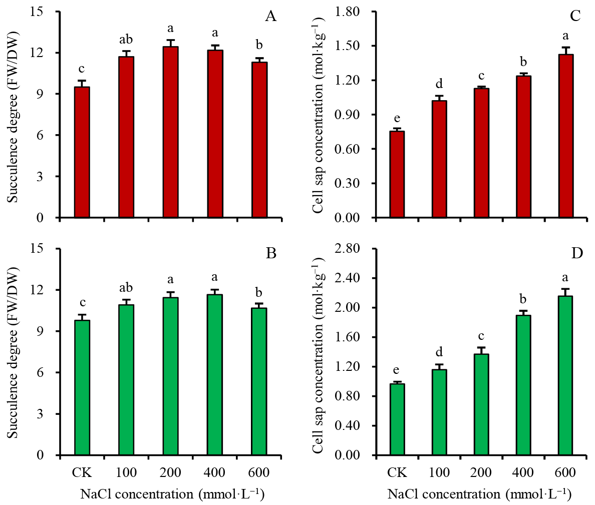
The water status of succulent plants is often expressed as the degree of succulence. Salt treatment enhanced the succulence degree of twigs of both euhalophytes. At NaCl concentrations of 100, 200, 400, and 600 mmol·L−1, the succulence degree of S. salsa twigs increased by 23.3%, 30.9%, 28.4%, and 19.0%, respectively, and that of S. bigelovii increased by 11.3%, 16.7%, 18.9%, and 8.9% relative to the control (Figure 4A,B). Even at 600 mmol·L−1 NaCl, the succulence degree of both species remained significantly higher than that of the control, indicating effective osmotic regulation. The cell sap concentration (CSC) increased gradually with increasing salinity. The CSC of S. salsa and S. bigelovii twigs at 100, 200, 400, and 600 mmol·L−1 NaCl was 1.35, 1.50, 1.64, 1.89 and 1.54, 1.83, 2.51, 2.86 times that of the control, respectively (Figure 4C,D). Under the same salt concentration, S. bigelovii exhibited higher CSC than S. salsa, suggesting that S. bigelovii has a stronger osmotic regulation capacity.
Figure 4.
The water status and osmoregulation of twigs of S. salsa and S. bigelovii seedlings treated with 5 (CK), 100, 200, 400 and 600 mmol·L−1 NaCl for 28 d. (A) The succulence degree (FW/DW) of S. salsa. (B) The succulence degree of S. bigelovii. (C) The cell sap concentration (CSC) of S. salsa. (D) The CSC of S. bigelovii. The data are means ± SD (n = 6). The means labeled with different letters denote significant differences (p < 0.05).
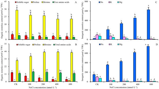
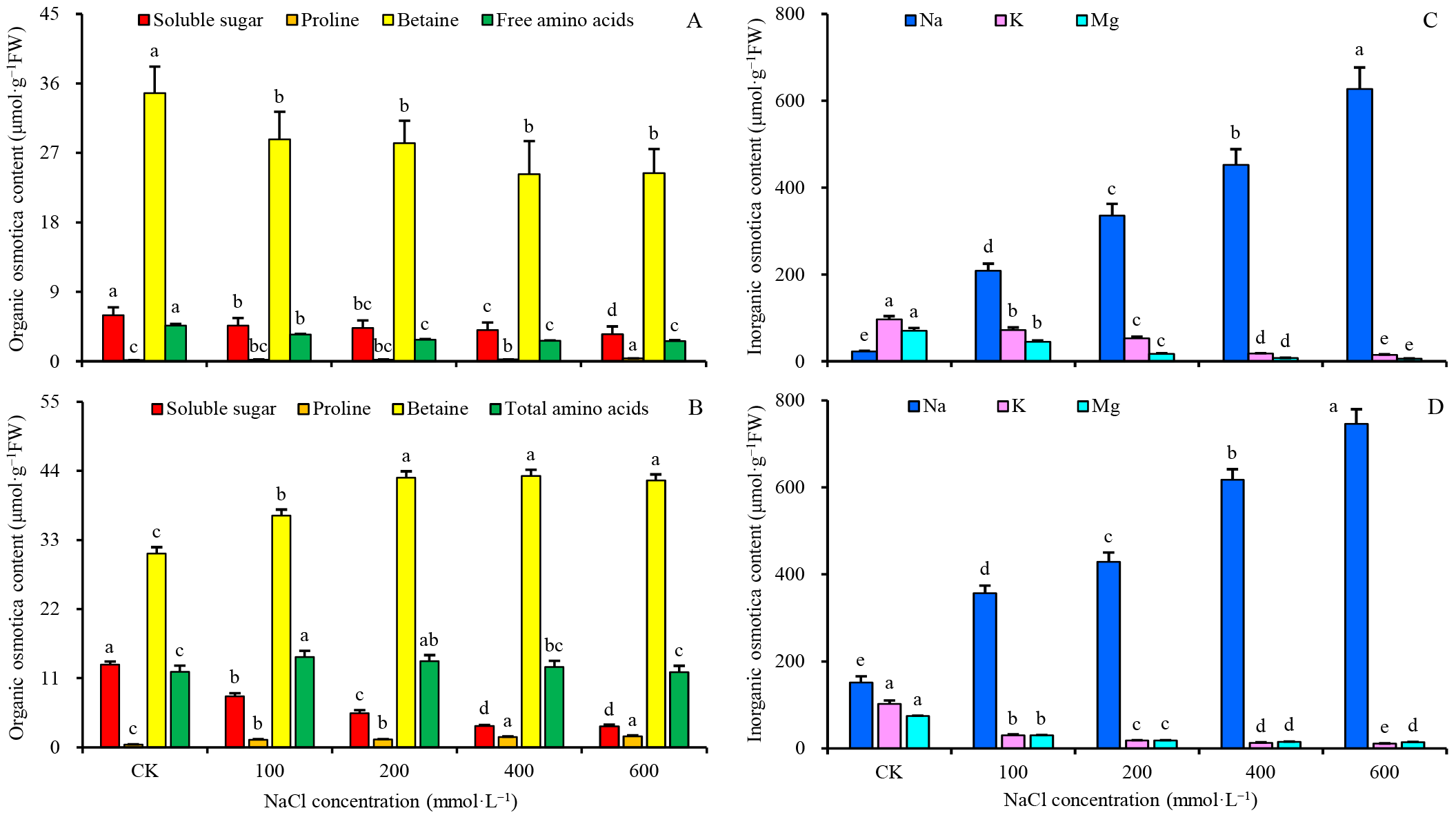
Soluble sugars, free amino acids (including proline), and betaine are the three most common organic osmotica in plant cells. Soluble sugar content decreased with increasing salinity in both euhalophytes. At NaCl concentrations of 100, 200, 400, and 600 mmol·L−1, the soluble sugar content in S. salsa twigs decreased by 21.6%, 27.1%, 31.8%, and 40.3%, respectively, and a larger decline (38.4%, 58.6%, 74.3%, and 74.5%) was observed in S. bigelovii twigs (Figure 5A,B). Among the three kinds of organic osmotica, betaine was the most abundant under all salt treatments. However, its content decreased significantly in S. salsa after salt treatment, whereas it increased markedly in S. bigelovii (Figure 5A,B). The response of free amino acids to salinity also differed between the two species: their content showed a decreasing trend in S. salsa with increasing salinity, while in S. bigelovii it first increased and then decreased (Figure 5A,B). Among free amino acids, proline content increased in both species under saline conditions, but its absolute content remained negligible, particularly in S. salsa.
Figure 5.
The organic and inorganic osmotica contents in twigs of S. salsa and S. bigelovii seedlings treated with 5 (CK), 100, 200, 400 and 600 mmol·L−1 NaCl for 28 d. (A) The organic osmotica contents of S. salsa. (B) The organic osmotica contents of S. bigelovii. (C) The inorganic osmotica contents of S. salsa. (D) The inorganic osmotica contents of S. bigelovii. The data are means ± SD (n = 6). The means labeled with different letters denote significant differences (p < 0.05).
Inorganic ions (Na+, K+, and Mg2+) also serve as important osmotica in plant cells. Under control condition, K+ was the predominant osmotic ion in S. salsa twigs, with content 4.29- and 1.38-fold that of Na+ and Mg2+, respectively (Figure 5C,D). Following NaCl treatments at 100, 200, 400, and 600 mmol·L−1, K+ content in S. salsa declined to 74.9%, 54.7%, 18.2%, and 15.4% of the control, while Mg2+ content decreased more sharply to 63.9%, 23.8%, 10.5%, and 8.4% of the control. In S. bigelovii, Na+ was the dominant cation under control condition (Figure 5C,D). Both K+ and Mg2+ contents in S. bigelovii decreased with increasing salinity: at 100, 200, 400, and 600 mmol·L−1 NaCl, K+ fell to 29.2%, 17.5%, 12.7%, and 10.6% of the control, respectively, and Mg2+ to 40.3%, 24.3%, 20.1%, and 19.1%. Salt treatment thus reduced K+ and Mg2+ uptake in both species, likely due to competition with Na+ uptake. Under CK, 100, 200, 400, and 600 mmol·L−1 NaCl conditions, Na+ content in S. salsa was 22.4, 208.2, 335.7, 452.4, and 626.8 μmol·g−1 FW respectively, whereas in S. bigelovii, it was 151.7, 357.0, 428.5, 617.6, and 745.5 μmol·g−1 FW (Figure 5C,D). The Na+ transfer factors for S. salsa and S. bigelovii were 4.48, 2.08, 1.68, 1.13, 1.09, and 30.34, 3.57, 2.14, 1.54, 1.24, respectively. These results indicate that both euhalophytes possess strong salt-accumulation capacity, which can be modulated in response to external salinity.
3.5. Chl Fluorescence and Gas-Exchange Parameters of Two Euhalophytes Under Saline Conditions
The net photosynthetic rate (Pn) of both euhalophytes decreased with increasing salinity. At NaCl concentrations of 100, 200, 400, and 600 mmol·L−1, Pn of S. salsa twigs decreased by 9.5%, 15.3%, 28.4%, and 35.3% relative to the control, respectively; and Pn of S. bigelovii decreased by 16.8%, 25.0%, 36.1%, and 44.7%, respectively (Table 1). Under the same salt concentration, Pn was consistently lower in S. bigelovii than in S. salsa, which may partly explain the slower growth of S. bigelovii. However, in both euhalophytes, the whole-plant total net photosynthetic capacity (Pt) increased initially and then decreased with increasing salinity. Compared with the control, Pt was significantly enhanced under 100–200 mmol·L−1 NaCl in S. salsa and under 100–400 mmol·L−1 NaCl in S. bigelovii. Stomatal conductance (Gs), a key factor affecting photosynthesis, decreased significantly with increasing salinity. At NaCl concentrations of 100, 200, 400, and 600 mmol·L−1, Gs in S. salsa twigs decreased by 19.1%, 31.9%, 32.4%, and 55.4%, respectively, relative to the control. A similar decline (12.7%, 20.0%, 30.5%, and 47.7%) was observed in S. bigelovii. Transpiration rate (Tr), which was proportional to Gs, showed a similar response to salinity. Reduced Tr may limit salt water uptake and thus prevent excessive salt accumulation in tissues. The parallel declines in Pn and Gs suggested that stomatal limitation might reduce photosynthesis in both euhalophytes. However, the indistinctive Ci values across different salt treatments indicated that stomatal limitation was not the primary cause of salinity-induced photosynthetic inhibition. This non-stomatal limitation was not attributable to the inhibition of light reaction activity, as the maximum photochemical efficiency of PSII (Fv/Fm) and the photosynthetic performance index (PI) did not decrease after salt treatment, except for a slight reduction in S. salsa at 600 mmol·L−1 NaCl. Notably, PI in S. bigelovii even increased after salt treatment. Furthermore, the inhibition was not due to increased respiration, as the respiration rate (Rr) of both species was significantly lower than that of the control. At 100, 200, 400, and 600 mmol·L−1 NaCl, Rr decreased by 36.7%, 37.0%, 47.1%, and 43.4% in S. salsa, and 34.4%, 34.2%, 44.5%, and 29.7% in S. bigelovii, respectively. There are other reasons for salinity-induced photosynthetic inhibition.
Table 1.
Chl fluorescence and gas-exchange parameters of S. salsa and S. bigelovii seedlings growing under different saline conditions.
3.6. Chl and Protein Content in the Twigs of Two Euhalophytes Under Saline Conditions
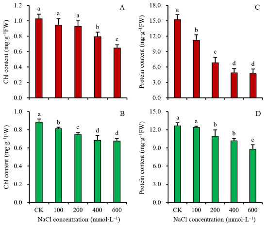
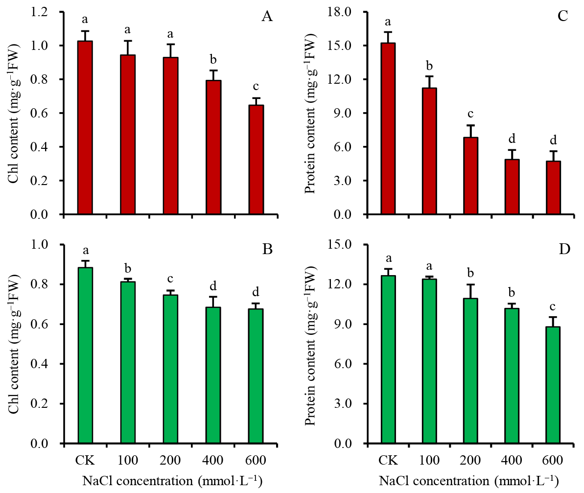
Chl and protein contents were examined to investigate salinity-induced photosynthetic inhibition in the two euhalophytes. Chl content in both species decreased with increasing salinity. At NaCl concentrations of 100, 200, 400, and 600 mmol·L−1, Chl content in S. salsa decreased to 91.9%, 90.5%, 77.3%, and 63.0% of the control, respectively, and that in S. bigelovii decreased to 91.9%, 84.4%, 77.4%, and 76.5% of the control (Figure 6A,B). The reduction in Chl content may reduce light energy absorption and thus affect photosynthetic performance. Protein content also decreased after salt treatment in both species. At 100, 200, 400, and 600 mmol·L−1 NaCl, protein content in S. salsa declined to 73.7%, 44.9%, 32.0%, and 31.0% of the control, respectively, and to 97.9%, 86.3%, 80.4%, and 69.5% in S. bigelovii, with a significantly smaller reduction in the latter (Figure 6C,D).
Figure 6.
The Chl content and protein contents in twigs of S. salsa and S. bigelovii seedlings treated with 5 (CK), 100, 200, 400 and 600 mmol·L−1 NaCl for 28 d. (A) The Chlorophyll (Chl) content of S. salsa. (B) The Chl content of S. bigelovii. (C) The protein content of S. salsa. (D) The protein content of S. bigelovii. The data are means ± SD (n = 6). The means labeled with different letters denote significant differences (p < 0.05).
4. Discussion
4.1. Moderate Salinity Provides a Favorable Environment for Euhalophytes in Terms of Growth and Cellular Homeostasis
The stability of plasma membrane is often used to detect whether plants have suffered environmental damage [34]. After salt treatment, the plasma membrane of the two euhalophytes remained stable, with PMP not increasing even at 600 mmol·L−1 NaCl (Figure 2A,B). Similarly, Tamarix species (recretohalophytes) have been reported to maintain membrane stability under saline conditions [35]. The MDA content in the twigs of both euhalophytes decreased after salt treatment, further suggesting that membrane damage occurred under non-saline conditions, whereas 100–400 mmol·L−1 NaCl represented a beneficial environment for their membrane stability (Figure 1 and Figure 2). A similar pattern has been observed in the recretohalophyte Limonium bicolor [20]. Therefore, the two euhalophytes did not require activation of the antioxidant enzyme system under 100–400 mmol·L−1 NaCl. After salt treatment, the activities of SOD, POD, and CAT in S. salsa twigs were all significantly lower than those of the control (Figure 3), differing from previous reports [36]. S. bigelovii differed from S. salsa in that its SOD activity decreased to a smaller extent with increasing salinity, while its CAT activity increased correspondingly (Figure 3). After treatment with 100–200 mmol·L−1 NaCl, although the growth of Atriplex portulacoides and L. bicolor was promoted, their antioxidant enzyme activities all increased [20,37]. Therefore, changes in the activity of a single antioxidant enzyme alone cannot determine whether a plant has been subjected to adverse conditions. The Chl a fluorescence results demonstrated that the photosystems of both euhalophytes were not damaged under saline conditions, although photosystems are highly sensitive to various stresses [38]. In vitro chloroplast experiments demonstrated that PSII activity in both glycophytes and halophytes decreased after treatment with low salinity [39]. In contrast, our results showed that 100–400 mmol·L−1 NaCl was harmless to the maximum photochemical efficiency (Fv/Fm) and performance index (PI) of PSII in the two euhalophytes (Table 1). Moreover, the PI of S. bigelovii significantly increased even under 400–600 mmol·L−1 NaCl (Table 1). The Fv/Fm of Nitraria tangutorum (euhalophyte), was also unaffected by saline environments [40]. Taken together, salinity is not a harmful habitat for the two euhalophytes regarding their growth, membrane stability, antioxidant response and photosystem performance.
4.2. Saline Environment Promotes the Growth of Euhalophytes
Morphologically, the two euhalophytes grew luxuriantly under saline conditions. In terms of biomass, salt promoted increases in both whole-plant FW and ODW (Figure 1). These results showed that salinity was beneficial to both species, with optimal growth at 100–200 mmol·L−1 NaCl for S. salsa and up to 200–400 mmol·L−1 NaCl for S. bigelovii (Figure 1). Similar results have been reported for other Suaeda and Salicornia species [41,42,43]. Compared with the non-saline condition, the euhalophytes grown under the optimal salt concentrations exhibited taller stems, more branches, and thicker and longer leaves (Figure 1G). This morphological change could be attributed to the fact that Na+ enhances the succulence of leaves and stems by increasing leaf water content and expanding epidermal cell size [44]. Salt treatment also altered the biomass distribution strategy among organs in the two euhalophytes, as evidenced by a significant decrease in the root-to-shoot ratio after salt treatment (Figure 1E,F). Similar salt-promoting growth phenotypes have also been observed in many recretohalophytes, with larger and more succulent leaves being a common feature. For example, the optimal NaCl concentration for the growth of some Tamarix plants was approximately 100–200 mmol·L−1 [35], about 100 mmol·L−1 for Atriplex centralasiatica [45], and about 50–100 mmol·L−1 for two Limonium species [46]. However, the growth of non-halophytes was inhibited under saline conditions, as was that of some pseudo-halophytes. For example, the growth rate of reed (Phragmites communis Trinius), a salt-excluding halophyte, was significantly reduced after treatment with 25 mmol·L−1 NaCl [12], although it could survive up to 600 mmol·L−1 NaCl [47]. Among the three types of halophytes, the two euhalophytes exhibited a higher optimal growth salinity and a more pronounced growth-promoting effect of salt.
4.3. The Mechanism of Euhalophytes Adapting to High Salinity
Salt treatment promoted the growth of the two euhalophytes, which was associated with increased tissue succulence degree (Figure 4). Recent studies have revealed that plant cells can directly sense and respond to water status, thereby actively promoting or suppressing growth [48]. Under saline conditions, increasing tissue water content appears to be an important mechanism for activating growth in the two euhalophytes. Recretohalophytes also exhibit a similar correlation between growth and water content [35,45,46]. Under high salinity, water uptake requires efficient osmotic regulation. The results in Figure 4 showed that the CSC of S. salsa and S. bigelovii reached 1.423 and 2.156 mol·kg−1, respectively, at 600 mmol·L−1 NaCl, which was sufficient to drive water absorption. At each NaCl concentration, S. bigelovii consistently exhibited higher CSC than S. salsa (Figure 4), indicating a stronger osmotic adjustment capacity. This may explain why S. bigelovii possesses greater salt tolerance than S. salsa.
A common feature of osmotic regulation in the two euhalophytes is that betaine plays a major role among the three organic osmotica (Figure 4). However, after salt treatment, the contents of soluble sugars, free amino acids, and betaine in S. salsa twigs all decreased (Figure 5A,B), suggesting that accumulation of organic osmotica is not the primary salt tolerance mechanism in the two euhalophytes. Among inorganic cations, Na+ played a major role in osmotic regulation among the three cations (Na+, K+, Mg2+) in both euhalophytes under saline conditions (Figure 5C,D). The contribution of Na+ in cellular osmoregulation was estimated as the Na+ content (mmol·kg−1 FW) divided by the cell sap concentration (mmol·kg−1) (Figure 4C,D and Figure 5). Na+ contribution in S. salsa was 20.4%, 29.8%, 36.6%, and 44.0%, respectively; and 30.8%, 31.3%, 32.6% and 34.6% in S. bigelovii, respectively. The higher the salt treatment concentration, the greater the contribution of Na+ in cellular osmoregulation. The actual contribution of Na+ should be slightly greater than the above results, because green twigs also contain a small amount of water-insoluble substances. In salt-treated plant tissues, the contribution of Cl− in cellular osmoregulation was slightly lower than that of Na+ [49]. Based on this estimation, the combined contribution of Na+ and Cl− to osmoregulation in the two euhalophytes under 100–600 mmol·L−1 NaCl was approximately 40–80%. K+ content declined markedly with increasing salinity, suggesting that the two euhalophytes do not enhance osmotic adjustment by increasing K+ uptake. Even in non-halophytes, Na+ and Cl− are the primary osmotic regulators under saline conditions, but they fail to increase tissue water content [50]. The salt tolerance mechanism of euhalophytes via water uptake is intimately associated with vacuolar Na+ compartmentalization and aquaporin-mediated water transport [10,51,52].
4.4. The Mechanism Underlying Rapid Growth of Euhalophytes Under Saline Conditions
Under appropriate salinity, the two euhalophytes showed enhanced growth, despite their net photosynthetic rate (Pn) being significantly lower than that of the control (Table 1). Photosynthetic performance depends heavily on leaf water status [53]. In the two euhalophytes, increased succulence degree of photosynthetic organs did not enhance photosynthetic capacity per unit biomass. Instead, they achieved higher whole-plant photosynthetic capacity (Pt) by increasing the biomass of photosynthetic organs (more branches and larger twigs and leaves) (Figure 1G,H). Under 100–200 mmol·L−1 NaCl conditions, the Pt values of individual S. salsa and S. bigelovii seedlings were about 1.44–1.64 times those of the control seedlings (Table 1). A similar adaptive strategy has been documented in Salicornia europaea and Arthrocnemum macrostachyum [43,54], which is the fundamental reason why salt promotes the growth of euhalophytes.
The salinity-induced inhibition of photosynthesis in euhalophytes is closely associated with decreased stomatal conductance (Gs) (Table 1). Reduced Gs benefits euhalophytes by limiting transpirational water loss and thereby reducing salt uptake. Glycophytes and other types of halophytes exhibit similar adaptive mechanisms [55]. However, intercellular CO2 concentration (Ci) did not change significantly after salt treatment, indicating that the decline in Pn in the two euhalophytes was not due to stomatal limitation (Table 1). In response to limited Ci under saline conditions, the two euhalophytes lowered photosynthetic capacity by actively reducing chlorophyll and protein contents, a strategy that may protect against photo-oxidative stress from excess light energy [43]. The two euhalophytes do not need to synthesize large amounts of photosynthetic enzymes and photosynthetic pigments under saline conditions (Figure 6C,D), thus conserving organic resources. Moreover, appropriate salinity did not induce stronger membrane lipid peroxidation in the two species (Figure 2C,D), nor did it require full activation of their antioxidant enzyme systems (Figure 3). Stress responses typically consume substantial metabolic energy [56]. In contrast, the respiratory consumption (Rr) of the two euhalophytes under 100–400 mmol·L−1 NaCl was significantly lower than that of the control (Table 1). This implied that appropriate salinity exerts a positive effect on the two euhalophytes from the perspective of respiration. In other types of halophytes, the changes in Rr follow a pattern opposite to that reported in this study [57]. Reduced metabolic expenditure appears to be a hallmark of salt tolerance in both euhalophytes.
5. Conclusions
The rapid growth of S. salsa and S. bigelovii under high salinity can be attributed to several common characteristics. First, compared with nonsaline conditions, the two euhalophytes exhibited better membrane stability and lower membrane lipid peroxidation under appropriate salinity, with no significant induction of antioxidant enzymes (SOD, POD). Thus, moderate salinity provides a favorable environment for euhalophytes in terms of growth and cellular homeostasis. Second, the two euhalophytes did not accumulate large amounts of organic osmotica after salt treatment. Instead, they mainly used NaCl as an osmotic regulator, turning a potential stressor into a beneficial factor to increase tissue water content. Improved water status stimulated the two euhalophytes to produce more branches and enlarge their photosynthetic organs. Third, the two euhalophytes are characterized by their ability to absorb water and grow rapidly to dilute internal salt concentrations while simultaneously reducing salt uptake by decreasing stomatal conductance (Gs) and transpiration. Although reduced Gs inevitably limits gas exchange and affects photosynthesis, the two euhalophytes compensate for the reduction in Pn by increasing the biomass of photosynthetic organs. Fourth, in adaptation to limited gas exchange, the two euhalophytes actively reduced their chlorophyll and protein contents. Reduced metabolic and respiratory consumption is an important salt tolerance trait in both euhalophytes. These characteristics offer new insights for genetic engineering research aimed at improving salt tolerance in crops.
Author Contributions
Conceptualization, N.Q., and R.W.; methodology, N.Q. and Y.Y.; software, N.Q. and B.L.; validation, N.Q., Y.Y. and R.W.; formal analysis, N.Q. and Y.Y.; investigation, Y.Y., J.S., W.D. and B.L.; resources, Y.Y., J.S., W.D. and B.L.; data curation, N.Q.; writing—original draft preparation, N.Q. and Y.Y.; writing—review and editing, N.Q. and R.W.; visualization, N.Q. and Y.Y.; supervision, N.Q.; project administration, R.W.; funding acquisition, R.W. All authors have read and agreed to the published version of the manuscript.
Funding
This research was funded by the National Natural Science Foundation of China (grant No. 42377474).
Data Availability Statement
The original contributions presented in this study are included in this article. Further inquiries can be directed to the corresponding authors.
Conflicts of Interest
The authors declare no conflicts of interest.
References
- Flowers, T.J.; Colmer, T.D. Salinity tolerance in halophytes. New Phytol. 2008, 179, 945–963. [Google Scholar] [CrossRef] [Scilit]
- Munns, R.; Tester, M. Mechanisms of salinity tolerance. Annu. Rev. Plant Biol. 2008, 59, 651–681. [Google Scholar] [CrossRef] [Scilit]
- Li, L.Y.; Zhang, Y.; Han, G.L.; Guo, J.R.; Meng, Z.; Chen, M. Progress in the study and use of seawater vegetables. J. Agric. Food Chem. 2020, 68, 5998–6006. [Google Scholar] [CrossRef] [Scilit]
- Ismail, A.M.; Horie, T. Genomics, physiology, and molecular breeding approaches for improving salt tolerance. Annu. Rev. Plant Biol. 2017, 68, 405–434. [Google Scholar] [CrossRef] [Scilit]
- Fita, A.; Rodríguez-Burruezo, A.; Boscaiu, M.; Prohens, J.; Vicente, O. Breeding and domesticating crops adapted to drought and salinity: A new paradigm for increasing food production. Front. Plant Sci. 2015, 6, 978. [Google Scholar] [CrossRef] [Scilit]
- Munns, R.; Gilliham, M. Salinity tolerance of crops-what is the cost? New Phytol. 2015, 208, 668–673. [Google Scholar] [CrossRef] [Scilit]
- Shabala, S.; Bose, J.; Hedrich, R. Salt bladders: Do they matter? Trends Plant Sci. 2014, 19, 687–691. [Google Scholar] [CrossRef] [Scilit]
- Breckle, S.W. How do halophytes overcome salinity? In Biology of Salt-Tolerant Plants; Khan, M.A., Ungar, I.A., Eds.; Book Crafters: Chelsea, MI, USA, 1995; pp. 199–213. [Google Scholar]
- Flowers, T.J.; Munns, R.; Colmer, T.D. Sodium chloride toxicity and the cellular basis of salt tolerance in halophytes. Ann. Bot. 2015, 115, 419–431. [Google Scholar] [CrossRef] [Scilit]
- Song, J.; Wang, B.S. Using euhalophytes to understand salt tolerance and to develop saline agriculture: Suaeda salsa as a promising model. Ann. Bot. 2015, 115, 541–553. [Google Scholar] [CrossRef] [Scilit]
- Zhao, K.F.; Fan, H.; Ungar, I.A. Survey of halophyte species in China. Plant Sci. 2002, 163, 491–498. [Google Scholar] [CrossRef] [Scilit]
- Matsushita, N.; Matoh, T. Characterization of Na+ exclusion mechanisms of salt-tolerant reed plants in comparison with salt-sensitive rice plants. Physiol. Plant. 1991, 83, 170–176. [Google Scholar] [CrossRef] [Scilit]
- Kumari, A.; Das, P.; Parida, A.K.; Agarwal, P.K. Proteomics, metabolomics, and ionomics perspectives of salinity tolerance in halophytes. Front. Plant Sci. 2015, 6, 537. [Google Scholar] [CrossRef] [Scilit]
- Benjamin, J.J.; Lucini, L.; Jothiramshekar, S.; Parida, A. Metabolomic insights into the mechanisms underlying tolerance to salinity in different halophytes. Plant Physiol. Biochem. 2019, 135, 528–545. [Google Scholar] [CrossRef] [Scilit]
- Geilfus, C.M. Chloride-from nutrient to toxicant. Plant Cell Physiol. 2018, 59, 877–886. [Google Scholar] [CrossRef] [Scilit]
- Ganie, S.A.; Molla, K.A.; Henry, R.J.; Bhat, K.V.; Mondal, T.K. Advances in understanding salt tolerance in rice. Theor. Appl. Genet. 2019, 132, 851–870. [Google Scholar] [CrossRef] [Scilit]
- Carden, D.E.; Walker, D.J.; Flowers, T.J.; Miller, A.J. Single-cell measurements of the contributions of cytosolic Na+ and K+ to salt tolerance. Plant Physiol. 2003, 131, 676–683. [Google Scholar] [CrossRef] [Scilit]
- Shabala, S. Learning from halophytes: Physiological basis and strategies to improve abiotic stress tolerance in crops. Ann. Bot. 2013, 112, 1209–1221. [Google Scholar] [CrossRef] [Scilit]
- Ding, G.H.; Yang, Q.; Ruan, X.Y.; Si, T.T.; Yuan, B.X.; Zheng, W.W.; Xie, Q.L.; Souleymane, O.A.; Wang, X.C. Proteomics analysis of the effects for different salt ions in leaves of true halophyte Sesuvium portulacastrum. Plant Physiol. Biochem. 2022, 170, 234–248. [Google Scholar] [CrossRef] [Scilit]
- Li, Y. Kinetics of the antioxidant response to salinity in the halophyte Limonium bicolour. Plant Soil Environ. 2008, 54, 493–497. [Google Scholar] [CrossRef] [Scilit]
- Yuan, F.; Lyu, M.J.A.; Leng, B.Y.; Zhu, X.G.; Wang, B.S. The transcriptome of NaCl-treated Limonium bicolor leaves reveals the genes controlling salt secretion of salt gland. Plant Mol. Biol. 2016, 91, 241–256. [Google Scholar] [CrossRef] [Scilit]
- Wei, X.C.; Yan, X.; Yang, Z.; Han, G.L.; Wan, L.; Yuan, F.; Wang, B.S. Salt glands of recretohalophyte Tamarix under salinity: Their evolution and adaptation. Ecol. Evol. 2020, 10, 9384–9395. [Google Scholar] [CrossRef] [Scilit]
- Orlova, Y.V.; Myasoedov, N.A.; Kirichenko, E.B.; Balnokin, Y.V. Contributions of inorganic ions, soluble carbohydrates, and multiatomic alcohols to water homeostasis in Artemisia lerchiana and A-pauciflora. Russ. J. Plant Physiol. 2009, 56, 200–210. [Google Scholar] [CrossRef] [Scilit]
- Arif, Y.; Singh, P.; Siddiqui, H.; Bajguz, A.; Hayat, S. Salinity induced physiological and biochemical changes in plants: An omic approach towards salt stress tolerance. Plant Physiol. Biochem. 2020, 156, 64–77. [Google Scholar] [CrossRef] [Scilit]
- Qiu, N.W.; Wang, R.J.; Sun, Y.; Wang, X.S.; Jiang, D.C.; Meng, Y.T.; Zhou, F. Toxic effects and mechanism of 2,2′,4,4′-tetrabromodiphenyl ether (BDE-47) on Lemna minor. Chemosphere 2018, 193, 711–719. [Google Scholar] [CrossRef] [Scilit]
- Landi, M. Commentary to: "Improving the thiobarbituric acid-reactive-substances assay for estimating lipid peroxidation in plant tissues containing anthocyanin and other interfering compounds" by Hodges et al., Planta (1999) 207: 604–611. Planta 2017, 245, 1035–1036. [Google Scholar] [CrossRef] [Scilit]
- Babić, M.; Radić, S.; Cvjetko, P.; Roje, V.; Pevalek-Kozlina, B.; Pavlica, M. Antioxidative response of Lemna minor plants exposed to thallium(I)-acetate. Aquat. Bot. 2009, 91, 166–172. [Google Scholar] [CrossRef] [Scilit]
- Bates, L.S.; Waldren, R.P.; Teare, I.D. Rapid determination of free proline for water-stress studies. Plant Soil 1973, 39, 205–207. [Google Scholar] [CrossRef] [Scilit]
- Jermyn, M.A. Increasing the sensitivity of the anthrone method for carbohydrate. Anal. Biochem. 1975, 68, 332–335. [Google Scholar] [CrossRef] [Scilit]
- Yang, X.H.; Lu, C.M. Photosynthesis is improved by exogenous glycinebetaine in salt-stressed maize plants. Physiol. Plant. 2005, 124, 343–352. [Google Scholar] [CrossRef] [Scilit]
- Kowalska, S.; Szłyk, E.; Jastrzębska, A. Simple extraction procedure for free amino acids determination in selected gluten-free flour samples. Eur. Food Res. Technol. 2022, 248, 507–517. [Google Scholar] [CrossRef] [Scilit]
- Yue, D.; Chao, E.; Deng, Y.; Chen, K.; Wang, Z.; Qiu, N.; Zhang, H. Natural physiological changes on overwintering and spring recovery of needles of Pinus densiflora Siebold & Zucc. Forests 2023, 14, 168. [Google Scholar] [CrossRef] [Scilit]
- Qiu, N.W.; Zhou, F.; Liu, Q.; Zhao, W.Q. Determination of photosynthetic parameters in two seawater tolerant vegetables. Chin. J. Oceanol. Limnol. 2016, 34, 412–415. [Google Scholar] [CrossRef] [Scilit]
- Shajar, F.; Nabi, A.U.; Manzoor, A.; Saleem, S.; Mushtaq, N.U.; Manzoor, S.; Rehman, R.U. Membrane-associated mechanisms in plant responses to abiotic stress. Plant Sci. 2025, 360, 112726. [Google Scholar] [CrossRef] [Scilit]
- Wang, H.B.; Zheng, L.J.; Ding, D.; Ding, F.J.; Guo, Y.C. Growth and physiological response to NaCl stress and salt tolerance differences of seven Tamarix Linn. species. Shandong Agric. Sci. 2022, 54, 31–38. (In Chinese) [Google Scholar] [CrossRef]
- Li, H.; Wang, H.; Wen, W.J.; Yang, G.W. The antioxidant system in Suaeda salsa under salt stress. Plant Signal. Behav. 2020, 15, 1771939. [Google Scholar] [CrossRef] [Scilit]
- Benzartri, M.; Rejeb, K.B.; Debez, A.; Messedi, D.; Abdelly, C. Photosynthetic activity and leaf antioxidative responses of Atriplex portulacoides subjected to extreme salinity. Acta Physiol. Plant. 2012, 34, 1679–1688. [Google Scholar] [CrossRef] [Scilit]
- Stirbet, A.; Lazár, D.; Kromdijk, J.; Govindjee. Chlorophyll a fluorescence induction: Can just a one-second measurement be used to quantify abiotic stress responses? Photosynthetica 2018, 56, 86–104. [Google Scholar] [CrossRef] [Scilit]
- Percey, W.J.; McMinn, A.; Bose, J.; Breadmore, M.C.; Guijt, R.M.; Shabala, S. Salinity effects on chloroplast PSII performance in glycophytes and halophytes. Funct. Plant Biol. 2016, 43, 1003–1015. [Google Scholar] [CrossRef] [Scilit]
- Cheng, T.L.; Chen, J.H.; Zhang, J.B.; Shi, S.Q.; Zhou, Y.W.; Lu, L.; Wang, P.K.; Jiang, Z.P.; Yang, J.C.; Zhang, S.G.; et al. Physiological and proteomic analyses of leaves from the halophyte Tangut Nitraria reveals diverse response pathways critical for high salinity tolerance. Front. Plant Sci. 2015, 6, 30. [Google Scholar] [CrossRef] [Scilit]
- Shekhawat, V.P.S.; Kumar, A.; Neumann, K.H. Effect of sodium chloride salinity on growth and ion accumulation in some halophytes. Commun. Soil Sci. Plant Anal. 2006, 37, 1933–1946. [Google Scholar] [CrossRef] [Scilit]
- Ahmad, S.T.; Sima, N.A.K.K.; Mirzaei, H.H. Effects of sodium chloride on physiological aspects of Salicornia persica growth. J. Plant Nutr. 2013, 36, 401–414. [Google Scholar] [CrossRef] [Scilit]
- Cárdenas-Pérez, S.; Rajabi Dehnavi, A.; Leszczyński, K.; Lubińska-Mielińska, S.; Ludwiczak, A.; Piernik, A. Salicornia europaea L. functional traits indicate its optimum growth. Plants 2022, 11, 1051. [Google Scholar] [CrossRef] [Scilit]
- Zhang, D.; Tian, C.; Mai, W. Succulence is a key trait involved in the response of Suaeda salsa to salt stress. Plants 2024, 13, 721. [Google Scholar] [CrossRef] [Scilit]
- Qiu, N.W.; Zhou, F.; Wang, Y.; Peng, X.Y.; Hua, C. The strategy of Na+ compartmentation and growth of Atriplex centralasiatica in adaptation to saline environments. Russ. J. Plant Physiol. 2014, 61, 238–245. [Google Scholar] [CrossRef] [Scilit]
- Leng, B.Y.; Yuan, F.; Dong, X.X.; Wang, J.; Wang, B.S. Distribution pattern and salt excretion rate of salt glands in two recretohalophyte species of Limonium (Plumbaginaceae). S. Afr. J. Bot. 2018, 115, 74–80. [Google Scholar] [CrossRef] [Scilit]
- Gorai, M.; Vadel, A.M.; Neffati, M.; Khemira, H. The effect of sodium chloride salinity on the growth, water status and ion content of Phragmites communis Trin. Pak. J. Biol. Sci. 2007, 10, 2225–2230. [Google Scholar] [CrossRef] [Scilit]
- Wang, Y.; Zhu, L.; Yang, Y.; Li, X.; Zhang, X.; Fang, X. Cellular water-potential sensing through biomolecular condensation. Nature 2026, 655, 1320–1329. [Google Scholar] [CrossRef] [Scilit]
- Wang, N.; Zhao, Z.Y.; Zhang, X.Y.; Liu, S.H.; Zhang, K.; Hu, M.F. Plant growth, salt removal capacity, and forage nutritive value of the annual euhalophyte Suaeda salsa irrigated with saline water. Front. Plant Sci. 2023, 13, 1040520. [Google Scholar] [CrossRef] [Scilit]
- Wang, J.; Qiu, N.W.; Wang, P.; Zhang, W.R.; Yang, X.Y.; Chen, M.; Wang, B.S.; Sun, J.K. Na+ compartmentation strategy of Chinese cabbage in response to salt stress. Plant Physiol. Biochem. 2019, 140, 151–157. [Google Scholar] [CrossRef] [Scilit]
- Mendis, C.L.; Padmathilake, R.E.; Attanayake, R.N.; Perera, D. Learning from Salicornia: Physiological, biochemical, and molecular mechanisms of salinity tolerance. Int. J. Mol. Sci. 2025, 26, 5936. [Google Scholar] [CrossRef] [Scilit]
- Yu, W.C.; Wu, W.W.; Zhang, N.; Wang, L.P.; Wang, Y.H.; Wang, B.; Lan, Q.K.; Wang, Y. Research advances on molecular mechanism of salt tolerance in Suaeda. Biology 2022, 11, 1273. [Google Scholar] [CrossRef] [Scilit]
- Xiong, D.L.; Nadal, M. Linking water relations and hydraulics with photosynthesis. Plant J. 2020, 101, 800–815. [Google Scholar] [CrossRef] [Scilit]
- Redondo-Gómez, S.; Mateos-Naranjo, E.; Figueroa, M.E.; Davy, A.J. Salt stimulation of growth and photosynthesis in an extreme halophyte, Arthrocnemum macrostachyum. Plant Biol. 2010, 12, 79–87. [Google Scholar] [CrossRef] [Scilit]
- Hedrich, R.; Shabala, S. Stomata in a saline world. Curr. Opin. Plant Biol. 2018, 46, 87–95. [Google Scholar] [CrossRef] [Scilit]
- Erofeeva, E.A. Plant hormesis: The energy aspect of low and high-dose stresses. Plant Stress 2024, 14, 100628. [Google Scholar] [CrossRef] [Scilit]
- Gao, Z.W.; Liu, J.; Lu, X.L.; Li, X.; Qin, Y.Q.; Gao, G.; He, L.J.; Cai, M.Z.; Chen, C.; Zhang, J.T.; et al. NaCl and NaHCO3 mediate tradeoffs in growth, leaf traits, and clonal reproduction of reed (Phragmites communis) under saline-alkali stress. Not. Bot. Horti Agrobo. 2025, 53, 14282. [Google Scholar] [CrossRef] [Scilit]

Disclaimer/Publisher’s Note: The statements, opinions and data contained in all publications are solely those of the individual author(s) and contributor(s) and not of MDPI and/or the editor(s). MDPI and/or the editor(s) disclaim responsibility for any injury to people or property resulting from any ideas, methods, instructions or products referred to in the content. |
© 2026 by the authors. Licensee MDPI, Basel, Switzerland. This article is an open access article distributed under the terms and conditions of the Creative Commons Attribution (CC BY) license.