Next Issue
Volume 11, February
Previous Issue
Volume 10, December


Dent. J., Volume 11, Issue 1 (January 2023) – 27 articles

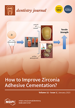
Cover Story (view full-size image): Several viewpoints were reported regarding the effect of temporary cements, different surface pretreatment protocols before adhesive cementation and predictive factors. The in vitro study tested if temporary cement, pretreatment of the tooth surface or the size of enamel or dentin influence adhesive cementation to zirconia ceramics. The mean tensile bond strength was significantly (p = 0.005) higher in the pretreated specimens (337N) than in the untreated ones (204N). The multivariable linear regression model was significant (p = 0.003): size of enamel (β = 0.506; p = 0.049), pretreatment effect (β = 0.478; p = 0.001) and size of dentin area (β = −0.105; p = 0.022). We conclude that the bonding strength can be determined by the size of enamel and dentin and the surface pretreatment prior to adhesive cementation. View this paper
  • Issues are regarded as officially published after their release is announced to the table of contents alert mailing list.
  • You may sign up for e-mail alerts to receive table of contents of newly released issues.
  • PDF is the official format for papers published in both, html and pdf forms. To view the papers in pdf format, click on the "PDF Full-text" link, and use the free Adobe Reader to open them.
Order results
Result details
Section
Select all
Export citation of selected articles as:
14 pages, 2479 KiB  
Article
In Vitro Trueness and Precision of Intraoral Scanners in a Four-Implant Complete-Arch Model
by Dimitrios Spagopoulos, George Kaisarlis, Foteini Spagopoulou, Demetrios J. Halazonetis, Jan-Frederik Güth and Efstratios Papazoglou
Dent. J. 2023, 11(1), 27; https://doi.org/10.3390/dj11010027 - 12 Jan 2023
Cited by 3 | Viewed by 2763
Abstract
(1) Background: New intraoral (IOS) and laboratory scanners appear in the market and their trueness and precision have not been compared. (2) Methods: Seven IOS and two laboratory scanners were used to scan a mandibular edentulous model with four parallel internal hexagon implant [...] Read more.
(1) Background: New intraoral (IOS) and laboratory scanners appear in the market and their trueness and precision have not been compared. (2) Methods: Seven IOS and two laboratory scanners were used to scan a mandibular edentulous model with four parallel internal hexagon implant analogues and PEEK scan bodies. Digital models in Standard Tessellation Language (STL) were created. The master model with the scan bodies was scanned (×10) with a computerized numerical control 3D Coordinate Measuring Machine (CMM). The short (distances of adjacent scan posts) and long distances (distances of the scan posts with non-adjacent sites in the arch) among the centroids of the four analogues were calculated using CMM special software. Trueness (comparisons with the master model) and precision (intragroup comparisons) were statistically compared with ANOVA, chi-square and Tukey tests. (3) Results: Laboratory scanners had the best trueness and precision compared to all IOSs for long distances. Only iTero (Align Technologies Inc., Milpitas, CA, USA) had comparable trueness with one laboratory scanner in short and long distances. For short distances, CS3600 (Carestream Health, Inc., Rochester, NY, USA), Omnicam, Primescan (Sirona Dental Sys-tems GmbH, Bens-heim, Germany) and TRIOS 4 (3Shape A/S, Copen-hagen, Denmark) had similar trueness to one laboratory scanner. From those, only Omnicam and Primescan had similar precision as the same laboratory scanner. Most IOSs seem to work better for smaller distances and are less precise in cross-arch distances. (4) Conclusions: The laboratory scanners showed significantly higher trueness and precision than all IOSs tested for the long-distance group; for the short distance, some IOSs were not different in trueness and precision than the laboratory scanners. Full article
Show Figures

Figure 1

16 pages, 325 KiB  
Review
Different Treatment Modalities of Oral Lichen Planus—A Narrative Review
by Ana Andabak-Rogulj, Ema Vindiš, Lorena Horvat Aleksijević, Ivana Škrinjar, Danica Vidović Juras, Anastazija Aščić and Božana Lončar Brzak
Dent. J. 2023, 11(1), 26; https://doi.org/10.3390/dj11010026 - 12 Jan 2023
Cited by 13 | Viewed by 6269
Abstract
Oral lichen planus (OLP) is a chronic inflammatory disease of unknown etiology which affects the oral mucosa. OLP varies in its clinical features from a reticular form that is, in most cases, asymptomatic, to atrophic–erosive, and is accompanied by symptoms of burning sensation [...] Read more.
Oral lichen planus (OLP) is a chronic inflammatory disease of unknown etiology which affects the oral mucosa. OLP varies in its clinical features from a reticular form that is, in most cases, asymptomatic, to atrophic–erosive, and is accompanied by symptoms of burning sensation and pain followed by difficulty in eating. Given the fact that OLP is a disease of unknown etiology, the treatment is symptomatic and involves suppressing the signs and symptoms of the disease using various topical and systemic drugs. The first line of therapy for treating symptomatic OLP is topical corticosteroids, whereas systemic corticosteroids are used for treating persistent lesions that do not respond to local treatment. However, the lack of convincing evidence on the efficacy of previous therapies, including topical corticosteroids, and numerous side effects that have appeared over recent years has resulted in the emergence and development of new therapeutic options. Some of the therapies mentioned are tacrolimus, efalizumab, dapson, interferon, retinoic acid, photochemotherapy with psoralen and ultraviolet A rays (PUVA), aloe vera, antimalarials, antibiotics and others. These therapies only partially meet the properties of efficacy and safety of use, thus justifying the continuous search and testing of new treatment methods. Full article
10 pages, 546 KiB  
Review
Minimally Invasive Non-Surgical Technique in the Treatment of Intrabony Defects—A Narrative Review
by Styliani Anoixiadou, Andreas Parashis and Ioannis Vouros
Dent. J. 2023, 11(1), 25; https://doi.org/10.3390/dj11010025 - 11 Jan 2023
Cited by 5 | Viewed by 3408
Abstract
Intrabony defects occur frequently in periodontitis and represent sites that, if left untreated, are at increased risk for disease progression. Although resective or repair procedures have been used to treat intrabony defects, aiming at their elimination, the treatment of choice is surgical periodontal [...] Read more.
Intrabony defects occur frequently in periodontitis and represent sites that, if left untreated, are at increased risk for disease progression. Although resective or repair procedures have been used to treat intrabony defects, aiming at their elimination, the treatment of choice is surgical periodontal regeneration. The development of periodontal regeneration in the last 30 years has followed two distinctive, though totally different, paths. The interest of researchers has so far focused on regenerative materials and products on one side, and on novel surgical approaches on the other side. In the area of materials and products, three different regenerative concepts have been explored namely, barrier membranes, bone grafts, and wound healing modifiers/biologics, plus many combinations of the aforementioned. In the area of surgical approaches, clinical innovation in flap design and handling, as well as minimally invasive approaches, has radically changed regenerative surgery. Recently, a minimally invasive non-surgical technique (MINST) for the treatment of intrabony defects was proposed. Initial clinical trials indicated comparable results to the surgical minimally invasive techniques in both clinical and radiographic outcomes. These results support the efficacy of this treatment approach. The aim of this review is to present the evidence on the application of minimally invasive non-surgical techniques and their efficacy in the treatment of intrabony defects. Full article
(This article belongs to the Special Issue Periodontal Health: Disease Prevention and Treatment)
Show Figures

Figure 1

21 pages, 1589 KiB  
Review
Wearable Orofacial Technology and Orthodontics
by Sabarinath Prasad, Sivakumar Arunachalam, Thomas Boillat, Ahmed Ghoneima, Narayan Gandedkar and Samira Diar-Bakirly
Dent. J. 2023, 11(1), 24; https://doi.org/10.3390/dj11010024 - 10 Jan 2023
Cited by 6 | Viewed by 3714
Abstract
Wearable technology to augment traditional approaches are increasingly being added to the arsenals of treatment providers. Wearable technology generally refers to electronic systems, devices, or sensors that are usually worn on or are in close proximity to the human body. Wearables may be [...] Read more.
Wearable technology to augment traditional approaches are increasingly being added to the arsenals of treatment providers. Wearable technology generally refers to electronic systems, devices, or sensors that are usually worn on or are in close proximity to the human body. Wearables may be stand-alone or integrated into materials that are worn on the body. What sets medical wearables apart from other systems is their ability to collect, store, and relay information regarding an individual’s current body status to other devices operating on compatible networks in naturalistic settings. The last decade has witnessed a steady increase in the use of wearables specific to the orofacial region. Applications range from supplementing diagnosis, tracking treatment progress, monitoring patient compliance, and better understanding the jaw’s functional and parafunctional activities. Orofacial wearable devices may be unimodal or incorporate multiple sensing modalities. The objective data collected continuously, in real time, in naturalistic settings using these orofacial wearables provide opportunities to formulate accurate and personalized treatment strategies. In the not-too-distant future, it is anticipated that information about an individual’s current oral health status may provide patient-centric personalized care to prevent, diagnose, and treat oral diseases, with wearables playing a key role. In this review, we examine the progress achieved, summarize applications of orthodontic relevance and examine the future potential of orofacial wearables. Full article
Show Figures

Graphical abstract

17 pages, 2410 KiB  
Review
Osteonecrosis of the Jaw
by Božana Lončar Brzak, Lorena Horvat Aleksijević, Ema Vindiš, Iva Kordić, Marko Granić, Danica Vidović Juras and Ana Andabak Rogulj
Dent. J. 2023, 11(1), 23; https://doi.org/10.3390/dj11010023 - 9 Jan 2023
Cited by 9 | Viewed by 6336
Abstract
Osteonecrosis of the jaw is a condition in which bone cells die due to various causes. It is classified as drug-induced jaw osteonecrosis, osteoradionecrosis, traumatic, non-traumatic, and spontaneous osteonecrosis. Antiresorptive or antiangiogenic drugs cause drug-induced osteonecrosis. The combination of medications, microbial contamination, and [...] Read more.
Osteonecrosis of the jaw is a condition in which bone cells die due to various causes. It is classified as drug-induced jaw osteonecrosis, osteoradionecrosis, traumatic, non-traumatic, and spontaneous osteonecrosis. Antiresorptive or antiangiogenic drugs cause drug-induced osteonecrosis. The combination of medications, microbial contamination, and local trauma induces this condition. Osteoradionecrosis is a severe radiation therapy side effect that can affect people with head and neck cancer. It is described as an exposed bone area that does not heal for longer than three months after the end of radiation treatment with the absence of any indications of an original tumor, recurrence, or metastasis. Trauma (tooth extraction), tumor site, radiation dose that the patient receives, the area of the bone which is irradiated, oral hygiene, and other factors are risk factors for the development of osteonecrosis. Less frequently, osteonecrosis can also be induced by non-traumatic and traumatic causes. Non-traumatic osteonecrosis is brought on by infections, acquired and congenital disorders, as well as the impact of chemicals. Traumatic osteonecrosis is brought on by thermal, mechanical, or chemical damage. The treatment of osteonecrosis can be conservative, which aims to be beneficial for the patient’s quality of life, and surgical, which involves debridement of the necrotic bone. Full article
(This article belongs to the Special Issue Oral and Dental Clinical Trials)
Show Figures

Graphical abstract

13 pages, 15273 KiB  
Article
Effect of EDTA Gel on Residual Subgingival Calculus and Biofilm: An In Vitro Pilot Study
by Charles M. Cobb, Stephen K. Harrel, Donggao Zhao and Paulette Spencer
Dent. J. 2023, 11(1), 22; https://doi.org/10.3390/dj11010022 - 9 Jan 2023
Cited by 4 | Viewed by 2881
Abstract
Background: Residual calculus, following scaling and root planing (SRP), is associated with persistent inflammation and the progression of periodontitis. This study examined the effects of a 24% neutral ethylenediaminetetraacetic acid (EDTA) gel on subgingival calculus and biofilms. Methods: Eleven single-rooted teeth extracted because [...] Read more.
Background: Residual calculus, following scaling and root planing (SRP), is associated with persistent inflammation and the progression of periodontitis. This study examined the effects of a 24% neutral ethylenediaminetetraacetic acid (EDTA) gel on subgingival calculus and biofilms. Methods: Eleven single-rooted teeth extracted because of severe periodontal disease were randomly assigned to the following treatment groups: (1) three teeth served as untreated controls; (2) three teeth were treated by scaling and root planing (SRP) only; and (3) three teeth were treated by SRP + EDTA. The remaining two teeth, one SRP only and the other SRP + EDTA were designated for energy-dispersive X-ray spectroscopy (EDS) analysis. EDTA gel was placed on the SRP surface for 2 min and then burnished with a sterile cotton pellet. Results: SRP + EDTA treated specimens exhibited severely damaged biofilm and the disruption of the extracellular polymeric matrix. EDS scans of the smear layer and calculus featured reductions in the Weight % and Atomic % for N, F, Na, and S and increases in Mg, P, and Ca. Conclusions: A 25% neutral EDTA gel was applied after SRP severely disrupted the residual biofilm and altered the character of dental calculus and the smear layer as shown by reductions in the Weight % and Atomic % for N, F, Na, and S and increases in Mg, P, and Ca. Full article
(This article belongs to the Special Issue Periodontal Health: Disease Prevention and Treatment)
Show Figures

Graphical abstract

11 pages, 448 KiB  
Article
Awareness of Students and Dentists on Sustainability Issues, Safety of Use and Disposal of Dental Amalgam
by Andreas Spaveras and Maria Antoniadou
Dent. J. 2023, 11(1), 21; https://doi.org/10.3390/dj11010021 - 8 Jan 2023
Cited by 7 | Viewed by 3039
Abstract
Among the pillars of sustainability in health care units is environmental protection. Although an EU-wide dental amalgam phase-out legislation exists, quantities of this material are still to be found in the market, dental offices or in the mouths of patients. The purpose of [...] Read more.
Among the pillars of sustainability in health care units is environmental protection. Although an EU-wide dental amalgam phase-out legislation exists, quantities of this material are still to be found in the market, dental offices or in the mouths of patients. The purpose of this study is to record the views of dentists and dental students in Greece regarding the use and safety of dental amalgam for people and the environment as well as their attitudes towards its restriction and disposal. Materials and methods: Two different questionnaires, through Google forms, were filled by each group. Descriptive statistics were used to describe the variables. The chi-square test or the chi-square test with Yates correction was used to examine potential differences per group (p-value = 0.05). Overall, 564 people participated in this study; 462 (81.9%) dentists (N1) and 102 (18.1%) dental students (N2). Results: Both groups agreed that they no longer use dental amalgam often. Dentists (39.8%) and students (36.4%) consider amalgam to have a moderate burden on the environment. This answer differed significantly per year of profession and year of undergraduate studies, respectively, with dentists from 6–25 years in the profession and 4th-year students, being the least aware on the environmental footprint of dental amalgam. Further, professionals (70%) and students (60%) believe that dental amalgam has a hazardous impact on patient’s health, at all or to a small extent. For staff health, dentists reported at a moderate degree dangerous impact (32.9%) while students (36.4%), respectively. The impact on patients and staff health, were found to differ significantly per region of practicing dentistry for both groups. Finally, there were suggestions made from both groups about the necessity of information sharing on amalgam and mercury safety and the impact on the environment at the level of professional organizations. Conclusions: Students, younger dentists and those living in non-urban regions seem to be more sensitive to the environmental impact of amalgam use, disposal, and health of people. Environmental issues should be addressed thoroughly by professional organizations, enhancing relevant activities for all people involved. Full article
Show Figures

Figure 1

13 pages, 2829 KiB  
Article
Monitoring the Calibration of In-Office 3D Printers
by Esha Mukherjee, Luke Malone, Edward Tackett, Bakeerathan Gunaratnam and Gerald Thomas Grant
Dent. J. 2023, 11(1), 20; https://doi.org/10.3390/dj11010020 - 5 Jan 2023
Cited by 1 | Viewed by 2006
Abstract
Most desktop 3D printers lack features that allow manual calibration of printer parameters. It is crucial to assess the accuracy of printing to minimize the margin of error and variance between each print. Therefore, this study aimed to develop a method for monitoring [...] Read more.
Most desktop 3D printers lack features that allow manual calibration of printer parameters. It is crucial to assess the accuracy of printing to minimize the margin of error and variance between each print. Therefore, this study aimed to develop a method for monitoring the calibration of in-office 3D printers. A calibration coupon was designed to have a tolerance and dimensions that define nominal geometry and allow the measurement of variances occurring in X–Y axes and curvature. Ten printing cycles were run on two stereolithography (SLA) 3D printers with two different resins. Additionally, the coupons were positioned in five positions on the build platform to assess errors caused by differences in positioning. Measurements were made on the X and Y axes. No statistical difference was noted between the coupons being printed in different positions on the build platform and between the two resins at both X and Y axes of measurement (p > 0.05). Desktop 3D printers currently lack a standardized calibration protocol, which provides a closed loop for design and manufacturing of printed parts. The coupon in this study will allow monitoring the calibration of desktop 3D printers to ensure high-quality printing. Full article
(This article belongs to the Section Digital Technologies)
Show Figures

Figure 1

11 pages, 2597 KiB  
Article
Effect of Temporary Cement, Surface Pretreatment and Tooth Area on the Bond Strength of Adhesively Cemented Ceramic Overlays—An In Vitro Study
by Sanita Grinberga, Evaggelia Papia, Jolanta Aleksejuniene, Vita Zalite, Janis Locs and Una Soboleva
Dent. J. 2023, 11(1), 19; https://doi.org/10.3390/dj11010019 - 5 Jan 2023
Viewed by 2266
Abstract
Several viewpoints have been reported regarding the effect of temporary cements, different surface pretreatment protocols before adhesive cementation, and predictive factors. This in vitro study tested if temporary cement, pretreatment of the tooth surface, the size of enamel or dentine influence adhesive cementation [...] Read more.
Several viewpoints have been reported regarding the effect of temporary cements, different surface pretreatment protocols before adhesive cementation, and predictive factors. This in vitro study tested if temporary cement, pretreatment of the tooth surface, the size of enamel or dentine influence adhesive cementation to zirconia ceramics. Twenty premolars were prepared for determination of enamel and dentin area, bond strength test and failure analysis. The samples were divided into two groups: untreated prior adhesive cementation (n = 10) and with temporary cementation done, pretreated prior adhesive cementation (n = 10). Zirconia overlays (Katana Zirconia STML) were cemented on the grounded flat teeth surfaces using Panavia V5. An additional six premolars underwent dentine tubule analysis with SEM to detect temporary cement residues after temporary cementation on an untreated tooth surface (n = 3) and on a pretreated surface (n = 3). The independent sample t-test was used to compare the two groups and the means of the total tooth, dentin or enamel areas did not differ significantly between the untreated and pretreated specimens. The mean tensile bond strength was significantly (p = 0.005) higher in the pretreated specimens (337N) than in the untreated ones (204N). The overall multivariable linear regression model with three predictors (surface pre-treatment, enamel area and dentine area) was significant (p = 0.003), among which the size of enamel was the strongest predictor (β = 0.506; p = 0.049), followed by the pretreatment effect (β = 0.478; p = 0.001) and the size of dentin area (β = −0.105; p = 0.022). Full article
(This article belongs to the Section Dental Materials)
Show Figures

Graphical abstract

18 pages, 585 KiB  
Review
State of the Art of Different Zirconia Materials and Their Indications According to Evidence-Based Clinical Performance: A Narrative Review
by Alexia María Arellano Moncayo, Lissethe Peñate, María Arregui, Luis Giner-Tarrida and Rosario Cedeño
Dent. J. 2023, 11(1), 18; https://doi.org/10.3390/dj11010018 - 4 Jan 2023
Cited by 14 | Viewed by 5630
Abstract
The aim of this study was to perform a narrative review to identify the modifications applied to the chemical structure of third- and fourth-generation zirconia ceramics and to determine the influence of these changes on the mechanical and optical properties. A bibliographical search [...] Read more.
The aim of this study was to perform a narrative review to identify the modifications applied to the chemical structure of third- and fourth-generation zirconia ceramics and to determine the influence of these changes on the mechanical and optical properties. A bibliographical search using relevant keywords was conducted in the PubMed® and EBSCO databases. The abstracts and full texts of the resulting articles were reviewed for final inclusion. Fifty-four articles were included in this review. The analyzed topics were: (1) the composition of first- and second-generation zirconia materials (Y-TZP), (2) the behavior of the studied generations in relation to mechanical and optical properties, and (3) the modifications that were carried out on third-generation (5Y-TZP) and fourth-generation (4Y-TZP) zirconia materials. However, studies focusing on these specific characteristics in third- and fourth-generation zirconia materials are scarce. The review shows that there is a lack of sufficient knowledge about the chemical modifications of zirconia in the new generations. Full article
(This article belongs to the Special Issue Dentistry Journal: 10th Anniversary)
Show Figures

Graphical abstract

15 pages, 1808 KiB  
Article
The Effect of General Bone Mineral Density on the Quantity and Quality of the Edentulous Mandible: A Cross-Sectional Clinical Study
by Anda Slaidina, Baiba Springe, Andris Abeltins, Sergio E. Uribe and Aivars Lejnieks
Dent. J. 2023, 11(1), 17; https://doi.org/10.3390/dj11010017 - 3 Jan 2023
Cited by 5 | Viewed by 2566
Abstract
Background: Osteoporosis is a disease which is characterized by a decrease in general bone mineral density (BMD), resulting in decreased bone strength and an increased risk of bone fractures. The effect of reduced BMD on the jawbones is still not fully understood. The [...] Read more.
Background: Osteoporosis is a disease which is characterized by a decrease in general bone mineral density (BMD), resulting in decreased bone strength and an increased risk of bone fractures. The effect of reduced BMD on the jawbones is still not fully understood. The purpose of the study was to evaluate the impact of BMD on the quality and quantity of the edentulous mandible. Methods: The present study included 127 edentulous postmenopausal women who underwent cone beam computed tomography (CBCT) examinations. BMD measurements of the lumbar spine and femoral necks were performed by dual-energy X-ray absorptiometry. In the cross-sectional CBCT images, three different areas of the mandible (lateral incisor, first premolar, and first molar) were selected. The complete mandibular, trabecular, and cortical bone volumes were measured. All measurements were performed on the total mandibular area, and the basal and alveolar parts of the mandible. Results: The volume of the cortical bone was reduced for females with reduced BMD in the lateral incisor and first premolar regions, both in the total mandibular area and in the basal part of the mandible. The trabecular bone volume statistically significantly increased when the BMD decreased in the complete mandibular area and the basal part of the mandible (linear regression). The total bone volume significantly decreased with a decrease in BMD in the basal part of the mandible. Conclusions: Reduced BMD has a negative effect on the quantity and quality of bone in the basal part of the edentulous mandible. Full article
(This article belongs to the Topic Diagnostic Imaging in Oral and Maxillofacial Diseases)
Show Figures

Figure 1

13 pages, 4345 KiB  
Case Report
Temporomandibular Joint Ankylosis in a Girl Child: Immunochemical Evaluation of Tissue Material Obtained from Repeated Arthroplasty Surgeries
by Nityanand Jain, Mara Pilmane, Andrejs Skagers, Shivani Jain and Pavlo Fedirko
Dent. J. 2023, 11(1), 16; https://doi.org/10.3390/dj11010016 - 3 Jan 2023
Viewed by 1801
Abstract
Temporomandibular joint ankylosis (TMJA) is a rare, but debilitating, condition that leads to TMJ joint hypomobility. Surgery is the mainstay for treatment, which is accompanied by rehabilitative and psychological support. Despite the advances in surgical techniques, the recurrence of TMJA post-surgery has been [...] Read more.
Temporomandibular joint ankylosis (TMJA) is a rare, but debilitating, condition that leads to TMJ joint hypomobility. Surgery is the mainstay for treatment, which is accompanied by rehabilitative and psychological support. Despite the advances in surgical techniques, the recurrence of TMJA post-surgery has been reported as a common complication. Therefore, it becomes essential to investigate and understand the histo-morpho-pathological processes governing these ankylotic changes. Given the lack of such studies in the literature, herein we present a case of a girl child who underwent primary surgery at the age of six years, followed by a second surgery at the age of twelve years. Ankylotic tissue samples collected during both surgeries were studied using various immunohistochemical markers for tissue remodeling, inflammation, antimicrobial activity, and transcriptional regulation. The expression of MMP-2 and -9 was downregulated in repeated surgery materials, whilst MMP-13 was rarely detected in both tissues. Strong MMP-8, TIMP-2, and TIMP-4 expressions were noted in both tissues, showing their anti-inflammatory and protective roles. Moderately strong expression of bFGF, FGFR-1, IL-1α, and TNF-α could indicate sustained tissue growth in the background of inflammation (wound healing). Interestingly, the expression of β-defensin-2 was found to be constant in both tissues, thereby indicating possible ECM remodeling and collagen breakdown. Finally, a moderate expression of RUNX-2, coupled with a low expression of WNT-1 and -3a, could indicate a slow and delayed bone regeneration process. Our results showcase the complex myriad of pathways that could be involved in the progression of TMJA and post-surgery healing processes. Immunopathological studies could aid in improving the diagnosis, treatment, and prognosis for patients affected with TMJA. Full article
(This article belongs to the Special Issue Oral Health Care in Paediatric Dentistry Volume 2)
Show Figures

Figure 1

9 pages, 42300 KiB  
Case Report
Digital Planning for Immediate Implants in Anterior Esthetic Area: Immediate Result and Follow-Up after 3 Years of Clinical Outcome—Case Report
by Saulo Henrique Salviano, João Carlos Amorim Lopes, Igor da Silva Brum, Kelly Machado, Marco Tulio Pedrazzi and Jorge José de Carvalho
Dent. J. 2023, 11(1), 15; https://doi.org/10.3390/dj11010015 - 3 Jan 2023
Cited by 2 | Viewed by 2850
Abstract
In this case report, we demonstrate how the correct positioning of implants, associated with optimal gingival conditioning, and the correct choice of biomaterial can yield very predictable and fantastic aesthetic results. Objective: We aimed to use dental implants to rehabilitate the area of [...] Read more.
In this case report, we demonstrate how the correct positioning of implants, associated with optimal gingival conditioning, and the correct choice of biomaterial can yield very predictable and fantastic aesthetic results. Objective: We aimed to use dental implants to rehabilitate the area of elements #11 and #21 in a satisfactory surgical and prosthetic manner, using guided surgery, connective tissue, nano-biomaterials, and a porcelain prosthesis. Case Report: A 32-year-old male patient presented with bone loss of elements #11 and #21, which was proven radiographically and clinically. Thus, oral rehabilitation with the use of dental implants was required. It was decided to proceed via digital planning with the DSD program (Digital smile design) and with the software Exoplan, (Smart Dent-Germany) whenever it was possible to plan immediate provisional and accurate dental implant positioning through reverse diagnostics (Software Exoplan, Smart Dent-German). The dental elements were extracted atraumatically; then, a guide was established, the implants were positioned, the prosthetic components were placed, the conjunctive tissue was removed from the palate and redirected to the vestibular wall of the implants, the nano-graft (Blue Bone®) was conditioned in the gaps between the vestibular wall and the implants, and, finally, the cemented provision was installed. Results: After a 5-month accompaniment, an excellent remodeling of the tissues had been achieved by the implants; consequently, the final prosthetic stage could begin, which also achieved a remarkable aesthetic result. Conclusions: This report demonstrates that the correct planning of dental implants, which is associated with appropriate soft tissue and bone manipulation, allows for the achievement of admirable clinical results. Full article
(This article belongs to the Special Issue Oral Implantology and Rehabilitation)
Show Figures

Figure 1

10 pages, 618 KiB  
Communication
Challenge-Based Learning in Dental Education
by Mohammed Zahedul Islam Nizami, Vicky Wenqing Xue, Amy Wai Yee Wong, Ollie Yiru Yu, Conson Yeung and Chun Hung Chu
Dent. J. 2023, 11(1), 14; https://doi.org/10.3390/dj11010014 - 3 Jan 2023
Cited by 2 | Viewed by 3505
Abstract
Challenge-based learning (CBL) is a novel learning framework for a collaborative and multidisciplinary learning experience. It allows students, teachers, stakeholders, researchers, families, and society to work together to identify and solve real-world challenges. CBL helps students develop a deeper knowledge of the subjects [...] Read more.
Challenge-based learning (CBL) is a novel learning framework for a collaborative and multidisciplinary learning experience. It allows students, teachers, stakeholders, researchers, families, and society to work together to identify and solve real-world challenges. CBL helps students develop a deeper knowledge of the subjects they are studying. The concepts of CBL originate from a variety of educational theories and approaches, such as problem-based learning and inquiry-based learning. The precursor to the CBL framework is problem-based learning. However, unlike in problem-based learning and other approaches, students formulate the challenges they will address in CBL. Furthermore, students need to create a solution resulting in concrete action. CBL takes into account the social impact of an idea rather than just the corporate benefits. Therefore, it can help students expand the scope and depth of learning, encourage teamwork capabilities, and raise their awareness about considering quality and ethics in decision-making. CBL is implemented in universities, schools, and institutions worldwide and its use is well-recognized in science, engineering, and medicine, but it has not been translated into dentistry. The aim of this paper is to describe the concept of inclusion, principles and design, implementation, and supervision of the CBL framework in a dental course for the adaption of this learning framework to dental education. Full article
(This article belongs to the Special Issue Preventive Dental Care, Chairside and Beyond)
Show Figures

Figure 1

9 pages, 600 KiB  
Article
Isolation of Clinical Microbial Isolates during Orthodontic Aligner Therapy and Their Ability to Form Biofilm
by Oleg Baybekov, Yaroslav Stanishevskiy, Nadezhda Sachivkina, Anna Bobunova, Natallia Zhabo and Marina Avdonina
Dent. J. 2023, 11(1), 13; https://doi.org/10.3390/dj11010013 - 3 Jan 2023
Cited by 3 | Viewed by 3070
Abstract
The purpose of this study is to calculate microbiological composition of aligners after a day of wearing them. To date, the dental market for orthodontists offers many ways to correct bites. Aligners are transparent and almost invisible from the teeth. They are used [...] Read more.
The purpose of this study is to calculate microbiological composition of aligners after a day of wearing them. To date, the dental market for orthodontists offers many ways to correct bites. Aligners are transparent and almost invisible from the teeth. They are used for everyday wear to correct the incorrect position of the teeth, which was once considered the prerogative of braces. Scientists worldwide have repeatedly considered questions regarding the interaction between aligners and the oral cavity’s microflora; however, the emphasis has mainly shifted toward species composition and antibiotic resistance. The various properties of these microorganisms, including biofilm formation, adhesion to various cells, and the ability to phagocytize, have not been studied so widely. In addition, these characteristics, as well as the microorganisms themselves, have properties that change over time, location, and in certain conditions. In this regard, the problem of biofilm formation in dental practice is always relevant. It requires constant monitoring since high contamination of orthodontic materials can reduce the effectiveness of local anti-inflammatory therapy and cause relapses in caries and inflammatory diseases of the oral cavity. Adhesive properties, one of the key factors in forming the architectonics of biofilms, provide the virulence factors of microorganisms and are characterized by an increase in optical density, determining the duration and retrospectivity of diagnostic studies. This paper focuses on the isolation of clinical microbial isolates during aligner therapy and their ability to form biofilms. In the future, we plan to use the obtained strains of microorganisms to create an effective and safe biofilm-destroying agent. We aimed to study morphometric and densitometric indicators of biofilms of microorganisms persisting on aligners. Full article
(This article belongs to the Special Issue Oral Microbiology and Related Research)
Show Figures

Figure 1

13 pages, 2053 KiB  
Article
Marginal Fit of Porcelain Laminate Veneer Materials under Thermocycling Condition: An In-Vitro Study
by Zanbaq Azeez Hanoon, Huda Abbas Abdullah, Zahraa Abdulaali Al-Ibraheemi, Rasha A. Alamoush, Suha Mohammad Sami and Julfikar Haider
Dent. J. 2023, 11(1), 12; https://doi.org/10.3390/dj11010012 - 1 Jan 2023
Cited by 1 | Viewed by 2940
Abstract
Objective: The purpose of this study was to evaluate the cervical marginal fit of porcelain laminate veneer (PLV) restorations made from two different types of CAD/CAM ceramic laminates: CEREC C PC and E.max (LD). Materials and Methods: This in-vitro experiment used [...] Read more.
Objective: The purpose of this study was to evaluate the cervical marginal fit of porcelain laminate veneer (PLV) restorations made from two different types of CAD/CAM ceramic laminates: CEREC C PC and E.max (LD). Materials and Methods: This in-vitro experiment used a total of 32 human maxillary first premolars that were clean and free of any cracks or caries, extracted for orthodontic purposes. The samples were divided in a random way into two study groups: A and B (n = 16). Each sample was mounted on a dental surveyor and a silicon impression was made to create a silicone index for each tooth in both groups. Standardized preparation was carried out for all the samples by using preparation bur kit for the ceramic veneer system. Subsequently, digital impressions were made for all the samples by using Trios 3 shape intraoral camera (Sirona Dental Systems). The design of veneer restorations was made using Sirona inLab CAD SW 16.1 with CEREC inLab MC XL (Dentsply, Sirona Dental Systems, Bensheim, Germany). The veneer restorations were cemented using 3M RelyX veneer resin cement (3M ESPE, Seefeld, Germany) and the samples kept in distilled water for two weeks at 37 °C. All the specimens were subjected to thermocycling in a water bath with temperature varying between 5 °C and 55 °C for 500 cycles. The cervical marginal fit of veneers was evaluated by a digital microscope after sectioning the embedded teeth in acrylic resin. Results: The lowest mean of cervical marginal gap was recorded for Group A (91.59431 ± 1.626069) which was restored with CEREC CAD/CAM veneers, while the highest mean value of the gap was recorded for Group B (106.48863 ± 2.506684) which was restored with IPS E.max CAD. The t-test showed that the type of porcelain veneer restoration had a highly significant effect on the cervical marginal fit (p ≤ 0.01). Conclusions: CEREC CAD/CAM veneers showed smaller cervical marginal gaps, indicating a better fit compared to the IPS E.max CAD. Full article
(This article belongs to the Collection Novel Ceramic Materials in Dentistry)
Show Figures

Figure 1

2 pages, 170 KiB  
Editorial
Modern Endodontics
by Alfredo Iandolo
Dent. J. 2023, 11(1), 11; https://doi.org/10.3390/dj11010011 - 30 Dec 2022
Cited by 1 | Viewed by 1754
Abstract
The goal of modern endodontics is the complete removal of damaged tissue and bacteria from the endodontic space [...] Full article
(This article belongs to the Special Issue Modern Endodontics)
12 pages, 693 KiB  
Review
Systemic Diseases and Biological Dental Implant Complications: A Narrative Review
by Luca Sbricoli, Elissar Bazzi, Edoardo Stellini and Christian Bacci
Dent. J. 2023, 11(1), 10; https://doi.org/10.3390/dj11010010 - 29 Dec 2022
Cited by 12 | Viewed by 3904
Abstract
The relationship between periodontitis and such systemic disorders as diabetes, cardiovascular disease and obesity has been extensively investigated. There is less scientific evidence available, however, regarding the influence of systemic diseases on the risk of late failure of dental implant rehabilitation due to [...] Read more.
The relationship between periodontitis and such systemic disorders as diabetes, cardiovascular disease and obesity has been extensively investigated. There is less scientific evidence available, however, regarding the influence of systemic diseases on the risk of late failure of dental implant rehabilitation due to peri-implantitis. The aim of the present study was to review the literature on the role of several common systemic disorders (diabetes, obesity, cardiovascular disease, hypertension and osteoporosis) in the onset of peri-implantitis. A database search initially yielded 2787 studies of potential interest published up to 1 March 2022 (993 in PubMed; 908 in Web of Science; and 886 in Scopus). After removing 1190 duplicate articles and checking the titles, abstracts and full texts for relevance, 70 articles were selected for the present analysis. Only cohort, case-control studies and clinical case series were considered. Most of the literature concludes for no association between diabetes, cardiovascular disease, hypertension or osteoporosis and the risk of peri-implantitis. On the other hand, almost all the studies that investigated obesity as a risk factor for implant rehabilitation found a positive association between the two. Further longitudinal studies are needed to better understand the effects of systemic diseases on rehabilitation with dental implants. Full article
Show Figures

Graphical abstract

10 pages, 801 KiB  
Article
Pandemic Financial Stress in Dental Medicine in Croatia
by Edi Orlic, Stjepan Spalj, Natasa Ivancic Jokic, Danko Bakarcic, Odri Cicvaric and Renata Grzic
Dent. J. 2023, 11(1), 9; https://doi.org/10.3390/dj11010009 - 27 Dec 2022
Viewed by 2236
Abstract
The aim of this cross-sectional research was to investigate how the COVID-19 pandemic affected the activity of dental medicine in the Republic of Croatia in 2020. It included 136 doctors of dental medicine who completed an online survey regarding their personal and professional [...] Read more.
The aim of this cross-sectional research was to investigate how the COVID-19 pandemic affected the activity of dental medicine in the Republic of Croatia in 2020. It included 136 doctors of dental medicine who completed an online survey regarding their personal and professional information; work in dental offices; and level of fear for their own health, the health of others, and financial existence; and their attitude about vaccination. There was a significantly higher decrease in patient visits in dental offices that do not have a contract with public health insurance (70% vs. 37%; p < 0.001) and in dental offices that have a higher percentage of profit from dental tourism (32% vs. 14%; p < 0.001). Fear of financial existence was significantly higher in the group of dentists who do not have a contract with public health insurance (p = 0.0) and is positively correlated with the percentage of profit from dental tourism (r = 0.299; p < 0.001). Dentists with a higher level of fear that they or their loved ones would get infected due to the nature of their job are more likely to get vaccinated (p ≤ 0.007). The decision to get vaccinated and wearing a disposable coat/apron was related to fear when all other parameters were controlled for (R = 0.44; p = 0.037). In conclusion, the COVID-19 pandemic had a minimal impact on the profession of dental medicine in Croatia but represented a larger financial stress for dentists working in dental offices that do not have a contract with public health insurance and have a higher percentage of income from dental tourism. Full article
(This article belongs to the Special Issue Dentistry Journal: 10th Anniversary)
Show Figures

Figure 1

10 pages, 1150 KiB  
Article
Occlusal Plane Modification in Clear Aligners Treatment: Three Dimensional Retrospective Longitudinal Study
by Domenico Ciavarella, Carlotta Fanelli, Carmela Suriano, Angela Pia Cazzolla, Alessandra Campobasso, Laura Guida, Michele Laurenziello, Gaetano Illuzzi and Michele Tepedino
Dent. J. 2023, 11(1), 8; https://doi.org/10.3390/dj11010008 - 27 Dec 2022
Cited by 4 | Viewed by 2702
Abstract
The purpose of the present study was to evaluate: (i) maxillary occlusal plane changes after clear aligners therapy with a 3D measurement technique; and (ii) as a secondary outcome, if such changes were correlated to the patient’s 1axilla-mandibular divergence. 3D maxillary models of [...] Read more.
The purpose of the present study was to evaluate: (i) maxillary occlusal plane changes after clear aligners therapy with a 3D measurement technique; and (ii) as a secondary outcome, if such changes were correlated to the patient’s 1axilla-mandibular divergence. 3D maxillary models of 32 patients (7 males and 25 females; mean age 22.3 +/− 3.4 year) treated with clear aligners were analyzed. The angle (α) between a reference palatine plane and a maxillary occlusal plane was measured. Five angular cephalometric measurements (NSL/MP; PP-OP; OP-MP; PP-MP; PFH/AFH%) were performed and related to Δα. The subjects were further divided into three groups according to facial divergence. After aligner treatment, Δα increased in hyperdivergent patients and decreased in hypodivergent patients (p < 0.05). Δα showed a significant positive correlation with NSL/MP (rho = 0.44) and negative correlation with PFH/AFH% (rho = −0.53). Aligners treatment produced a counterlockwise rotation of the maxillary occlusal plane, even if this rotation occurs differently depending on divergence. Full article
(This article belongs to the Special Issue Orthodontics and New Technologies)
Show Figures

Figure 1

10 pages, 927 KiB  
Article
Brazilian Multiethnic Association Study of Genetic Variant Interactions among FOS, CASP8, MMP2 and CRISPLD2 in the Risk of Nonsyndromic Cleft Lip with or without Cleft Palate
by Renato Assis Machado, Lilianny Querino Rocha de Oliveira, Ana Lúcia Carrinho Ayroza Rangel, Silvia Regina de Almeida Reis, Rafaela Scariot, Daniella Reis Barbosa Martelli, Hercílio Martelli-Júnior and Ricardo D. Coletta
Dent. J. 2023, 11(1), 7; https://doi.org/10.3390/dj11010007 - 26 Dec 2022
Cited by 3 | Viewed by 2074
Abstract
Associations of CRISPLD2 (cysteine-rich secretory protein LCCL domain containing 2) and genes belonging to its activation pathway, including FOS (Fos proto-oncogene), CASP8 (caspase 8) and MMP2 (matrix metalloproteinase 2), with nonsyndromic orofacial cleft risk, have been reported, but the results are yet unclear. [...] Read more.
Associations of CRISPLD2 (cysteine-rich secretory protein LCCL domain containing 2) and genes belonging to its activation pathway, including FOS (Fos proto-oncogene), CASP8 (caspase 8) and MMP2 (matrix metalloproteinase 2), with nonsyndromic orofacial cleft risk, have been reported, but the results are yet unclear. The aim of this study was to evaluate single nucleotide polymorphisms (SNPs) in FOS, CASP8 and MMP2 and to determine their SNP-SNP interactions with CRISPLD2 variants in the risk of nonsyndromic cleft lip with or without cleft palate (NSCL±P) in the Brazilian population. The SNPs rs1046117 (FOS), rs3769825 (CASP8) and rs243836 (MMP2) were genotyped using TaqMan allelic discrimination assays in a case-control sample containing 801 NSCL±P patients (233 nonsyndromic cleft lip only (NSCLO) and 568 nonsyndromic cleft lip and palate (NSCLP)) and 881 healthy controls via logistic regression analysis adjusted for the effects of sex and genomic ancestry proportions with a multiple comparison p value set at ≤0.01. SNP-SNP interactions with rs1546124, rs8061351, rs2326398 and rs4783099 in CRISPLD2 were performed with the model-based multifactor dimensionality reduction test complemented with a 1000 permutation-based strategy. Although the association between FOS rs1046117 and risk of NSCL±P reached only nominal p values, NSCLO risk was significantly higher in carriers of the FOS rs1046117 C allele (OR: 1.28, 95% CI: 1.10–1.64, p = 0.004), TC heterozygous genotype (OR: 1.59, 95% CI: 1.16–2.18, p = 0.003), and in the dominant model (OR: 1.50, 95% CI: 1.10–2.02, p = 0.007). Individually, no significant associations between cleft risk and the SNPs in CASP8 and MMP2 were observed. SNP-SNP interactions involving CRISPLD2 variants and rs1046117 (FOS), rs3769825 (CASP8) and rs243836 (MMP2) yielded several significant p values, mostly driven by FOS rs1046117 and CASP8 rs3769825 in NSCL±P, FOS rs1046117 in NSCLO and CRISPLD2 rs8061351 in NSCLP. Our study is the first in the Brazilian population to reveal the association of FOS rs1046117 with NSCLO risk, and to support that CRISPLD2, CASP8, FOS and MMP2 interactions may be related to the pathogenesis of this common craniofacial malformation. Full article
(This article belongs to the Special Issue Dentistry Journal: 10th Anniversary)
Show Figures

Figure 1

9 pages, 1046 KiB  
Article
Convenient Decision Criteria for Surgery in Elderly Patients with Oral Squamous Cell Carcinoma
by Ryo Takasaki, Kenji Yamagata, Satoshi Fukuzawa, Fumihiko Uchida, Naomi Ishibashi-Kanno and Hiroki Bukawa
Dent. J. 2023, 11(1), 6; https://doi.org/10.3390/dj11010006 - 26 Dec 2022
Cited by 1 | Viewed by 2647
Abstract
Elderly patients with oral squamous cell carcinoma (OSCC) have difficulty undergoing curative surgical treatment due to various factors besides age. The purpose of the present study was to study the factors determining surgery in elderly patients with OSCC. We designed and implemented a [...] Read more.
Elderly patients with oral squamous cell carcinoma (OSCC) have difficulty undergoing curative surgical treatment due to various factors besides age. The purpose of the present study was to study the factors determining surgery in elderly patients with OSCC. We designed and implemented a retrospective cohort study. The study sample included elderly patients aged ≥ 70 years with OSCC and they were statistically compared between the surgery and non-surgery groups. The primary outcome variable was selecting surgery as the treatment plan, while the secondary outcome was the prognosis of each group. The sample comprised 76 patients aged ≥ 70 years with OSCC, of whom 52 treated with surgery and 24 patients treated with non-surgery. As decision factors, performance status (PS), clinical stage, serum Alb level, body mass index (BMI), and Geriatric Nutritional Risk Index (GNRI) were significantly associated with the selection of surgery. Logistic multivariate analysis identified three independent predictive factors for selecting surgery: Alb (≥3.5 vs. <3.5), PS (0, 1, 2, 3), and clinical stage. According to the decision tree analysis, curative surgery is the recommended treatment strategy for elderly patients with Alb ≥ 3.5 g/dL, PS 0, and stage I, II. In conclusion, Alb, PS, and clinical stage may be the criteria for selecting surgery in elderly patients. Full article
(This article belongs to the Special Issue Oral and Maxillofacial Surgery: Latest Advances and Prospects)
Show Figures

Figure 1

14 pages, 2470 KiB  
Article
Implementation of a Full Digital Workflow by 3D Printing Intraoral Splints Used in Dental Education: An Exploratory Observational Study with Respect to Students’ Experiences
by Pablo Kraemer-Fernandez, Sebastian Spintzyk, Ebru Wahl, Fabian Huettig and Andrea Klink
Dent. J. 2023, 11(1), 5; https://doi.org/10.3390/dj11010005 - 26 Dec 2022
Cited by 3 | Viewed by 3672
Abstract
Fully digital workflows gained acceptance in dental practice and thereby are of interest for undergraduate education. An exploratory clinical observation was designed to track the implementation of such a workflow with novice digital users in order to describe its feasibility, time investment, and [...] Read more.
Fully digital workflows gained acceptance in dental practice and thereby are of interest for undergraduate education. An exploratory clinical observation was designed to track the implementation of such a workflow with novice digital users in order to describe its feasibility, time investment, and pitfalls. Methods: Students were invited to provide feedback for their experiences with a training module that consisted of the following: intraoral scanning, computer-aided design (CAD), manual finishing, and insertion of a 3D-printed bite splint for the lower jaw. Results: A total of 82 fourth-year students participated in the module. The average time required to perform an intraoral scan was 17 m 5 s, and all students were able to design a splint with an average time of 2 h 38 m. Students who indicated prior experience with CAD seem to outperform inexperienced students in both CAD task completion and intraoral scanning. The initial fit was reported as clinically acceptable by 68.5% of the participants, while 79% rated the workflow as very good to satisfactory and indicated that the training was helpful for dental practice. Conclusions: The implementation of a digital workflow in undergraduate dental education is feasible and has acceptable clinical results. However, CAD is time-intensive, and the experience can be challenging. Full article
(This article belongs to the Special Issue New Trends in Dental Education and Dental Care)
Show Figures

Figure 1

13 pages, 4501 KiB  
Article
Implant-Supported Prostheses in the Edentulous Mandible: Biomechanical Analysis of Different Implant Configurations via Finite Element Analysis
by Eduardo Anitua, Naiara Larrazabal Saez de Ibarra and Luis Saracho Rotaeche
Dent. J. 2023, 11(1), 4; https://doi.org/10.3390/dj11010004 - 23 Dec 2022
Cited by 4 | Viewed by 2299
Abstract
This study explores the implant-supported prosthetic treatment alternatives of the edentulous mandible from a biomechanical point of view by means of a Finite Element Analysis (FEA). Finite element (FE) models were used to simulate cases treated with six, five, and four, implants and [...] Read more.
This study explores the implant-supported prosthetic treatment alternatives of the edentulous mandible from a biomechanical point of view by means of a Finite Element Analysis (FEA). Finite element (FE) models were used to simulate cases treated with six, five, and four, implants and a fixed prosthesis with a cantilever. In the four implant treatments, three cases were analyzed; the posterior implants were placed in axial positions, angled at 30° and 45°. Cases with six and four axially placed implants were also analyzed by placing the posterior implants distally to the foramen, thus eliminating the cantilever in the prostheses. In the cases with implants between foramina, the highest values for the principal strains and von Mises stresses were observed in the case with four implants where the posterior implants were angled at 45°. Cases with implants placed distally to the foramen and without a cantilever showed much lower bone stress and strain levels compared to cases with implants between foramina. From a biomechanical point of view, it seems to be a better option to use implants positioned distally to the foramen, eliminating cantilevers. Full article
(This article belongs to the Special Issue Dentistry Journal: 10th Anniversary)
Show Figures

Graphical abstract

9 pages, 2531 KiB  
Case Report
Successful Treatment of Myofascial Pain Syndrome (MPS) with Surgical Cauterization of Temporalis Muscle Trigger Points: A Case Report
by Craig Pearl, Brendan Moxley, Andrew Perry and Nagi Demian
Dent. J. 2023, 11(1), 3; https://doi.org/10.3390/dj11010003 - 23 Dec 2022
Cited by 1 | Viewed by 3860
Abstract
For patients suffering from myofascial pain syndrome (MPS) affecting muscles of mastication, traditional trigger point therapy treatment regimens can prove inconvenient, due to the short duration of pain relief after each injection and expense of repeated visits which are often not covered by [...] Read more.
For patients suffering from myofascial pain syndrome (MPS) affecting muscles of mastication, traditional trigger point therapy treatment regimens can prove inconvenient, due to the short duration of pain relief after each injection and expense of repeated visits which are often not covered by insurance. We present a case of a patient treated using an alternative technique that could develop into an additional modality for treating MPS patients who are refractory to conservative treatment. This technique involves identifying and marking the patient’s trigger points and surgically cauterizing each location using a Bovie electrosurgical unit. While traditional trigger point injection therapy for myofascial pain syndrome is a well-described technique with acceptable pain relief expected for a period of 8–12 weeks, this technique provided up to 24 months of adequate pain relief in a patient. While further studies are indicated before widespread adoption can be recommended, this patient’s response suggests that this technique may be useful in offering longer-term pain relief compared with trigger point injection therapy. Full article
(This article belongs to the Special Issue Oral and Maxillofacial Surgery: Latest Advances and Prospects)
Show Figures

Figure 1

9 pages, 1158 KiB  
Article
Transepithelial Gingival Depigmentation Using a New Protocol with Q-Switched Nd:YAG: An In Vivo Observational Study
by Niccolò Giuseppe Armogida, Carlo Rengo, Mariangela Cernera, Flavia Iaculli and Gianrico Spagnuolo
Dent. J. 2023, 11(1), 2; https://doi.org/10.3390/dj11010002 - 21 Dec 2022
Cited by 6 | Viewed by 3769
Abstract
Gingival melanin hyperpigmentation is a para-physiological condition that may have a negative impact on smile esthetics. In the present study, the use of the Q-Switched Nd:YAG laser, according to a defined protocol, was proposed to treat Gingival Melanin Hyperpigmentation with a transepithelial approach. [...] Read more.
Gingival melanin hyperpigmentation is a para-physiological condition that may have a negative impact on smile esthetics. In the present study, the use of the Q-Switched Nd:YAG laser, according to a defined protocol, was proposed to treat Gingival Melanin Hyperpigmentation with a transepithelial approach. A total of 10 Patients with different grades of gingival hyperpigmentation were treated with Q-Switched Nd:YAG in one to four laser sessions without local anesthesia. The grade of depigmentation was evaluated by comparing Oral Pigmentation Index (OPI) and Melanin Pigmentation Index (MPI) at baseline and three weeks after the laser session. Additionally, oral discomfort rated by the Numeric Rating Scale (NRS) was recorded one, three, and five days after the procedure. Complete depigmentation was achieved in all cases. Patients reported no-little discomfort (NRS 0 to 3) during the laser session that lasted a maximum of five days. No major complications were reported, and no recurrences were observed at least after one year of follow-up. In addition, patients were available to be re-treated if necessary. These findings suggested that the Q-Switched Nd:YAG could be an effective and well-tolerated approach in the treatment of gingival melanin hyperpigmentation. Full article
(This article belongs to the Special Issue Dentistry Journal: 10th Anniversary)
Show Figures

Graphical abstract

14 pages, 3745 KiB  
Article
Machine Learning Predictive Model as Clinical Decision Support System in Orthodontic Treatment Planning
by Jahnavi Prasad, Dharma R. Mallikarjunaiah, Akshai Shetty, Narayan Gandedkar, Amarnath B. Chikkamuniswamy and Prashanth C. Shivashankar
Dent. J. 2023, 11(1), 1; https://doi.org/10.3390/dj11010001 - 20 Dec 2022
Cited by 13 | Viewed by 3069
Abstract
Diagnosis and treatment planning forms the crux of orthodontics, which orthodontists gain with years of expertise. Machine Learning (ML), having the ability to learn by pattern recognition, can gain this expertise in a very short duration, ensuring reduced error, inter–intra clinician variability and [...] Read more.
Diagnosis and treatment planning forms the crux of orthodontics, which orthodontists gain with years of expertise. Machine Learning (ML), having the ability to learn by pattern recognition, can gain this expertise in a very short duration, ensuring reduced error, inter–intra clinician variability and good accuracy. Thus, the aim of this study was to construct an ML predictive model to predict a broader outline of the orthodontic diagnosis and treatment plan. The sample consisted of 700 case records of orthodontically treated patients in the past ten years. The data were split into a training and a test set. There were 33 input variables and 11 output variables. Four ML predictive model layers with seven algorithms were created. The test set was used to check the efficacy of the ML-predicted treatment plan and compared with that of the decision made by the expert orthodontists. The model showed an overall average accuracy of 84%, with the Decision Tree, Random Forest and XGB classifier algorithms showing the highest accuracy ranging from 87–93%. Yet in their infancy stages, Machine Learning models could become a valuable Clinical Decision Support System in orthodontic diagnosis and treatment planning in the future. Full article
Show Figures

Graphical abstract

Previous Issue
Next Issue
Back to TopTop