


Abstract
Catalytic acceleration of urea decomposition offers a route to artificial urea lyases. Because urease-inspired complexes do not reproduce the enzymatic hydrolysis pathway but instead promote urea decomposition, we developed four dinuclear nickel catalysts for urea lyase activity. The complexes are phenolate-bridged in the solid state, with triazole units bearing butyl ((Ni,Ni)L1), n-propanol ((Ni,Ni)L2), or phenyl ((Ni,Ni)L3 and (Ni,Ni)L3″) groups at C4 to modulate the electronic and secondary coordination environment. Characterization by microanalysis, MALDI-TOF, FTIR, UV-Vis, 1H NMR, TGA, conductivity, and magnetic susceptibility indicates high-spin Ni(II) centers in (Ni,Ni)L1-L3 and a nearly diamagnetic species in (Ni,Ni)L3″. All complexes catalyzed urea decomposition to isocyanate and ammonia. Stopped-flow pH monitoring showed that (Ni,Ni)L1 and (Ni,Ni)L3 complexes gave the greatest rate enhancements (kcat/knon-cat = 7.7 × 10¹¹ for (Ni,Ni)L1 and 4.6 × 10¹² for (Ni,Ni)L3). These differences were linked to distinct active species in solution in which aquation breaks the phenolate bridge and generates species with different electrolyte compositions. The most active complexes formed 2:1 electrolytes, whereas the less active ones formed 1:1 or 3:1 electrolytes. These results relate solution-state speciation to urea lyase activity and highlight the role of coordination-sphere reorganization in controlling catalytic urea decomposition.
1. Introduction
Enzymes achieve extraordinary rate accelerations through precisely organized active sites that stabilize reactive intermediates and transition states [1]. Although the relative contributions of conformational dynamics and electrostatic preorganization remain debated, electrostatic effects provide an attractive framework for understanding how enzymes lower reaction barriers through strategically positioned interactions [2,3,4,5,6,7]. Reproducing these principles in synthetic systems offers both mechanistic insight and opportunities for catalyst design. While numerous enzyme mimics have been developed [8,9,10], only a small number achieve rate enhancements approaching those of natural enzymes [11,12,13,14,15,16,17,18,19,20,21], highlighting the difficulty of identifying the minimal features required for enzyme-like catalysis [22].
Urea is an unusually stable molecule, and its spontaneous decomposition occurs extremely slowly [23,24]. In contrast, the natural catalyst urease is able to hydrolyze urea through a carbamate intermediate, ultimately producing ammonia and carbonate, with a reported rate enhancement of approximately 1015-fold [24,25]. This remarkable rate acceleration by urease has inspired many researchers to synthesize urease mimics [26,27,28]. However, the mechanism and reaction products of these mimics differ from the biological route, forming isocyanate as an intermediate [29,30,31,32,33] in an elimination rather than a hydrolytic mechanism [28,34]. Thus, although these systems are often described as urease mimics, they do not reproduce the chemical transformation directly catalyzed by urease. Instead, they can be more appropriately regarded as abiotic urea lyases, in which urea decomposition proceeds through an elimination pathway.
Synthetic catalysts likely favor elimination because they lack the highly organized water network that directs urease toward hydrolysis. In urease, the bridged coordinated water between two nickel centers is intimately involved in directing the reaction toward hydrolysis, whereas in synthetic systems lacking an analogous environment, elimination can become the kinetically preferred pathway [28,34]. Despite their mechanistic differences from urease, these abiotic urea lyases provide an opportunity to investigate the fundamental factors that control urea activation and elimination. However, their catalytic efficiencies remain substantially below that of a natural enzyme. The highest rate enhancement reported for a synthetic molecular based urea-decomposing system is approximately 102-fold [31], leaving a gap of nearly thirteen orders of magnitude relative to urease.
Recently, inspired by the active site of urease, we reported the synthesis and characterization of two dinuclear nickel complexes supported by phenolate-based ligands. Combined experimental and theoretical studies demonstrated that an aqua intermediate plays a key role in enabling urea coordination through an outer-sphere mechanism, ultimately facilitating isocyanate release through the action of an activated water molecule acting as a base [35]. The complex that formed a 1:1 electrolyte in solution exhibited substantially higher urea lyase activity than its non-electrolytic analog. We attributed this difference to ligand dissociation being involved in the rate-determining step, highlighting the sensitivity of urea activation to the coordination environment surrounding the dinuclear nickel center.
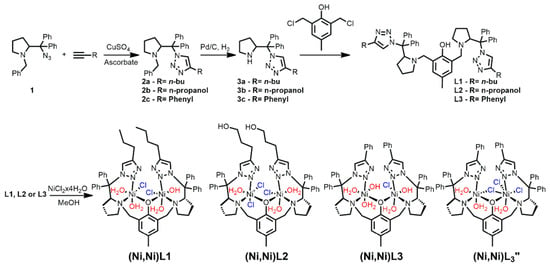
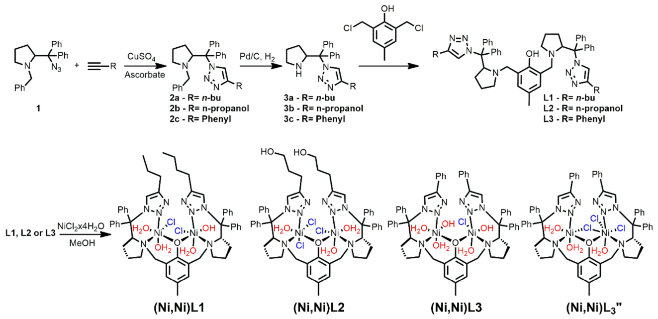
However, unlike the active site of urease, in which histidine residues coordinate the nickel ions, our previously reported mimics featured alcohol donor groups in the first coordination sphere. Because the identity and electronic properties of first-sphere ligands can strongly influence the electrostatic environment and reactivity of a metal center, we hypothesized that introducing a more histidine-like donor would provide an additional means of controlling urea activation. Here, we therefore designed and synthesized a series of dinuclear nickel complexes supported by bis-triazole ligands, in which the substituent at the C4 position of the triazole was systematically varied (Scheme 1). The triazole donors provide a closer electronic analog to the histidine coordination environment of urease while the C4 substituents enable modulation of the electrostatic environment surrounding the dinuclear metal center. Remarkably, these modifications produced a pronounced enhancement in urea lyase activity. Complexes exhibiting a 2:1 electrolyte behavior in solution promoted the rapid formation of isocyanate and ammonia, with a rate enhancement of up to 1011-fold relative to the uncatalyzed reaction. This represents, to our knowledge, the highest rate enhancement reported for an abiotic urea lyase and approaches the catalytic efficiency characteristic of enzymes. The systematic dependence of reactivity on the ligand environment further indicates that electrostatic effects play a key role in controlling urea activation and elimination. These findings demonstrate that enzyme-like rate acceleration can be achieved in a synthetic catalyst even through a reaction pathway distinct from that employed by the natural enzyme, highlighting electrostatic control as a powerful design principle for abiotic catalysis.
Scheme 1.
Synthesis of the ligands and their corresponding complexes.
2. Results
2.1. Characterization of the Complexes
To investigate how the second coordination sphere influences urea activation, we synthesized four dinuclear nickel complexes supported by bis-triazole ligands bearing different C4 substituents (Scheme 1). The protocols to obtain the intermediates and the ligands along with their characterization is described in the Supporting Information (Figures S1–S51 for NMR, S52–S59 for HRMS, S60 for sc-XRD of 2a). The complexes were designed to provide closely related dinuclear nickel environments while systematically modulating the electronic and electrostatic properties of the ligand framework. The complexes were obtained in moderate-to-good yields and characterized by elemental analysis, HRMS, UV-Vis spectroscopy, conductivity measurements, and magnetic studies (See Supporting Information, S62–S70 for MALDI-TOF, S71–S74 for thermogravimetry, S75–S78 for FTIR spectroscopy, S79–S82 for UV-Vis spectroscopy). During the synthesis of the complexes containing the phenyl-radical-containing ligand, two distinct products were obtained in independent experiments. The complex isolated in the first synthesis was green, whereas the product obtained in the second synthesis was orange. Upon repeating the syntheses, it was observed that, despite using the same drying time, the color of the hydrated nickel chloride salt varied after heat/vacuum drying. In some experiments, the salt became pale yellow, yielding a green complex. In others, the salt developed a yellow color with a slight orange tint, and the resulting complex was orange. These observations suggest that the extent of dehydration of the nickel salt may influence the color of the resulting complexes and, potentially, their coordination geometries, as water molecules can also coordinate to the metal center. Because the heating was not temperature-controlled, the actual temperature reached during this process was unknown. The dehydration of NiCl2.6H2O may occur in multiple stages. The first mass loss takes place between 24 and 120 °C and corresponds to approximately 30% of the initial mass, equivalent to the loss of four water molecules. Complete dehydration requires temperatures above 250 °C [36]. Therefore, the nickel salt was likely not fully dehydrated under the conditions employed. Nevertheless, the differences in salt coloration observed during the syntheses of (Ni,Ni)L3 and (Ni,Ni)L3″ suggest that the last may have involved a greater degree of dehydration. This difference in hydration state may have affected the coordination environment of the nickel centers, potentially accounting for the distinct colors observed in the resulting complexes.
Thermogravimetric analysis provided additional support for the proposed compositions of the complexes (Figures S71–S74). The thermogravimetric profiles and residual masses are consistent with the formulations derived from elemental analysis, supporting the different ligand and chloride contents assigned to the complexes (Table 1). The mass loss between 35 and 120 °C is attributed to loosely bound solvent: two ethanol molecules for (Ni,Ni)L1 (7.1%), six water molecules for (Ni,Ni)L2 (9%), and three water molecules for (Ni,Ni)L3 (5%) and (Ni,Ni)L3″ (4%). Between 120 and 280 °C, more tightly bound water and chloride are lost. (Ni,Ni)L1 loses three water molecules (4.3%, 120–200 °C), followed by 2 Cl−, one OH− and one H2O (8%, 200–260 °C). (Ni,Ni)L2 loses three H2O and three Cl− (12%), (Ni,Ni)L3 loses two OH− and one Cl− (6%), and (Ni,Ni)L3″ loses three Cl− (9%). At higher temperatures, the organic ligand decomposes (55–75% of the mass), leaving residues of about 21% for (Ni,Ni)L1, 24% for (Ni,Ni)L2, 14% for (Ni,Ni)L3, and 15% for (Ni,Ni)L3″. Because thermal analysis cannot establish connectivity, these data support the composition of the complexes but not their structure.
Table 1.
Tentative assignment of the TGA/DTG experiment for each complex.
The TGA data also help rationalize an observation made during the synthesis of the phenyl-substituted complexes. Two distinct products, (Ni,Ni)L3 (green) and (Ni,Ni)L3″ (orange), were obtained in independent experiments, which differ in their chloride/hydroxide content (Table 1), indicating that water is retained or taken up during isolation and purification, and that residual hydration of the nickel salt may contribute to the variability between batches.
Although the ligand substitution occurs at a position remote from the metal center, the complexes exhibited markedly different solution properties. Conductivity measurements revealed that all complexes behave as non-electrolyte species in dichloromethane, whereas significant differences emerged in methanol solution. Complexes (Ni,Ni)L1 and (Ni,Ni)L3 displayed conductivities characteristic of 2:1 electrolytes, (Ni,Ni)L2 behaved as a 3:1 electrolyte, and (Ni,Ni)L3″ remained largely non-electrolytic. These observations indicate that subtle changes in ligand structure strongly influence ligand lability and ionization in coordinating solvents.
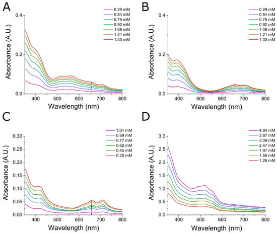
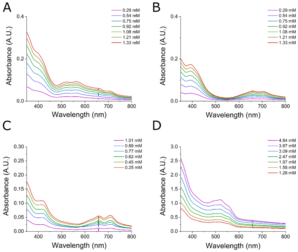
UV-Vis spectroscopy further revealed important differences among the dinuclear Ni(II) complexes. Complexes (Ni,Ni)L1-(Ni,Ni)L3 display broadly similar spectral signatures, characterized by weak d-d transitions typical of high-spin Ni(II) centers in distorted octahedral environments (Figure 1) [35]. Bands assigned to the 3A2g→3T1g(P) transition are observed at 348 (εmax ~ 175 mol−1 cm−1 L), 392 (εmax ~ 131 mol−1 cm−1 L), and 410 nm (εmax ~ 110 mol−1 cm−1 L) for (Ni,Ni)L1, (Ni,Ni)L2, and (Ni,Ni)L3, respectively. Additional low-intensity absorptions in the 520–710 nm region are consistent with the 3A2g→3T1g(F) and 3A2g→3T2g transitions expected for octahedral Ni(II) (522 nm (εmax ~ +76 mol−1 cm−1 L), 562 nm (εmax ~ 78 mol−1 cm−1 L), 652 nm (εmax ~ 54 mol−1 cm−1 L) and 702 nm (εmax ~ 44 mol−1 cm−1 L) for (Ni,Ni)L1; 662 nm (εmax ~ 35 mol−1 cm−1 L) and 702 nm (εmax ~ 33 mol−1 cm−1 L) for (Ni,Ni)L2; and (εmax ~ 52 mol−1 cm−1 L) and 708 nm (εmax ~ 55 mol−1 cm−1 L) for (Ni,Ni)L3). While (Ni,Ni)L1 exhibits a more complex pattern with four partially resolved bands, likely reflecting inequivalent Ni centers and/or the presence of multiple species in solution, the spectra of (Ni,Ni)L2 and (Ni,Ni)L3 are remarkably similar. The close correspondence of both band positions and intensities suggests that modification of the ligand substituent has only a minor effect on the coordination environment of the dinuclear Ni center.
Figure 1.
Electronic spectroscopy of complexes (Ni,Ni)L1-(Ni,Ni)L3″ in the 350–800 nm range. (A) Complex (Ni,Ni)L1 was analyzed from 0.29 mM to 1.33 mM concentrations, (B) complex (Ni,Ni)L2 was analyzed from 0.29 mM to 1.33 mM concentrations, (C) complex (Ni,Ni)L3 was analyzed from 0.25 mM to 1.01 mM concentrations and (D) complex (Ni,Ni)L3″ was analyzed from 1.26 mM to 4.84 mM concentrations. All complexes were dissolved in dichloromethane.
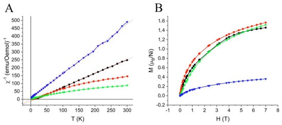
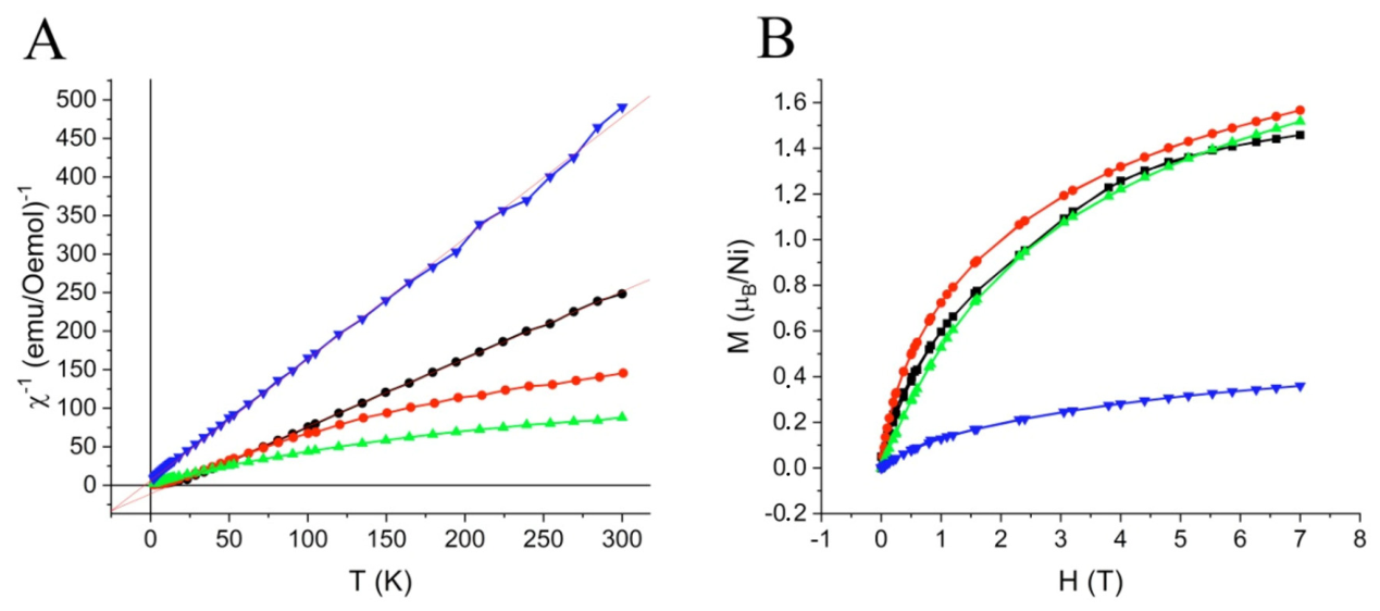
In contrast, (Ni,Ni)L3″ exhibits a markedly different electronic spectrum, displaying an intense absorption at 330 nm (εmax ≈ 1777 mol−1 cm−1 L) together with a series of partially resolved bands centered between 500 and 650 nm (518 = εmax ~212). The increased intensity of the visible absorptions relative to those observed for (Ni,Ni)L1-(Ni,Ni)L3 is consistent with a reduction in local symmetry around the Ni(II) centers, leading to a relaxation of Laporte selection rules. Attempts to characterize the complex structure by 1H NMR failed due to the paramagnetic nature of these complexes. All of them presented broadening of the bands (Figure S52), and complex (Ni,Ni)L3″ was the only one with a less broadening effect (Figure S53). This behavior is supported by magnetic measurements, which reveal a substantially different magnetic ground state for (Ni,Ni)L3″ (Figure 2). Whereas (Ni,Ni)L1-(Ni,Ni)L3 display χMT values of approximately 90–250 cm3 K mol−1 at 300 K, (Ni,Ni)L3″ reaches nearly 500 cm3 K mol−1 and exhibits significantly reduced field-dependent saturation, attaining only ~0.35 μB per Ni at 7 T. These features are indicative of enhanced magnetic anisotropy and substantially stronger ferromagnetic exchange interactions between the Ni(II) centers in (Ni,Ni)L3″. The combination of more intense d-d transitions and pronounced ferromagnetic coupling is consistent with a tetrahedral or strongly tetrahedrally distorted coordination environment, which is expected to generate a markedly different ligand-field splitting pattern and magnetic anisotropy relative to the distorted octahedral geometries proposed for (Ni,Ni)L1-(Ni,Ni)L3.
Figure 2.
Magnetization of the complexes. Complex (Ni,Ni)L1 is shown as red line, (Ni,Ni)L2 as green, (Ni,Ni)L3 as black and (Ni,Ni)L3″ as blue. Graphic of the reverse magnetic susceptibility in function of temperature (A) and magnetization curves in function of the magnetic field at 2K (B).
Dinuclear nickel(II) complexes supported by phenolate-bridged compartmental ligands have been structurally characterized extensively as urease models, typically showing hexacoordinate high-spin Ni(II) centers with Ni···Ni distances of 2.9–3.5 Å, a μ-phenolate bridge, and additional exogenous bridges or terminal solvent and chloride ligands [37,38,39,40]. Likewise, 1,2,3-triazoles coordinate Ni(II) through N [3] with Ni-N distances of 2.0–2.2 Å [41,42]. However, we have evidenced the N [2] coordination in similar mononuclear triazole complexes [43]. Our spectroscopic, magnetic, and conductometric data are consistent with the proposed coordination mode as the Ni(II) centers are high-spin and octahedral, the triazole bands in the FTIR shift upon coordination (ex: 1675 cm−1 to 1653 cm−1), and the chloride content determined by elemental analysis and conductivity agrees with the proposed chlorides in the inner sphere. Without a crystal structure, we propose the structures in Scheme 1 on this basis.
2.2. Catalysis
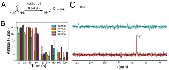
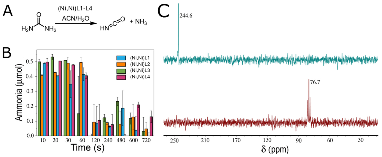
We next investigated whether the dinuclear nickel complexes promote urea decomposition under aqueous acetonitrile conditions (15.8% water, 10.5% dichloromethane, 73.7% acetonitrile). Using the Berthelot assay to monitor ammonia formation [44], all four complexes were found to catalyze the conversion of urea, producing detectable ammonia within the first seconds of the reaction (Figure 3B). Control with NiCl2 did not exhibit the formation of ammonia (Figure S83). To confirm the nature of the transformation, reactions with 15N-labeled urea were analyzed by 15N NMR spectroscopy. Disappearance of the urea resonance (76 ppm) and formation of a new signal corresponding to isocyanate (244 ppm) confirmed that the complexes promote urea decomposition through an elimination pathway rather than the hydrolytic mechanism employed by urease (Figure 3C).
Figure 3.
Quantification of ammonia formed by nickel complexes during the urea degradation reaction. Urea is decomposed into isocyanate and ammonia (A). Concentration of ammonia over reaction time was measured by Berthelot method [44], using 0.27 µmol of the complex; 15.8% of water and 9 µmol of urea (B) at 5 °C. The 15N NMR evidenced that the urea signal (76.7 ppm) was shown to disappear upon complex addition, resulting in the formation of isocyanate (244.6 ppm) (C).
As we did not observe any other product, this data suggest that the reaction stopped at isocyanate and ammonia. The highest concentration of ammonia was detected in the first 10 s of the reaction and after that, the ammonia concentration decreased, indicating that ammonia was lost by volatilization (Figure 3B). A similar temporal profile has been reported for copper-mediated urea decomposition in acetonitrile/water mixtures, where the catalytic activity was closely linked to water-induced formation of hydrated copper species. In that system, water promoted ligand exchange and dissociation processes that generated more reactive monomeric copper centers, thereby accelerating urea hydrolysis [45]. The analogous dependence on water observed here suggests that changes in the nickel site speciation may also contribute to the burst of ammonia formation at early reaction times, while the subsequent decline in measured ammonia reflects its physical loss from solution rather than a reversal of the hydrolysis reaction.
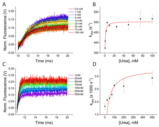
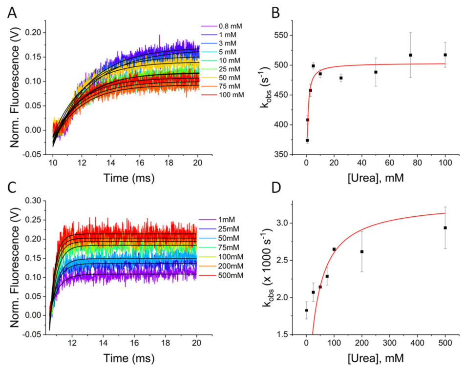
Although the ammonia-quantification experiments established that all four complexes promote urea decomposition, the relatively low temporal resolution of the assay did not allow a rigorous comparison of their intrinsic catalytic activities. We therefore employed stopped-flow fluorescence measurements to determine the kinetic parameters associated with urea decomposition and to compare the reactivity of the complexes under identical conditions. Fluorescein was used as a fluorescent pH indicator, and the increase in pH was observed by the increase in fluorescence at 520 nm. The comparison between fluorescence intensity change in a catalytic reaction and control pHs (Figure S84) enabled the determination of the ammonia produced in the reaction. Complexes (Ni,Ni)L1 and (Ni,Ni)L3 exhibited a Michaelis–Menten behavior (Figure 4) and using Equation (1), we could determine the KM and k2 of the reaction, in which k2 is defined as the rate constant of the transformation of the [ES] complex into products. Hence, (Ni,Ni)L1 and (Ni,Ni)L3 exhibited KM of 0.26 mM and 12 mM, respectively, and k2 of 500 s−1 and 3000 s−1.
Figure 4.
Kinetics of ammonia decomposition by complexes (Ni,Ni)L1 and (Ni,Ni)L3. (A) Stopped-flow assays at different urea concentrations for complex (Ni,Ni)L1 and (B) the Michaelis–Menten curve obtained with the rates extracted from the stopped-flow measurements of (Ni,Ni)L1. The experimental values are shown as dots and the red line is the computed exponential curve using these values. (C) Stopped-flow assays at different urea concentrations for complex (Ni,Ni)L3 and (D) the Michaelis–Menten curve obtained with the rates extracted from the stopped-flow measurements of (Ni,Ni)L3.The experimental values are shown as dots and the red line is the computed exponential curve using these values Stopped-flow assays were carried out with 30 μM nickel complexes, 9 μM fluorescein and variable concentrations of urea. All assays were performed in a 50/50 v/v water/acetonitrile mixture.
In contrast, complex (Ni,Ni)L2 exhibited substantially lower turnover rate (k2 = 0.024 s−1), while (Ni,Ni)L3″ was inhibited at increasing urea concentrations (Figure S85). The several-orders-of-magnitude differences in intrinsic catalytic activity suggest that subtle changes in solution structure profoundly affect substrate activation.
The kinetic parameters obtained from the stopped-flow experiments allowed estimation of the catalytic rate enhancement relative to the uncatalyzed decomposition of urea (k = 6.5 × 10⁻¹⁰ s−1). Using the experimentally determined k2 values, rate enhancements of 7.7 × 10¹¹ and 4.6 × 10¹² were obtained for complexes (Ni,Ni)L1 and (Ni,Ni)L3, respectively, whereas (Ni,Ni)L2 exhibited a considerably lower enhancement of 3.7 × 10⁷. These results highlight the high activity of (Ni,Ni)L1 and (Ni,Ni)L3 in promoting urea decomposition. For comparison, Table 2 summarizes selected abiotic catalytic systems reported to promote urea decomposition. Although numerous synthetic urease mimics have been described, quantitative kinetic data are scarce, and differences in reaction conditions and kinetic parameters further complicate direct comparisons with previously reported systems.
Table 2.
Selected examples of urea catalyzed decomposition and their estimated rate enhancement.
Notably, in our case, the largest rate enhancements were observed exclusively for the complexes that form 2:1 electrolyte species under catalytic conditions. This correlation suggests that hydration-induced ionization and reorganization of the coordination sphere are important features of catalyst activation. A possible explanation is the differing propensity of the complexes to disrupt the phenolate-bridged dinuclear core. Previous studies on related urease-inspired systems have shown that more labile bridges are readily displaced in solution [35], generating coordinatively accessible metal centers, whereas strongly bridged species remain catalytically less competent [45]. In this context, it is possible that catalytic efficiency may depend not on nuclearity itself, but on the equilibrium between closed and open forms capable of generating accessible nickel sites for urea coordination and activation.
2.3. Identification of the Catalytically Active Species
The kinetic analyses revealed that complexes (Ni,Ni)L1 and (Ni,Ni)L3 accelerate urea decomposition by more than eleven orders of magnitude, whereas the complexes (Ni,Ni)L2 and (Ni,Ni)L3″ are substantially less active. These pronounced differences suggest that the isolated complexes may not represent the catalytically competent species under the reaction conditions. We therefore investigated whether changes in catalyst speciation occur in the presence of water and whether such changes correlate with catalytic activity. Before examining the identity of the catalytically competent species, we considered whether the observed activity could instead arise from heterogeneous nickel species. The strong ligand dependence of the catalytic activity argues against a common heterogeneous nickel species, such as Ni(0) nanoparticles or Ni(OH)2 precipitates, as the origin of the observed reactivity. First, the pH changes by only ~0.5 unit during the reaction, owing to the equilibrium between isocyanate (pKa = 3.5) [49] and ammonium (pKa = 9.35) [50]. Second, the reaction proceeds at room temperature, well below the hydrothermal conditions typically associated with Ni(OH)2 formation [51,52]. Additionally, no reducing agent is present, making the formation of Ni(0) particles unlikely. Lastly, catalytic activity can be restored by addition of further urea solution, indicating that the active species persists in solution (S87).
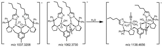
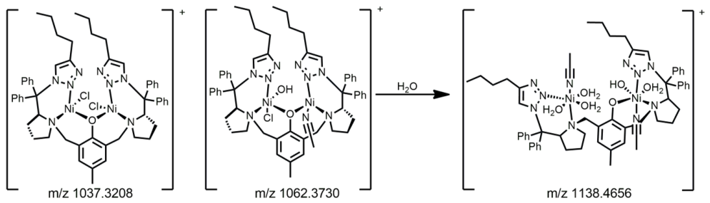
We have noticed that in the absence of water, no detectable ammonia formation was observed (Figure S87), indicating that hydration is required for catalyst activation and that the initially coordinated waters do not suffice for catalysis. Because catalytic activity was only observed in the presence of water, we first examined the effect of hydration on the solution properties of the complexes. To investigate whether water induces changes in catalyst speciation, conductivity measurements were performed in ACN/H2O mixtures; Figure S88. These measurements revealed the progressive ionization of (Ni,Ni)L1 upon addition of water. In acetonitrile, the complex behaved as a 1:1 electrolyte, consistent with displacement of one anionic ligand by the coordinating solvent. Increasing the water content resulted in a gradual increase in conductivity, reaching values characteristic of a 2:1 electrolyte above 4.5% H2O and remaining in this region under catalytic conditions (15% H2O). These observations suggest that water promotes displacement of a second labile anionic ligand, increasing the overall ionic character of the complex. High-resolution mass spectrometry (HRMS, Figure S89) analysis of complex (Ni,Ni)L1 in different conditions provided additional support for this interpretation. For instance, in acetonitrile, HRMS of (Ni,Ni)L1 reveals the presence of peaks at m/z 1037.3208 and 1049.3489, assigned to species containing two chloride ligands and one coordinated acetonitrile molecule, respectively C55H63Cl2N8Ni2O (m/ztheor. 1037.3208) and C59H69N10Ni2O (m/ztheor. 1049.4362). Upon addition of water, the intensities of these signals decreased concomitantly with the emergence of a new signal at m/z 1136.4734 (C59H76N10Ni2O6, m/ztheor. 1136.4656). The composition of this species is consistent with extensive ligand exchange upon hydration. Considering the octahedral coordination requirements of both Ni(II) centers, the observed composition cannot be accommodated while retaining the original phenolate-bridged dinuclear connectivity. We therefore propose that this species arises from a water-induced reorganization involving disruption of the phenolate bridge and formation of distinct hydrated nickel coordination environments (Scheme 2).
Scheme 2.
Proposed species that are found in solution based on the HRMS spectrum of a DCM/ACN solution and after addition of water.
We subsequently extended the conductivity measurements to the complete series. (Ni,Ni)L3 exhibited conductivity consistent with a 2:1 electrolyte (Figure S88), whereas (Ni,Ni)L2 and (Ni,Ni)L3′ remained predominantly within the 1:1 electrolyte range. This behavior indicates that, in the latter complexes, water promotes the displacement of only one labile anionic ligand, which may occur without the disruption of the phenolate bridge. Consistent with this interpretation, the HRMS spectrum of (Ni,Ni)L2 showed no significant changes upon addition of water (Figure S90).
These results reveal a correlation between catalytic activity and the extent of water-induced ionization. This correlation suggests that extensive hydration and the associated reorganization of the dinuclear coordination framework may be important for generating the catalytically competent species and facilitating catalytic turnover.
The apparent structural reorganization observed for the most active catalysts may have important mechanistic implications. Urease-catalyzed hydrolysis relies on cooperative activation of the substrate by the metal center, whereas β-elimination reactions catalyzed by lyases typically involve substrate binding to a metal site and abstraction of a β-proton by a nearby basic residue [53]. If hydration disrupts the dinuclear phenolate-bridged structure and generates two independent nickel coordination environments, one metal center could serve as the substrate-binding site while the second site could facilitate proton abstraction.
As urea coordination was not observed in our previous study, we sought to determine whether urea is able to coordinate to the complex under the anhydrous conditions in which no catalytic activity is observed. For this purpose, (Ni,Ni)L3 was reacted with urea under anhydrous conditions, and the resulting product was analyzed by FTIR spectroscopy (Figure S91). Comparison of the spectra before and after reaction revealed the emergence of a band at 1770 cm−1, which is consistent with the coordination of urea through one of its nitrogen atoms [54]. Additional bands at 1574, 1259, 1098, 1050, and 800 cm−1 can be assigned to ν(CO), δ(NH), ρ(NH), ν′(CN), β(OCNN) of urea, respectively.
These results demonstrate that urea can coordinate to (Ni,Ni)L3 in the absence of water. However, because no detectable catalysis occurs under anhydrous conditions, urea coordination alone is insufficient to generate the catalytically competent species. These observations indicate that substrate binding and catalyst activation are distinct processes. While urea coordination can occur in the absence of water, catalytic turnover requires hydration-induced reorganization of the nickel coordination sphere. This interpretation is consistent with the conductivity and HRMS results, which indicate substantial water-dependent changes in the coordination environment of the most active complexes. Further evidence for this water-induced reorganization was obtained by difference FTIR spectroscopy of (Ni,Ni)L3 before and after water addition (Figure S92). Upon hydration, new bands emerged at 1630 and 808 cm−1, consistent with water coordination and Ni–O/Ni–OH vibrational modes, respectively, concomitant with a decrease in the intensity of bands associated with the phenolate framework at 1488, 1444, and 1240 cm−1. These changes are consistent with the reorganization of the phenolate-containing coordination sphere upon hydration. Thus, the combined data support a mechanism in which urea binding and formation of the catalytically competent nickel species are distinct events, with hydration playing a critical role in generating the latter.
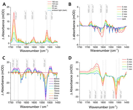
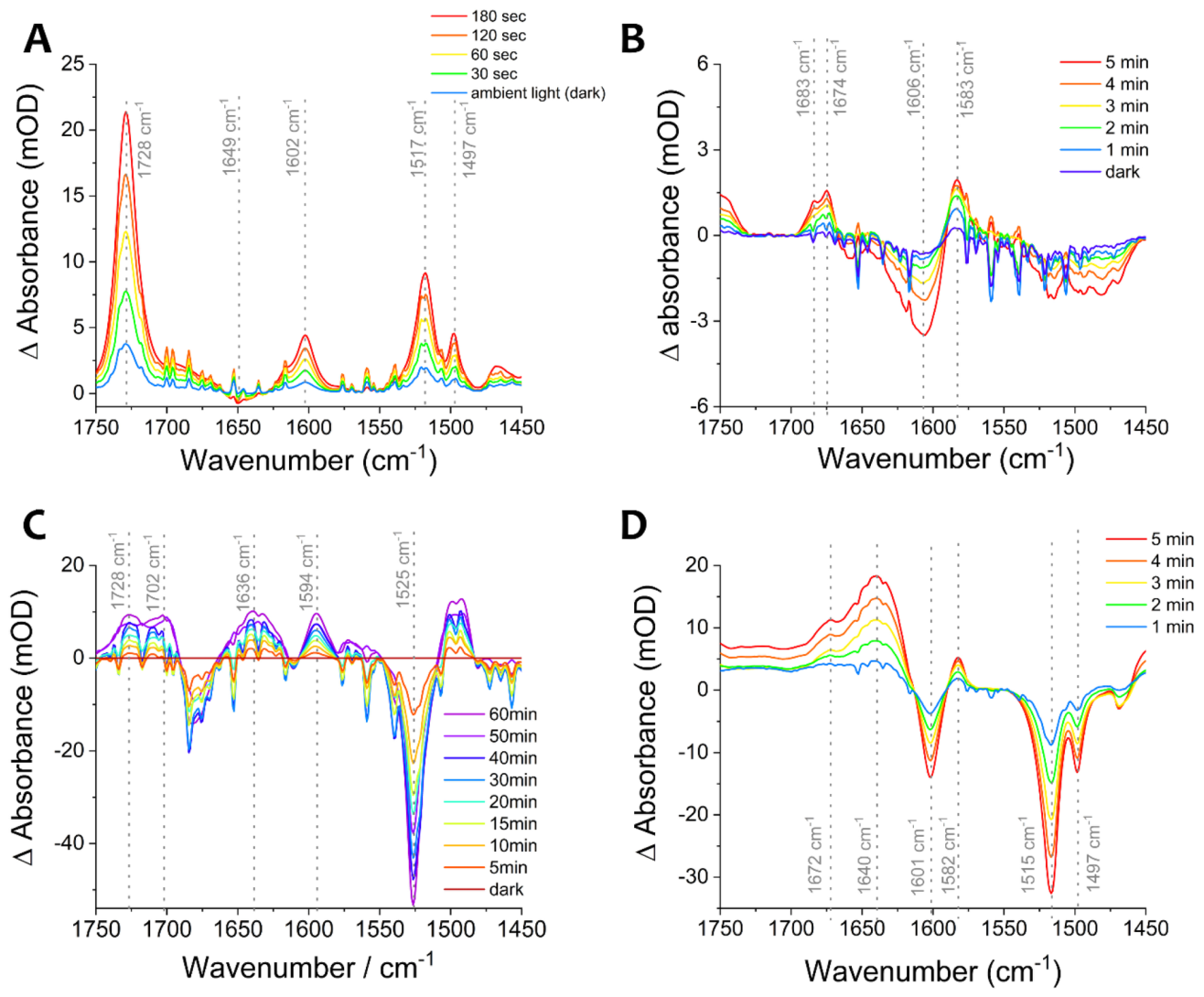
To determine whether urea coordination to the complex could also be detected in solution, we investigated the reaction between the complex and a ruthenium-caged urea [55], which upon photolysis generates free urea. As acetonitrile is a coordinating solvent, it can easily displace the coordinated urea, which results in not-optimal dark subtractions. However, it is evident that the control reaction of the caged urea in acetonitrile reveals the emergence of bands at 1728, 1602, 1517 and 1497 cm−1, which can be attributed to δs NH2, ν(C=O) [56], bpy (ν(C=N) and ν(C=C) stretching modes) [57] and ν(C-N) [56] (Figure 5). The blueshift of the carbonyl bands from the solid state is mostly due to the disruption of standard hydrogen-bonding aggregation [58]. In the presence of the complex ((Ni,Ni)L3) we can analyze the bands that emerge relative to the control experiment performed in the absence of caged urea. In an anhydrous condition, the 1728 cm−1 and the 1497 cm−1 bands are easily visible, revealing that free urea is in solution. Interestingly, bands at 1702, 1636 and 1594 cm−1 are distinct from the complex-only control. These bands could be related to urea coordination to the complex in anhydrous conditions. For instance, N-coordination normally shifts ν(C=O) modes to higher wavenumbers [59], whereas O-coordination shifts to lower. Considering the observed shifts from 1728 to 1702 cm−1 and 1602 to 1636 cm−1, it is possible that we have a O-bond coordination. An unambiguous assignment is not possible, however, because the 1636 cm−1 band could also arise from release of water from the complex. An attempt to identify urea coordination via HRMS in anhydrous conditions failed, most probably due to labile binding and possibly in-source dissociation due to the competition by acetonitrile solvent for coordination (S92).
Figure 5.
Photolysis experiments of Ru-caged urea and (Ni,Ni)L3 using a diode laser (λ-532 nm). The Ru-caged urea [55] (A) and the complex (B) were dissolved in acetonitrile. The anhydrous reaction between photocleaved urea and the complex is shown in (C), whereas the reaction in the presence of water is shown in (D). Conditions: Caged urea (10 mM), complex (5 mM). The dark spectrum is the subtraction of a spectrum in the dark versus the initial spectrum before photolysis. The time of irradiation of each experiment is shown in the insets.
In the presence of water, all urea-related bands except 1728 cm−1 appear as negative features, indicating that urea is consumed. The absence of a negative band at 1728 cm−1 may reflect overlap with a new band at a similar wavenumber. The most prominent new band is at 1640 cm−1. Although this is consistent with O-bound urea at nickel [60,61], it more likely reflects water release (δ(H2O) falls in this region), given that the complex is highly active in water and no intermediate could be isolated at room temperature. We were unable to detect isocyanate or other potential reaction intermediate because the strong absorption of acetonitrile obscures the nitrile/cyanate region of the spectrum. These solution data therefore support that in anhydrous conditions, urea coordinates to the nickel centers, corroborating to the solid-state assay.
Although these experiments do not allow definitive assignment of the solution-state binding mode under catalytic conditions, the combined results from the anhydrous coordination studies and the photolysis experiments with caged urea provide evidence that urea can interact directly with the nickel center. Urea coordination can be detected even under anhydrous conditions, where no catalytic activity is observed. In contrast, catalysis occurs only in the presence of water, suggesting that water-induced reorganization of the nickel coordination sphere, rather than urea binding itself, is critical for generating the catalytically competent species. Moreover, these observations suggest that at least part of the catalytic cycle may involve inner-sphere coordination of urea to the nickel center, representing a departure from our previously proposed purely outer-sphere elimination pathway [35].
3. Experimental Part
Synthesis of the Complexes
In a 50 mL Schlenk flask, 79 mg of NiCl2.6H2O (~0.3 mmol) were added and kept under vacuum and mild heating (~100 °C) until a complete change in color of the solids from green to pale yellow (in the synthesis of complex (Ni,Ni)L3″, the nickel chloride was dried till an orange tint). Meanwhile, the ligand solution was prepared under argon atmosphere in another 50 mL Schlenk flask by addition of 100 mg of the bis-triazoles (5–7, ~0.1 mmol) and 6 mg of sodium methoxide (~0.1 mmol) to 4 mL of anhydrous methanol. This solution was stirred at 50 °C for two hours, after which, it was added by syringe to the NiCl2-containing Schlenk flask. The reaction was kept under argon atmosphere and 50 °C for 48 h. After this period, the solvent was removed by vacuum. The solid was dissolved with 5 mL of dichloromethane followed by centrifugation to remove the excess of inorganic impurities (nickel chloride and sodium chloride). The solution was dried under vacuum and the solid was washed with ethyl acetate to remove any excess of ligand. Finally, the resulting solid was dried over two days under high vacuum.
(Ni,Ni)L1: Yield 238 mg (63%), κ (1 mM, DCM) = 2.8 µS cm−1, κ (1 mM, MeOH) = 186 µS cm−1, HRMS (MALDI-TOF) m/z calcd for (C75H75N10Ni2O7) 1343.453 found 1343.478, CHN calcd for C58H78Cl2N8Ni2O8 57.88, 6.53, 9.31 found 57.85, 6.54, 9.17. The higher mass observed for the complexes is due to the coordination of the matrix to the complex.
(Ni,Ni)L2: Yield 275 mg (74%), κ (1 mM, DCM) = 2.3 µS cm−1, κ (1 mM, MeOH) = 250 µS cm−1, HRMS (MALDI-TOF) m/z calcd for (C74H75N10Ni2O8) 1347.447 found 1347.544, CHN calcd for C53H77Cl3N8Ni2O12 51.25, 6.25, 9.02 found 51.03, 5.83, 8.95. The higher mass observed for the complexes is due to the coordination of the matrix to the complex.
(Ni,Ni)L3: Yield 134 mg (58%), κ (1 mM, DCM) = 2.5 µS cm−1, κ (1 mM, MeOH) = 137 µS cm−1, HRMS (MALDI-TOF) m/z calcd for (C80H69N9Ni2O7) 1383.402 found 1383.239, CHN calcd for C59H63ClN8Ni2O6 62.54, 5.60, 9.89 found 62.24, 5.90, 9.47. The higher mass observed for the complexes is due to the coordination of the matrix to the complex.
(Ni,Ni)L3″: Yield 105 mg (46%), κ (1 mM, DCM) = 2.4 µS cm−1, κ (1 mM, MeOH) = 47 µS cm−1, HRMS (MALDI-TOF) m/z calcd for (C80H69N9Ni2O7) 1383.402 found 1383.409, CHN calcd for C59H61Cl3N8Ni2O4 60.54, 5.60, 9.57 found 60.60, 5.56, 9.36. The higher mass observed for the complexes is due to the coordination of the matrix to the complex.
4. Conclusions
We have developed a series of dinuclear nickel complexes that function as highly efficient artificial urea lyases, promoting the decomposition of urea to ammonia and isocyanate. Systematic modification of the ligand environment produced large differences in catalytic activity, with complexes (Ni,Ni)L1 and (Ni,Ni)L3 displaying rate enhancements of up to 1012-fold relative to the uncatalyzed reaction, representing among the highest values reported for synthetic urea-decomposing catalysts. The most active complexes undergo extensive hydration and ligand exchange, generating 2:1 electrolyte species under catalytic conditions, whereas the less active systems remain predominantly in the 1:1 region. The strong correlation between ionization, structural reorganization, and catalytic performance suggests that catalyst flexibility and water-induced activation are essential for efficient turnover. These findings demonstrate that enzyme-like rate enhancements can be achieved in relatively simple synthetic systems through control of coordination dynamics and the electrostatic environment surrounding the metal center. The relationship between electrolyte formation and catalytic activity provides a useful design principle for the development of future artificial metalloenzymes and abiotic catalysts capable of approaching the efficiencies of biological systems.
Supplementary Materials
The following supporting information can be downloaded at https://www.mdpi.com/article/10.3390/inorganics14110277/s1. The supplementary materials contain synthesis and catalysis protocols, characterization protocols, NMR spectra of intermediates and ligands, mass spectra of intermediates and ligands, mass spectra of the complexes, elemental analyses of the complexes, thermogravimetric analysis, infrared spectroscopic analysis, UV–Vis spectroscopic analysis, and catalytic assays [62,63,64].
Author Contributions
Conceptualization, C.G.C.M.N.; methodology, C.G.C.M.N., L.K.S. and R.S.F.; validation, L.K.S., G.R.C., R.G.S. and R.S.F.; formal analysis, C.G.C.M.N., L.K.S., R.G.S. and R.S.F.; investigation, L.K.S., R.G.S. and R.S.F.; resources, C.G.C.M.N. and R.S.F.; data curation, L.K.S., R.G.S. and R.S.F.; writing—original draft preparation, C.G.C.M.N.; writing—review and editing, C.G.C.M.N. and R.S.F.; visualization, L.K.S. and R.S.F.; supervision, C.G.C.M.N.; project administration, C.G.C.M.N.; funding acquisition, C.G.C.M.N. All authors have read and agreed to the published version of the manuscript.
Funding
This research was funded by FAPESP grants number: 2008/10177-7; 2014/22332-8; 2016/01622-3. This research was also partially funded by Coordenação de Aperfeiçoamento de Pessoal de Nivel Superior-Brasil (CAPES) 001.
Data Availability Statement
The original contributions presented in this study are included in the article/Supplementary Materials. Further inquiries can be directed to the corresponding author.
Acknowledgments
We kindly thank Antonio Gilberto Ferreira from UFSCar for the 15N NMR experiments.
Conflicts of Interest
The authors declare no conflicts of interest.
References
- Quinn, D.M.; Sikorski, R.S. Enzymatic Rate Enhancements; John Wiley & Sons: Hoboken, NJ, USA, 2014. [Google Scholar]
- Richard, J.P. Enzymatic Rate Enhancements: A Review and Perspective. Biochemistry 2013, 52, 2009. [Google Scholar] [CrossRef] [Scilit] [PubMed]
- Warshel, A.; Sharma, P.K.; Kato, M.; Xiang, Y.; Liu, H.; Olsson, M.H.M. Electrostatic basis for enzyme catalysis. Chem. Rev. 2006, 106, 3210. [Google Scholar] [CrossRef] [Scilit] [PubMed]
- Hay, S.; Scrutton, N.S. Good vibrations in enzyme-catalysed reactions. Nat. Chem. 2012, 4, 161. [Google Scholar] [CrossRef] [Scilit] [PubMed]
- Agarwal, P.K. Role of Protein Dynamics in Reaction Rate Enhancement by Enzymes. J. Am. Chem. Soc. 2005, 127, 15248. [Google Scholar] [CrossRef] [Scilit] [PubMed]
- Warshel, A.; Bora, R.P. Perspective: Defining and quantifying the role of dynamics in enzyme catalysis. J. Chem. Phys. 2016, 144, 180901. [Google Scholar] [CrossRef] [Scilit] [PubMed]
- Nagel, Z.D.; Klinman, J.P. A 21st century revisionist’s view at a turning point in enzymology. Nat. Chem. Biol. 2009, 5, 543. [Google Scholar] [CrossRef] [Scilit] [PubMed]
- Breslow, R. Biomimetic Chemistry and Artificial Enzymes: Catalysis by Design. Acc. Chem. Res. 1995, 28, 146. [Google Scholar] [CrossRef] [Scilit]
- Krämer, R. Bioinorganic models for the catalytic cooperation of metal ions and functional groups in nuclease and peptidase enzymes. Coord. Chem. Rev. 1999, 182, 243. [Google Scholar] [CrossRef] [Scilit]
- Kuah, E.; Toh, S.; Yee, J.; Ma, Q.; Gao, Z. Enzyme Mimics: Advances and Applications. Chem. Eur. 2016, 22, 8404. [Google Scholar] [CrossRef] [Scilit] [PubMed]
- Liu, L.; Breslow, R. A Potent Polymer/Pyridoxamine Enzyme Mimic. J. Am. Chem. Soc. 2002, 124, 4978. [Google Scholar] [CrossRef] [Scilit] [PubMed]
- Neverov, A.A.; Lu, Z.-L.; Maxwell, C.I.; Mohamed, M.F.; White, C.J.; Tsang, J.S.W.; Brown, R.S. Combination of a Dinuclear Zn2+ Complex and a Medium Effect Exerts a 1012-Fold Rate Enhancement of Cleavage of an RNA and DNA Model System. J. Am. Chem. Soc. 2006, 128, 16398. [Google Scholar] [CrossRef] [Scilit] [PubMed]
- Brown, R.S.; Lu, Z.-L.; Liu, C.T.; Tsang, W.Y.; Edwards, D.R.; Neverov, A.A. Dinuclear Zn(II) catalysts as biomimics of RNA and DNA phosphoryl transfer enzymes: Changing the medium from water to alcohol provides enzyme-like rate enhancements. J. Phys. Org. Chem. 2010, 23, 1–15. [Google Scholar] [CrossRef] [Scilit]
- Mukherjee, G.; Lee, C.W.Z.; Nag, S.S.; Alili, A.; Reinhard, F.G.C.; Kumar, D.; Sastri, C.V.; Visser, S.P.D. Dramatic rate-enhancement of oxygen atom transfer by an iron(IV)-oxo species by equatorial ligand field perturbations. Dalton Trans. 2018, 47, 14945. [Google Scholar] [CrossRef] [Scilit] [PubMed]
- Batha, S.; Arman, H.; Larionov, O.V.; Musie, G.T. Zinc(II) complexes of a versatile heptadentate ligand as phosphohydrolase structural and functional mimics. Inorg. Chim. Acta 2019, 497, 119077. [Google Scholar] [CrossRef] [Scilit]
- Park, H.I.; Ming, L.J. A 1010 Rate Enhancement of Phosphodiester Hydrolysis by a Dinuclear Aminopeptidase-Transition-State Analogues as Substrates? Angew. Chem. Int. Ed. 1999, 38, 2914. [Google Scholar] [CrossRef] [Scilit]
- Lyu, Y.; Scrimin, P. Mimicking Enzymes: The Quest for Powerful Catalysts from Simple Molecules to Nanozymes. ACS Catal. 2021, 11, 11501. [Google Scholar] [CrossRef] [Scilit]
- Sreedhara, A.; Freed, J.D.; Cowan, J.A. Efficient Inorganic Deoxyribonucleases. Greater than 50-Million-Fold Rate Enhancement in Enzyme-Like DNA Cleavage. J. Am. Chem. Soc. 2000, 122, 8814. [Google Scholar] [CrossRef] [Scilit]
- Liu, F. The Upside of Downsizing: Asymmetric Trifunctional Organocatalysts as Small Enzyme Mimics for Cooperative Enhancement of Both Rate and Enantioselectivity With Regulation. Chirality 2013, 25, 675. [Google Scholar] [CrossRef] [Scilit] [PubMed]
- Nothling, M.D.; Xiao, Z.; Hill, N.S.; Blyth, M.T.; Bhaskaran, A.; Sani, M.-A.; Espinosa-Gomez, A.; Ngov, K.; White, J.; Buscher, T.; et al. A multifunctional surfactant catalyst inspired by hydrolases. Sci. Adv. 2020, 6, eaaz0404. [Google Scholar] [CrossRef] [Scilit] [PubMed]
- Erxleben, A. Mechanistic Studies of Homo- and Heterodinuclear Zinc Phosphoesterase Mimics: What Has Been Learned? Front. Chem. 2019, 7, 82. [Google Scholar] [CrossRef] [Scilit] [PubMed]
- Prier, C.K.; Arnold, F.H. Chemomimetic Biocatalysis: Exploiting the Synthetic Potential of Cofactor-Dependent Enzymes To Create New Catalysts. J. Am. Chem. Soc. 2015, 137, 13992. [Google Scholar] [CrossRef] [Scilit] [PubMed]
- Estiu, G.; Merz, K.M. The Hydrolysis of Urea and the Proficiency of Urease. J. Am. Chem. Soc. 2004, 126, 6932. [Google Scholar] [CrossRef] [Scilit] [PubMed]
- Callahan, B.P.; Yuan, Y.; Wolfenden, R. The Burden Borne by Urease. J. Am. Chem. Soc. 2005, 127, 10828–10829. [Google Scholar] [CrossRef] [Scilit] [PubMed]
- Mazzei, L.; Cianci, M.; Ciurli, S. Inhibition of Urease by Hydroquinones: A Structural and Kinetic Study. Chem. Eur. J. 2022, 28, e202201770. [Google Scholar] [CrossRef] [Scilit] [PubMed]
- Lee, W.-Z.; Tseng, H.-S.; Wang, T.-L.; Tsai, H.-L.; Kuo, T.-S. Bioinspired Catalytic Conjugate Additions of Thiophenols to α,β-Enones by a Disubstituted Benzoate-Bridged Nickel Mimic for the Active Site of Urease. Organometallics 2010, 29, 2874. [Google Scholar] [CrossRef] [Scilit]
- Lee, W.-Z.; Tseng, H.-S.; Ku, M.-Y.; Kuo, T.-S. Dinickel complexes of disubstituted benzoate polydentate ligands: Mimics for the active site of urease. Dalton Trans. 2008, 69, 2538. [Google Scholar] [CrossRef] [Scilit] [PubMed]
- Kundu, B.K.; Biswas, S.; Mondal, A.; Mazumdar, S.; Mobin, S.M.; Mukhopadhyay, S. Unveiling the urease like intrinsic catalytic activities of two dinuclear nickel complexes towards the in situ syntheses of aminocyanopyridines. Dalton Trans. 2021, 50, 4848. [Google Scholar] [CrossRef] [Scilit] [PubMed]
- Uprety, B.; Arderne, C.; Bernal, I. Catalytic Cleavage of the Amide Bond in Urea Using a Cobalt(III) Amino-Based Complex. Eur. J. Inorg. Chem. 2018, 2018, 5058–5067. [Google Scholar] [CrossRef] [Scilit]
- Meyer, F.; Kaifer, E.; Kircher, P.; Heinze, K.; Pritzkow, H. Cooperative Transformations of Small Molecules at a Dinuclear Nickel(II) Site. Chem. Eur. J. 1999, 5, 1617–1630. [Google Scholar] [CrossRef]
- Barrios, A.M.; Lippard, S.J. Interaction of Urea with a Hydroxide-Bridged Dinuclear Nickel Center: An Alternative Model for the Mechanism of Urease. J. Am. Chem. Soc. 2000, 122, 9172–9177. [Google Scholar] [CrossRef] [Scilit]
- Barrios, A.M.; Lippard, S.J. Decomposition of Alkyl-Substituted Urea Molecules at a Hydroxide-Bridged Dinickel Center. Inorg. Chem. 2001, 40, 1250–1255. [Google Scholar] [CrossRef] [Scilit] [PubMed]
- Koga, T.; Furutachi, H.; Nakamura, T.; Fukita, N.; Ohba, M.; Takahashi, K.; Ōkawa, H. Dinuclear Nickel(II) Complexes of Phenol-Based “End-Off” Compartmental Ligands and Their Urea Adducts Relevant to the Urease Active Site. Inorg. Chem. 1998, 37, 989. [Google Scholar] [CrossRef] [Scilit]
- Alexandrova, A.N.; Jorgensen, W.L. Why Urea Eliminates Ammonia Rather than Hydrolyzes in Aqueous Solution. J. Phys. Chem. B 2007, 111, 720. [Google Scholar] [CrossRef] [Scilit] [PubMed]
- Martins, C.O.; Sebastiany, L.K.; Lopez-Castillo, A.; Freitas, R.S.; Andrade, L.H.; Toma, H.E.; Netto, C.G.C.M. Urea Decomposition Mechanism by Dinuclear Nickel Complexes. Molecules 2023, 28, 1659. [Google Scholar] [CrossRef] [Scilit] [PubMed]
- Mishra, S.K.; Kanungo, S.B. Thermal dehydration and decomposition of nickel chloride hydrate (NiCl2·xH2O). J. Therm. Anal. 1992, 38, 2417. [Google Scholar] [CrossRef] [Scilit]
- Adams, H.; Clunas, S.; Fenton, D.E.; Spey, S.E. Dinuclear nickel(II) complexes of reduced asymmetric compartmental ligands. Dalton Trans. 2003, 32, 625. [Google Scholar] [CrossRef] [Scilit]
- Paital, A.R.; Wong, W.T.; Aromí, G.; Ray, D. New [LNiII2]+ complexes incorporating 2-formyl or 2,6-diformyl-4-methyl phenol as inhibitors of the hydrolysis of the ligand L3-: Ni⋯Ni ferromagnetic coupling and S = 2 ground states. Inorg. Chem. 2007, 46, 5727. [Google Scholar] [CrossRef] [Scilit] [PubMed]
- Mahapatra, T.S.; Chaudhury, S.; Dasgupta, S.; Bertolasi, V.; Ray, D. Dinuclear nickel complexes of divergent Ni⋯Ni separation showing ancillary ligand addition and bio-macromolecular interaction. New J. Chem. 2015, 40, 2268. [Google Scholar] [CrossRef] [Scilit]
- Horn, A., Jr.; Fim, L.; Bortoluzzi, A.J.; Szpoganicz, B.; Silva, M.D.S.; Novak, M.A.; Neto, M.B.; Eberlin, L.S.; Catharino, R.R.; Eberlin, M.N.; et al. Solid state and solution characterization of a new dinuclear nickel (II) complex: The search for synthetic models for urease. J. Mol. Struct. 2006, 797, 154. [Google Scholar] [CrossRef] [Scilit]
- Schweinfurth, D.; Krzystek, J.; Schapiro, I.; Demeshko, S.; Klein, J.; Telser, J.; Ozarowski, A.; Su, C.-Y.; Meyer, F.; Atanasov, M.; et al. Electronic Structures of Octahedral Ni(II) Complexes with “Click” Derived Triazole Ligands: A Combined Structural, Magnetometric, Spectroscopic, and Theoretical Study. Inorg. Chem. 2013, 52, 6880. [Google Scholar] [CrossRef] [Scilit] [PubMed]
- Schweinfurth, D.; Su, C.-Y.; Wei, S.-C.; Braunsteind, P.; Sarkar, B. Nickel complexes with “click”-derived pyridyl-triazole ligands: Weak intermolecular interactions and catalytic ethylene oligomerisation. Dalton Trans. 2012, 41, 12984. [Google Scholar] [CrossRef] [Scilit] [PubMed]
- Ferreira, M.P.; Castro, C.B.; Honorato, J.; He, S.; Júnior, W.G.G.; Esmieu, C.; Castellano, E.E.; Moura, A.F.D.; Truzzi, D.R.; Nascimento, O.R.; et al. Biomimetic catalysis of nitrite reductase enzyme using copper complexes in chemical and electrochemical reduction of nitrite. Dalton Trans. 2023, 52, 11254. [Google Scholar] [CrossRef] [Scilit] [PubMed]
- Rhine, E.D.; Mulvaney, R.L.; Pratt, E.J.; Sims, G.K. Improving the Berthelot Reaction for Determining Ammonium in Soil Extracts and Water. Soil. Sci. Soc. Am. J. 1998, 62, 473–480. [Google Scholar] [CrossRef] [Scilit]
- Castro, C.B.; Silveira, R.G.; Colombari, F.M.; Moura, A.F.D.; Nascimento, O.R.; Netto, C.G.C.M. Solvent Effect on the Regulation of Urea Hydrolysis Reactions by Copper Complexes. Chemistry 2020, 2, 525. [Google Scholar] [CrossRef] [Scilit]
- Daramola, D.A.; Singh, D.; Botte, G.G. Dissociation Rates of Urea in the Presence of NiOOH Catalyst: A DFT Analysis. J. Phys. Chem. A 2010, 114, 11513. [Google Scholar] [CrossRef] [Scilit] [PubMed]
- Kaminskaia, N.V.; Kostić, N.M. Kinetics and Mechanism of Urea Hydrolysis Catalyzed by Palladium(II) Complexes. Inorg. Chem. 1997, 36, 5917. [Google Scholar] [CrossRef] [Scilit] [PubMed]
- Biswas, S.; Karim, S.; Adhikary, J.; Das, D. Solvent-Driven Self-Assembly of Discrete Ni(II)-Azide Complexes: Unraveling Unusual Behavior in Mimicking Jack Bean Urease. Inorg. Chem. 2025, 64, 986. [Google Scholar] [CrossRef] [Scilit] [PubMed]
- Borduas, N.; Place, B.; Wentworth, G.R.; Abbatt, J.; Murphy, J.G. Solubility and reactivity of HNCO in water: Insights into HNCO’s fate in the atmosphere. Atmos. Chem. Phys. 2016, 16, 703. [Google Scholar] [CrossRef] [Scilit]
- Lide, D.R. CRC Handbook of Chemistry and Physics; Internet Version; CRC Press: Boca Raton, FL, USA, 2005. [Google Scholar]
- Soler-Illia, G.J.D.A.A.; Jobbágy, M.; Regazzoni, A.E.; Blesa, M.A. Synthesis of nickel hydroxide by homogeneous alkalinization. Chem. Matter 1999, 11, 3140. [Google Scholar] [CrossRef] [Scilit]
- Hall, D.S.; Lockwood, D.J.; Bock, C.; MacDougall, B.R. Nickel hydroxides and related materials: A review of their structures, synthesis and properties. Proc. Math. Phys. Eng. Sci. 2015, 471, 20140792. [Google Scholar] [CrossRef] [Scilit] [PubMed]
- Villiers, M.D.; Veetil, V.P.; Raj, H.; Villiers, J.D.; Poelarends, G.J. Catalytic Mechanisms and Biocatalytic Applications of Aspartate and Methylaspartate Ammonia Lyases. ACS Chem. Biol. 2012, 7, 1618. [Google Scholar] [CrossRef] [Scilit] [PubMed]
- Woon, T.C.; Wickramasinghe, W.A.; Fairlie, D.P. Oxygen versus nitrogen coordination of a urea to (diethylenetriamine)platinum(II). Inorg. Chem. 2002, 32, 2190. [Google Scholar]
- Netto, C.G.C.M.; Bell, S.; Castro, C.B.; Machado, P.H.; Purohit, V.; Davis, K.M.; Batista, A.P.L.; Dyer, R.B. Photocontrolled Release of Urea Enables the Detection of Urea–Urease Intermediates by Cryo-FTIR. Angew. Chem. Int. Ed. 2025, 64, e202504332. [Google Scholar] [CrossRef] [Scilit] [PubMed]
- Grdadolnik, J.; Maréchal, Y. Urea and urea–water solutions—An infrared study. J. Mol. Struct. 2002, 615, 177. [Google Scholar] [CrossRef] [Scilit]
- Omberg, K.M.; Schoonover, J.R.; Treadway, J.A.; Leasure, R.M.; Dyer, R.B.; Meyer, T.J. Mid-Infrared Spectrum of [Ru(bpy)3]2+. J. Am. Chem. Soc. 1997, 119, 7013. [Google Scholar] [CrossRef] [Scilit]
- Otte, X.; Eurard, B.; Oelattre, L.; Thunus, L. Development and validation of a new Fourier transform infrared spectrometric method for the quantification of urea in creams and ointments. Anal. Chim. Acta 2002, 451, 323. [Google Scholar] [CrossRef] [Scilit]
- Penland, B.; Mizushima, S.; Curran, C.; Quagliano, J.V. Infrared Absorption Spectra of Inorganic Coordination Complexes. X. Studies of Some Metal-Urea Complexes. J. Am. Chem. Soc. 1957, 79, 1575. [Google Scholar] [CrossRef] [Scilit]
- Konrad, M.; Meyer, F.; Jacobi, A.; Kircher, P.; Rutsch, P.; Zsolnai, L. Coordination and Conversion of Urea at Dinuclear μ-Acetato Nickel(II) Complexes with Symmetric and Asymmetric Cores. Inorg. Chem. 1999, 38, 4559–4566. [Google Scholar] [CrossRef] [Scilit] [PubMed]
- Mochizuki, K.; Takahashi, J.; Ishima, Y.; Shindo, T. Synthesis and structures of urea-coordinated dinickel(II) complexes with binucleating ligand 1,3-bis(N-(2-(dimethylamino)ethyl)-N-methylamino)propan-2-ol (HL1) and its analogs. Inorg. Chem. Acta 2013, 400, 151–158. [Google Scholar] [CrossRef] [Scilit]
- Rostovtsev, V.V.; Green, L.G.; Fokin, V.V.; Sharpless, K.B. A Stepwise Huisgen Cycloaddition Process: Copper(I)-Catalyzed Regioselective TMLigation∫ of Azides and Terminal Alkynes. Angew. Chem. Int. Ed. 2002, 41, 2596–2599. [Google Scholar]
- Kocie´nski, K.J.; P. Protecting Groups. J. Chem. Soc. Perkin Trans. 1999, 1, 1589–1615. [Google Scholar] [CrossRef] [Scilit]
- Kurosawa, W.; Kan, T.; Fukuyama, T. Preparation of Secondary Amines from Primary Amines via 2-Nitrobenzenesulfonamides: N-(4-Methoxybenzyl)-3-phenylpropylamine. Org. Synth. Coll. 2014, 10, 482. [Google Scholar] [CrossRef] [Scilit]

Disclaimer/Publisher’s Note: The statements, opinions and data contained in all publications are solely those of the individual author(s) and contributor(s) and not of MDPI and/or the editor(s). MDPI and/or the editor(s) disclaim responsibility for any injury to people or property resulting from any ideas, methods, instructions or products referred to in the content. |
© 2026 by the authors. Licensee MDPI, Basel, Switzerland. This article is an open access article distributed under the terms and conditions of the Creative Commons Attribution (CC BY) license.